1. Introduction
Achieving an adequate primary stability (PS) is crucial for the long-term survival and success of dental implants, as well to obtain a fast implant osseointegration [
1]. As a matter of fact, before proceeding with the functional loading of the dental implant, an intimate contact between the implant surface and the surrounding bone has to be established [
2]. Currently, different clinical strategies have been adopted to increase PS and the bone-to-implant contact in a post-extractive condition, such as site under-preparation by means of surgical rotating drills, piezo electric or electromagnetic devices, osseodensifying burs, bone expanders, osteotomes, and cutters [
3,
4,
5]. Besides these, the implant micro- and macro-geometry, the surface characteristics, the thread profile and pitch together with their cutting ability have all been reported to affect the biomechanics of the bone-to-implant contact and the bone anchorage, improving implant stability [
6,
7,
8].
However, the effects of the shape of implant apical threads on osseointegration and PS are not still completely elucidated [
9,
10] and the relationship between the implant apical design and clinical results does not appear to be clear [
11]. For example, limited data are present in literature about the influence of cutting flute implant designs on PS and on mineralized bone formation at the interface with implants can be found [
12]. In their study, Hsieh et al. [
11] demonstrated that self-tapping implants with self-cutting flutes may determine an improvement of implant stability, especially in immediate post-extraction sites, when the implant stability is generated by the apex anchoring of the bone only in a few mm (3–5 mm). Moreover, new implant macro-designs, also with innovative apical portions, have been proposed to improve PS and long-term implant survival and success [
13].
Bone density is also recognized to represent another key factor in determining implants PS, being correlated with the bone-to-implant contact. In this regard, Misch classification [
14] categorized natural bone, according to the density and the cortical and trabecular microstructure:
- -
D1: dense cortical bone and poor/absent trabecular bone (mandible symphysis);
- -
D2: cortical and dense trabecular bone tissue (mandible and anterior maxilla);
- -
D3: thin cortical and trabecular bone (mandible and anterior/posterior maxilla);
- -
D4: poor/absent cortical and thin trabecular tissues (posterior maxilla).
In recent years, the polyurethane study model has been widely used as an alternative material to perform biomechanical tests in different medical fields, and also to test dental implants [
15,
16,
17]. Indeed, this artificial bone has been considered to be similar to natural bone tissue by the American Society for Testing and Materials (ASTM F-1839-08 [
18]), as it presents consistent mechanical and physical characteristics that closely resemble those of different bone tissue densities, with high reliability and stability, without any special handling or human variables. In this way, easy and clinically applicable non-invasive tests could be performed in order to evaluate implant stability and the relationship between the fixture and different qualities of bone [
5,
6,
17,
19].
Therefore, assessing the influence of the shape and structure of different implant apexes and threads on the biomechanical properties of implants inserted in different polyurethane foam densities could be useful to corroborate other preclinical and clinical studies. For this reason, the aim of the present in vitro study was an evaluation of the Insertion Torque (IT), the Removal Torque (RT), and the Resonance Frequency Analysis (RFA) of different latest generation implant apical threads in cylindrical and conical implants inserted in low-density polyurethan foam sheets.
2. Materials and Methods
2.1. Dental Implants
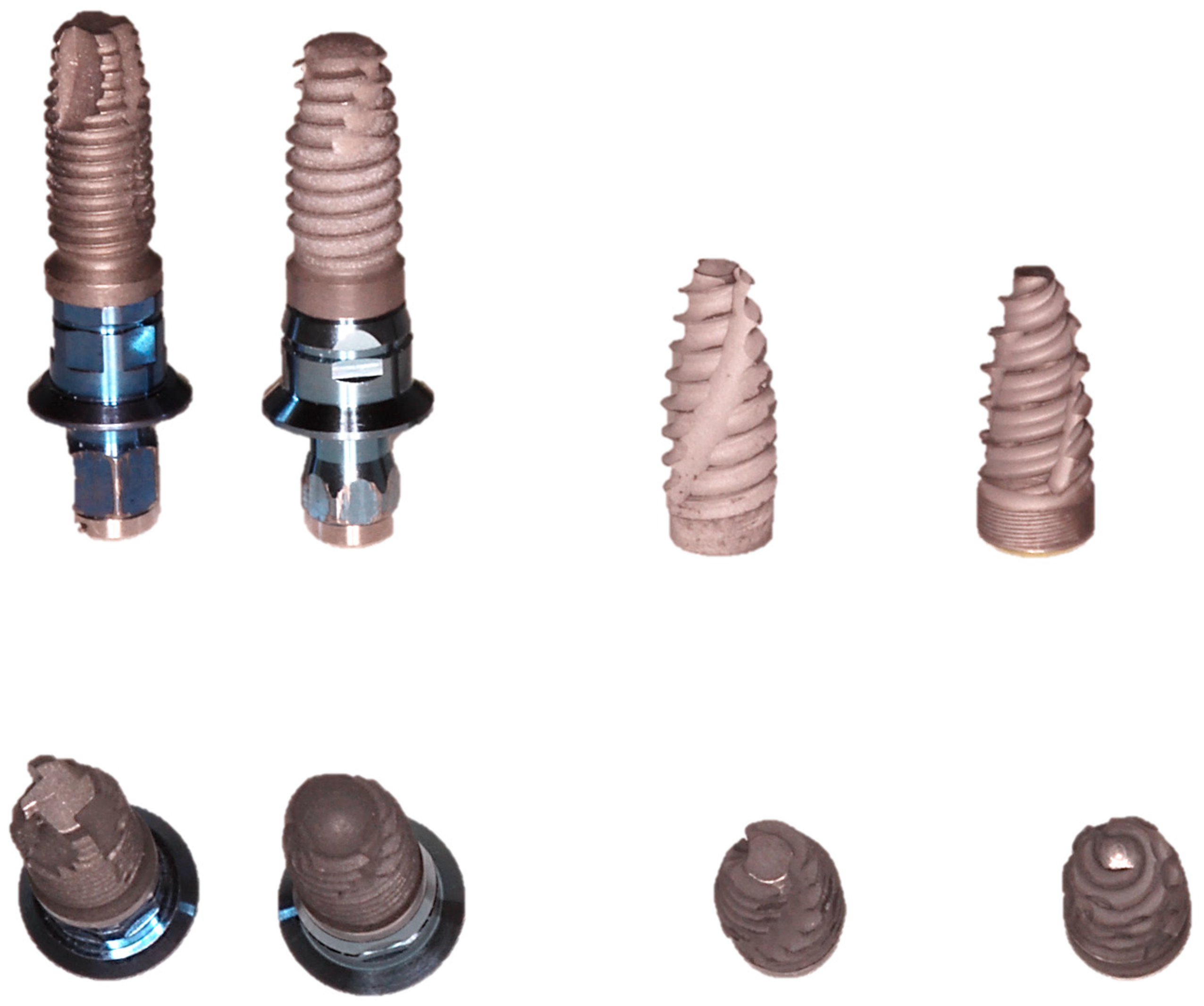
The characteristics of the different dental implants used for this in vitro study are listed below:
- -
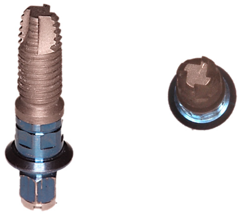
Cylindrical implants with V-shaped threads, flat apex, and an external hexagon connection (EE: 3i Osseotite, Biomax SpA, Vicenza, Italy) (
Figure 1);
- -
Conical tapered implants with V-shaped threads, round apex, and an external hexagon connection (T3: 3i T3 Implant, Biomax SpA, Vicenza, Italy) (
Figure 2);
- -
Conical implants with Smooth Design threads, a cutting apex with a reduced diameter, and a Cone Morse self-locking connection (TAC: Total Approach Concept, Aon Implants, Grisignano di Zocco, Italy) (
Figure 3);
- -
Conical cutting tapered implants with the CT cutting design of the threads, an atraumatic apex with a reduced diameter, and a Cone Morse connection (Intra-lock: Intra-lock System Europa SpA, Salerno, Italy) (
Figure 4).
These two latter implants had new apical threads designs. The Smooth Design of TAC threads and the reduced apex were designed to perfectly adapt to the mini-invasive preparation of the implant site and to respect as much as possible the hard tissue. On the other hand, the conical shape, reduced apex, and the Blossom threadform of Intra-lock implants have been developed to have a high cutting action, distributing the native bone over the entire implant surface, and with an ideal angulation for type III and IV bones.
All the implants used had the same dimensions: a diameter of 4 mm and a length of 10 mm. A representation of all the implants tested is reported in
Figure 5.
2.2. Study Design
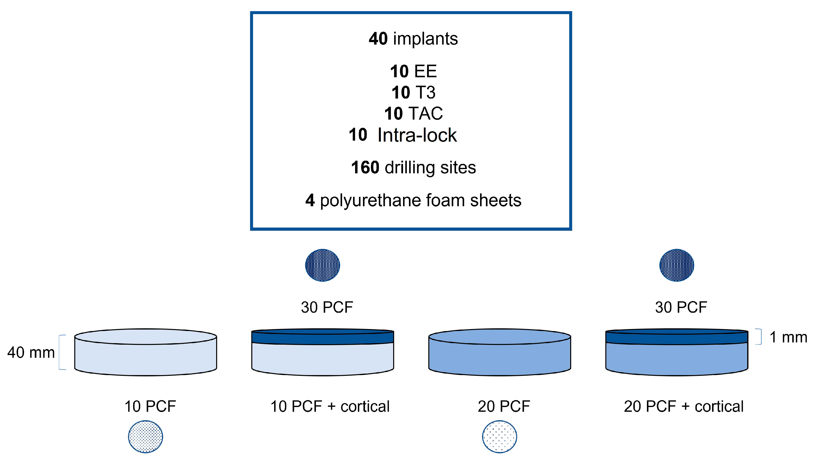
A total of 40 dental implants (10 EE, 10 T3, 10 TAC, and 10 Intra-lock) were used in this investigation to perform a total of 160 drilling sites on 10 and 20 PCF polyurethane foam sheets, with or without the presence of a cortical foam sheet of 30 PCF in density (10 osteotomies per implant for each experimental condition) (
Figure 6).
Different 40 mm thickness polyurethane foam sheets with densities of 10 and 20 PCF, with and without the addition of a 1 mm thickness cortical sheet with a density of 30 PCF (13 cm × 18 cm, Sawbones Europe AB, Malmö, Sweden), were used to perform this in vitro test. The 10 PCF density sheet corresponds to a density of 0.16 g/cm
3 and has been used to mime the D3 bone quality, whereas the 20 PCF density sheet has a density of 0.32 g/cm
3, similar to a D2 bone type. Moreover, a 1 mm thickness sheet with a density of 0.48 g/cm
3 (30 PCF) was added in order to simulate a layer of D1 cortical bone [
15,
16] (
Figure 7).
The polyurethane foams are available with different proportions of cortical and trabecular components, which reproduce the bone microstructure, obtaining a realistic simulation of the clinical condition.
2.3. Drilling Protocol
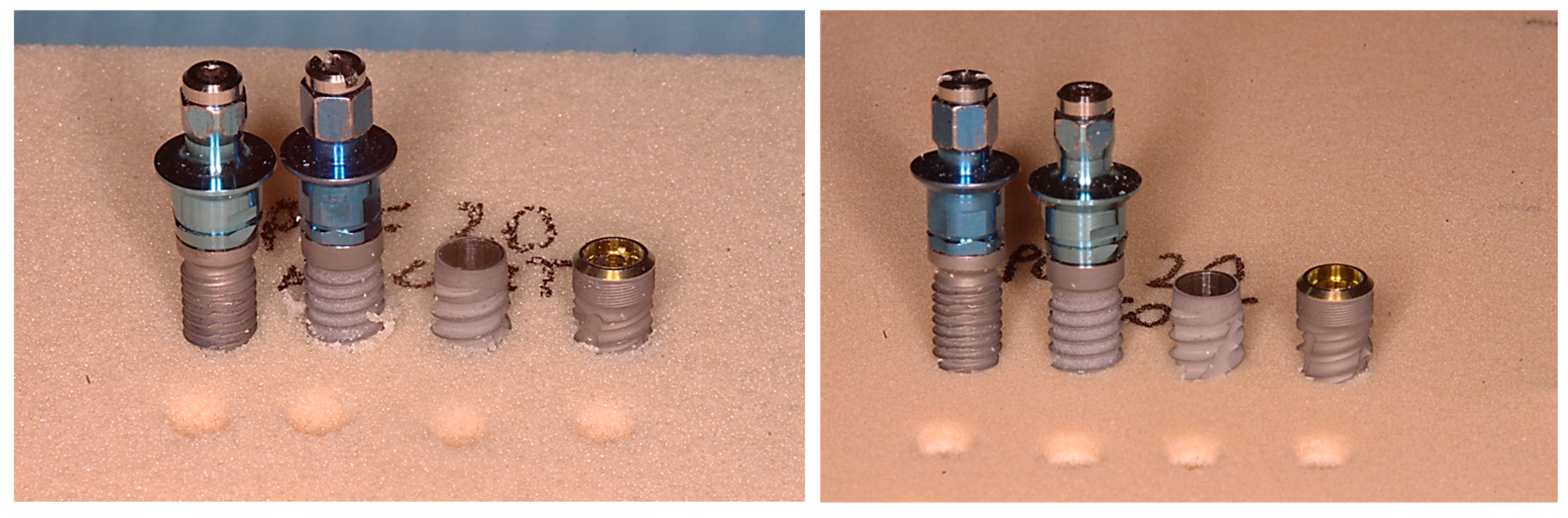
The drilling and the insertion of the implants into all the experimental polyurethane sheets were conducted only for the first 4–5 mm in length, in order to properly evaluate the apical portion of all the implants (
Figure 8 and
Figure 9).
Implants were positioned in the polyurethane sheets of any densities, following the corresponding manufacturer’s instructions and kit, but standardizing the use of the initial lanceolate drill at 300 rpm. After this, a 2 mm bur and a 2.8 mm bur were used for TAC and Intra-lock implants, respectively, while for EE and T3 implants a further 3 mm bur was used, always using a surgical implant motor at 300 rpm (Chiropro, Bien Air Dental SA, Bienne, Switzerland), following the manufacturer’s protocol established for critical conditions such as the present test.
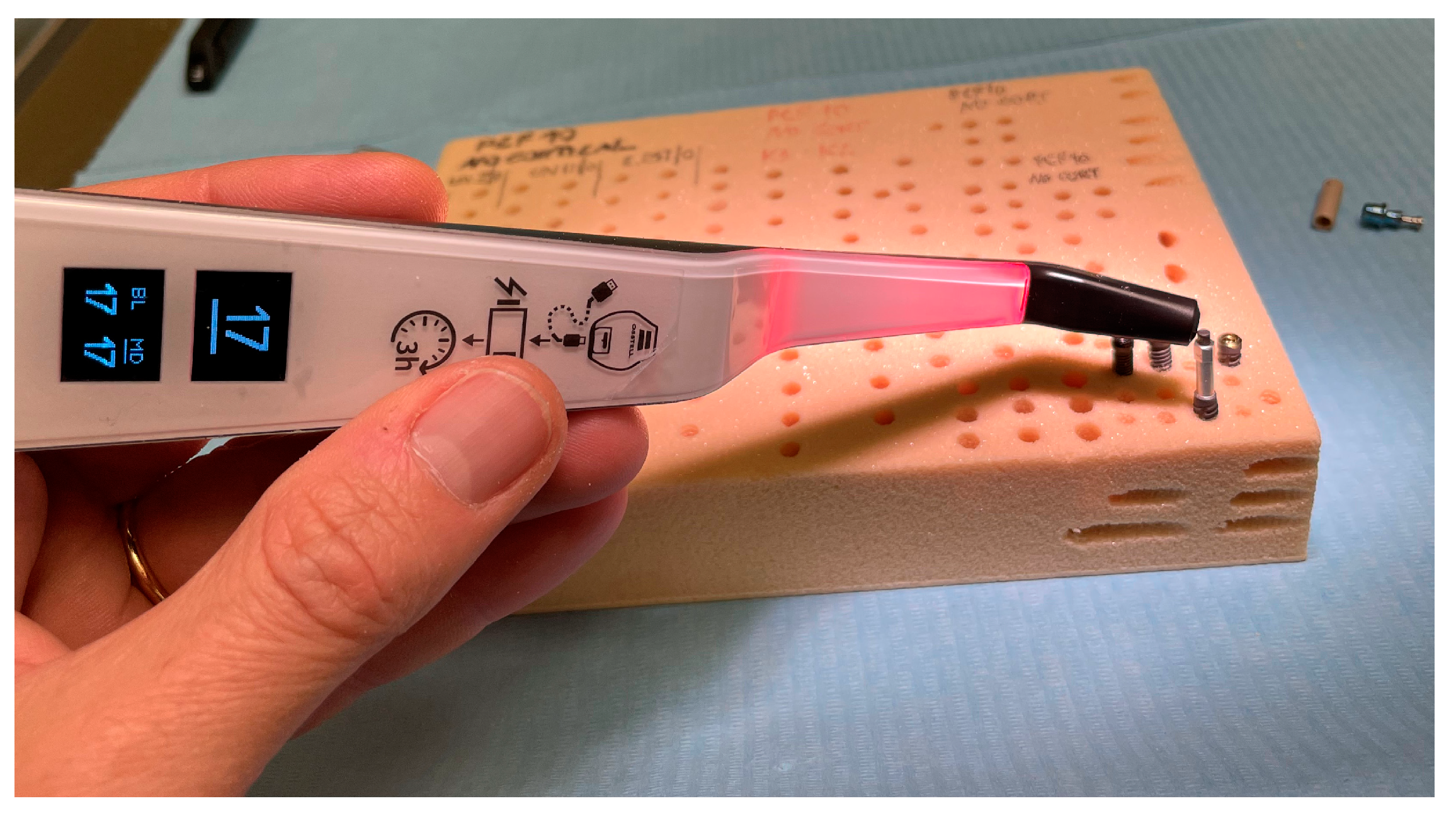
The investigation was conducted by a single operator (Dr. Luca Comuzzi), measuring the IT, RT, and RFA values of all implants tested. Particularly, the final implant insertion was performed at 30 rpm with a torque set at 50 N and then, the final 1mm IT and RT values were recorded by dynamometric analysis using a calibrated torque meter during screw positioning and removal. At the end, the RFA was measured recording the ISQ values in two different orientations at 90 degrees (bucco-lingual: BL, and mesio-distal: MD) (
Figure 10), by using the SmartPeg n.78 (Ostell Inc., Göteborg, Schweden) for TAC and Intra-lock implants, and the SmartPeg n.1 (Ostell Inc., Göteborg, Schweden) for EE and T3 implants.
2.4. Statistical Analysis
Power analysis and sample size calculation were performed using the ANCOVA statistical test (effect size: 0.4, α err: 0.05; power (1 − β): 0.95; numerator df: 9; number of groups: 4; number of covariates: 4), using the program G*Power 3.1.9.7. The minimum total sample size necessary to achieve a statistically significant output was 157 implant sites. The Kolmogorov-Smirnov test was used to assess the normality distribution of data. Then, Kruskal-Wallis nonparametric test was performed to evaluate the IT, RT, and RFA values, considering a p-value < 0.05 statistically significant. These data were analyzed by using the statistical software package GraphPad 9.0 (Prism, San Diego, CA, USA).
3. Results
3.1. Insertion Torque Evaluation
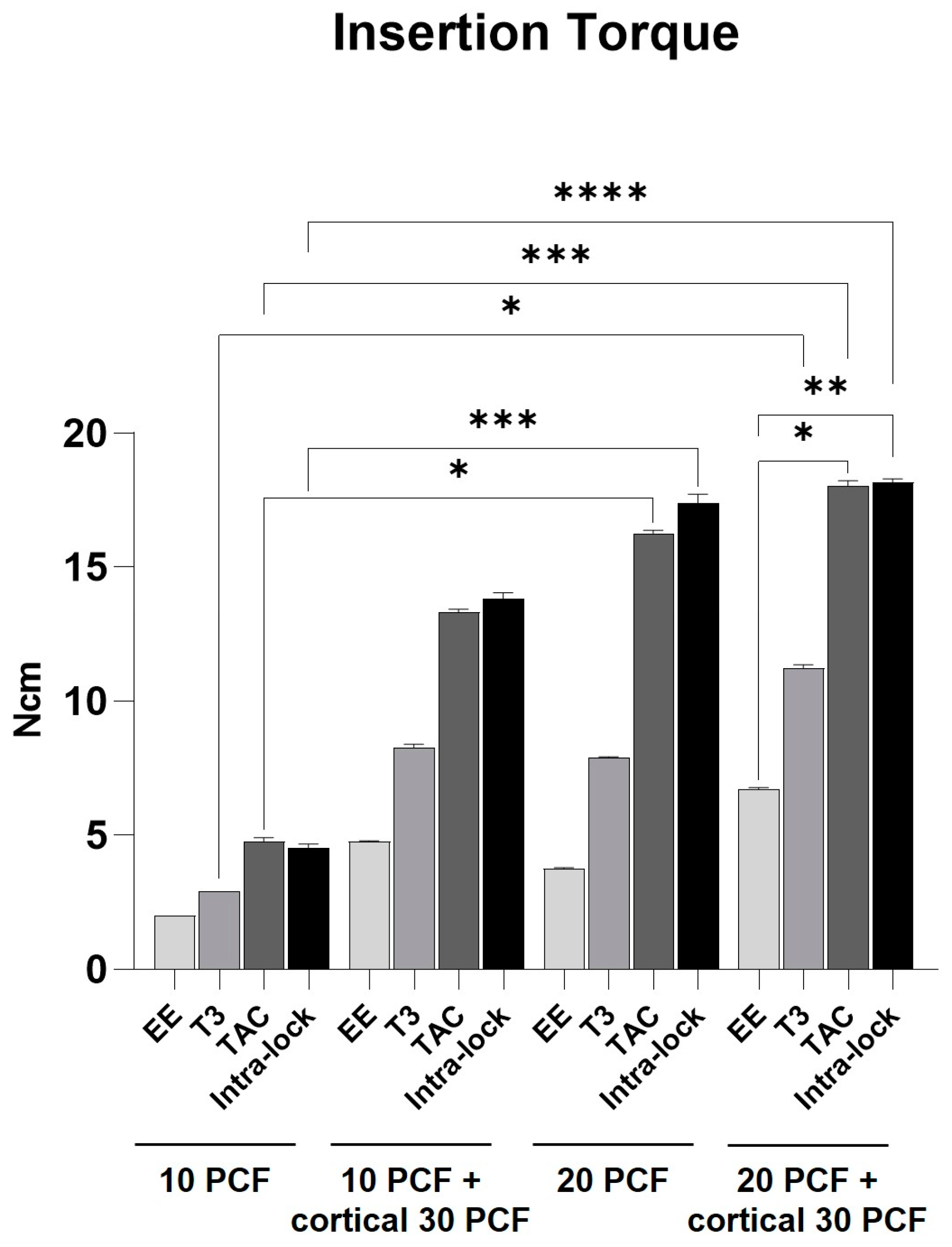
The IT values of all the implants tested proportionally increased with the increase of the polyurethane density and with the presence of the cortical sheet, especially for TAC and Intra-lock implants that showed statistically significant higher values in both the thickest sheets when compared to a 10 PCF density sheet without the cortical. Indeed, the lowest values were reported by EE implants in the 10 PCF density sheet without the cortical sheet (2 Ncm), while the highest values for TAC and Intra-lock implants were in the 20 PCF density sheet with the cortical sheet (18.6 Ncm).
There was not a significant difference among IT values of the four implants tested in the same polyurethane condition, except for TAC and Intra-lock implants in comparison with EE implants when inserted in the 20 PCF density sheet with the cortical sheet, although in all the experimental conditions the first two implants have always showed higher results. Moreover, also T3 implants reported statistically significant higher values in the thickest sheet in comparison with the 10 PCF density sheet without the cortical sheet, even if the highest IT value was slightly sufficient (11.8 Ncm).
Overall, the worst results were found in EE implants when tested in all the polyurethane conditions. The highest value reached by this implant was 6.9 Ncm in the 20 PCF density sheet with the addition of the cortical sheet (
Figure 11).
3.2. Removal Torque
The RT values were proportional to the sheet densities and to the presence of the cortical sheet as well. In fact, all the implants, except for EE, have shown significantly higher results in presence of the cortical sheet on the 10 and 20 PCF density sheets.
The highest values were always expressed by TAC and Intra-lock implants, with a RT of 10.8 Ncm for TAC implants and 13.7 Ncm for Intra-lock ones in the 20 PCF density sheet with the cortical sheet. On the other hand, the lowest results were always reported by EE and T3 implants, with 2 Ncm for both in the 10 PCF density sheet without the cortical sheet. In particular, Intra-lock implants showed statistically significant higher values in respect to EE implants in almost all the conditions, except for the 10 PCF density sheet without the cortical sheet, as well as TAC implants in the 20 PCF density cortical sheet without the cortical sheet.
The RT values were always lower than the corresponding IT values for all the implants tested, with the highest difference registered for TAC implants (
Figure 12).
3.3. Resonance Frequency Analysis
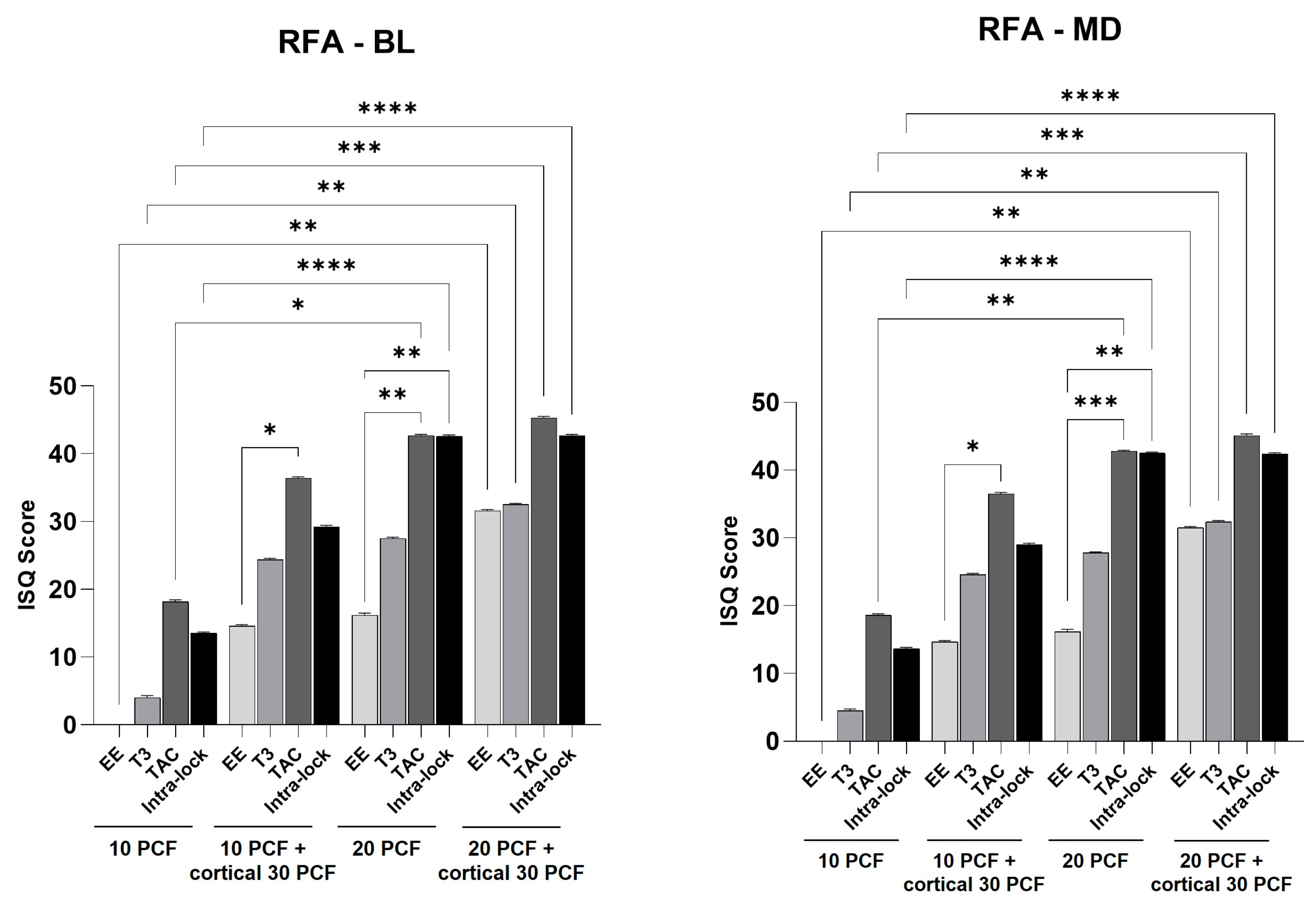
The BL and MD RFA values were expressed by the Implant Stability Quotient (ISQ). TAC and Intra-lock values never showed significant differences, but they were always higher in respect to EE and T3 implants, reaching statistical significances in the 10 PCF density with the cortical sheet (TAC implants) and the 20 PCF density sheet without the cortical sheet (Intra-lock and TAC implants) in respect to EE implants.
The highest values were reported by TAC implant in the 20 PCF density sheet with the cortical sheet (46 ISQ), while in the lowest density sheet (10 PCF without the cortical sheet), the EE implants could be not even measurable by the device.
All the ISQ values registered by the four implants in the thickest sheet were significantly higher in respect to the corresponding values found in the lowest density sheet (
Figure 13).
4. Discussion
The implant design and the surgical technique are important parameters that can be improved, possibly influencing implant stability [
4]. Nowadays several novel implant designs that may facilitate the achievement of initial implant PS have been proposed [
13]. Nevertheless, the effect of the apical portion on the initial implant stability has not been completely elucidated yet [
9,
10,
20].
In relation to this, homogeneous synthetic solid rigid polyurethane foam sheets have been produced as an alternative test medium for human cancellous/cortical bone. Even though polyurethane foam cannot perfectly replicate the same human bone structure, the ASTM F-1839-08 standard stated that it exhibits mechanical properties in the range of human cancellous bone. Artificial bones are regularly used to perform pre-clinical tests on dental implants and several studies have already shown findings on the influence of the threads design on PS [
15,
16,
21].
For example, Romanos et al. [
20] in a similar study on polyurethane foam sheets reported that the apical implant stability contributed to nearly 30–43% of the entire implant stability and thus it was clear that the apical portion of an implant may play a fundamental role. In particular, their study evaluated the role of the apical threads design on implant stability after being placed in type II dense artificial bone (D2). The implants were divided into two groups: a test group (with the aim to evaluate the apical stability of implants inserted with only 3 mm of the apical portion into the artificial bone and without lateral contact between the residual implant length and the osteotomy walls) and a control group (for the assessment of full implant stability with implants inserted with full contact between the osteotomy walls and the implant surface). Another polyurethane in vitro study was conducted by Sciasci et al. [
22] to evaluate the mechanical behavior of geometric changes in the apex region of dental implants. Implants without apical cut (Wc), apical bi-split cut, apical tri-split cut, apical quadri-split cut (Qs), and without any modifications, have been evaluated. At the end it was demonstrated that implants without any modifications and Qs had shown a significant increase of the IT, but those without any modifications and Wc had the greatest ISQ values. In this way, the authors concluded that the proposed geometries at the apical third of implants have greatly influenced their IT and PS. In animal experimental studies as well [
8], it was reported that the apical portion design of implants had an effect on PS and on the amount of mineralized bone present at the implant-bone interface. In particular, immediately loaded fully tapered implants with deep apical threads had similar high survival rates and bone-to-implant contact when inserted in healed sites and fresh extraction sockets [
13].
In the present study, T3 conical tapered implants seemed to have an insufficient IT and PS in all the experimental conditions, reporting higher results only in the 20 PCF density sheet with the cortical sheet (11.8 Ncm and 33 ISQ for the IT and RFA, respectively) and, together with EE cylindrical implants, registering slightly measurable results in the lowest density sheet. Indeed, the EE macro-geometry appeared to not properly suit the post-extraction condition and thus to neither allow implant stabilization nor the correct implant positioning into the osteotomy. All the tested measurements confirmed this data with very low IT, RT, and RFA values in all the artificial bone densities. The cylindrical design and the apical shape of these implants may be more suitable for higher mandibular symphysis densities (D1–D2) and less for low-quality and quantity of bone, like in post-extraction sites, where implant orientation is also important to consider. The T3 implant shape helped these implants to improve some parameters showed by the EE design, but the round apex did not suit with the post-extraction condition, because a spinning effect that stripped the osteotomy bottom was noticed during the insertion in the 20 PCF density sheets, when the operator had to force the implant insertion, thereby jeopardizing the IT and RFA values. Thus, a high cutting design was needed to increase implant penetration without stripping and spinning, and without the loss of all the mechanical characteristics. Indeed, in a finite elements study on the possible relationship between implant apex and maxillary sinus floor, Yan et al. [
23] showed that the different positions of the apex (in contact with, piercing or breaking through bone) had an important effect on the implant PS.
Moreover, other clinical studies investigated the influence of a cutting flute shape on implant stability, reporting a higher PS for implants without cutting flutes and an important effect to withstand lateral loads with the straight flute design [
11], whereas other ex vivo works compared the PS of implants with apically located screw threads or a flat tip, demonstrating a significantly higher stability for implants with apically located screw threads [
9]. These and other authors [
8,
10] concluded that factors, such as implant geometry (also at the implant tip), should be taken into account for biomechanical evaluations. In this regard, the present in vitro study observed a clear different behavior between old generation apical shapes and the latest generation ones tested in post-extraction conditions. In particular, the threads design and the conical macro-geometries of TAC and Intra-lock implants showed a better stability in anchoring the polyurethane material (up to 46 ISQ), even during the first inserting rotations, and the particular tip designs resulted to be favorable in the management of the direction, stability, and depth of the implants.
Jimbo et al. [
12] evaluated implants with two different apical geometries, one with a cutting flute design and the other with a self-tapping design. These authors found that implants with the modified cutting flute design had a significantly reduced IT with an improved organization of the peri-implant bone in an animal study. In the present work, the RFA values were proportional to the IT values, thus TAC and Intra-lock implants always presented a higher PS, even if in the lowest density sheet the apical portion was able to direct the insertion of the implant, but not to guarantee a good initial stability and an intimate contact of the fixture to the artificial bone (19 and 14 ISQ for TAC and Intra-lock, respectively). In any case, in the other experimental conditions their apical and threads designs allowed a fast and efficient implant insertion and a good contact with the material, progressively increasing the IT with the thickness of the sheet (up to 18.6 Ncm in the 20 PCF density sheet with the cortical sheet). The RT values were always lower than the corresponding IT with higher results for Intra-lock implants (up to 13.7 Ncm in the 20 PCF density sheet with the cortical sheet). Overall, although the only apical portion of TAC and Intra-lock implants did not guarantee an adequate ISQ value to allow the immediate loading of these implants, it showed an ISQ score that adequately favored the implant insertion until its final placement and reduced micromovements, which may promote osseointegration. These findings could be clinically relevant, because they proved that the apical morphology allows a good bone-to-implant contact from the first mm of insertion, without compromising the artificial bone structure, stressing the implant connection or the insertion at high IT values.
Nevertheless, the in vitro nature of this study evidently constitutes a strong limitation, although it provided substantial in vitro mechanical knowledge on the influence of different apical and thread implant shapes on PS. There is need of further experimental animal and clinical studies to corroborate these results on implant stability with histomorphometric and biomechanical measurements that also reflect the condition of the peri-implant bone [
10,
11,
13]. In addition, the IT, RT, and RFA values expressed by different implants may be influenced in vivo by the biological conditions and by multiple variabilities that could affect the density and nature of the bone, such as the presence of graft materials or the concurrent medical conditions of the patient affecting bone density.
In future, it would be interesting to verify if high ISQ values could be detected and maintained also in clinical situations up to the first month, a crucial moment for the biological stability, when ISQ values could lower and sometimes lead to implant stability failures. This data could allow the immediate loading of implants only by means of the RFA measurement, as an alternative to the current prosthetic loading protocols.

 ,
, 


