Article

The Role of Cisternostomy in the Management of Severe Traumatic Brain Injury: A Triple-Center Study

by
Manuel Encarnación Ramirez
1,
Ismael Peralta Baez
2,
Harmonie Marszal Mangbel’ Mikorska
3,
Jeff Natalaja Mukengeshay
4,
Renat Nurmukhametov
1,
Matias Baldoncini
5,
Jesus Lafuente
6,
Carlos Ovalles
7,
Michael David López Santos
8,
Gennady E. Chmutin
1 and
Nicola Montemurro
9,*
1
Department of Neurosurgery, RUDN University, 121359 Moscow, Russia
2
Department of Neurosurgery, Hospital Regional Alejandro Cabral, San Juan de la Maguana 72000, Dominican Republic
3
Department Neurosurgery, Centre Hospitalier Initiative Plus, Kinshasa 0364, Democratic Republic of the Congo
4
Department Neurosurgery, Clinique Ngaliema, Kinshasa 3089, Democratic Republic of the Congo
5
Laboratory of Microsurgical Neuroanatomy, School of Medicine, University of Buenos Aires, Buenos Aires B1430, Argentina
6
Spine Center Hospital del Mar, Sagrat Cor University Hospital, 08029 Barcelona, Spain
7
General Hospital 450, Durango 34030, Mexico
8
Hospital Regional Universitario José María Cabral y Baez, Santiago 72000, Dominican Republic
9
Department of Neurosurgery, Azienda Ospedaliero Universitaria Pisana (AOUP), University of Pisa, 56100 Pisa, Italy
*
Author to whom correspondence should be addressed.
Surgeries 2023, 4(2), 283-292; https://doi.org/10.3390/surgeries4020029
Submission received: 11 April 2023 / Revised: 1 June 2023 / Accepted: 6 June 2023 / Published: 7 June 2023

Abstract

:
Background: Traumatic brain injury (TBI) is a critical public health problem worldwide, constituting a major cause of mortality and morbidity for people of all ages, but especially in the younger population. Decompressive craniectomy (DC) and cisternostomy are surgical procedures commonly used in the management of severe TBI, but their effectiveness in improving outcomes remains controversial. Methods: We conducted a prospective longitudinal study on patients who underwent surgical treatment for severe TBI between 2021 and 2022. The extended Glasgow outcome scale (GOS-E) was used to assess clinical outcome at 2 weeks, 3 months, and 6 months after surgery. Results: The study included 30 patients (21 men and 9 women) who met the inclusion criteria. Among them, 24 patients (80%) underwent DC combined with cisternostomy, while 6 patients (20%) underwent cisternostomy alone. The initial Glasgow Coma Scale (GCS) score at admission ranged from 4 to 8 points, with an average score of 5.9. The overall mortality and overall morbidity was 13.3% and 20%, respectively. The mortality rate was 12.5% and 16.7% in the DC + cisternostomy group and in the cisternostomy alone group, respectively. No statistically significant difference was seen between the two groups in terms of mortality, morbidity and favorable outcome at 2 weeks, 3 months and 6 months. Conclusions: Our preliminary multi-center study shows a good clinical outcome in patients who underwent DC + cisternostomy or cisternostomy alone in both early and long-term follow-up. Larger multi-center randomized trials are needed to establish the effectiveness of cisternostomy in the management of TBI.

1. Introduction

Traumatic brain injury (TBI) is a critical public health problem worldwide and a major cause of mortality and morbidity for people of all ages, but especially in the younger population [1,2]. Statistical projections show that TBI could become one of the major causes of death and disability by the year 2030 [1].
Decompressive craniectomy (DC) has long been considered to be the treatment of choice to reduce intracranial pressure (ICP) in patients with refractory intracranial hypertension after severe TBI [3]. The DECRA trial, the largest randomized trial in diffuse TBI, failed to show the effectiveness of DC in adults with severe TBI [4], even if subsequent studies established the role of DC when ICP continues to increase to values ≥20 mm Hg [5,6]. A secondary analysis of the RESCUEicp Randomized Clinical Trial [6] reported that patients with post-traumatic refractory intracranial hypertension who were surgically treated had a sustained reduction in mortality and higher rates of vegetative state, severe disability and moderate disability at 24 months compared to the medical group. Although DC brings the ICP to atmospheric pressure, it does not counteract the intracerebral pressure, which causes severe brain swelling and herniation.
Cisternostomy, which has been routinely used in aneurysm surgeries and skull base tumors, has recently been reintroduced as an adjuvant surgical procedure for reducing brain edema and resistant intracranial hypertension [7]. Cisternostomy is defined as opening the basal cisterns to atmospheric pressure and the drainage of the subarachnoid basal cistern. This technique helps to reduce the ICP in severe head trauma as well as other conditions when so-called sudden “brain swelling” troubles the surgeon [8]. Cisternostomy is a technique that incorporates knowledge of skull base surgery and microvascular surgery, as, by opening the basal cisterns to atmospheric pressure, this technique could decrease the ICP due to a backshift of the cerebrospinal fluid (CSF) from the swollen brain to the cisterns through the Virchow Robin spaces [9,10]. Traumatic subarachnoid hemorrhage causes cisternal blockage, which, in accordance with the CSF-shift process, causes an increase in cisternal pressure above brain parenchymal pressure. As a result, there is a pressure gradient between the brain parenchyma and the cistern, which is at a lower pressure [10]. According to recent research, the development of edema may also be linked to CSF entry into the brain parenchyma through the low-resistance para-arterial space and decreased interstitial fluid efflux, or a combination of the two processes [8,11]. As a result, after a severe TBI, CSF may be transferred from the cerebral cisterns to the brain, resulting in severe brain swelling. A traumatic subarachnoid bleed, which is frequently linked to severe TBI and results in a pressure gradient that is higher in the cisterns and lower in the brain, may be one of the causes of this rapid shift [12].
Even if Timofeev et al. [12] demonstrated that an external ventricular drain (EVD) is effective in rapidly reducing ICP, almost half of their patients experienced a later increase in ICP (20 mm Hg) despite the EVD; therefore, some considerations should be made about the decision to place a cisternal drain rather than a ventricular drain, which is currently the standard procedure in severe TBI [13,14]. The role of cisternostomy in the management of TBI is a topic of ongoing research and debate. To better understand the potential benefits and risks of this procedure, a triple-center study was conducted to evaluate the outcomes of patients who underwent cisternostomy for TBI. This study involved the collaboration of three medical centers, each one with important experience in the management of TBI.
The primary objective of this paper is to analyze the role of cisternostomy in the management of TBI. Specifically, we will discuss surgical indications for cisternostomy; the surgical technique used; and the mortality, morbidity and the surgical outcome of patients who underwent cisternostomy in combination with DC or alone in severe TBI to better understand how to manage patients with severe TBI.

2. Materials and Methods

2.1. Patients Selection

We performed a prospective longitudinal study in a cohort of patients who were surgically treated for severe TBI between 2021 and 2022 in one of these three different medical centers: RUDN University (Moscow, Russia), General Hospital (Durango, Mexico) and Hospital Regional Universitario José María Cabral y Baez (Santiago, Dominican Republic). The inclusion criteria were patients between 18 and 70 years old without severe systemic disease, GCS ≤ 8, presenting with a concomitant predominantly unilateral mass effect such as an acute subdural hematoma (SDH) greater than 20 mm or a midline shift greater than 5 mm on a computed tomography (CT) scan, which are indications for DC. The exclusion criteria were patients with GCS 3, moderate and mild TBI, axonal diffuse injury, cardiopulmonary instability, infarcts of brainstem and multi-organ injuries. Table 1 shows all the details.
A consent form was signed according to the legal responsibilities, and the procedure were performed within a maximum of 4 h after emergency room admission. The extended Glasgow outcome scale (GOS-E) at 2 weeks and 3 and 6 months after surgery was used to determine the clinical outcome. The pre-operative and post-operative neurological exams were evaluated. Those patients that could not be contacted by phone were excluded as well, due to a lack of clinical/radiological follow-up. Patients were treated according to the protocol for the management of severe TBI, in line with the current recommended guidelines. All patients were sedated and mechanically ventilated.
The primary objective was to determine the clinical outcome of these patients surgically treated with DC and cisternostomy at two weeks and three and six months after a TBI.

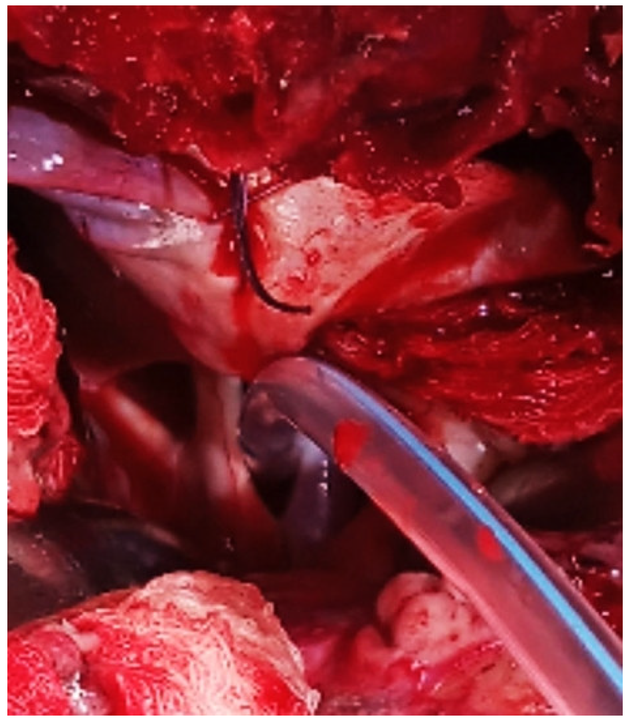
2.2. Surgical Procedure

For DC, a classic trauma flap with an inverted question mark incision measuring no less than 12 × 15 cm or 15 cm in diameter was created [14]. The principal steps for cisternostomy can be summarized in the following ten steps: (1) drilling the sphenoid ridge until it meets the meningeal–orbital arteries; (2) the identification of the superior orbital fissure and the meningo-orbital band; (3) the division of the meningo-orbital band, which marks the lateral edge of the superior orbital fissure; (4) exposing the inferior aspect of the anterior clinoid process; (5) the elevation of the temporal lobe from the superior orbital fissure exposing the anterior clinoid process; (6) the elevation of the frontal lobe from the anterior clinoid process, which can easily be removed; (7) opening the dura; (8) the identification and opening of the sylvian, chiasmatic, optico-carotid and lateral carotid cisterns; (9) approaching the Liliequist membrane through the optico-carotid window or the lateral carotid window; (10) drilling the posterior clinoid process in the case of difficulty in opening the Liliequist membrane [15].
The main step in the procedure consists, after the evacuation of clots and contusions, if any, of CSF being released from the basal cisterns by opening the sylvian, chiasmatic, lateral carotid cisterns, Liliequist membrane and prepontine cistern. A saline wash ensures the satisfactory removal of subarachnoid blood. A silicone drain is placed in the prepontine cistern (Figure 1) and is taken out of the wound for CSF drainage in the post-operative period. The CSF diversion allows the brain to relax, and it is limited to 150–200 mL per day by maintaining the drainage bag at the level of the head for 3–5 days.

2.3. Data Collection

Clinical data extracted from the records included the patient demographic data, GCS at presentation, pupillary size and reactivity and duration of surgery. The early clinical outcome measures studied were the post-operative duration of ventilation, the duration of intensive care unit (ICU) stay, GCS at discharge from ICU, early mortality (during ICU stay) and length of hospital stay. The long-term clinical outcome (6 months after surgery) was assessed by using the extended Glasgow outcome scale (GOS-E) which was dichotomized as “favorable” (GOS-E was ≥ 5) and “unfavorable” (GOS-E < 5).
CT scans at admission were analyzed to evaluate: (1) midline shift in millimeters; (2) presence and size of a mass lesion such as SDH; and (3) surgical complications such as neurovascular injuries, iatrogenic contusions and post-surgical hematomas.
The study was conducted in accordance with the Declaration of Helsinki and approved by the Institutional Review Board of Department of Neurosurgery, Hospital Regional Alejandro Cabral, San Juan de la Maguana, Dominican Republic (n° 05/2020).
Statistical analysis was performed using SPSS software (version 25.0, IBM Corp, Armonk, NY, USA). The descriptive analysis was performed for all variables, and the results were expressed as the mean ± standard deviation (SD) or median (interquartile range, IQR). The comparison between groups was performed using the chi-squared test or Fisher’s exact test for categorical variables and the Student’s t-test or Mann–Whitney U test for continuous variables, as appropriate. The statistical significance was defined as p-value < 0.05.

3. Results

The present study included a total of 30 patients (21 men and 9 women) who met the inclusion criteria. A total of 24 patients (80%) underwent DC + cisternostomy, whereas 6 patients (20%) underwent cisternostomy exclusively. GCS at admission ranged from 4 to 8 points, with an average score of 5.9. All patients who underwent cisternostomy had also an acute SDH, which was evacuated. The mortality rate was 12.5% (3 patients) and 16.7% (1 case) in the DC + cisternostomy group and in the cisternostomy alone group, respectively. The overall mortality was 13.3%, with a morbidity rate of 50% in patients with initial GCS scores of 4 and a mortality rate of 16.7% in patients with GCS scores of 6.
Our results show that there was a clinical relationship between mortality and morbidity outcome and pupil examination at the admission in the emergency room. As you might think, patients with midposition and fixed pupils (BP) reported 100% mortality, whereas patients with bilateral minimally reactive dilated pupils (BDU) and bilateral reactive pupils (BR) reported a mortality rate of 50% and 20%, respectively. Patients with anisocoria (ANSC) reported a mortality rate of 5.2%. Similarly, morbidity rate is correlated with preoperative GCS and pupil examination at admission in the emergency room. Patients with BDU reported a morbidity rate of 50%. Figure 2 shows all the details.
Surgically, the Liliequist membrane was opened in a total of 18 patients (60%). In particular, the Liliequist membrane was opened in 14 patients (58.3%) who underwent DC + cisternostomy and in 4 patients (66.7%) who underwent cisternostomy alone. The mortality and surgical morbidity rates were 12.5% and 20.8% in the DC + cisternostomy group of patients, respectively, and 16.7% in the cisternostomy alone group of patients. A favorable (GOS-E ≥ 5) outcome increased significantly up to 87.5% and 83.3% in the DC + cisternostomy group and in the cisternostomy alone group, respectively, at the 3-month follow-up. No statistically significant difference was seen in the two groups in terms of mortality, morbidity and favorable outcome at 2 weeks, 3 months and 6 months (Table 2). The mean length of hospital stay was 12.2 days, with a mean ICU stay of 4.8 days and a mean time to the successful extubation of patients of 2.9 days.

4. Discussion

TBI is a major cause of morbidity and mortality all over the world, especially in developing countries. Focused injuries, such as hematomas, brain contusions and skull fractures, are often caused by direct TBI [16]. According to recent research, the development of edema is also linked to CSF entry into the brain parenchyma via the low-resistance para-arterial space, decreased interstitial fluid outflow or a combination of the two in cases of severe TBI. After a severe TBI, the hydrodynamics of the brain is disrupted, and this is accompanied by diffuse secondary injuries to this process. The recent hypothesis that the glymphatic removal of excess interstitial fluid avoids a second lesion, allowing the passage of CSF from the cerebral cisterns to the brain after TBI is well described [15]. Cisternostomy has since been suggested to enable the opening of the basal cisterns to atmospheric pressure and decrease levels of cerebral edema, thus allowing for the successful replacement of bone flaps [17]. Although cisternostomy for the management of traumatic brain injuries has increasingly been recognized during the past ten years, there have not been extensive randomized controlled experiments on cisternostomy up until this point [18]. However, a 2022 study comparing cisternostomy to craniotomy in a sample of 50 patients showed a significant reduction in ICP, satisfactory GOS ratings and a low rate of complications, supporting the use of cisternostomy [18]. Cisternostomy has also been shown to reduce the time spent in the intensive care unit (ICU), the number of days spent on a ventilator and the requirement for a cranioplasty [18]. Considering this, basal cisternostomy seems to be a promising surgical operation, but its execution is still challenging and requires strong surgical expertise in procedures involving the skull [18].
Although other pathologies can also increase the ICP [19,20,21,22], cisternostomy is a potential surgical treatment for patients with TBI-related prolonged ICP elevation [21]. A previous case report also demonstrated the effect of cisternostomy in decreasing ICP and its potential role as a surgical treatment for pseudotumor cerebri [23].
In 2013, Cherian and Munakomi [24] first introduced the concept of cisternostomy in severe TBI as a more elegant procedure that can replace the DC, with low morbidity and mortality, even if a learning curve is needed. Cherian and Munakomi [24] similarly presented the results of a single-surgeon series of patients treated with DC + cisternostomy and cisternostomy alone in patients with severe TBI, with a mean GOS at 6 weeks of 3.7 and 3.9, respectively.
It is challenging to open cisterns in a small brain because, in diffuse TBI, the cisterns are frequently compressed, and the CSF flow is changed [25]. For the prognosis and mortality estimation of TBI, several categorization scores, including Marshall and Rotterdam, take the cisternal condition into account. The basal cisternae opening is a common technique performed in skull base and vascular neurosurgeries. It is useful for brain relaxation and helps improve CSF flow and brain hydrodynamics [9]. Nevertheless, after the opening of the inter-optic, optic-carotid and lateral carotid cisterns, the brain becomes lax. This happens as a result of a reversed cisternal pressure gradient, which allows CSF to flow back into the cisterns and lowers the ICP [9,14,26]. Hoz et al. [8] proposed a classification system for cisternostomy in terms of its mechanisms of action. Cisternostomy may serve as either an outflow-one way or inflow-two way corridor, the so-called cisternostomy proper, which is further subcategorized into convexity and basal cisternostomy. As for its indications, cisternostomy can either be a planned or unplanned procedure. A planned cisternostomy is often applied in skull base and microvascular surgeries, whereas unplanned cisternostomy is used to control intra-operative brain swelling. Cherian et al. [26] described a case series of cisternostomy for the treatment of TBI, comparing it with DC, and they achieved good results. Cisternostomy presented decreased morbidity and mortality rates when compared with DC. The idea of offering cisternostomy to TBI patients is related to the discovery of the glymphatic system. The glymphatic system is a network of perivascular channels that promote the entry and exit of substances within the CNS [27,28]. The interstitial fluid collected within perivenous spaces is then drained to the cervical lymphatic circulation [29]. Basal cisternostomy is a safe and feasible microsurgical procedure for the management of TBI that can effectively evacuate CSF and relax the brain [30]. Compared to DC, basal cisternostomy depends on the use of microsurgical techniques. Cisternostomy, a novel technique that incorporates knowledge of the skull base and microvascular surgery, has been proposed to decrease brain swelling, mortality and morbidity [31,32,33].
Goyal et al. [34] published a study with a cohort of nine patients who underwent both cisternostomy and DC, showing a significant difference between opening and closing parenchymal pressures, supporting a CSF-shift edema hypothesis and suggesting that both cisternostomy and DC should be provided for head injuries with severe edema.
Chandra et al. [33] compared outcomes of cisternostomy with DC in a randomized controlled trial and studied the effectiveness of cisternostomy in decreasing cerebral edema in 50 patients with TBI, reporting a mortality rate of 32% in the cisternostomy group and a mortality rate of 44% in the DC group. Masoudi et al. [25] reported a case of severe TBI in a 13-year-old boy with diffuse brain edema, left frontal contusion and posterior interhemispheric subdural hematoma, with a 26 mmHg mean value of ICP that was surgically treated using the cisternostomy technique. The patient showed progressive improvement in the neurological conditions in the following hours and completely recovered at the 3-month follow-up. Similarly, Giammattei et al. [13] reported a case of a of 50-year-old man with refractory intracranial hypertension successfully managed with cisternostomy.
In a recent study, Partiban and colleagues [35] performed cisternostomy alone rather than combining both cisternostomy and DC, showing a favorable outcome in the cisternostomy group (77.8%) compared to the cisternostomy + DC group (72.7%). In addition, Kumar et al. [7] reported in their study on 40 patients a higher 30-day mortality rate for the cisternostomy + DC group (66.6%) compared to the DC alone group (32.2%), with a mean GCS at discharge that was better in the cisternostomy + DC group (11.7 ± 2.9) compared to the DC alone group (10.5 ± 3.7). Abdulla et al. [36] drew attention to the fact that complications linked with cisternostomy in TBI are not well documented, as cisternostomy in aneurysm surgery has caused iatrogenic vascular damage perforation, due to poor technique [37,38]. However, cisternostomy seems to decrease the number of days of ventilator support and the length of ICU stay and avoids the need for second surgery in the form of cranioplasty and its associated morbidity [33]. Cisternostomy could be an appropriate alternative to DC for the management of intracranial hypertension in patients with severe TBI.
We observed that the association of opening the Liliequist membrane with the surgical procedure resulted in a higher percentage of favorable clinical outcome (53.3%). Our study also highlights the importance of the GCS score and pupil examination at admission in the emergency room in predicting mortality and morbidity. In terms of the treatment group, we found no significant difference in mortality rate, surgical morbidity and clinical outcome between patients undergoing DC + cisternostomy and those undergoing cisternostomy alone. However, patients undergoing DC + cisternostomy had a higher rate of good outcomes at discharge, with 29.2% compared to 0% in the cisternostomy alone group. However, this difference was not statistically significant. According to Cherian et al. [24], the average time for cisternostomy from dural opening is approximately 20 min, with extra time needed in the case of posterior clinoid drilling or any other additional unforeseen circumstances associated with severe head injuries. In another study [33], the mean duration of surgery was 3.28 ± 0.52 h in the cisternostomy group and 2.90 ± 0.38 h in the DC group. The use of a surgical microscope or exoscope is preferred for magnification when working around the cisterns, and also, the knowledge of skull base surgical techniques is of aid when working on these spaces [39,40,41,42].
Recently, Servadei et al. [43] warned us about the use of cisternostomy for TBI as a new procedure, but without sufficient evidence to warrant its usage outside of research settings. Neurosurgery and innovation have been closely related since its start. Innovations such as the operating microscope, CT and MR imaging and endoscopy put us at the forefront of technological advancements, and they quickly translated into better patient outcomes, including safety [31]. Telehealth interventions and programs are promising avenues for healthcare delivery and may represent windows of opportunity, combining traditional treatment with rehabilitation to increase symptom management self-efficacy among TBI patients during recovery [44,45]. Prospective, randomized, controlled studies that will clarify whether cisternostomy is effective or not are needed.

Limitations of the Study

The main limitations of the study are the small sample size, the non-randomization of the study and the impossibility of ICP measurement. The measurement of ICP should be performed for better monitoring and to avoid excessive drainage. Due to the limited number of patients, statistical analysis can be performed, but its value is low. Further clinical studies with larger cohorts are needed to determine the role of cisternostomy in TBI.

5. Conclusions

Cisternostomy, a novel technique that incorporates knowledge of the skull base and microvascular surgery, has been proposed to decrease brain swelling, mortality and morbidity after severe TBI [31]. We believe that the procedure of adjuvant cisternostomy is safe and feasible in the context of severe TBI. The opening of the Liliequist membrane was performed in a significant proportion of our patients and was associated with a higher rate of complete clinical recovery without adding morbidity to the surgical procedure. Our preliminary multi-center data indicate a clinically favorable outcome both in early and long-term follow-up in patients with severe TBI who underwent DC + cisternostomy or cisternostomy alone. The results of this study have important implications for the treatment of patients with severe TBI. By providing a comprehensive analysis of the role of cisternostomy in TBI management, this paper aims to contribute to the ongoing discussion surrounding this emerging treatment option. Basal cisternostomy appears to be a promising surgery, but its execution remains difficult and demands proficient surgical skills in skull-based procedures. To fully determine the efficacy of cisternostomy in the therapy of neurotrauma, further randomized and controlled studies are necessitated. Our study shows that the treatment of traumatic acute subdural hematoma with DC and cisternostomy when needed, with or without the opening of the Liliequist membrane, can lead to a lower mortality rate compared to subdural hematoma evacuation alone. Larger multi-center randomized trials are needed to establish the effectiveness of cisternostomy in the management of TBI.

Author Contributions

Conceptualization, M.E.R., I.P.B., H.M.M.M. and N.M.; methodology, N.M. and M.E.R.; validation, M.E.R., I.P.B., C.O., M.D.L.S., G.E.C. and N.M.; formal analysis, M.E.R., J.N.M., R.N., M.B., J.L. and N.M.; investigation, M.E.R., I.P.B. and H.M.M.M.; data curation, N.M.; writing—original draft preparation, M.E.R., I.P.B., C.O., M.D.L.S. and N.M.; writing—review and editing, N.M.; visualization, M.E.R.; supervision, M.E.R. and N.M. All authors have read and agreed to the published version of the manuscript.

Funding

This research received no external funding.

Institutional Review Board Statement

The study was conducted in accordance with the Declaration of Helsinki and approved by the Institutional Review Board of Department of Neurosurgery, Russia Hospital Regional Alejandro Cabral, San Juan de la Maguana, Dominican Republic (n° 05/2020).

Informed Consent Statement

Informed consent was obtained from all subjects involved in the study.

Data Availability Statement

Not applicable.

Conflicts of Interest

The authors declare no conflict of interest.

References

  1. Mathers, C.D.; Loncar, D. Projections of global mortality and burden of disease from 2002 to 2030. PLoS Med. 2006, 3, e442. [Google Scholar] [CrossRef] [PubMed] [Green Version]
  2. Montemurro, N.; Santoro, G.; Marani, W.; Petrella, G. Posttraumatic synchronous double acute epidural hematomas: Two craniotomies, single skin incision. Surg. Neurol. Int. 2020, 11, 435. [Google Scholar] [CrossRef] [PubMed]
  3. Cooper, D.J.; Rosenfeld, J.V.; Murray, L.; Arabi, Y.M.; Davies, A.R.; D’Urso, P.; Kossmann, T.; Ponsford, J.; Seppelt, I.; Reilly, P.; et al. Decompressive craniectomy in diffuse traumatic brain injury. N. Engl. J. Med. 2011, 364, 1493–1502. [Google Scholar] [CrossRef] [PubMed] [Green Version]
  4. Chi, J.H. Craniectomy for traumatic brain injury: Results from the DECRA trial. Neurosurgery 2011, 68, N19–N20. [Google Scholar]
  5. Honeybul, S.; Ho, K.M.; Lind, C.R. What can be learned from the DECRA study. World Neurosurg. 2013, 79, 159–161. [Google Scholar]
  6. Kolias, A.G.; Adams, H.; Timofeev, I.S.; Corteen, E.A.; Hossain, I.; Czosnyka, M.; Timothy, J.; Anderson, I.; Bulters, D.O.; Belli, A.; et al. Evaluation of Outcomes Among Patients with Traumatic Intracranial Hypertension Treated with Decompressive Craniectomy vs Standard Medical Care at 24 Months: A Secondary Analysis of the RESCUEicp Randomized Clinical Trial. JAMA Neurol. 2022, 79, 664–671. [Google Scholar] [CrossRef]
  7. Kumar, P.; Goyal, N.; Chaturvedi, J.; Arora, R.K.; Singh, P.R.; Shakya, J.; Rekapalli, R.; Sadhasivam, S.; Sihag, R.; Bahurupi, Y. Basal Cisternostomy in Head Injury: More Questions than Answers. Neurol. India 2022, 70, 1384–1390. [Google Scholar]
  8. Hoz, S.S.; Alramadan, A.H.; Hadi, A.Q.; Moscote Salazar, L.R. Cisternostomy in Neurosurgery: A New Proposed General Classification Based on Mechanism and Indications of the Cisternostomy Proper. J. Neurosci. Rural. Pract. 2018, 9, 650–652. [Google Scholar]
  9. Espaillat, A.; Encarnacion Ramirez, M.D.J.; Barrientos, R.; Nurmukhametov, R.; Peralta, I.; Coats, C.; Marmol, F.; Jimenez, O.; Fermin, R.; Valenzuela, S.; et al. Prevalece and Topography Anatomy of Dilated Virchow-Robin Spaces. World J. Med. Case Rep. 2022, 3, 11–20. [Google Scholar]
  10. Cherian, I.; Grasso, G.; Bernardo, A.; Munakomi, S. Anatomy and physiology of cisternostomy. Chin. J. Traumatol. 2016, 19, 7–10. [Google Scholar] [CrossRef]
  11. Iliff, J.J.; Wang, M.; Liao, Y.; Plogg, B.A.; Peng, W.; Gundersen, G.A.; Benveniste, H.; Vates, G.E.; Deane, R.; Goldman, S.A.; et al. A paravascular pathway facilitates CSF flow through the brain parenchyma and the clearance of interstitial solutes, including amyloid β. Sci. Transl. Med. 2012, 4, 147ra111. [Google Scholar] [CrossRef] [Green Version]
  12. Timofeev, I.; Dahyot-Fizelier, C.; Keong, N.; Nortje, J.; Al-Rawi, P.G.; Czosnyka, M.; Menon, D.K.; Kirkpatrick, P.J.; Gupta, A.K.; Hutchinson, P.J. Ventriculostomy for control of raised ICP in acute traumatic brain injury. Acta Neurochir. Suppl. 2008, 102, 99–104. [Google Scholar]
  13. Giammattei, L.; Messerer, M.; Oddo, M.; Borsotti, F.; Levivier, M.; Daniel, R.T. Cisternostomy for Refractory Posttraumatic Intracranial Hypertension. World Neurosurg. 2018, 109, 460–463. [Google Scholar] [CrossRef]
  14. Carney, N.; Totten, A.M.; O’Reilly, C.; Ullman, J.S.; Hawryluk, G.W.; Bell, M.J.; Bratton, S.L.; Chesnut, R.; Harris, O.A.; Kissoon, N.; et al. Guidelines for the management of severe traumatic brain injury, fourth edition. Neurosurgery 2017, 80, 6–15. [Google Scholar] [CrossRef]
  15. Grasso, G.; Cherian, I. Cisternostomy for Traumatic Brain Injury: A New Era Begins. Bull. Emerg. Trauma 2016, 4, 119–120. [Google Scholar]
  16. Badke, G.L.; Araujo, J.L.V.; Miura, F.K.; Guirado, V.; Saade, N.; Paiva, A.L.C.; Avelar, T.M.; Pedrozo, C.A.G.; Veiga, J.C.E. Analysis of direct costs of decompressive craniectomy in victims of traumatic brain injury. Arq. Neuropsiquiatr. 2018, 76, 257–264. [Google Scholar] [CrossRef]
  17. Valle, D.; Villarreal, X.P.; Lunny, C.; Chalamgari, A.; Wajid, M.; Mahmood, A.; Buthani, S.; Lucke-Wold, B. Surgical Management of Neurotrauma: When to Intervene. J. Clin. Trials Regul. 2022, 4, 41–55. [Google Scholar]
  18. Iliff, J.J.; Chen, M.J.; Plog, B.A.; Zeppenfeld, D.M.; Soltero, M.; Yang, L.; Singh, I.; Deane, R.; Nedergaard, M. Impairment of glymphatic pathway function promotes tau pathology after traumatic brain injury. J. Neurosci. 2014, 34, 16180–16193. [Google Scholar] [CrossRef] [Green Version]
  19. Encarnacion Ramirez, M.; Barrientos Castillo, R.E.; Musa, G.; Nurmukhametov, R.; Igorevich, B.O. ‘Brain Tissue Out’ in a Train Struck Victim: A Case Report. Neurol. Neurobiol. 2022, 1, 2613–7828. [Google Scholar]
  20. Prasad, G.L.; Gupta, D.K.; Mahapatra, A.K.; Sharma, B.S. Surgical results of decompressive craniectomy in very young children: A level one trauma centre experience from India. Brain Inj. 2015, 29, 1717–1724. [Google Scholar] [CrossRef]
  21. Eraky, A.M.; Treffy, R.; Hedayat, H.S. Cisternostomy as a Surgical Treatment for Traumatic Brain Injury-Related Prolonged and Delayed Intracranial Pressure Elevation: A Case Report. Cureus 2023, 15, e37508. [Google Scholar] [CrossRef] [PubMed]
  22. Montemurro, N.; Benet, A.; Lawton, M.T. Julius Caesar’s Epilepsy: Was It Caused by A Brain Arteriovenous Malformation? World Neurosurg. 2015, 84, 1985–1987. [Google Scholar] [CrossRef] [PubMed]
  23. Eraky, A.M.; Treffy, R.; Hedayat, H.S. Cisternotomy and Liliequist’s Membrane Fenestration as a Surgical Treatment for Idiopathic Intracranial Hypertension (Pseudotumor Cerebri): A Case Report. Cureus 2022, 14, e31363. [Google Scholar] [CrossRef] [PubMed]
  24. Cherian, I.; Yi, G.; Munakomi, S. Cisternostomy: Replacing the age old decompressive hemicraniectomy? Asian J. Neurosurg. 2013, 8, 132–138. [Google Scholar] [CrossRef] [Green Version]
  25. Masoudi, M.S.; Rezaee, E.; Hakiminejad, H.; Tavakoli, M.; Sadeghpoor, T. Cisternostomy for Management of Intracranial Hypertension in Severe Traumatic Brain Injury; Case Report and Literature Review. Bull. Emerg. Trauma 2016, 4, 161–164. [Google Scholar]
  26. Cherian, I.; Bernardo, A.; Grasso, G. Cisternostomy for traumatic brain injury: Pathophysiological mechanisms and surgical technical notes. World Neurosurg. 2016, 89, 51–57. [Google Scholar] [CrossRef]
  27. Hutchinson, P.J.; Kolias, A.G.; Timofeev, I.S.; Corteen, E.A.; Czosnyka, M.; Timothy, J.; Anderson, I.; Bulters, D.O.; Belli, A.; Eynon, C.A.; et al. RESCUEicp Trial Collaborators. Trial of Decompressive Craniectomy for Traumatic Intracranial Hypertension. N. Engl. J. Med. 2016, 375, 1119–1130. [Google Scholar] [CrossRef] [Green Version]
  28. Jessen, N.A.; Munk, A.S.F.; Lundgaard, I.; Nedergaard, M. The glymphatic system: A beginner’s guide. Neurochem. Res. 2015, 40, 2583–2599. [Google Scholar] [CrossRef] [Green Version]
  29. Johnston, M.; Zakharov, A.; Papaiconomou, C.; Salmasi, G.; Armstrong, D. Evidence of connections between cerebrospinal fluid and nasal lymphatic vessels in humans, non-human primates and other mammalian species. Cereb. Fluid Res. 2004, 1, 2. [Google Scholar] [CrossRef] [Green Version]
  30. Encarnacion Ramirez, M.D.J.; Barrientos Castillo, R.E.; Vorobiev, A.; Kiselev, N.; Aquino, A.A.; Efe, I.E. Basal cisternostomy for traumatic brain injury: A case report of unexpected good recovery. Chin. J. Traumatol. 2022, 25, 302–305. [Google Scholar] [CrossRef]
  31. Grasso, G. Surgical treatment for traumatic brain injury: Is it time for reappraisal? World Neurosurg. 2015, 84, 594. [Google Scholar] [CrossRef]
  32. Volovici, V.; Haitsma, I.K. Cisternostomy in Traumatic Brain Injury: Time for the World to Listen-Cerebrospinal Fluid Release: Possibly the Missing Link in Traumatic Brain Injury. World Neurosurg. 2022, 162, 3–5. [Google Scholar] [CrossRef]
  33. Chandra, V.V.R.; Mowliswara Prasad, B.C.; Banavath, H.N.; Chandrasekhar Reddy, K. Cisternostomy versus Decompressive Craniectomy for the Management of Traumatic Brain Injury: A Randomized Controlled Trial. World Neurosurg. 2022, 162, e58–e64. [Google Scholar] [CrossRef]
  34. Goyal, N.; Kumar, P. Putting ‘CSF-Shift Edema’ Hypothesis to Test: Comparing Cisternal and Parenchymal Pressures After Basal Cisternostomy for Head Injury. World Neurosurg. 2021, 148, e252–e263. [Google Scholar] [CrossRef]
  35. Parthiban, J.K.B.C.; Sundaramahalingam, S.; Rao, J.B.; Nannaware, V.P.; Rathwa, V.N.; Nasre, V.Y.; Prahlad, S.T. Basal Cisternostomy—A Microsurgical Cerebro Spinal Fluid Let Out Procedure and Treatment Option in the Management of Traumatic Brain Injury. Analysis of 40 Consecutive Head Injury Patients Operated with and without Bone Flap Replacement Following Cisternostomy in a Tertiary Care Centre in India. Neurol. India 2021, 69, 328–333. [Google Scholar]
  36. Abdulla, E.; Ravindra, J.; Rahman, S.; Rahman, M.M. Letter to the Editor Regarding “Cisternostomy in Traumatic Brain Injury: Time for the World to Listen. Cerebrospinal Fluid Release: Possibly the Missing Link in TBI”. World Neurosurg. 2022, 162, 203–204. [Google Scholar] [CrossRef]
  37. Marani, W.; Mannará, F.; Noda, K.; Kondo, T.; Ota, N.; Perrini, P.; Montemurro, N.; Kinoshita, Y.; Tsuji, S.; Kamiyama, H.; et al. Management of an Uncommon Complication: Anterior Choroidal Artery Occlusion by Posterior Clinoid Process Detected Through Intraoperative Monitoring After Clipping of Paraclinoid Aneurysm: 2-Dimensional Operative Video. Oper. Neurosurg. 2021, 21, E124–E125. [Google Scholar] [CrossRef]
  38. Horiuchi, T.; Yamamoto, Y.; Kuroiwa, M.; Rahmah, N.N.; Hongo, K. Pontine infarction caused by medial branch injury of the basilar artery as a rare complication of cisternal drain placement. J. Clin. Neurosci. 2012, 19, 592–593. [Google Scholar] [CrossRef] [Green Version]
  39. Montemurro, N.; Scerrati, A.; Ricciardi, L.; Trevisi, G. The Exoscope in Neurosurgery: An Overview of the Current Literature of Intraoperative Use in Brain and Spine Surgery. J. Clin. Med. 2021, 11, 223. [Google Scholar] [CrossRef]
  40. Lizana, J.; Montemurro, N.; Aliaga, N.; Marani, W.; Tanikawa, R. From textbook to patient: A practical guide to train the end-to-side microvascular anastomosis. Br. J. Neurosurg. 2023, 37, 116–120. [Google Scholar] [CrossRef]
  41. Rush, B.; Rousseau, J.; Sekhon, M.S.; Griesdale, D.E. Craniotomy Versus Craniectomy for Acute Traumatic Subdural Hematoma in the United States: A National Retrospective Cohort Analysis. World Neurosurg. 2016, 88, 25–31. [Google Scholar] [CrossRef] [PubMed] [Green Version]
  42. Encarnacion Ramirez, M.; Ramirez Pena, I.; Barrientos Castillo, R.E.; Sufianov, A.; Goncharov, E.; Soriano Sanchez, J.A.; Colome-Hidalgo, M.; Nurmukhametov, R.; Cerda Céspedes, J.R.; Montemurro, N. Development of a 3D Printed Brain Model with Vasculature for Neurosurgical Procedure Visualisation and Training. Biomedicines 2023, 11, 330. [Google Scholar] [CrossRef] [PubMed]
  43. Servadei, F.; Kolias, A.; Kirollos, R.; Khan, T.; Hutchinson, P. Cisternostomy for traumatic brain injury-rigorous evaluation is necessary. Acta Neurochir. 2020, 162, 481–483. [Google Scholar] [CrossRef] [PubMed] [Green Version]
  44. Montemurro, N. Telemedicine: Could it represent a new problem for spine surgeons to solve? Glob. Spine J. 2022, 12, 1306–1307. [Google Scholar] [CrossRef]
  45. Suarilah, I.; Zulkarnain, H.; Saragih, I.D.; Lee, B.-O. Effectiveness of telehealth interventions among traumatic brain injury survivors: A systematic review and meta-analysis. J. Telemed. Telecare 2022, 3, 1357633X221102264. [Google Scholar] [CrossRef]
Figure 1. The insertion of a catheter through the optic nerve and the internal carotid artery until the prepontine cistern is reached.
Surgeries 04 00029 g001
Figure 2. Mortality and morbidity outcome in relation with pupil examination at admission in the emergency room. BP, midposition and fixed pupils; BDU, bilateral minimally reactive dilated pupils; BR, bilateral reactive pupils; ANSC, anisocoria.
Surgeries 04 00029 g002
Table 1. Study population.
Hospital CenterN° Patients (%)Male/Female RatioGCS at Admission, Mean (Range)
RUDN University (Moscow, Russia)14 (46.7)13/15.9 (4–8)
General Hospital (Durango, Mexico)10 (33.3)6/46.2 (4–8)
Hospital Regional Universitario José María
Cabral y Baez (Santiago, Dominican Republic)
6 (20)2/45.3 (4–6)
Table 2. Mortality, morbidity and surgical outcome at 2 weeks and at 3 and 6 months.
Treatment GroupMortality RateSurgical MorbidityFavorable Outcome at DischargeFavorable
Recovery at
2 Weeks
Favorable Recovery at 3 MonthsFavorable
Recovery at 6 Months
DC + Cisternostomy (n = 24)12.5%20.8%29.2%54.5%87.5%91.7%
Cisternostomy Alone (n = 6)16.7%16.7%0%50%83.3%100%
p-value0.820.820.160.480.600.46
p-values were calculated using chi-squared tests or Fisher’s exact tests where appropriate, with a significance level of 0.05. DC, decompressive craniectomy.
Disclaimer/Publisher’s Note: The statements, opinions and data contained in all publications are solely those of the individual author(s) and contributor(s) and not of MDPI and/or the editor(s). MDPI and/or the editor(s) disclaim responsibility for any injury to people or property resulting from any ideas, methods, instructions or products referred to in the content.

