Case Report

ATTR Variant Amyloidosis in Patients with Dysphagia

by
Christina Hui Lee Ng
1,*,
Gerald J. Berry
2 and
Edward J. Damrose
3
1
Department of Otolaryngology/Head & Neck Surgery, Sengkang General Hospital, Singapore 544886, Singapore
2
Department of Pathology, Stanford University School of Medicine, Stanford, CA 95035, USA
3
Department of Otolaryngology–Head & Neck Surgery, Division of Laryngology, Stanford University Medical Center, Stanford, CA 94305, USA
*
Author to whom correspondence should be addressed.
Surgeries 2023, 4(2), 275-282; https://doi.org/10.3390/surgeries4020028
Submission received: 8 May 2023 / Revised: 28 May 2023 / Accepted: 2 June 2023 / Published: 6 June 2023

Abstract

:
Amyloidosis is a rare disease characterized by the accumulation of misfolded extracellular proteins in various organs. Over 30 precursor proteins have been identified that can form amyloid deposits in different parts of the body. The most frequently encountered amyloidosis variant is the immunoglobulin light chain amyloid (AL). In this report, we present a unique case of a patient with biopsy-confirmed hypopharyngeal amyloidosis caused by transthyretin (ATTR). While hypopharyngeal involvement has been hypothesized in the past, conclusive reports are lacking, although rare instances of hypopharyngeal involvement by the AL variant of amyloidosis have been reported. We present the first case of biopsy-proven ATTR systemic amyloidosis with cardiomyopathy and hypopharyngeal involvement.

1. Introduction

Amyloidosis is a rare disease that results from the extracellular deposition of amyloid, a misfolded extracellular protein in the form of non-soluble fibrils derived from various precursor proteins. These abnormal proteins can be deposited pathologically in any tissue [1]. Amyloid is recognizable by apple-green birefringence on the Congo red stain viewed under polarized light. Amyloid typing is conducted with immunohistochemistry, immunoelectron microscopy, laser capture microscopy, and mass spectrometry from a fixed histological specimen [1]. Variants of amyloidosis are named using the amyloid fibril protein (designated protein A) followed by the precursor protein name. For example, AL designates amyloid fibril derived from immunoglobulin light chains and ATTR designates amyloid fibril derived from transthyretin [2]. Amyloid deposition can be localized or systemic; the four main types of systemic amyloidosis are AL (light chain), AA (serum amyloid A), ATTR (transthyretin), and Aβ2M (β2-microglobulin) [3,4]. Amyloidosis can be found anywhere within the body; the larynx is the most common location in the head and neck area, commonly presenting as localized AL subtype [5]. The larynx is estimated to be involved in 9–15% of all amyloidosis cases [2,6,7].
ATTR amyloidosis is the third most common systemic amyloidosis type after AL and AA. ATTR amyloidosis can be categorized into a familial form (autosomal dominant inheritance) and a nonfamilial acquired form. The familial form, also called ATTR variant or mutant, is caused by mutations in the transthyretin (TTR) gene, which encodes a protein that helps transport thyroid hormone and vitamin A in the bloodstream. The name transthyretin is an acronym for transports thyroxine and retinol [8]. TTR is synthesized by the liver, the choroid plexus of the brain, and the retina [9]. TTR has important roles in behavior, cognition, neuropeptide amidation, nerve regeneration, and axonal growth [10]. The TTR mutation was first reported in Portugal, and later in Japan and Sweden [11]. Over the years, 100 TTR gene mutations and protein variants have been identified, and the Val30Met mutation is the most reported globally. The misfolding of the TTR protein can form insoluble amyloid deposits, damaging organs and tissues, leading to peripheral and autonomic neuropathy, cardiomyopathy, renal failure, and vitreous opacities [4]. Nonfamilial ATTR, also called ATTR wildtype or senile systemic amyloidosis (SSA), is sporadic, with aging-related protein misfolding, without an identifiable cause or biomarkers for its diagnosis [4,8,12]. The deposits can be found throughout the body, and the heart and the carpal tunnels are commonly affected. The disease predominately affects men over the age of 70 [12], and commonly presents as progressive cardiomyopathy. ATTR wild-type amyloidosis can lead to heart failure and accounts for one third of patients with heart failure in whom the ejection fraction is preserved [13]. Carpal tunnel syndrome, a condition where the median nerve becomes compressed as it passes through the carpal tunnel affecting the hand and wrist, is also a common initial symptom of ATTR wild-type amyloidosis and often precedes the definitive diagnosis of amyloidosis by several years [14].

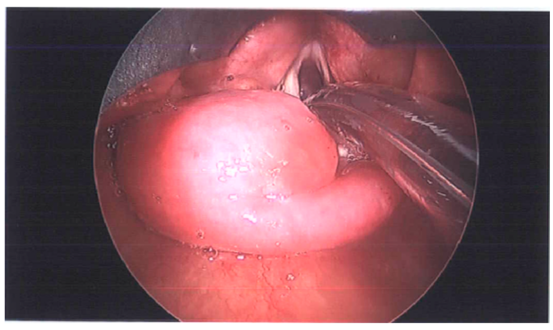
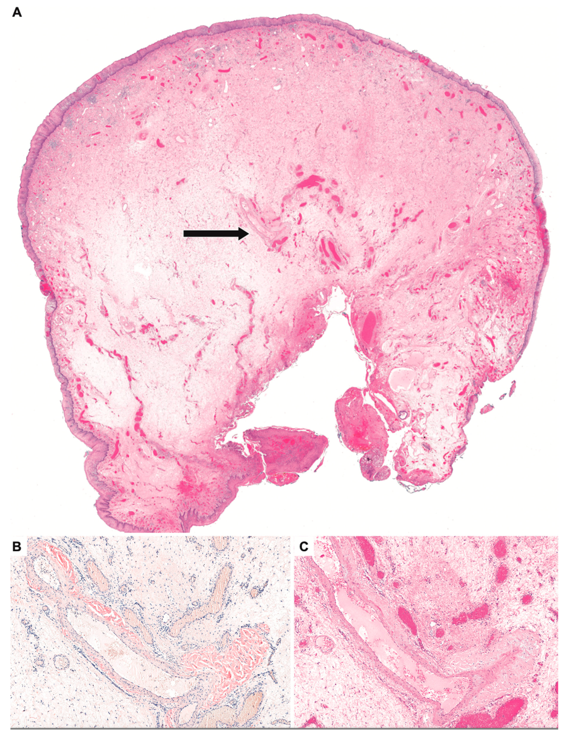
2. Detailed Case Description

An 85-year-old male presented with dysphagia, weight loss, cough, and exertional dyspnea. His past medical history was significant for atrial fibrillation, cardiomyopathy, congestive heart failure (CHF), and pacemaker placement. Interestingly, angiography had shown no evidence of coronary artery atherosclerosis, and the etiology of his cardiomyopathy was unclear. Previous echocardiography showed a normal ejection fraction of 50% and left ventricular hypertrophy with a dilated left atrium. His Eating Assessment Tool 10 (EAT-10) score was 13; a score greater than 3 is abnormal. Upon flexible laryngoscopic examination, there was a large, soft, smooth mass arising from the posterior pharyngeal wall obstructing the esophageal inlet and prolapsing into the glottis. CT scan showed a 1.7 × 3.0 × 2.2 cm soft tissue mass at the esophageal inlet. He underwent endoscopic and excision of a submucosal mass of the posterior hypopharyngeal wall (Figure 1). Histopathological evaluation demonstrated amyloid deposition in the walls of small- and medium-sized arteries (Figure 2). Mass spectrometry confirmed the protein subtype as ATTR variant. Cardiac re-evaluation in light of this new information was performed and the patient was diagnosed with ATTR cardiomyopathy. He expired due to heart failure shortly after confirmation of the disease.

3. Discussion

The biopsy-proven ATTR variant of hypopharyngeal amyloidosis has not been reported previously. In our case, the patient presented with dysphagia and endoscopic findings of a hypopharyngeal mass. Cases of hypopharyngeal AL involvement have been reported previously in association with multiple myeloma [5,15] or as localized hypopharyngeal AL amyloidosis [16], with patients presenting with dysphagia and endoscopic findings of submucosal infiltration of the posterior hypopharyngeal wall. Our patient had a similar presentation but secondary to the ATTR variant of hypopharyngeal amyloidosis, which represents the first biopsy-proven case of this variant affecting the hypopharynx. ATTR amyloidosis commonly presents with systemic amyloid deposition with predominantly peripheral and autonomic neuropathy. Other target tissues are the heart, eye, and ligaments. Patients with ATTR amyloidosis predominantly present with peripheral sensory polyneuropathy, autonomic neuropathy, cardiomyopathy, renal failure, carpal tunnel syndrome, and eye vitreous opacities.
Dysphagia in the setting of amyloidosis is usually secondary to macroglossia from AL amyloidosis [17]. Macroglossia occurs due to extracellular, perivascular, and intramuscular amyloid deposition, affecting the oral phase of swallowing [18,19]. In our patient, his dysphagia was likely secondary to the direct mechanical obstruction of the mass, though in ATTR amyloidosis, autonomic and peripheral sensory neuropathy are also possible mechanisms [5]. Other rare causes of dysphagia in the setting of amyloidosis can be secondary to esophageal or gastric involvement. Esophageal involvement by amyloidosis has been associated with achalasia, spasm, non-specific motility disorders, malabsorption, and esophagitis. A study of 30 patients with systemic amyloidosis showed abnormalities in esophageal manometry such as decreased resting lower esophageal sphincter pressure and abnormalities in esophageal motility [20]. These presentations and findings are consistent with the postulation that the deposition of amyloid in smooth and striated muscle as well as the nervous system affects the swallowing mechanism [21,22].
Besides dysphagia, gastrointestinal (GI) amyloidosis may also present with other symptoms such as diarrhea, esophageal reflux, nausea, and abdominal pain. These symptoms are attributed to autonomic neuropathy. Gastric amyloidosis commonly manifests as systemic amyloidosis compared to localized amyloidosis. According to a 13-year retrospective study, with a total of 76 gastric amyloidosis patients, 79% had systemic gastric amyloidosis and 21% had localized gastric amyloidosis [23]. Endoscopic findings in esophageal and gastric amyloidosis are nonspecific, including erosions, ulcers, erythema, nodularity, strictures, a fine granular appearance, polypoidal protrusions, mucosal friability, and submucosal tumor-like lesions [22,24]. Interestingly, a study by Tada et al. showed that the two endoscopic findings characteristic of amyloidosis are fine granular appearance and polypoid protrusion. They also found that different gastrointestinal tract biopsy sites have different frequencies of amyloid deposition in the biopsy specimens, where the degree of amyloid deposition is highest in the duodenum (100%) and the lowest in the esophagus (72%). Thus, the study concludes with the importance of examining the upper gastrointestinal tract, especially the duodenum [24].
The diagnosis of amyloidosis is based on histological confirmation of amyloid in biopsies from affected tissues. A high index of suspicion is necessary for a clinician to offer a biopsy when a hypopharyngeal mass is seen on laryngoscopy. Once the diagnosis of amyloidosis is proven from a biopsy, whole-body screening for systemic amyloidosis is recommended before diagnosing localized amyloidosis. Systemic amyloidosis is diagnosed when two or more sites of the body are involved. In systemic ATTR amyloidosis, cardiac infiltration is common. This infiltration results in poor diastolic relaxation and impaired left ventricular diastolic filling, resulting in right-sided heart failure. In patients presenting with heart failure and preserved ejective fraction, amyloid cardiomyopathy should be strongly suspected [8]. In addition to heart failure, conduction disturbances or arrhythmias requiring pacemaker placement are also frequently encountered in patients with ATTR amyloidosis [25]. Systemic ATTR amyloidosis is becoming increasingly recognized as a cause of heart failure and is likely underdiagnosed [12]. The diagnosis of amyloidosis cardiomyopathy is challenging because the symptoms are often attributed to other more common cardiac conditions such as ischemic heart disease, hypertensive heart disease, hypertrophic cardiomyopathy, and idiopathic restrictive cardiomyopathy. An invasive cardiac biopsy is essential in the diagnosis of ATTR cardiac amyloidosis if there is no involvement of other more accessible sites and suspicion remains high. Other more accessible sites are the subcutaneous abdominal wall fatty tissue, salivary gland, rectum, kidney, or gastric mucosa. The abdominal wall fatty tissue is usually chosen as it is readily accessible with low morbidity of the procedure. However, while the specificities are reported as high (almost 100%), the diagnostic sensitivities are variable, with a report as low as 12% overall sensitivity on ATTR amyloidosis [4,26]. Biopsy of the involved organ remains the gold standard for diagnosis, with 100% sensitivity and specificity [4,27]. There are also a lack of screening criteria contributing to the underdiagnosis of ATTR amyloidosis cardiomyopathy.
A study by Bartier et al. shows that patients with ATTR amyloidosis with cardiac involvement have a high prevalence of pharyngolaryngeal impairments without any macroscopic organic lesions seen. The pharyngolaryngeal impairments are manifested by hoarseness and dysphagia, with prevalence of 47% and 17%, respectively [17]. Several pathophysiological hypotheses in the literature include the nerve dysfunction hypothesis and the amyloid infiltration tissue hypothesis. Amyloid deposits infiltrate nerves as evidenced by histologic studies showing the amyloid deposition of walls of cranial nerves [17,28]. Cranial nerves involved in swallowing and phonation (V, VII, IX, X, XI) can have amyloid deposition affecting their function [17,29]. Amyloid deposits’ infiltration in the brainstem and cortex can potentially result in polyneuropathy and compromised coordination of various swallowing phases, ultimately causing dysphagia. Muscles and mucosa can also be infiltrated with amyloid, affecting the function of pharyngolaryngeal organs. Synovial deposits in the joints of pharyngolaryngeal organs can also lead to ankylosis, affecting the swallowing mechanisms [17].
ATTR amyloidosis treatment options are rapidly expanding. Multiple clinical trials are ongoing to develop new treatments to improve the outcomes of patients with ATTR amyloidosis. Treatments to improve outcomes can be divided into drug therapies for ATTR amyloidosis and drugs for ATTR amyloidosis comorbidities and complications. Drug therapies for ATTR amyloidosis are classified into inhibitors of TTR gene expression, tetramer stabilizers, inhibitors of oligomer aggregation, and inhibitors of degradation and reabsorption of amyloid fibers. Most medications focus on preventing the formation of TTR amyloid protein or inhibiting the degradation of TTR tetramers into monomers [12]. ATTR amyloidosis, with common comorbidities such as heart failure, requires medications such as diuretics, beta-blockers, and angiotensin-converting enzyme inhibitors. In ATTR-cardiomyopathy-related atrial fibrillation, patients require anticoagulation and potentially cardioversion and a pacemaker to maintain sinus rhythm. In ATTR-cardiomyopathy-related end-stage heart failure with no significant extracardiac involvement, left ventricular assist device and cardiac transplantations are options. The prognosis of patients with ATTR amyloidosis is determined by the extent of cardiac involvement [30]. The early diagnosis and treatment of ATTR amyloidosis are crucial in improving outcomes for patients.
From the perspective of the case reported in this article, there are several important lessons to be considered. Amyloid infiltration of the hypopharynx and esophagus should be considered in the differential diagnosis of dysphagia, whether gross disease is apparent or not. In order to evaluate the presence of disease, it may be important to biopsy affected sites, even if they appear morphologically normal, and to obtain a representative biopsy that can include an evaluation of muscle and nerve tissue. Moreover, suspicion of the presence of the ATTR variant should increase when evaluating patients in the eighth decade of life who present with increasing dysphagia, especially in the absence of a gross/mechanical lesion. Even more importantly, otolaryngologists should be wary of the clinical presentation of elderly patients with heart failure presenting with dysphagia. As heart failure is most commonly due to coronary artery disease, and because dysphagia commonly increases with age, it is normal to consider these two issues separately when evaluating elderly patients with dysphagia. However, in this clinical scenario, it is important to understand the etiology of the heart failure. When heart failure is considered idiopathic, or when angiography has failed to demonstrate the presence of significant coronary artery disease, ATTR variant amyloidosis should be strongly suspected as the etiology of the dysphagia. This should prompt further evaluation of heart function, through serum evaluation of troponin levels as well as B-type natriuretic peptide (BNP) and N-terminal-pro-BNP. Given the poor prognosis and clinical fragility of patients with heart failure in the context of ATTR amyloidosis, invasive interventions, especially those that require general anesthesia, should be carefully considered. Non-invasive, supportive measures such as feeding tube placement may be more appropriate for this patient population.
Amyloidosis should be suspected in findings of unexplained signs such as organomegaly, proteinuria, renal failure, right-sided heart failure, biventricular hypertrophic cardiac walls, peripheral axonal polyneuropathy, and autonomic neuropathy [4]. ATTR amyloidosis is likely underdiagnosed in many patients with cardiomyopathy and heart failure. The overall prevalence of amyloid cardiomyopathy found in patients with heart failure is 13.7% [31]. Amyloidosis cardiomyopathy is more commonly described in the literature compared to head and neck amyloidosis. Dysphagia with a history of amyloidosis should also alert clinicians to evaluate the entire aerodigestive tract with a lower threshold for biopsy when findings such as fine granular appearance, polypoid protrusion, erosion, ulcers, erythema, nodularity, or mucosa friability are seen. In the absence of an organic lesion seen on the endoscope, dysphagia or dysphonia experienced by patients with ATTR amyloidosis can be due to amyloid deposition on nerves, muscles, mucosa, and joints involved in swallowing and phonation. The management of amyloidosis should also be multidisciplinary, with a team of experts caring for the organs involved in systemic amyloidosis. With better awareness and early recognition of the disease, early intervention can be instilled to modify the underlying pathologic process to improve prognosis.

4. Conclusions

Otolaryngologists should be alert to the possibility that patients with dysphagia and cardiomyopathy, with or without the presence of a submucosal hypopharyngeal mass, may have underlying ATTR variant amyloidosis. ATTR variant amyloidosis with cardiomyopathy is unique in its high prevalence of dysphagia and dysphonia, even without an organic lesion seen. The diagnostic challenges remain due to its rarity and non-specific symptoms with multi-organ involvement. The current literature showing terminal prognosis is likely contributed by the diagnostic challenge and delayed diagnosis. High clinical suspicion, early biopsy, disease confirmation, and prompt treatment are important to improve outcomes given the wide array of new medications targeting various phases of amyloidogenesis to change the disease’s natural history. With more awareness of this ATTR variant amyloidosis in patients with dysphagia, better clinical practice with improved clinical outcomes can be extended to our patients.

Author Contributions

Conceptualization, E.J.D.; writing—original draft preparation, C.H.L.N.; writing—review and editing, E.J.D.; histology figures and description, G.J.B.; supervision, E.J.D. All authors have read and agreed to the published version of the manuscript.

Funding

This research received no external funding.

Institutional Review Board Statement

This study was conducted in accordance with the Declaration of Helsinki, and approved by the Institutional Review Board of Stanford University (protocol code 69525 and date of approval 29 March 2023.

Informed Consent Statement

Not applicable.

Data Availability Statement

Not applicable.

Conflicts of Interest

The authors declare no conflict of interest.

References

  1. Wechalekar, A.D.; Gillmore, J.D.; Hawkins, P.N. Systemic amyloidosis. Lancet 2016, 387, 2641–2654. [Google Scholar] [CrossRef] [PubMed]
  2. Rudy, S.F.; Jeffery, C.C.; Damrose, E.J. Clinical characteristics of laryngeal versus nonlaryngeal amyloidosis. Laryngoscope 2018, 128, 670–674. [Google Scholar] [CrossRef]
  3. Falk, R.H.; Comenzo, R.L.; Skinner, M. The Systemic Amyloidoses. N. Engl. J. Med. 1997, 337, 898–909. [Google Scholar] [CrossRef] [PubMed]
  4. Hazenberg, B.P.C. Amyloidosis: A clinical overview. Rheum. Dis. Clin. 2013, 39, 323–345. [Google Scholar] [CrossRef] [Green Version]
  5. Hammami, B.; Mnejja, M.; Kallel, S.; Bouguecha, L.; Chakroun, A.; Charfeddine, I.; Ghorbel, A. Hypopharyngeal amyloidosis: A case report. Eur. Ann. Otorhinolaryngol. Head Neck Dis. 2010, 127, 83–85. [Google Scholar] [CrossRef] [Green Version]
  6. Stevenson, R.; Witteles, R.; Damrose, E.; Arai, S.; Lafayette, R.A.; Schrier, S.; Afghahi, A.; Liedtke, M. More Than a Frog in the Throat: A Case Series and Review of Localized Laryngeal Amyloidosis. Arch. Otolaryngol. Neck Surg. 2012, 138, 509–511. [Google Scholar] [CrossRef] [PubMed]
  7. Harris, G.; Lachmann, H.; Hawkins, P.; Sandhu, G. One Hundred Cases of Localized Laryngeal Amyloidosis—Evidence for Future Management. Laryngoscope 2021, 131, E1912–E1917. [Google Scholar] [CrossRef]
  8. Gertz, M.A.; Benson, M.D.; Dyck, P.J.; Grogan, M.; Coelho, T.; Cruz, M.; Berk, J.L.; Plante-Bordeneuve, V.; Schmidt, H.H.; Merlini, G. Diagnosis, Prognosis, and Therapy of Transthyretin Amyloidosis. J. Am. Coll. Cardiol. 2015, 66, 2451–2466. [Google Scholar] [CrossRef] [Green Version]
  9. Connors, L.H.; Lim, A.; Prokaeva, T.; Roskens, V.A.; Costello, C.E. Tabulation of human transthyretin (TTR) variants, 2003. Amyloid 2003, 10, 160–184. [Google Scholar] [CrossRef]
  10. Vieira, M.; Saraiva, M.J. Transthyretin: A multifaceted protein. Biomol. Concepts 2014, 5, 45–54. [Google Scholar] [CrossRef]
  11. Hellman, U.; Alarcon, F.; Lundgren, H.E.; Suhr, O.B.; Bonaiti-PelliÉ, C.; Planté-Bordeneuve, V. Heterogeneity of penetrance in familial amyloid polyneuropathy, ATTR Val30Met, in the Swedish population. Amyloid 2008, 15, 181–186. [Google Scholar] [CrossRef] [PubMed]
  12. Teng, C.; Li, P.; Bae, J.Y.; Pan, S.; Dixon, R.A.F.; Liu, Q. Diagnosis and treatment of transthyretin-related amyloidosis cardiomyopathy. Clin. Cardiol. 2020, 43, 1223–1231. [Google Scholar] [CrossRef] [PubMed]
  13. Puig-Carrion, G.D.; Reyentovich, A.; Katz, S.D. Diagnosis and treatment of heart failure in hereditary transthyretin amyloidosis. Clin. Auton. Res. 2019, 29, 45–53. [Google Scholar] [CrossRef] [PubMed] [Green Version]
  14. Nakagawa, M.; Sekijima, Y.; Yazaki, M.; Tojo, K.; Yoshinaga, T.; Doden, T.; Koyama, J.; Yanagisawa, S.; Ikeda, S.-I. Carpal tunnel syndrome: A common initial symptom of systemic wild-type ATTR (ATTRwt) amyloidosis. Amyloid 2016, 23, 58–63. [Google Scholar] [CrossRef] [Green Version]
  15. Chadwick, M.A.; Buckland, J.R.; Mason, P.; Randall, C.J.; Theaker, J. A rare case of dysphagia: Hypopharyngeal amyloidosis masquerading as a post-cricoid tumour. J. Laryngol. Otol. 2002, 116, 54–56. [Google Scholar] [CrossRef]
  16. Ghekiere, O.; Desuter, G.; Weynand, B.; Coche, E. Hypopharyngeal Amyloidoma. Am. J. Roentgenol. 2003, 181, 1720–1721. [Google Scholar] [CrossRef]
  17. Bartier, S.; Bodez, D.; Kharoubi, M.; Canouï-Poitrine, F.; Chatelin, V.; Henrion, C.; Coste, A.; Damy, T.; Béquignon, E. Pharyngo-laryngeal involvement in systemic amyloidosis with cardiac involvement: A prospective observational study. Amyloid 2019, 26, 216–224. [Google Scholar] [CrossRef]
  18. Ohki, M.; Kikuchi, S. Dysphagia due to systemic light chain amyloidosis revealed by videoendoscopic and videofluorographic swallowing examinations. Otolaryngol. Case Rep. 2018, 6, 4–6. [Google Scholar] [CrossRef]
  19. Muchtar, E.; Derudas, D.; Mauermann, M.; Liewluck, T.; Dispenzieri, A.; Kumar, S.K.; Dingli, D.; Lacy, M.Q.; Buadi, F.K.; Hayman, S.R.; et al. Systemic Immunoglobulin Light Chain Amyloidosis–Associated Myopathy: Presentation, Diagnostic Pitfalls, and Outcome. Mayo Clin. Proc. 2016, 91, 1354–1361. [Google Scholar] [CrossRef]
  20. Rubinow, A.; Burakoff, R.; Cohen, A.S.; Harris, L.D. Esophageal manometry in systemic amyloidosis. A study of 30 patients. Am. J. Med. 1983, 75, 951–956. [Google Scholar] [CrossRef]
  21. Abbas, Z.; Abid, S.; Rehman, N.U.; Pervez, S. Primary amyloidosis of gut presenting with dysphagia. J. Pak. Med. 1995, 45, 274–275. [Google Scholar]
  22. Iida, T.; Yamano, H.; Nakase, H. Systemic amyloidosis with gastrointestinal involvement: Diagnosis from endoscopic and histological views. J. Gastroenterol. Hepatol. 2018, 33, 583–590. [Google Scholar] [CrossRef] [PubMed] [Green Version]
  23. Andrew, J. Cowan, Martha Skinner, David C. Seldin; et al. Amyloidosis of the gastrointestinal tract: A 13-year, single-center, referral experience. Haematologica 2013, 98, 141–146. [Google Scholar] [CrossRef] [Green Version]
  24. Tada, S.; Lida, M.; Iwashita, A.; Matsui, T.; Fuchigami, T.; Yamamoto, T.; Yao, T.; Fujishima, M. Endoscopic and biopsy findings of the upper digestive tract in patients with amyloidosis. Gastrointest. Endosc. 1990, 36, 10–14. [Google Scholar] [CrossRef] [PubMed]
  25. Coelho, T.; Maurer, M.S.; Suhr, O.B. THAOS—The Transthyretin Amyloidosis Outcomes Survey: Initial report on clinical manifestations in patients with hereditary and wild-type transthyretin amyloidosis. Curr. Med. Res. Opin. 2013, 29, 63–76. [Google Scholar] [CrossRef]
  26. Garcia, Y.; Collins, A.B.; Stone, J.R. Abdominal fat pad excisional biopsy for the diagnosis and typing of systemic amyloidosis. Human Pathol. 2018, 72, 71–79. [Google Scholar] [CrossRef]
  27. Witteles, R. Cardiac Amyloidosis. 2016. Available online: https://www.acc.org/latest-in-cardiology/articles/2016/07/07/14/59/cardiac-amyloidosis (accessed on 9 May 2023).
  28. Conaghan, P.; Chung, D.; Vaughan, R. Recurrent laryngeal nerve palsy associated with mediastinal amyloidosis. Thorax 2000, 55, 436. [Google Scholar] [CrossRef] [Green Version]
  29. Batista, J.A.d.S.; Carrera, L.R.; Viriato, A.R.; Novaes, M.A.C.; de Morais, R.J.L.; Oliveira, F.T.; Marques, W.; Costa, M.C.M. Involvement of cranial nerves in ATTR Ile127Val amyloidosis. Eur. J. Med. Genet. 2022, 65, 104524. [Google Scholar] [CrossRef]
  30. Ruberg, F.L.; Grogan, M.; Hanna, M.; Kelly, J.W.; Maurer, M.S. Transthyretin Amyloid Cardiomyopathy: JACC State-of-the-Art Review. J. Am. Coll. Cardiol. 2019, 73, 2872–2891. [Google Scholar] [CrossRef]
  31. See, A.S.Y.; Ho, J.S.-Y.; Chan, M.Y.; Lim, Y.C.; Yeo, T.-C.; Chai, P.; Wong, R.C.; Lin, W.; Sia, C.-H. Prevalence and Risk Factors of Cardiac Amyloidosis in Heart Failure: A Systematic Review and Meta-Analysis. Hear. Lung Circ. 2022, 31, 1450–1462. [Google Scholar] [CrossRef]
Figure 1. Intraoperative view of posterior hypopharyngeal mass.
Surgeries 04 00028 g001
Figure 2. Histopathology showing ATTR variant amyloidosis. Panel (A): Low-power magnification of broad-based polypoid mass lined by squamous mucosa. Arrow indicates affected vessel highlighted in images (H&E ×10). Panel (B): Congo red staining showing orangophilic deposits within the walls of the arteries (Congo red × 100). Panel (C): Scanning magnification showing an edematous stroma and intact artery with mural amyloid accumulations (H&E ×100).
Surgeries 04 00028 g002
Disclaimer/Publisher’s Note: The statements, opinions and data contained in all publications are solely those of the individual author(s) and contributor(s) and not of MDPI and/or the editor(s). MDPI and/or the editor(s) disclaim responsibility for any injury to people or property resulting from any ideas, methods, instructions or products referred to in the content.

