Communication

Prevalence of Lumbosacral Transition Vertebrae in Symptomatic Adults and the Levels of Degeneration in the Suprajacent Disc

by
Joshua Song
1,
Brjan Kaiji Betzler
2,
Arun-Kumar Kaliya-Perumal
2,* and
Jacob Yoong-Leong Oh
1,*
1
Division of Spine, Department of Orthopaedic Surgery, Tan Tock Seng Hospital, Singapore 308433, Singapore
2
Lee Kong Chian School of Medicine, Nanyang Technological University, Singapore 636921, Singapore
*
Authors to whom correspondence should be addressed.
Surgeries 2023, 4(1), 120-126; https://doi.org/10.3390/surgeries4010013
Submission received: 10 February 2023 / Revised: 6 March 2023 / Accepted: 9 March 2023 / Published: 15 March 2023

Abstract

:
Lumbosacral transition vertebra (LSTV) is a common occurrence and its prevalence ranges from 2.6% to 35.6%. Our aim is to study this phenomenon in the adult Singaporean population and assess the level of degeneration of the suprajacent disc in those with LSTV. Retrospectively, 545 patients (Age = 57.6 ± 18.3 years; Male = 277; Female = 268) who underwent radiographic evaluation of the lumbar spine for lower back pain or radicular symptoms were shortlisted. LSTV was found to be present in 106 patients (19.4%) with sacralization of L5 and lumbarization of S1 occurring in 96 patients (17.6%) and 10 patients (1.8%), respectively. The most common form of LSTV was Castellvi type IIA (46 patients; 43.4%). Based on Pfirrmann grading, Grade IV disc degeneration was most common in both the LSTV level (63%) and the unfused suprajacent level (77%) in those with LSTV. There was a significantly higher number of patients with grade IV and above degeneration in the suprajacent disc level among those with LSTV when compared to the last unfused (L5-S1) disc level in those without LSTV (84% vs. 65%; p = 0.0001). This suprajacent disc degeneration seen in patients with LSTV may contribute to low back pain and related problems in these patients.

1. Introduction

Lumbosacral transitional vertebra (LSTV) is a common anatomical abnormality that is often seen at the L5/S1 level of the spine [1]. This can either present as lumbarization of S1 vertebra or sacralization of L5 vertebra [2]. The lumbarization of S1 is characterized by various features, including a squared shaped S1 vertebra with well-formed lumbar type facet joints, anomalous articulation, and a fully developed disc below S1. On the other hand, sacralization of L5 vertebra can range from elongated and broadened transverse processes to complete fusion with the sacrum [2,3]. Most commonly, the transitions are unilateral or incomplete [4]. Previous studies have shown the prevalence of LSTV to range from 2.6% to 35.6% [1,5,6,7,8]. French et al. described prevalence of LSTV to be 9.9% in the Australian population, while Tang et al. reported prevalence of LSTV to be 15.8% in the Han Chinese population [1,8].
The most common classification system for LSTV was devised by Castellvi et al. in 1984 and is based on the different morphological characteristics of the lower spine [3]. There is currently no standardized modality for identifying LSTV, but most authors agree that anteroposterior (AP) radiographs provide the best radiological evidence for LSTV [9]. It is essential to identify LSTV in patients requiring spinal surgery. Discrepancies in correlating patient symptoms with the correct spinal level have occurred previously and can be due to the presence of LSTV [2,10]. It is therefore imperative that spinal surgeons identify the presence of LSTV prior to spinal surgery. The presence of LSTV can also affect the load distribution in the spine and cause more degeneration in the disc cranial to the LSTV [11,12]. There have also been studies demonstrating the protective effects of the LSTV on the disc at the transitional level [13,14,15]. However, there is a lack of research on LSTV specifically focused on the Singaporean population. Thus, our aim is to examine the frequency of LSTV among symptomatic adults and assess the extent of intervertebral disc degeneration both at the transitional level and the level immediately above the LSTV.

2. Material and Methods

After institutional review board approval, retrospectively, a list of AP and lateral lumbar X-rays were reviewed. All participants provided written informed consent for participation in the study with due care to maintaining privacy. Included were consecutive adults older than 18 years of age irrespective of gender who visited our clinic with radiographic evidence of 12 ribs, and clearly visible vertebral body articulation of the last rib, lumbar transverse processes, and the entire sacral wing. Patients were all symptomatic, either with back pain or radicular pain at initial presentation and were subjected to radiographic evaluation. This included those who were to be managed conservatively or posted for surgery or had completed surgical intervention. Excluded were those in whom there was difficulty in identifying the transitional vertebra due to instrumentation or abdominal contents. Patient’s age at time of imaging, gender, race, and the number of lumbar vertebral bodies were noted.
The identification of the transitional vertebra was performed by first counting down from the last thoracic vertebra on the AP view X-ray and was further confirmed using the lateral view. If any hypoplastic ribs were observed, then the vertebra directly beneath it was designated as L1, which could be identified by the presence of transverse processes. “Sacralization of L5” was considered when the transverse process of the last lumbar vertebra formed either a bony bridge or a pseudoarthrosis with sacral ala. “Lumbarization of S1” was considered when there were five distinct lumbar vertebrae, in addition to a squared-shaped S1 vertebra with well-formed lumbar type facet joints, where the transverse process either fuses with or forms a pseudoarthrosis with the sacral ala, with or without a fully developed disc below S1. We considered Castellvi types II, III, and IV only as transitional states (Figure 1). Type I was excluded as it lacks clinical and surgical significance; furthermore, Castellvi type I has been considered as a variation of normal due to the presence of a mobile disc caudal to the vertebra in question [16]. If the morphology differed between the right and the left side, the transition was designated to the side that had the higher type numerically. Data were recorded as type II to IV. All suspected LSTV were jointly reviewed by two reviewers, and a consensus was reached on the classification.
For patients with LSTV, the intervertebral discs, located at the level of the LSTV, and the disc above (suprajacent) were evaluated using the Pfirrmann criteria to determine their grade of degeneration [17]. Additionally, the last unfused intervertebral disc at L5/S1 in normal patients was also evaluated using the same grading criteria. Measurements were performed by two independent reviewers. The Pfirrmann grading system uses MRI T2 weight imaging (T2WI) sagittal images of the spine to assess degeneration of the intervertebral discs by looking at the asymmetry in disc structure, distinction of the nucleus and the annulus, signal intensity of intervertebral discs and height of intervertebral discs. The degree of degeneration of the intervertebral discs were categorized into grade I to V based on the 5-level Pfirrmann grading system (Figure 2). A detailed description of the grading system can be found in the original work of Pfirrmann et al. [17] and validations by Griffith et al. [18] and Miyazaki et al. [19].
Statistical analyses were performed using SPSS version 28 (SPSS, Inc, an IBM Company, Chicago, IL, USA). The patients were chosen through convenience sampling, inclusive of all patients during a specific time frame. Intra- and interrater reliability of Pfirrmann grading were assessed using Cohen’s kappa on 35 patients. The calculated minimum sample size required for the reliability analysis was 14 based on power calculation, with expected reliability of 0.8 and precision of approximately 95% confidence interval [20]. The kappa coefficient (κ) has a maximum value of 1.0 and a minimum value of 0, indicating agreement no better than chance. Recommendation from Landis and Koch stated that κ values of more than 0.8 are considered excellent; 0.6–0.8 as good; 0.4–0.6 as moderate and <0.4 as poor correlation [21]. Categorical variables were expressed as frequencies. Comparisons of disc degeneration between the groups were performed using the Fischer’s Exact test. We utilized Spearman’s rank correlation coefficient to examine the relationship between age and the extent of disc degeneration. The coefficient can range from −1 to +1, where a score of +1 represents a complete positive correlation, a score of 0 denotes no correlation, and a score of −1 indicates a total negative correlation.

3. Results

A total of 580 symptomatic patients were evaluated. Based on our exclusion criteria, 19 patients whose radiographs were inadequately exposed, 11 patients who had previous instrumentation, two patients who underwent previous vertebroplasty, one who underwent radiologically guided aspiration of spondylodiscitis, another who had previous lumbar laminectomy, and one who was too young at time of radiography (16 years old) were all excluded. As a result, a total of 545 radiographs were shortlisted for this study. The mean age of the population in our study was 57.6 ± 18.3 years and LSTV was found in 106 patients (19.6%), the majority male (61 patients; 58%) (Table 1). Of the 277 included males, 61 had LSTV, whereas, among the 268 included females, 45 had LSTV. This difference was not statistically significant (p = 0.13).
Of the patients with LSTV, 10 (1.8%) had lumbarization of the S1 and 96 (17.6%) had sacralization of L5. The most common type of LSTV as per the Castellvi classification was type IIA in 46 patients (44.0%), while the least common was type IV for four patients (3.7%). The intra-rater reliability of Pfirrmann grading was excellent at 0.884, while the interrater reliability of Pfirrmann grading was moderate at 0.464. Based on Pfirrmann’s grading, Grade IV degeneration was most common in both the LSTV level (63%) and the unfused suprajacent level (77%) in those with LSTV. Grade IV degeneration was also common (275 patients; 63%) in the last unfused disc level (L5-S1) among those without LSTV. However, there was a significantly higher number of patients with grade IV and above degeneration in the last unfused disc level among those with LSTV when compared to those without (84% vs. 65%; p = 0.0001) (Table 2). We also noted a significant positive correlation between age and the grade of degeneration at the suprajacent level; nevertheless, the correlation coefficient indicated that the relationship was not particularly robust (Spearman’s rho: 0.421). In addition, gender did not seem to influence the grade of degeneration.

4. Discussion

Accurately identifying the level of the spinal nerve during spinal surgery is crucial, since operating on the wrong level can result in catastrophic consequences for the patient [22,23]. However, the presence of LSTV can make this task more difficult, which is why it is essential to comprehend the prevalence of this condition in the local context. This study showed that the prevalence of LSTV in our symptomatic population was 19.4%, with a higher number of patients with sacralization of L5 (17.6%) compared to lumbarization of S1 (1.8%). Furthermore, Castellvi type IIA was the most common type of LSTV present in our study population.

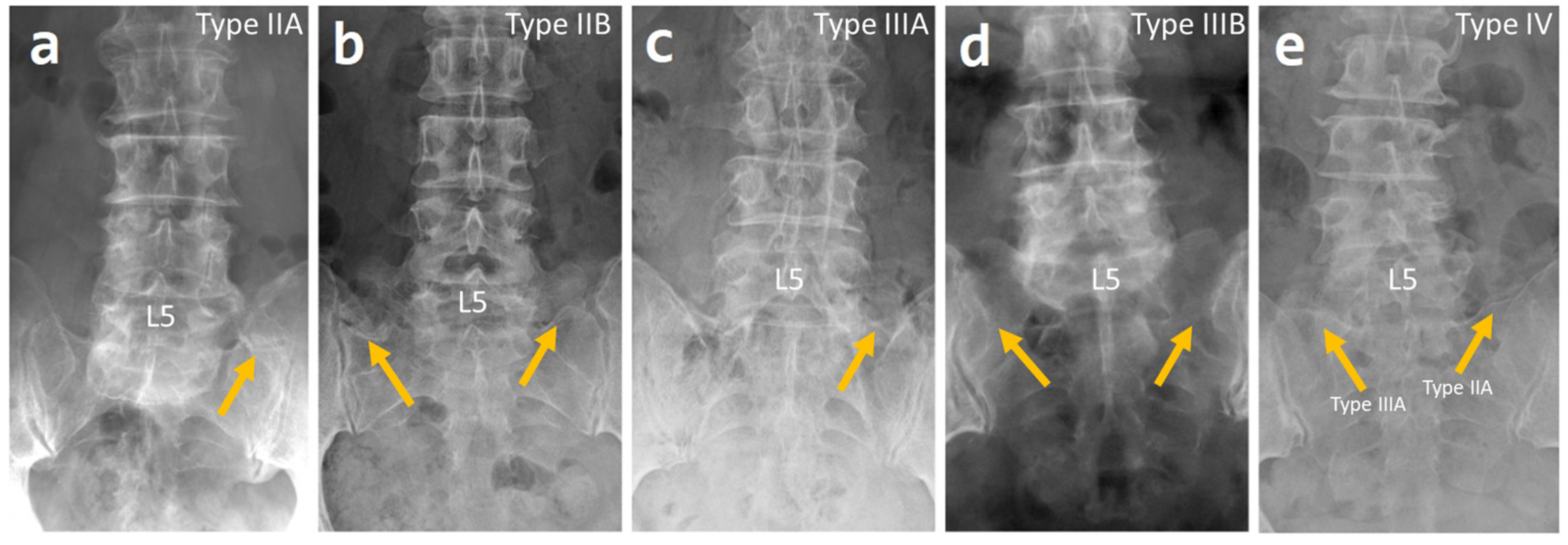
4.1. Prevalence of LSTV

The prevalence of LSTV varies widely, as stated in the literature, and previous studies have shown a range between 2.6% and 35.6% [1,5,6,7,8]. Tang et al. conducted a study on a Han Chinese population in 2014 and discovered that the prevalence of LSTV was 15.8%, with the most common being Castellvi type II [8]. French et al. found that the prevalence of LSTV in the Australian population was 9.9% [1]. Ucar et al. studied the Turkish population and found that prevalence of LSTV was 18.9% with sacralization of L5 and lumbarization of S1 occurring in 17.2% and 1.7% of the population, respectively [24]. Gopalan et al. studied the Indian population and found the prevalence of LSTV to be 24.3%, with sacralization of L5 and lumbarization of S1 at 20.9% and 3.3% respectively [25]. Multiple other studies on the Indian population revealed LSTV’s prevalence to be 22–26.8% [26,27]. Our results fall between the studies done by Tang et al. and Gopalan et al. This is likely due to the racial profiles of patients included in our study, with 7% of the population being Indians, 7% being Malays and the majority (77%) being Chinese. We also found that sacralization of L5 was more prevalent compared to lumbarization of S1 and that Castellvi IIA was the most common form of LSTV, which is consistent with many other studies [6,24,25,28].

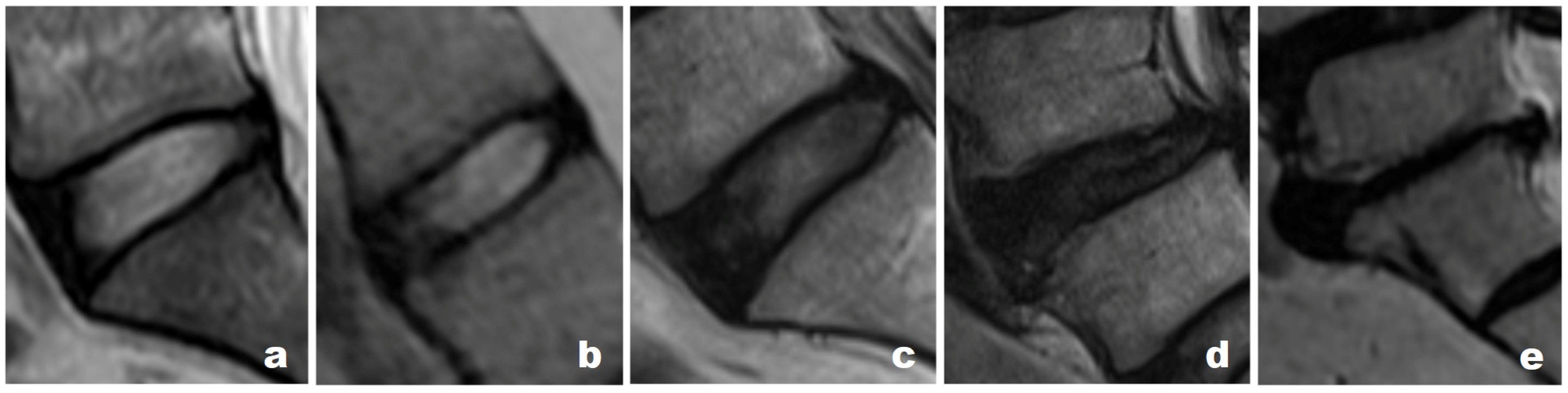
4.2. Degeneration of the Disc Compared to the Suprajacent Disc

Several studies have investigated the extent of intervertebral disc degeneration at the LSTV level and the disc above it. Notably, previous studies have reported accelerated degeneration of the suprajacent disc [13,14,15]. The proposed mechanisms include the suprajacent disc needing to bear more stress, as it is juxtaposed with a relatively non-mobile segment, with mechanisms similar to post-fusion adjacent segment disease [16]. Another mechanism includes instability of the suprajacent segment due to a weaker iliolumbar ligament [15]. This phenomenon was also observed in our cohort, as we saw a significantly higher number of patients with Grade IV degeneration of the suprajacent disc when compared to the disc at the level of transition.
On comparing the level of degeneration of the suprajacent disc in patients with LSTV and the last unfused (L5-S1) intervertebral disc in patients without LSTV, we found a significantly higher number of patients with grade IV and above degeneration in the suprajacent disc among those with LSTV. This finding is of clinical importance as the degeneration could be the cause of symptoms. Various studies have reported similar findings in patients with LSTV. To note, Hanhivaara et al. performed a study on 3855 abdominal CT scans and observed a greater amount of lumbar spine degeneration in patients with LSTV [11]. Furthermore, they found that patients with Castellvi IIA had the third highest amount of disc degeneration after Castellvi IIIB and IV [11]. This observation suggests that it is plausible to consider LSTV as a potential cause of back pain. However, this association remains controversial. While studies have associated lower back pain with LSTV [8,26,29], some have shown no association [2] and hence this result needs to be interpreted with caution.

4.3. Limitations of the Study

There are limitations to this study due to its retrospective nature. Firstly, the number of patients we included in this study is small compared to other studies. Moreover, there was an element of selection bias in our study as we did not analyze radiographs of asymptomatic patients. All our patients either had low back pain or radicular symptoms at first presentation. Hence, the selected cohort may not represent the general population. The interrater reliability of Pfirrmann grading of disc degeneration was moderate. This could be due to the discs which were difficult to differentiate, especially types 1, 2 and 3.

5. Conclusions

The prevalence of LSTV in the selected population was found to be 19.4%, with sacralization of L5 being the most common transitional state. Grade IV disc degeneration was most common in both the LSTV level and the unfused suprajacent level in those with LSTV. Grade IV degeneration was also common in the last unfused disc level among those without LSTV. However, there was a significantly higher number of patients with grade IV and above degeneration in the last unfused disc level among those with LSTV when compared to those without. Due to the increased tendency for suprajacent disc degeneration in LSTV patients, these patients may present with low back pain and related problems.

Author Contributions

Conceptualization, J.S., B.K.B., A.-K.K.-P. and J.Y.-L.O.; methodology, J.S., B.K.B., A.-K.K.-P. and J.Y.-L.O.; formal analysis, J.S. and B.K.B.; resources, J.S., B.K.B. and J.Y.-L.O.; data curation, J.S. and B.K.B.; writing—original draft preparation, J.S., B.K.B. and A.-K.K.-P.; writing—review and editing, A.-K.K.-P. and J.Y.-L.O.; supervision, J.Y.-L.O.; project administration, J.Y.-L.O. All authors have read and agreed to the published version of the manuscript.

Funding

This research received no external funding.

Institutional Review Board Statement

The study was conducted in accordance with the Declaration of Helsinki and approved by the National Healthcare Group (NHG), Domain Specific Review Board (DSRB), Singapore (2020/01435).

Informed Consent Statement

The requirement for informed consent was waived as this is a retrospective registry-based study.

Data Availability Statement

Not applicable.

Conflicts of Interest

The authors declare no conflict of interest.

References

  1. French, H.D.; Somasundaram, A.J.; Schaefer, N.R.; Laherty, R.W. Lumbosacral transitional vertebrae and its prevalence in the Australian population. Glob. Spine J. 2014, 4, 229–232. [Google Scholar] [CrossRef] [PubMed] [Green Version]
  2. Konin, G.P.; Walz, D.M. Lumbosacral transitional vertebrae: Classification, imaging findings, and clinical relevance. AJNR Am. J. Neuroradiol. 2010, 31, 1778–1786. [Google Scholar] [CrossRef] [PubMed] [Green Version]
  3. Castellvi, A.E.; Goldstein, L.A.; Chan, D.P. Lumbosacral transitional vertebrae and their relationship with lumbar extradural defects. Spine 1984, 9, 493–495. [Google Scholar] [CrossRef] [PubMed]
  4. Bron, J.L.; van Royen, B.J.; Wuisman, P.I. The clinical significance of lumbosacral transitional anomalies. Acta Orthop. Belg. 2007, 73, 687–695. [Google Scholar]
  5. Nevalainen, M.T.; McCarthy, E.; Morrison, W.B.; Zoga, A.C.; Roedl, J.B. Lumbosacral transitional vertebrae: Significance of local bone marrow edema at the transverse processes. Skelet. Radiol. 2018, 47, 1145–1149. [Google Scholar] [CrossRef]
  6. Paik, N.C.; Lim, C.S.; Jang, H.S. Numeric and morphological verification of lumbosacral segments in 8280 consecutive patients. Spine 2013, 38, E573–E578. [Google Scholar] [CrossRef]
  7. Apazidis, A.; Ricart, P.A.; Diefenbach, C.M.; Spivak, J.M. The prevalence of transitional vertebrae in the lumbar spine. Spine J. 2011, 11, 858–862. [Google Scholar] [CrossRef]
  8. Tang, M.; Yang, X.F.; Yang, S.W.; Han, P.; Ma, Y.M.; Yu, H.; Zhu, B. Lumbosacral transitional vertebra in a population-based study of 5860 individuals: Prevalence and relationship to low back pain. Eur. J. Radiol. 2014, 83, 1679–1682. [Google Scholar] [CrossRef]
  9. Hughes, R.J.; Saifuddin, A. Numbering of lumbosacral transitional vertebrae on MRI: Role of the iliolumbar ligaments. AJR Am. J. Roentgenol. 2006, 187, W59–W65. [Google Scholar] [CrossRef]
  10. Wigh, R.E.; Anthony, H.F., Jr. Transitional lumbosacral discs. probability of herniation. Spine 1981, 6, 168–171. [Google Scholar] [CrossRef]
  11. Hanhivaara, J.; Maatta, J.H.; Niinimaki, J.; Nevalainen, M.T. Lumbosacral transitional vertebrae are associated with lumbar degeneration: Retrospective evaluation of 3855 consecutive abdominal CT scans. Eur. Radiol. 2020, 30, 3409–3416. [Google Scholar] [CrossRef] [Green Version]
  12. Luoma, K.; Vehmas, T.; Raininko, R.; Luukkonen, R.; Riihimaki, H. Lumbosacral transitional vertebra: Relation to disc degeneration and low back pain. Spine 2004, 29, 200–205. [Google Scholar] [CrossRef]
  13. Farshad-Amacker, N.A.; Herzog, R.J.; Hughes, A.P.; Aichmair, A.; Farshad, M. Associations between lumbosacral transitional anatomy types and degeneration at the transitional and adjacent segments. Spine J. 2015, 15, 1210–1216. [Google Scholar] [CrossRef]
  14. Vergauwen, S.; Parizel, P.M.; van Breusegem, L.; Van Goethem, J.W.; Nackaerts, Y.; Van den Hauwe, L.; De Schepper, A.M. Distribution and incidence of degenerative spine changes in patients with a lumbo-sacral transitional vertebra. Eur. Spine J. 1997, 6, 168–172. [Google Scholar] [CrossRef]
  15. Aihara, T.; Takahashi, K.; Ogasawara, A.; Itadera, E.; Ono, Y.; Moriya, H. Intervertebral disc degeneration associated with lumbosacral transitional vertebrae: A clinical and anatomical study. J. Bone Jt. Surg. Br. 2005, 87, 687–691. [Google Scholar] [CrossRef]
  16. Sekharappa, V.; Amritanand, R.; Krishnan, V.; David, K.S. Lumbosacral transition vertebra: Prevalence and its significance. Asian Spine J. 2014, 8, 51–58. [Google Scholar] [CrossRef] [Green Version]
  17. Pfirrmann, C.W.; Metzdorf, A.; Zanetti, M.; Hodler, J.; Boos, N. Magnetic resonance classification of lumbar intervertebral disc degeneration. Spine 2001, 26, 1873–1878. [Google Scholar] [CrossRef]
  18. Griffith, J.F.; Wang, Y.X.; Antonio, G.E.; Choi, K.C.; Yu, A.; Ahuja, A.T.; Leung, P.C. Modified Pfirrmann grading system for lumbar intervertebral disc degeneration. Spine 2007, 32, E708–E712. [Google Scholar] [CrossRef]
  19. Miyazaki, M.; Hong, S.W.; Yoon, S.H.; Morishita, Y.; Wang, J.C. Reliability of a magnetic resonance imaging-based grading system for cervical intervertebral disc degeneration. J. Spinal Disord. Tech. 2008, 21, 288–292. [Google Scholar] [CrossRef]
  20. Cohen, J. A Coefficient of Agreement for Nominal Scales. Educ. Psychol. Meas. 2016, 20, 37–46. [Google Scholar] [CrossRef]
  21. Landis, J.R.; Koch, G.G. The measurement of observer agreement for categorical data. Biometrics 1977, 33, 159–174. [Google Scholar] [CrossRef] [PubMed] [Green Version]
  22. Lee, C.H.; Park, C.M.; Kim, K.A.; Hong, S.J.; Seol, H.Y.; Kim, B.H.; Kim, J.H. Identification and prediction of transitional vertebrae on imaging studies: Anatomical significance of paraspinal structures. Clin. Anat. 2007, 20, 905–914. [Google Scholar] [CrossRef] [PubMed]
  23. Malanga, G.A.; Cooke, P.M. Segmental anomaly leading to wrong level disc surgery in cauda equina syndrome. Pain Physician 2004, 7, 107–110. [Google Scholar] [CrossRef] [PubMed]
  24. Ucar, D.; Ucar, B.Y.; Cosar, Y.; Emrem, K.; Gumussuyu, G.; Mutlu, S.; Mutlu, B.; Cacan, M.A.; Mertsoy, Y.; Gumus, H. Retrospective cohort study of the prevalence of lumbosacral transitional vertebra in a wide and well-represented population. Arthritis 2013, 2013, 461425. [Google Scholar] [CrossRef]
  25. Gopalan, B.; Yerramshetty, J.S. Lumbosacral Transitional Vertebra-Related Low Back Pain: Resolving the Controversy. Asian Spine J. 2018, 12, 407–415. [Google Scholar] [CrossRef]
  26. Ravikanth, R.; Majumdar, P. Bertolotti’s syndrome in low-backache population: Classification and imaging findings. Ci Ji Yi Xue Za Zhi 2019, 31, 90–95. [Google Scholar] [CrossRef]
  27. Daniel, P.; Joel, J.J.; Rana, P. Lumbosacral transitional vertebrae in patients with low back pain: Radiological classification and morphometric analysis. J. Anat. Soc. 2019, 68, 123–128. [Google Scholar] [CrossRef]
  28. Nakagawa, T.; Hashimoto, K.; Tsubakino, T.; Hoshikawa, T.; Inawashiro, T.; Tanaka, Y. Lumbosacral Transitional Vertebrae Cause Spinal Level Misconception in Surgeries for Degenerative Lumbar Spine Disorders. Tohoku J. Exp. Med. 2017, 242, 223–228. [Google Scholar] [CrossRef] [Green Version]
  29. Landauer, F.; Trieb, K. Diagnostic Limitations and Aspects of the Lumbosacral Transitional Vertebrae (LSTV). Appl. Sci. 2022, 12, 830. [Google Scholar] [CrossRef]
Figure 1. Castellvi classification of LSTV at L4-5 level. (a) Type IIA—Unilateral pseudoarthrosis of the transverse process. (b) Type IIB—Bilateral pseudoarthrosis. (c) Type IIIA—Unilateral complete fusion of the transverse process. (d) Type IIIB—Bilateral complete fusion. (e) Type IV: Type IIA on one side and Type IIIA on the other side. Type 1 was considered as a variation of normal. Arrows point to the anomalous articulation.
Surgeries 04 00013 g001
Figure 2. Pfirrmann grading of intervertebral disc degeneration. (a) Grade I: Homogenous and bright white disc with clear distinction between the nucleus and annulus, with signal intensity being hyperintense/isointense to cerebrospinal fluid and maintaining normal height. (b) Grade II: Inhomogeneous disc with or without horizontal bands and a clear distinction between nucleus and annulus, with signal intensity being hyperintense/isointense to cerebrospinal fluid and maintaining normal height. (c) Grade III: Inhomogeneous gray disc with unclear distinction between nucleus and annulus with signal intensity being intermediate and having normal to slightly decreased height. (d) Grade IV: Inhomogeneous gray to black disc with loss of distinction between nucleus and annulus, and with signal intensity being intermediate to hypointense and having normal to moderately decreased height. (e) Grade V: Inhomogeneous black disc with loss of distinction between nucleus and annulus with signal intensity being hypointense and appearing collapsed.
Surgeries 04 00013 g002
Table 1. Baseline characteristics of patients.
VariablesDescriptive Statistics *
Number of Patients545
Age (years)57.6 ± 18.3
BMI (kg/m2)26.0 ± 5.3
Gender
Male 277 (50.8%)
Female268 (49.2%)
Race
Chinese420 (77.1%)
Malay40 (7.3%)
Indian37 (6.8%)
Others48 (8.8%)
Presence of LSTV106 (19.4%)
Male61 (58%)
Female45 (42%)
Castellvi Classification
Type IIA46 (44%)
Type IIB17 (16%)
Type IIIA9 (8.3%)
Type IIIB30 (27.5%)
Type IV4 (3.7%)
Pfirrmann grading at LSTV level (n = 106)
Grade II3 (3%)
Grade III26 (25%)
Grade IV67 (63%)
Grade V10 (9%)
* Values are presented as mean ± standard deviation or n (%).
Table 2. Comparison of disc degeneration at the suprajacent level.
Pfirrmann GradingNumber of Patients as per Pfirrmann Grading *Statistical Significance (p) c
LSTV Patients a (n = 106)Non-LSTV Patients b (n = 439)
Grade I0 (0%)2 (0.5%)1
Grade II1 (1%)31 (7%)0.01
Grade III16 (15%)121(28%)0.008
Grade IV82 (77%)275 (63%)0.004
Grade V7 (7%)10 (2%)0.03
* Values are presented as n (%); a Numbers represent Pfirrmann grading of the last non-transitional disc level in patients with LSTV; b Numbers represent Pfirrmann grading of the last unfused (L5-S1) disc level in those without LSTV; c A p value of less than 0.05 is considered statistically significant.
Disclaimer/Publisher’s Note: The statements, opinions and data contained in all publications are solely those of the individual author(s) and contributor(s) and not of MDPI and/or the editor(s). MDPI and/or the editor(s) disclaim responsibility for any injury to people or property resulting from any ideas, methods, instructions or products referred to in the content.

