
Cognitive Impairment in Multiple Sclerosis: An Update on Assessment and Management

Abstract
:1. Introduction
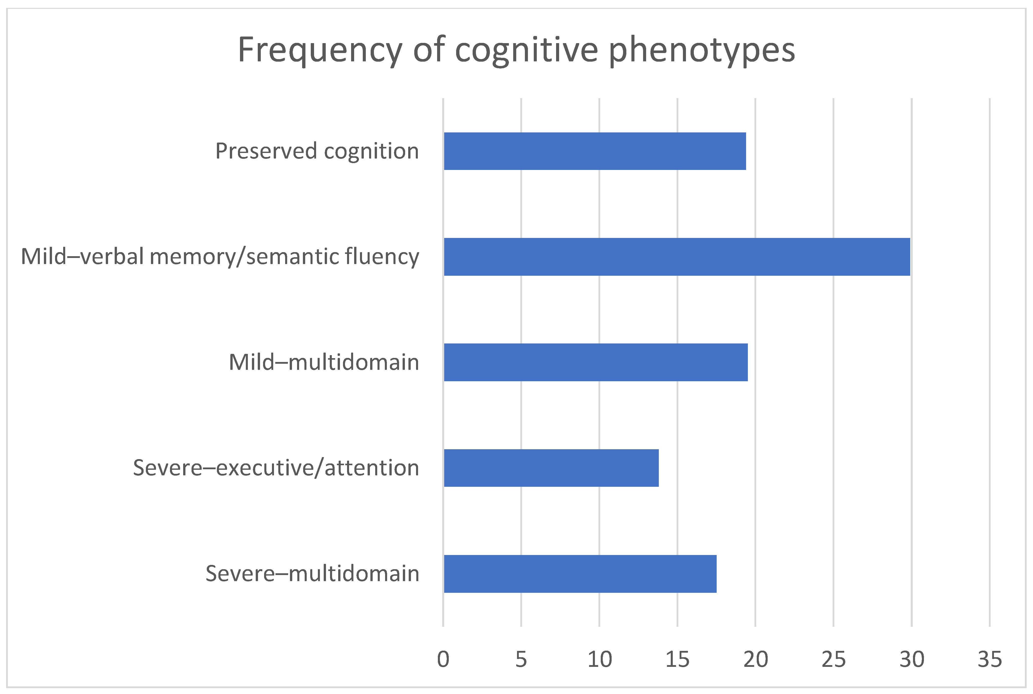
2. Prevalence and Profile of Cognitive Impairment in Multiple Sclerosis
3. Pathogenesis of Cognitive Impairment in Multiple Sclerosis
4. Neuropsychological Assessment
5. Treatment of Cognitive Impairment in Multiple Sclerosis
6. Conclusions and Future Directions
Funding
Conflicts of Interest
References
- McGinley, M.P.; Goldschmidt, C.H.; Rae-Grant, A.D. Diagnosis and Treatment of Multiple Sclerosis: A Review. JAMA 2021, 325, 765–779. [Google Scholar] [CrossRef] [PubMed]
- Kappos, L.; Wolinsky, J.S.; Giovannoni, G.; Arnold, D.L.; Wang, Q.; Bernasconi, C.; Model, F.; Koendgen, H.; Manfrini, M.; Belachew, S.; et al. Contribution of Relapse-Independent Progression vs Relapse-Associated Worsening to Overall Confirmed Disability Accumulation in Typical Relapsing Multiple Sclerosis in a Pooled Analysis of 2 Randomized Clinical Trials. JAMA Neurol. 2020, 77, 1132–1140. [Google Scholar] [CrossRef] [PubMed]
- Portaccio, E.; Bellinvia, A.; Fonderico, M.; Pastò, L.; Razzolini, L.; Totaro, R.; Spitaleri, D.; Lugaresi, A.; Cocco, E.; Onofrj, M.; et al. Progression is independent of relapse activity in early multiple sclerosis: A real-life cohort study. Brain 2022, 145, 2796–2805. [Google Scholar] [CrossRef] [PubMed]
- Lublin, F.D.; A Häring, D.; Ganjgahi, H.; Ocampo, A.; Hatami, F.; Čuklina, J.; Aarden, P.; Dahlke, F.; Arnold, D.L.; Wiendl, H.; et al. How patients with multiple sclerosis acquire disability. Brain 2022, 145, 3147–3161. [Google Scholar] [CrossRef] [PubMed]
- Charcot, J.M. Lectures on Diseases of the Nervous System; New Sydenham Society: London, UK, 1877. [Google Scholar]
- DeLuca, J.; Chiaravalloti, N.D.; Sandroff, B.M. Treatment and management of cognitive dysfunction in patients with multiple sclerosis. Nat. Rev. Neurol. 2020, 16, 319–332. [Google Scholar] [CrossRef]
- Benedict, R.H.B.; Amato, M.P.; DeLuca, J.; Geurts, J.J.G. Cognitive impairment in multiple sclerosis: Clinical management, MRI, and therapeutic avenues. Lancet Neurol. 2020, 19, 860–871. [Google Scholar] [CrossRef]
- Pardini, M.; Uccelli, A.; Grafman, J.; Yaldizli, Ö.; Mancardi, G.; Roccatagliata, L. Isolated cognitive relapses in multiple sclerosis. J. Neurol. Neurosurg. Psychiatry 2014, 85, 1035–1037. [Google Scholar] [CrossRef]
- Ruano, L.; Portaccio, E.; Goretti, B.; Niccolai, C.; Severo, M.; Patti, F.; Cilia, S.; Gallo, P.; Grossi, P.; Ghezzi, A.; et al. Age and disability drive cognitive impairment in multiple sclerosis across disease subtypes. Mult. Scler. J. 2016, 23, 1258–1267. [Google Scholar] [CrossRef] [Green Version]
- Zipoli, V.; Goretti, B.; Hakiki, B.; Siracusa, G.; Sorbi, S.; Portaccio, E.; Amato, M.P. Cognitive impairment predicts conversion to multiple sclerosis in clinically isolated syndromes. Mult. Scler. J. 2009, 16, 62–67. [Google Scholar] [CrossRef]
- Deloire, M.; Ruet, A.; Hamel, D.; Bonnet, M.; Brochet, B. Early cognitive impairment in multiple sclerosis predicts disability outcome several years later. Mult. Scler. J. 2010, 16, 581–587. [Google Scholar] [CrossRef]
- Moccia, M.; Lanzillo, R.; Palladino, R.; Chang, K.C.-M.; Costabile, T.; Russo, C.; De Rosa, A.; Carotenuto, A.; Sacca, F.; Maniscalco, G.T.; et al. Cognitive impairment at diagnosis predicts 10-year multiple sclerosis progression. Mult. Scler. J. 2016, 22, 659–667. [Google Scholar] [CrossRef] [PubMed] [Green Version]
- Cavaco, S.; Ferreira, I.; Moreira, I.; Santos, E.; Samões, R.; Sousa, A.P.; Pinheiro, J.; Teixeira-Pinto, A.; da Silva, A.M. Cognitive dysfunction and mortality in multiple sclerosis: Long-term retrospective review. Mult. Scler. J. 2021, 28, 1382–1391. [Google Scholar] [CrossRef] [PubMed]
- De Meo, E.; Portaccio, E.; Giorgio, A.; Ruano, L.; Goretti, B.; Niccolai, C.; Patti, F.; Chisari, C.G.; Gallo, P.; Grossi, P.; et al. Identifying the Distinct Cognitive Phenotypes in Multiple Sclerosis. JAMA Neurol. 2021, 78, 414–425. [Google Scholar] [CrossRef]
- Lin, X.; Zhang, X.; Liu, Q.; Zhao, P.; Zhong, J.; Pan, P.; Wang, G.; Yi, Z. Social cognition in multiple sclerosis and its subtypes: A meta-analysis. Mult. Scler. Relat. Disord. 2021, 52, 102973. [Google Scholar] [CrossRef]
- Battaglia, S.; Serio, G.; Scarpazza, C.; D’Ausilio, A.; Borgomaneri, S. Frozen in (e)motion: How reactive motor inhibition is influenced by the emotional content of stimuli in healthy and psychiatric populations. Behav. Res. Ther. 2021, 146, 103963. [Google Scholar] [CrossRef] [PubMed]
- Radlak, B.; Cooper, C.; Summers, F.; Phillips, L.H. Multiple sclerosis, emotion perception and social functioning. J. Neuropsychol. 2021, 15, 500–515. [Google Scholar] [CrossRef] [PubMed]
- Portaccio, E.; De Meo, E.; Bellinvia, A.; Amato, M. Cognitive Issues in Pediatric Multiple Sclerosis. Brain Sci. 2021, 11, 442. [Google Scholar] [CrossRef]
- Portaccio, E.; Bellinvia, A.; Razzolini, L.; Pastò, L.; Goretti, B.; Niccolai, C.; Fonderico, M.; Zaffaroni, M.; Pippolo, L.; Moiola, L.; et al. Long-term Cognitive Outcomes and Socioprofessional Attainment in People With Multiple Sclerosis With Childhood Onset. Neurology 2022, 98, e1626–e1636. [Google Scholar] [CrossRef]
- Jakimovski, D.; Weinstock-Guttman, B.; Roy, S.; Jaworski, M.I.; Hancock, L.; Nizinski, A.; Srinivasan, P.; Fuchs, T.A.; Szigeti, K.; Zivadinov, R.; et al. Cognitive Profiles of Aging in Multiple Sclerosis. Front. Aging Neurosci. 2019, 11, 105. [Google Scholar] [CrossRef]
- Butler Pagnotti, R.; Hua, L.H.; Miller, J.B. Cognition and disease characteristics in adult onset versus late onset multiple sclerosis. Mult Scler. 2022, 28, 933–941. [Google Scholar] [CrossRef]
- Branco, M.; Ruano, L.; Portaccio, E.; Goretti, B.; Niccolai, C.; Patti, F.; Chisari, C.; Gallo, P.; Grossi, P.; Ghezzi, A.; et al. Aging with multiple sclerosis: Prevalence and profile of cognitive impairment. Neurol. Sci. 2019, 40, 1651–1657. [Google Scholar] [CrossRef] [PubMed]
- Feinstein, A. Mood disorders in multiple sclerosis and the effects on cognition. J. Neurol. Sci. 2006, 245, 63–66. [Google Scholar] [CrossRef] [PubMed]
- Portaccio, E. Differential diagnosis, discerning depression from cognition. Acta Neurol. Scand. 2016, 134, 14–18. [Google Scholar] [CrossRef] [PubMed] [Green Version]
- Kalron, A.; Aloni, R.; Allali, G. The relationship between depression, anxiety and cognition and its paradoxical impact on falls in multiple sclerosis patients. Mult. Scler. Relat. Disord. 2018, 25, 167–172. [Google Scholar] [CrossRef]
- Oreja-Guevara, C.; Ayuso Blanco, T.; Brieva Ruiz, L.; Hernandez Perez, M.A.; Meca-Lallana, V.; Ramio-Torrenta, L. Cognitive Dysfunctions and Assessments in Multiple Sclerosis. Front. Neurol. 2019, 10, 581. [Google Scholar] [CrossRef]
- Andreasen, A.K.; Spliid, P.E.; Andersen, H.; Jakobsen, J. Fatigue and processing speed are related in multiple sclerosis. Eur. J. Neurol. 2009, 17, 212–218. [Google Scholar] [CrossRef]
- Patel, V.P.; Walker, L.; Feinstein, A. Processing speed and distractibility in multiple sclerosis: The role of sleep. Mult. Scler. Relat. Disord. 2017, 11, 40–42. [Google Scholar] [CrossRef]
- Braley, T.J.; Kratz, A.L.; Kaplish, N.; Chervin, R.D. Sleep and Cognitive Function in Multiple Sclerosis. Sleep 2016, 39, 1525–1533. [Google Scholar] [CrossRef] [Green Version]
- Riccitelli, G.C.; Pacifico, D.; Manconi, M.; Sparasci, D.; Sacco, R.; Gobbi, C.; Zecca, C. Relationship between cognitive disturbances and sleep disorders in multiple sclerosis is modulated by psychiatric symptoms. Mult. Scler. Relat. Disord. 2022, 64, 103936. [Google Scholar] [CrossRef]
- Sumowski, J.F.; Horng, S.; Brandstadter, R.; Krieger, S.; Leavitt, V.M.; Sand, I.K.; Fabian, M.; Klineova, S.; Graney, R.; Riley, C.S.; et al. Sleep disturbance and memory dysfunction in early multiple sclerosis. Ann. Clin. Transl. Neurol. 2021, 8, 1172–1182. [Google Scholar] [CrossRef]
- Di Filippo, M.; Portaccio, E.; Mancini, A.; Calabresi, P. Multiple sclerosis and cognition: Synaptic failure and network dysfunction. Nat. Rev. Neurosci. 2018, 19, 599–609. [Google Scholar] [CrossRef] [PubMed]
- Mandolesi, G.; Gentile, A.; Musella, A.; Fresegna, D.; De Vito, F.; Bullitta, S.; Sepman, H.; Marfia, G.A.; Centonze, D. Synaptopathy connects inflammation and neurodegeneration in multiple sclerosis. Nat. Rev. Neurol. 2015, 11, 711–724. [Google Scholar] [CrossRef] [PubMed]
- Dineen, R.A.; Vilisaar, J.; Hlinka, J.; Bradshaw, C.M.; Morgan, P.; Constantinescu, C.; Auer, D. Disconnection as a mechanism for cognitive dysfunction in multiple sclerosis. Brain 2009, 132, 239–249. [Google Scholar] [CrossRef] [PubMed]
- Rocca, M.A.; Pravatà, E.; Valsasina, P.; Radaelli, M.; Colombo, B.; Vacchi, L.; Gobbi, C.; Comi, G.; Falini, A.; Filippi, M. Hippocampal-DMN disconnectivity in MS is related to WM lesions and depression. Hum. Brain Mapp. 2015, 36, 5051–5063. [Google Scholar] [CrossRef] [Green Version]
- Rossi, F.; Giorgio, A.; Battaglini, M.; Stromillo, M.L.; Portaccio, E.; Goretti, B.; Federico, A.; Hakiki, B.; Amato, M.P.; De Stefano, N. Relevance of Brain Lesion Location to Cognition in Relapsing Multiple Sclerosis. PLoS ONE 2012, 7, e44826. [Google Scholar] [CrossRef]
- Foong, J.; Maier, M.; Barker, G.J.; Brocklehurst, S.; Miller, D.H.; A Ron, M. In vivo investigation of white matter pathology in schizophrenia with magnetisation transfer imaging. J. Neurol. Neurosurg. Psychiatry 2000, 68, 70–74. [Google Scholar] [CrossRef] [Green Version]
- Rovaris, M.; Iannucci, G.; Falautano, M.; Possa, F.; Martinelli, V.; Comi, G.; Filippi, M. Cognitive dysfunction in patients with mildly disabling relapsing–remitting multiple sclerosis: An exploratory study with diffusion tensor MR imaging. J. Neurol. Sci. 2002, 195, 103–109. [Google Scholar] [CrossRef]
- A Deloire, M.S.; Salort, E.; Bonnet, M.; Arimone, Y.; Boudineau, M.; Amieva, H.; Barroso, B.; Ouallet, J.-C.; Pachai, C.; Galliaud, E.; et al. Cognitive impairment as marker of diffuse brain abnormalities in early relapsing remitting multiple sclerosis. J. Neurol. Neurosurg. Psychiatry 2005, 76, 519–526. [Google Scholar] [CrossRef]
- Vrenken, H.; Geurts, J.J.G.; Knol, D.L.; Van Dijk, L.N.; Dattola, V.; Jasperse, B.; van Schijndel, R.; Polman, C.H.; Castelijns, J.A.; Barkhof, F.; et al. Whole-Brain T1 Mapping in Multiple Sclerosis: Global Changes of Normal-appearing Gray and White Matter. Radiology 2006, 240, 811–820. [Google Scholar] [CrossRef]
- Geurts, J.J.; Pouwels, P.J.; Uitdehaag, B.M.; Polman, C.H.; Barkhof, F.; Castelijns, J.A. Intracortical lesions in multiple sclerosis: Improved detection with 3D double inversion-recovery MR imaging. Radiology 2005, 236, 254–260. [Google Scholar] [CrossRef]
- Nelson, F.; Datta, S.; Garcia, N.; Rozario, N.L.; Perez, F.; Cutter, G.; Narayana, P.A.; Wolinsky, J.S. Intracortical lesions by 3T magnetic resonance imaging and correlation with cognitive impairment in multiple sclerosis. Mult. Scler. J. 2011, 17, 1122–1129. [Google Scholar] [CrossRef] [PubMed] [Green Version]
- Filippi, M.; Rocca, M.A.; Benedict, R.H.; DeLuca, J.; Geurts, J.J.; Rombouts, S.A.; Ron, M.; Comi, G. The contribution of MRI in assessing cognitive impairment in multiple sclerosis. Neurology 2010, 75, 2121–2128. [Google Scholar] [CrossRef] [PubMed]
- Amin, M.; Ontaneda, D. Thalamic Injury and Cognition in Multiple Sclerosis. Front. Neurol. 2020, 11, 623914. [Google Scholar] [CrossRef] [PubMed]
- Amato, M.P.; Bartolozzi, M.L.; Zipoli, V.; Portaccio, E.; Mortilla, M.; Guidi, L.; Siracusa, G.; Sorbi, S.; Federico, A.; De Stefano, N. Neocortical volume decrease in relapsing-remitting MS patients with mild cognitive impairment. Neurology 2004, 63, 89–93. [Google Scholar] [CrossRef]
- Amato, M.P.; Portaccio, E.; Goretti, B.; Zipoli, V.; Battaglini, M.; Bartolozzi, M.L.; Stromillo, M.L.; Guidi, L.; Siracusa, G.; Sorbi, S.; et al. Association of neocortical volume changes with cognitive deterioration in relapsing-remitting multiple sclerosis. Arch Neurol. 2007, 64, 1157–1161. [Google Scholar] [CrossRef] [Green Version]
- Calabrese, M.; Agosta, F.; Rinaldi, F.; Mattisi, I.; Grossi, P.; Favaretto, A.; Atzori, M.; Bernardi, V.; Barachino, L.; Rinaldi, L.; et al. Cortical Lesions and Atrophy Associated With Cognitive Impairment in Relapsing-Remitting Multiple Sclerosis. Arch. Neurol. 2009, 66, 1144–1150. [Google Scholar] [CrossRef] [Green Version]
- Eijlers, A.J.; Dekker, I.; Steenwijk, M.D.; Meijer, K.A.; Hulst, H.E.; Pouwels, P.J.; Uitdehaag, B.M.; Barkhof, F.; Vrenken, H.; Schoonheim, M.M.; et al. Cortical atrophy accelerates as cognitive decline worsens in multiple sclerosis. Neurology 2019, 93, e1348–e1359. [Google Scholar] [CrossRef]
- Di Filippo, M.; Mancini, A.; Bellingacci, L.; Gaetani, L.; Mazzocchetti, P.; Zelante, T.; La Barbera, L.; De Luca, A.; Tantucci, M.; Tozzi, A.; et al. Interleukin-17 affects synaptic plasticity and cognition in an experimental model of multiple sclerosis. Cell Rep. 2021, 37, 110094. [Google Scholar] [CrossRef]
- Brummer, T.; Muthuraman, M.; Steffen, F.; Uphaus, T.; Minch, L.; Person, M.; Zipp, F.; Groppa, S.; Bittner, S.; Fleischer, V. Improved prediction of early cognitive impairment in multiple sclerosis combining blood and imaging biomarkers. Brain Commun. 2022, 4, fcac153. [Google Scholar] [CrossRef]
- Yalachkov, Y.; Anschütz, V.; Jakob, J.; Schaller-Paule, M.A.; Schäfer, J.H.; Reiländer, A.; Friedauer, L.; Behrens, M.; Steffen, F.; Bittner, S.; et al. Brain-derived neurotrophic factor and neurofilament light chain in cerebrospinal fluid are inversely correlated with cognition in Multiple Sclerosis at the time of diagnosis. Mult. Scler. Relat. Disord. 2022, 63, 103822. [Google Scholar] [CrossRef]
- Tanaka, M.; Szabó, Á.; Spekker, E.; Polyák, H.; Tóth, F.; Vécsei, L. Mitochondrial Impairment: A Common Motif in Neuropsychiatric Presentation? The Link to the Tryptophan–Kynurenine Metabolic System. Cells 2022, 11, 2607. [Google Scholar] [CrossRef]
- Tanaka, M.; Vécsei, L. Monitoring the Redox Status in Multiple Sclerosis. Biomedicines 2020, 8, 406. [Google Scholar] [CrossRef] [PubMed]
- Komatsu, H.; Watanabe, E.; Fukuchi, M. Psychiatric Neural Networks and Precision Therapeutics by Machine Learning. Biomedicines 2021, 9, 403. [Google Scholar] [CrossRef] [PubMed]
- Schoonheim, M.M.; Broeders, T.A.; Geurts, J.J. The network collapse in multiple sclerosis: An overview of novel concepts to address disease dynamics. NeuroImage Clin. 2022, 35, 103108. [Google Scholar] [CrossRef] [PubMed]
- Rao, S.M.; Leo, G.J.; Bernardin, L.; Unverzagt, F. Cognitive dysfunction in multiple sclerosis. I. Frequency, patterns, and prediction. Neurology 1991, 41, 685–691. [Google Scholar] [CrossRef]
- Benedict, R.H.; DeLuca, J.; Phillips, G.; LaRocca, N.; Hudson, L.D.; Rudick, R. Multiple Sclerosis Outcome Assessments Consortium Validity of the Symbol Digit Modalities Test as a cognition performance outcome measure for multiple sclerosis. Mult. Scler. J. 2017, 23, 721–733. [Google Scholar] [CrossRef]
- Langdon, D.W.; Amato, M.P.; Boringa, J.; Brochet, B.; Foley, F.; Fredrikson, S.; Hämäläinen, P.; Hartung, H.-P.; Krupp, L.; Penner, I.-K.; et al. Recommendations for a Brief International Cognitive Assessment for Multiple Sclerosis (BICAMS). Mult. Scler. J. 2011, 18, 891–898. [Google Scholar] [CrossRef] [Green Version]
- Kalb, R.; Beier, M.; Benedict, R.H.; Charvet, L.; Costello, K.; Feinstein, A.; Gingold, J.; Goverover, Y.; Halper, J.; Harris, C.; et al. Recommendations for cognitive screening and management in multiple sclerosis care. Mult. Scler. J. 2018, 24, 1665–1680. [Google Scholar] [CrossRef] [Green Version]
- Wojcik, C.M.; Beier, M.; Costello, K.; DeLuca, J.; Feinstein, A.; Goverover, Y.; Gudesblatt, M.; Jaworski, M.; Kalb, R.; Kostich, L.; et al. Computerized neuropsychological assessment devices in multiple sclerosis: A systematic review. Mult. Scler. J. 2019, 25, 1848–1869. [Google Scholar] [CrossRef]
- Rao, S.M.; Losinski, G.; Mourany, L.; Schindler, D.; Mamone, B.; Reece, C.; Kemeny, D.; Narayanan, S.; Miller, D.M.; Bethoux, F.; et al. Processing speed test: Validation of a self-administered, iPad®-based tool for screening cognitive dysfunction in a clinic setting. Mult. Scler. J. 2017, 23, 1929–1937. [Google Scholar] [CrossRef]
- Ruet, A.; Deloire, M.S.; Charré-Morin, J.; Hamel, D.; Brochet, B. A new computerised cognitive test for the detection of information processing speed impairment in multiple sclerosis. Mult. Scler. J. 2013, 19, 1665–1672. [Google Scholar] [CrossRef]
- Benedict, R.H.; Cookfair, D.; Gavett, R.; Gunther, M.; Munschauer, F.; Garg, N.; Weinstock-Guttman, B. Validity of the minimal assessment of cognitive function in multiple sclerosis (MACFIMS). J. Int. Neuropsychol. Soc. 2006, 12, 549–558. [Google Scholar] [CrossRef]
- Benedict, R.H.B.; Fishman, I.; McClellan, M.M.; Bakshi, R.; Weinstock-Guttman, B. Validity of the Beck Depression Inventory-Fast Screen in multiple sclerosis. Mult. Scler. J. 2003, 9, 393–396. [Google Scholar] [CrossRef] [PubMed]
- Honarmand, K.; Feinstein, A. Validation of the Hospital Anxiety and Depression Scale for use with multiple sclerosis patients. Mult. Scler. J. 2009, 15, 1518–1524. [Google Scholar] [CrossRef] [PubMed]
- Landmeyer, N.C.; Bürkner, P.C.; Wiendl, H.; Ruck, T.; Hartung, H.P.; Holling, H.; Meuth, S.G.; Johnen, A. Disease-modifying treatments and cognition in relapsing-remitting multiple sclerosis: A meta-analysis. Neurology 2020, 94, e2373–e2383. [Google Scholar] [CrossRef] [PubMed]
- Chen, M.H.; Goverover, Y.; Genova, H.M.; DeLuca, J. Cognitive Efficacy of Pharmacologic Treatments in Multiple Sclerosis: A Systematic Review. CNS Drugs 2020, 34, 599–628. [Google Scholar] [CrossRef] [PubMed]
- DeLuca, J.; Schippling, S.; Montalban, X.; Kappos, L.; Cree, B.A.C.; Comi, G.; Arnold, D.L.; Hartung, H.P.; Sheffield, J.K.; Liu, H.; et al. Effect of Ozanimod on Symbol Digit Modalities Test Performance in Relapsing MS. Mult. Scler. Relat. Disord. 2021, 48, 102673. [Google Scholar] [CrossRef]
- Cree, B.A.; Arnold, D.L.; Fox, R.J.; Gold, R.; Vermersch, P.; Benedict, R.H.; Bar-Or, A.; Piani-Meier, D.; Rouyrre, N.; Ritter, S.; et al. Long-term efficacy and safety of siponimod in patients with secondary progressive multiple sclerosis: Analysis of EXPAND core and extension data up to >5 years. Mult. Scler. J. 2022, 28, 1591–1605. [Google Scholar] [CrossRef]
- Zhang, E.; Tian, X.; Li, R.; Chen, C.; Li, M.; Ma, L.; Wei, R.; Zhou, Y.; Cui, Y. Dalfampridine in the treatment of multiple sclerosis: A meta-analysis of randomised controlled trials. Orphanet J. Rare Dis. 2021, 16, 87. [Google Scholar] [CrossRef] [PubMed]
- Lampit, A.; Heine, J.; Finke, C.; Barnett, M.H.; Valenzuela, M.; Wolf, A.; Leung, I.H.K.; Hill, N.T.M. Computerized Cognitive Training in Multiple Sclerosis: A Systematic Review and Meta-analysis. Neurorehabil. Neural. Repair 2019, 33, 695–706. [Google Scholar] [CrossRef]
- Chiaravalloti, N.D.; Moore, N.B.; Nikelshpur, O.M.; DeLuca, J. An RCT to treat learning impairment in multiple sclerosis: The MEMREHAB trial. Neurology 2013, 81, 2066–2072. [Google Scholar] [CrossRef] [PubMed] [Green Version]
- Ghielen, I.; Rutten, S.; Boeschoten, R.E.; Gier, M.H.-D.; van Wegen, E.; Heuvel, O.A.V.D.; Cuijpers, P. The effects of cognitive behavioral and mindfulness-based therapies on psychological distress in patients with multiple sclerosis, Parkinson’s disease and Huntington’s disease: Two meta-analyses. J. Psychosom. Res. 2019, 122, 43–51. [Google Scholar] [CrossRef] [PubMed]
- Zarotti, N.; Eccles, F.; Broyd, A.; Longinotti, C.; Mobley, A.; Simpson, J. Third wave cognitive behavioural therapies for people with multiple sclerosis: A scoping review. Disabil. Rehabil. 2022. [Google Scholar] [CrossRef] [PubMed]


| Duration of Administration | Tests Included | Cognitive Functions Assessed | |
|---|---|---|---|
| Screening tests | |||
| Symbol Digit Modalities Test (SDMT) [57] | 5 min | Symbol Digit Modalities Test (SDMT) | Information processing speed |
| Processing Speed Test (PST) [61] | 5 min | Processing Speed Test (PST) | Information processing speed |
| Computerized Speed Cognitive Test (CSCT) [62] | 5 min | Computerized Speed Cognitive Test (CSCT) | Information processing speed |
| Brief Neuropsychological Batteries | |||
| Brief International Cognitive Assessment for Multiple Sclerosis (BICAMS) [58] | 15 min | Symbol Digit Modalities Test (SDMT) California Verbal Learning Test–2nd ed. (CVLT-II) Brief Visuospatial Memory Test–Revised (BVMTR) | Information processing speed Verbal learning and memory Visuospatial learning and memory |
| Brief Repeatable Neuropsychological Batter (BRNB) [56] | 45 min | Paced Auditory Serial Addition Test (PASAT) Symbol Digit Modalities Test (SDMT) Selective Reminding Test (SRT) 10/36 Spatial Recall Test (SPART) Controlled Oral Word Association Test (COWAT) | Attention Information processing speed Verbal learning and memory Visuospatial learning and memory Verbal fluency |
| Minimal Assessment of Cognitive Function in Multiple Sclerosis (MACFIMS) [63] | 90 min | Paced Auditory Serial Addition Test (PASAT) Symbol Digit Modalities Test (SDMT) California Verbal Learning Test–2nd ed. (CVLT-II) Brief Visuospatial Memory Test–Revised (BVMTR) Controlled Oral Word Association Test (COWAT) Judgement of Line Orientation Test (JLoT) Delis-Kaplan Executive Function System Sorting Test (D-KEFS-ST) | Attention Information processing speed Verbal learning and memory Visuospatial learning and memory Verbal fluency Visuospatial perception Executive functions |
| Cognitive impairment is now widely acknowledged as a prevalent core feature of the MS clinical picture, with critical consequences on patients’ lives. |
| The Symbol Digit Modalities Test is a quick, easy, and sensitive measure that should routinely be performed in MS clinical assessments. Wider cognitive screening should be considered in patients at high risk of cognitive impairment. |
| Cognitive screening should include evaluation of mood disorders and other factors impacting cognition, such as fatigue and sleep disorders. |
| Unfortunately, to date there are no approved medications for cognitive impairment in MS. It is conceivable that multimodal, personalized approaches, including pharmacological and non-pharmacological interventions, could allow better management of MS-related cognitive impairment. |
Publisher’s Note: MDPI stays neutral with regard to jurisdictional claims in published maps and institutional affiliations. |
© 2022 by the authors. Licensee MDPI, Basel, Switzerland. This article is an open access article distributed under the terms and conditions of the Creative Commons Attribution (CC BY) license (https://creativecommons.org/licenses/by/4.0/).
Share and Cite
Portaccio, E.; Amato, M.P. Cognitive Impairment in Multiple Sclerosis: An Update on Assessment and Management. NeuroSci 2022, 3, 667-676. https://doi.org/10.3390/neurosci3040048
Portaccio E, Amato MP. Cognitive Impairment in Multiple Sclerosis: An Update on Assessment and Management. NeuroSci. 2022; 3(4):667-676. https://doi.org/10.3390/neurosci3040048
Chicago/Turabian StylePortaccio, Emilio, and Maria Pia Amato. 2022. "Cognitive Impairment in Multiple Sclerosis: An Update on Assessment and Management" NeuroSci 3, no. 4: 667-676. https://doi.org/10.3390/neurosci3040048