Review

Percutaneous Vertebroplasty: A Minimally Invasive Procedure for the Management of Vertebral Compression Fractures

1
Department of Diagnostic and Interventional Radiology, Departmental Faculty of Medicine and Surgery, Fondazione Policlinico Universitario Campus Bio-Medico, 00128 Rome, Italy
2
Department of Orthopaedic and Trauma Surgery, Fondazione Policlinico Universitario Campus Bio-Medico, 00128 Rome, Italy
*
Author to whom correspondence should be addressed.
Osteology 2022, 2(4), 139-151; https://doi.org/10.3390/osteology2040017
Submission received: 25 June 2022 / Revised: 8 September 2022 / Accepted: 28 September 2022 / Published: 10 October 2022
(This article belongs to the Special Issue Feature Papers in Osteology)

Abstract

:
A vertebral compression fracture (VCF) is a pathological condition, which can be caused by osteoporotic degeneration or metastatic disease. It represents a socioeconomic burden on healthcare systems, due to increased pain, long-term morbidity, and disability. Vertebroplasty (VP) is an image-guided, minimally invasive, interventional procedure, in which bone cement is injected via a percutaneous approach into the vertebral soma, to provide structural support and to stabilize the weakened structure. The aim of this narrative review is to describe vertebral column biomechanics, as well as indications, contraindications, and techniques to successfully perform VP for the treatment of VCFs. Methods: We performed a narrative literature review on the main online databases regarding VP, and mainly focused on patient selection, preoperative imaging, procedural steps, complications, and outcomes. Results: The most recent evidence in the literature has shown that VP provides significant and sustained clinical benefits for patients with a VCF, and it is indicated in patients with comorbidities that make prolonged bed rest dangerous, patients with fractures that fail to heal, and as palliation in patients with a painful VCF due to metastatic disease. Conclusions: VP is considered to be a safe and effective treatment option for the treatment of osteoporotic and malignant VCFs that are resistant to adequate medical therapy. Patient selection, preprocedural evaluation, and proper technique execution are the key points to obtain the best outcomes and to minimize complications.

1. Introduction

A vertebral compression fracture (VCF) represents a common cause of debilitating back pain, which severely affects physical function, mental health, survival, and hence, quality of live [1,2]. Osteoporosis, neoplasms (e.g., myeloma, metastasis, lymphoma, and hemangioma), osteonecrosis, and trauma represent the most frequent etiologies [3].
Annually, osteoporotic VCFs involve approximately 1.4 million patients in the world [4], influencing patients’ quality of life and resulting in increased costs of public health care systems [5,6]. Regardless of etiology, the initial treatment of VCFs has always been considered to be conservative with administration of analgesic drugs [7]. The VERTOS III study, which evaluated a large group of symptomatic patients with VCFs, reported that 50% of patients experienced progressively decreased pain, especially during the first 3 months, while the other half of the patients reported insufficient reduction in pain at 12 months [8].
Bone metastases are often related to intense pain and pathological fractures, which result in functional alterations and increased disability, morbidity, and mortality. The handling of this condition is influenced by the therapeutic intent, the risk of fracture, or the eventuality of neurological compression [9].
Vertebra augmentation procedures (VAPs) include several minimally invasive interventional techniques that are used to treat VCFs, in which, under imaging guidance, bone cement is injected percutaneously to restore the strength of a vertebral body [10]. Percutaneous kyphoplasty (PKP) can restore vertebral height and correct kyphosis, by placing an inflatable bone tamp in a vertebral body subsequently filled with cement [11,12,13].
Before cement injection, percutaneous implant techniques (PIT) include the placement of different types of expandable bone implant systems in the vertebral body [7].
Percutaneous vertebroplasty (VP) is a minimally invasive technique that involves injection of bone cement into a partially collapsed vertebral body to improve stability and to obtain pain reduction. [11] VP initially entered the field of interventional radiology in the case of painful vertebral hemangioma, to obtain a reduction in pain [14]; subsequently, VP has been used in patients with a VCF to provide fast pain relief, and to improve functional capability, and hence, quality of life [15,16,17,18,19,20]. However, there is still controversy regarding the choice between VP and conservative management.
Conservative treatments commonly proposed in patients with a VCF include pharmacological medications, in particular, analgesics and behavioral interventions such as bed rest, bracing, and physiotherapy [21] with manual techniques and exercises in order to improve physical function and reduce disability [22].
In the literature on the use of orthosis for patients with a VCF, there is only one randomized controlled trial [23], in which, for most outcome measures, the differences between the kyphoplasty and the non-surgical groups were significant at one-month follow-up, but not at twelve-month follow-up [24].
However, the superiority of VAPs over conservative management is inconclusive, until the best conservative management for patients with a VCF is defined and standardized [21,25].
Recently published level I studies have shown significant advantages associated with VP as compared with conservative management. Klazen et al. [26], in a study involving 202 patients with acute osteoporotic VCFs, demonstrated immediate significantly greater pain relief after VP as compared with conservative management.
Another randomized controlled trial by Farrokhi et al. (2011) [27] demonstrated an instant five-point reduction in the pain visual analog scale (VAS) score after VP and better quality of life at 2, 6, 12, and 24 months, in association with a significantly better correction of spinal deformity and vertebral height restoration as compared with medical management.
A multidisciplinary strategy is mandatory for patients, who should go through this procedure with a proper adjuvant therapy and follow-up, correlating symptoms and the clinical signs with the imaging findings [1,28].
The purpose of this article is to present the vertebral biomechanics, indications and contraindications, and techniques to perform a successful percutaneous vertebroplasty.

2. Literature Search Method

A literature review on the use of VP for VCFs was performed using online databases. PubMed, MEDLINE, Google Scholar, and Web of Science were used to extract the articles, associated with the research keywords “percutaneous vertebroplasty” and “vertebral compression fractures”. Human studies written in English up to July 2022 were taken into consideration and included. In order to identify any missing publications, the reference lists of the relevant studies were screened. The literature search and study selection were performed independently by two investigators (G.P. and F.R.). First, the titles and abstracts were evaluated. Secondly, full texts were retrieved and evaluated. A third author (E.F.) resolved any discrepancies. Data on patient selection, preoperative imaging, procedural steps, complications, and outcomes of VP were extracted from each eligible study.

3. Results

3.1. Patient Selection

The indications and contraindications of percutaneous vertebroplasty that have been reported in the CIRSE guidelines are summarized in Table 1 [7].

3.2. Preoperative Imaging

Adequate preoperative imaging is essential because it is required to identify the vertebral fracture, to date the fracture, and to define the anatomy also assessing the integrity of the posterior vertebral somatic wall [1]; moreover, it excludes other differential diagnoses for back pain [2].
Plain spine radiographs, at least in an anteroposterior and lateral view, provide panoramic images of the levels affected in the disease process. An MRI is more accurate because it provides information essential to guide therapeutic management decisions, such as age and healing status of the lesion (acute vs. chronic, incompletely healed vs. consolidated). Due to bone marrow edema, acute, subacute and non-healed fractures are hypointense on T1W images and hyperintense on T2W and STIR sequences [2,29].
A CT scan can be executed [2] to acquire information concerning the presence of epidural or foraminal stenosis caused by extension of the tumor or retropulsed bone fragments, the location and extent of the lytic process, and the involvement of pedicles.

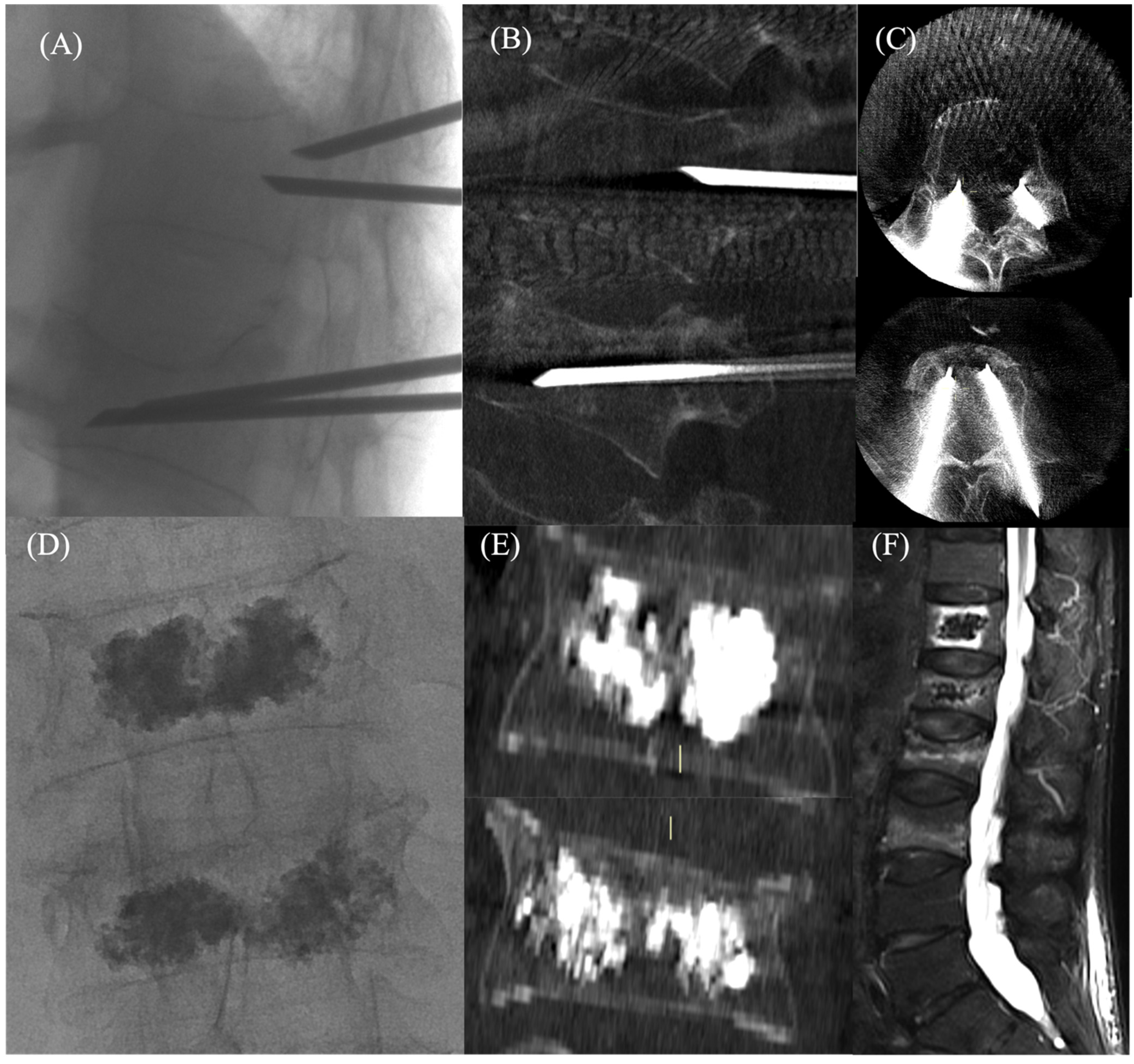
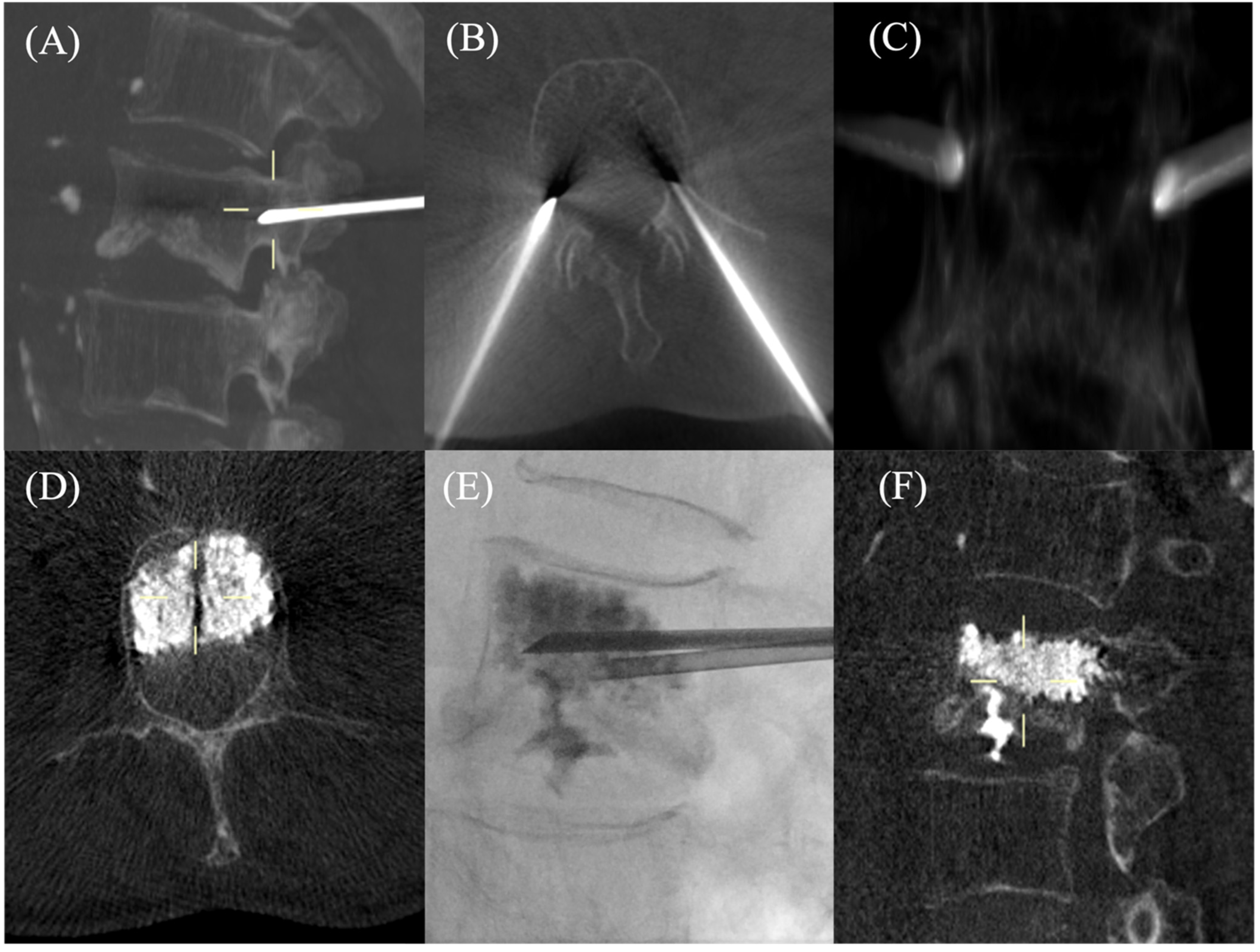
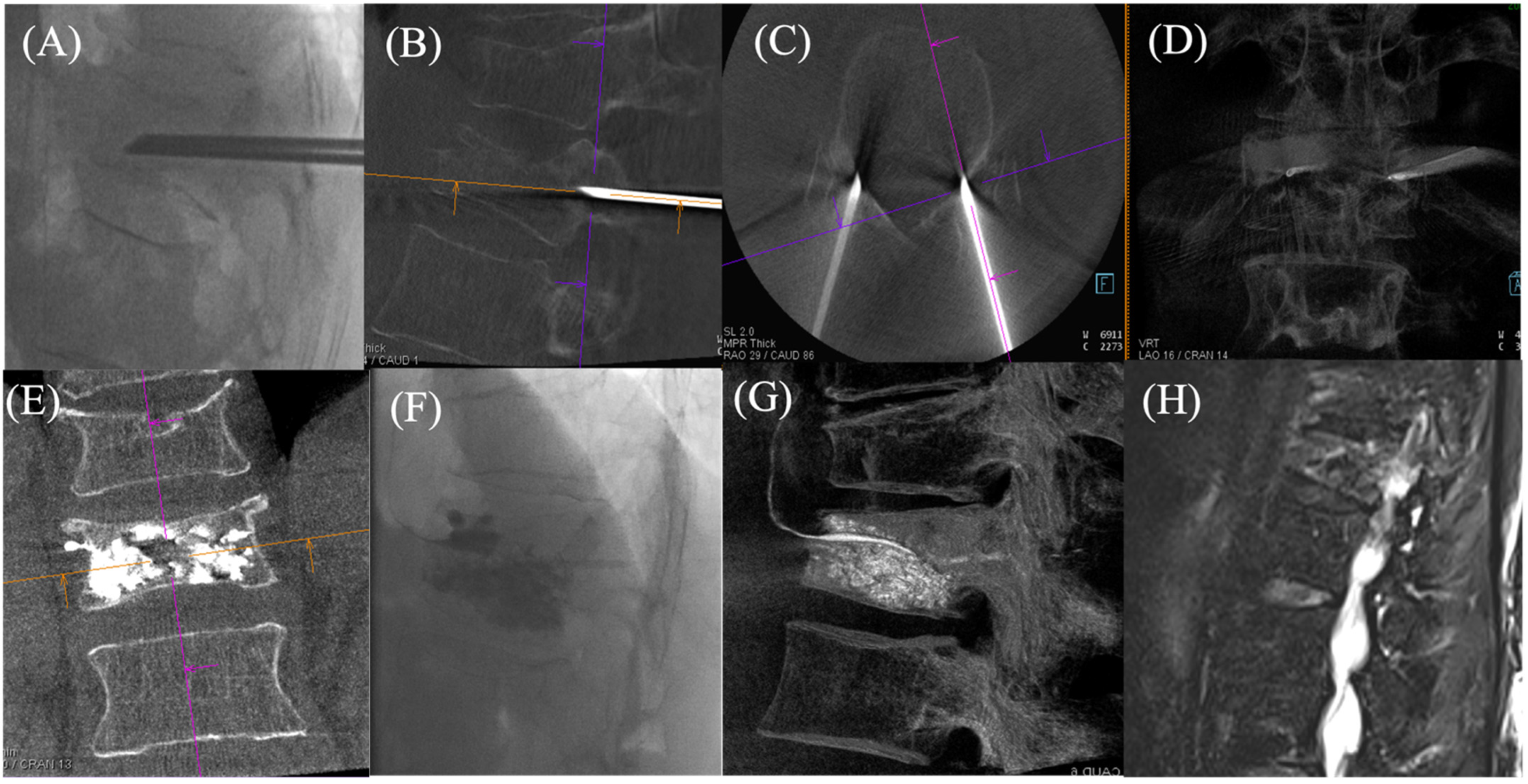
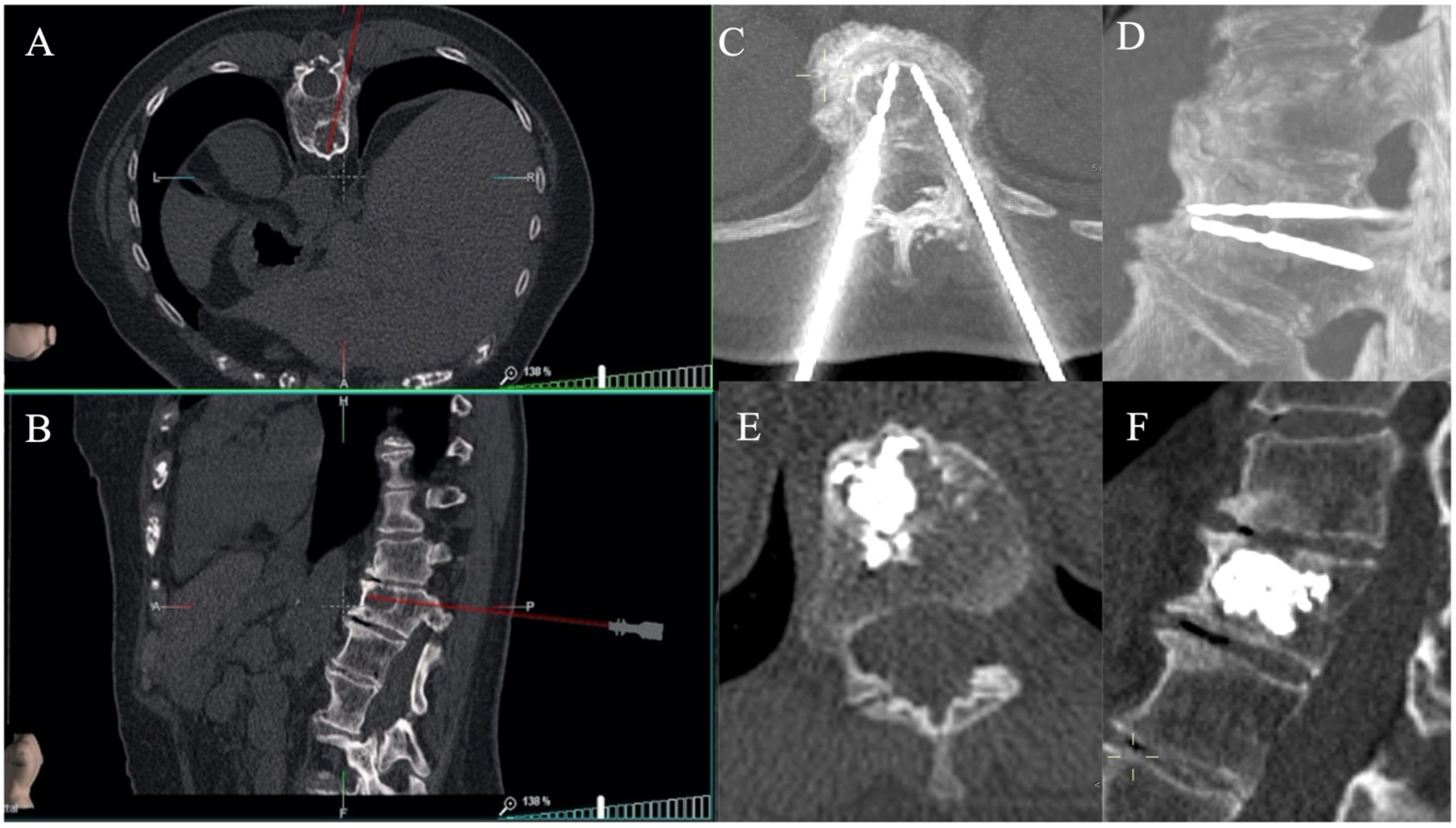
3.3. Procedural Steps

The procedure can be carried out under local anesthesia [30], conscious sedation, epidural/spinal anesthesia, or general anesthesia [31,32,33]. In all patients, a preprocedural antibiotic prophylaxis should be performed immediately before the procedure, accompanied by a mandatory intraprocedural antibiotic administration in the case of immuno-compromised patients [34,35]. The patient should assume a prone position, with padding to minimize spinal curvature [36].
Fluoroscopic guidance is the most widely used A CT scan is used especially in patients with lytic pathologic lesions and posterior wall cortical interruption [10]. Cone-beam CT, combined multidetector CT, or cone-beam CT and real-time fluoroscopy can be used to plan computer-assisted needle guidance [37,38]. In the absence of fluoroscopy, it can aid navigation systems for CT-guided percutaneous procedures.

Technique

Patients are usually in a prone position and a local anesthesia with a 22 gauge spinal needle is always performed during VP needle placement, under the skin and the periosteum. Identification of the pedicle is fundamental and it is usually performed using fluoroscopic guidance with an oblique plane [39].
A direct transoral approach should be performed to avoid neural and vascular structures in the case of C1 and C2 fractures; below that level, both anterolateral and posterior transpedicular approaches can be used [40]. Needle trajectory, planned in the case of the upper thoracic level, is through the horizontally orientated transverse process, using a unilateral transpedicular approach. As far as the lumbar spine is concerned, a unilateral transpedicular approach is indicated.
The trajectory of the needle is through the pedicle and the needle tip point to the midline and anterior third of the vertebral body. In the case of a bipedicular approach, the entrance of the needle is more medial and the trajectory is less oblique. For stable fractures of the sacral wings, a posterior approach is usually used, while for fractures of the sacral body (level S1, S2), the oblique approach through the SI joint is preferred [7]. Usually, 10–14 gauge needles with a blunt or diamond-tipped stylet are used [41]. The fluoroscopic C arm is arranged in a 20-/30-degree ipsilateral oblique view, and then it should be inclined cranially or caudally to obtain a “straight on” view of the vertebral body with a visual overlap of the anterior and posterior walls. With the aid of intermittent fluoroscopic images, the VP needle is advanced “down the barrel” of the pedicle, with a transpedicular approach, to reach the vertebral body. In order to limit the risk of nerve root or spinal cord injury, the needle should not violate the medial and inferior walls of the pedicle, hence, it should be advanced toward the upper and outer half of the face of the pedicle. Pedicle structures, especially in older degeneratively altered spines, are hardly recognizable in AP projection, and reliable identification of the safe zone is only possible in pedicle view to avoid misplacements.
To guarantee an adequate cranio-caudal inclination and to determine whether the needle has reached the posterior wall of the vertebral body, intermittent lateral views and AP (ipsilateral oblique) projections should be performed.
AP (ipsilateral oblique) projection is used to advance the needle and, once the posterior vertebral body has been reached, the needle should be advanced in the lateral projection with continuous visualization.
A bioptic sample can be obtained, if necessary, using a bone biopsy needle through the outer cannula [36].
Typically, the upper outer quadrant of the pedicle is targeted and the advancement of the needle into the vertebral body is monitored using alternating anteroposterior oblique and lateral fluoroscopy; the tip of the needle should be positioned sufficiently.
Close to the midline, at the junction of the anterior (ventral) and middle third of the vertebral body, the tip of the needle should be positioned sufficiently close to the midline, at the junction of the anterior (ventral) and middle third of the vertebral body [39]. Prior to definitive bone cement injection, final needle positioning should be confirmed in both the lateral and frontal projections (Figure 1).
Continuous lateral fluoroscopic guidance is used to monitor cement injection in order to detect any epidural leakage; while intermittent AP projections are used to evaluate cement distribution and depict any lateral leaks. During the injection, to allow the cement to homogenously distribute into the whole vertebral body, the needle is slowly pulled backward.
Specially designed bone cements exist and can be safely used for vertebral fractures. The viscosity, radiopacity, and polymerization time differ between the existing cements. The best cement is one that the user is familiar with, that is sufficiently radiopaque, and that allows enough working time.
Polymethyl methacrylate (PMMA) is the most common bone cement utilized for the treatment of osteoporotic and malignancy-related vertebral body fractures [36].
PMMA polymerizes and transitions from a liquid to a solid form via an exothermic reaction. The final state of the PMMA does not resorb over time and has an impressive capacity to sustain compressive forces. Other less commonly used bone cements include calcium phosphate cement, calcium sulfate cement, hydroxyapatite cement, and magnesium phosphate cement. In the United States, another type of cement is used, i.e., a bioactive calcium phosphate micro-glass cement (Cortoss; Stryker, Malvern, PA, USA). Cortoss is a non-resorbable composite; it consists of 33% difunctional methacrylates that form a cross-linked three-dimensional polymer reinforced with 67% of bioactive glass ceramic [42,43].
The risk of cement leakage is higher at the beginning of cement injection. If a leakage outside the vertebral body is observed, the injection should be temporarily stopped and the needle position and/or the bevel direction should be modified. Opacification of paravertebral or epidural veins need to be avoided.
Cement injection is considered to be completed when the anterior two-thirds of the vertebral body are filled and the cement is homogenously distributed. The volume of the cement injected depends on its consistency and the size of the vertebrae. The cannulas may be removed after an adequate amount of cement is injected (15–25% of the non-compressed vertebral body volume) and the cement has sufficiently hardened. Regardless of the viscosity of the bone cement, an injection volume >4 mL can provide patients with good improvement in clinical indicators and a low vertebral body re-collapse rate. The recommended volume of low-viscosity bone cement is 4–6 mL, while the optimal volume of high-viscosity bone cement is 6–8 mL [43,44].
It is important to remove the needle under fluoroscopy guidance to ensure no extraosseous cement deposition. If available, a CT scan or a cone-beam CT should be performed at the end of the procedure to further evaluate cement distribution and to detect leakage [7].
After the procedure, the patient should be in a prone position for about 2 h under adequate surveillance. A post-procedural neurological exam should be performed and compared with the patient preprocedural exam. [36]

3.4. Complications

The complication rates after VP are low, in particular, data published in the literature report complication rates of 2.2–3.9% related to osteoporotic fractures [45,46] and 11.5% in malignant fractures [47]. Moreover, the complications of VP are usually minor and and rarely require intervention.
Cement leakage from the vertebral body into adjacent structures represents one of the most common findings, with a rate of 30 to 80% [48]. A CT scan or cone-beam CT is superior to fluoroscopy for the detection of cement extravasation [49]. This event is usually asymptomatic, however, PMMA leakage into the epidural space can cause nerve root or spinal cord compression, with subsequential deficits and neurological symptoms [36]. Hence, radiculopathy can result from the contact of cement with an emerging nerve root. To mitigate this complication, an immediate infiltration with cool saline and steroids into the nerve foramina, could be performed, in order to reduce local inflammation and subsequent pain. In addition, cord compression is a serious complication that could require neurosurgical decompression [7]. Pain from exothermic heat or actual mass effect may result from extravasation into a disc or paravertebral tissues (Figure 2). In cases of severe osteoporosis, collapse of vertebral bodies can cause important adjacent disc leakage [7]. Cement leakage can also spread into the epidural and vertebral veins (Figure 3), thus, resulting in pulmonary emboli, which can be symptomatic or asymptomatic with an incidence ranging from 3.5 to 23% [50]. Usually, the embolus is lodged peripherally, hence, it is asymptomatic [51] and requires no treatment; rarely, a central pulmonary embolism could happen, leading to lung infarction [52,53]. Currently, there is no consensus on the management of cement pulmonary embolism, however, anticoagulation therapy for six months is recommended for an asymptomatic central or symptomatic peripheral embolism [7].
Other serious complications include infections and bleedings, however, their incidence is approximatively 1%. In particular, although vertebral infection is the rarest complication, it is associated with massive morbidity when it occurs since causal therapy involves a corpectomy of the cemented vertebral body. Moreover, fracture of ribs, posterior elements, or pedicle can occur in fewer than 1% of cases [7].
As already mentioned, VP can be associated with increased risk of collapse of the vertebral body immediately next to the treated level. However, data in the literature are contradictory, in fact, while some studies suggest that fractured vertebra augmentation can cause an increase in stress level and determine new adjacent vertebral fractures, others have not shown this correlation [54,55]. The VERTOS II open-label, randomized controlled clinical trial reported that there was no increment in the incidence of an adjacent vertebral body fracture after a VP as compared with conservative treatment [56].
Rare allergic reactions and transient hypotension related to the PMMA monomer have been described in the literature [36]. The risk of these complications could be minimized by correct positioning of the needle tip, limiting the number of treated levels to not more than five and using a high viscous cement, gradual injection of cement, and/or new strategies such as vertebral body lavage [31,53,57].

4. Discussion

Numerous studies have already demonstrated the safety and efficiency of VP procedures in the therapeutic management of osteoporotic and malignant VCFs that have failed to adequately respond to medical treatments [57,58,59,60,61,62,63].
An important reduction in pain following VP has been described in the literature in a substantial percentage of patients, in particular, in more than 90% of osteoporotic VCFs, 70% of malignant VCFs, and 80% of painful hemangiomas. A reduction in pain after VP, and therefore, less dependence on pain medications leads to an increase in physical mobility and independence, thus, resulting in a general improvement in the quality of life [64].
Recently, two randomized controlled trials were published in which patients were randomized to VP or a sham procedure [65,66]. These studies, with pain reduction as the primary outcome, have questioned the benefit of VP as compared with sham procedures and have received much criticism. Specifically, the main criticisms are the large discrepancy in the number of patients screened versus those who were ultimately enrolled, the need for a longer follow-up, and the fact that screening MRIs and bone scans were not required in one of the studies. Furthermore, persistence of pain after vertebroplasty may indicate etiologies for the pain other than fracture, and the use of cement helps to increase the structural stability of the treated vertebra in addition to a reduction in pain.
Adding support to the efficacy of VP and in contrast to the aforementioned two studies, one of the most recently published, multicenter, randomized, double-blind trials (VAPOUR) demonstrated that VP was strongly superior to placebo intervention for pain relief in patients with acute osteoporotic fractures for 6 weeks (between-group difference 23 percentage points, 95% CI 6–39, p = 0.011), with the highest benefit in the thoraco-lumbar spinal segment [67]. The VERTOS II trial, comparing VP with conservative treatments for acute VCF (less than 6 weeks of symptoms) showed an important improvement in pain regarding the VP group (the difference between groups in reduction of mean VAS score from baseline was 2.6 (95% CI 1.74–3.37, p < 0.0001) at 1 month and 2.0 (1.13–2.80, p < 0.0001) at 1 year) [26].
VP represents a minimally invasive percutaneous procedure that usually only requires a few days of hospitalization, thus, contributing to significantly reduced costs of VP in EU-member countries, in particular, in Italy as compared with other countries and in the USA [68,69].
Even in the case of malignant spinal disease, VP has been shown to result in reduced pain and improved disability [49,70]. In these cases, the use of a combined treatment with VP and thermal ablation (Figure 4), radiofrequency ablation, or cryoablation, is currently proposed and performed in several centers [71]. In fact, it is believed that this dual approach, using both cement and thermal ablation, can provide more lasting clinical results through wider ablation of the underlying tumor [39].

5. Conclusions

Percutaneous VP is a widely accepted interventional radiology technique that can be considered to be a safe and effective treatment, as well as cost-effective, in the therapeutic management of osteoporotic and malignant VFs that are resistant to non-invasive therapies. Appropriate patient selection, preprocedural evaluation, and proper technique execution are the key points to obtain best outcomes and to minimize complications.

Author Contributions

Conceptualization, E.F. and G.P.; methodology, E.F., G.P. and F.R.; validation, E.F and G.V.; investigation, C.A. and C.B.; data curation, M.S. and L.C.; writing—original draft preparation, G.P. and F.R.; writing—review and editing, E.F. and G.P.; visualization, G.V., B.B.Z. and R.F.G.; supervision, G.V., B.B.Z. and R.F.G. All authors have read and agreed to the published version of the manuscript.

Funding

This research received no external funding.

Institutional Review Board Statement

The study was conducted in accordance with the Declaration of Helsinki, and approved by the Institutional Review Board of the University Hospital of Campus Bio-Medico in Rome (https://www.unicampus.it/documents/UCBM_Codice_Etico.pdf, accessed on 29 January 2019) and we obtained a written informed consent from each enrolled patient.

Data Availability Statement

No new data were created or analyzed in this study. Data sharing is not applicable to this article.

Conflicts of Interest

The authors declare no conflict of interest.

References

  1. Phillips, F. Minimal invasive treatment of osteoporotic vertebral compression fractures. Spine 2003, 28, s45–s53. [Google Scholar] [CrossRef]
  2. Stallemeyer, M.; Zoarski, G.; Obuchowski, A. Bernadette optimizing patient selection in percutaneous vertebroplasty. Interv. Radiol. 2003, 14, 683–696. [Google Scholar] [CrossRef]
  3. Wade, S.W.; Strader, C.; Fitzpatrick, L.A.; Anthony, M.S. Sex- and age-specific incidence of non-traumatic fractures in selected industrialized countries. Arch. Osteoporos. 2012, 7, 219–227. [Google Scholar] [CrossRef]
  4. Johnell, O.; Kanis, J.A. An estimate of the worldwide prevalence and disability associated with osteoporotic fractures. Osteoporos. Int. 2006, 17, 1726–1733. [Google Scholar] [CrossRef] [PubMed]
  5. Kado, D.M.; Duong, T.; Stone, K.L.; Ensrud, K.E.; Nevitt, M.C.; Greendale, G.A.; Cummings, S.R.; Stone, K.L.; Ensrud, K.E. Incident vertebral fractures and mortality in older women: A prospective study. Osteoporos. Int. 2003, 14, 589–594. [Google Scholar] [CrossRef]
  6. Lau, E.; Ong, K.; Kurtz, S.; Schmier, J.; Edidin, A. Mortality following the diagnosis of a vertebral compression fracture in the medicare population. J. Bone Joint Surg. Am. 2008, 90, 1479–1486. [Google Scholar] [CrossRef] [Green Version]
  7. Tsoumakidou, G.; Too, C.W.; Koch, G.; Caudrelier, J.; Cazzato, R.L.; Garnon, J.; Gangi, A. CIRSE Guidelines on Percutaneous Vertebral Augmentation. Cardiovasc. Intervent. Radiol. 2017, 40, 331–342. [Google Scholar] [CrossRef] [PubMed]
  8. Venmans, A.; Lohle, P.N.M.; van Rooij, W.J. Pain course in conservatively treated patients with back pain and a VCF on the spine radiograph (VERTOS III). Skeletal. Radiol. 2014, 43, 13–18. [Google Scholar] [CrossRef] [PubMed]
  9. Laredo, J.D.; Chiras, J.; Kemel, S.; Taihi, L.; Hamze, B. Vertebroplasty and interventional radiology procedures for bone metastases. Joint Bone Spine 2018, 85, 191–199. [Google Scholar] [CrossRef] [PubMed]
  10. Khan, M.; Kushchayev, S.V. Percutaneous Vertebral Body Augmentations: The State of Art. Neuroimaging Clin. N. Am. 2019, 29, 495–513. [Google Scholar] [CrossRef] [PubMed]
  11. Lieberman, I.H.; Togawa, D.; Kayanja, M.M. Vertebroplasty and kyphoplasty: Filler materials. Spine J. 2005, 5, S305–S316. [Google Scholar] [CrossRef]
  12. Robinson, Y.; Heyde, C.E.; Fo¨rsth, P.; Olerud, C. Kyphoplasty in osteoporotic vertebral compression fractures—Guidelines and technical considerations. J. Orthop. Surg. Res. 2011, 6, 43. [Google Scholar] [CrossRef] [Green Version]
  13. Denaro, L.; Longo, U.G.; Denaro, V. Vertebroplasty and kyphoplasty: Reasons for concern? Orthop. Clin. N. Am. 2009, 40, 465–471. [Google Scholar] [CrossRef] [PubMed]
  14. Galibert, P.; Deramond, H.; Rosat, P.; Le Gars, D. Preliminary note on the treatment of vertebral angioma by percutaneous acrylic vertebroplasty. Neurochirurgie 1987, 33, 166–168. [Google Scholar] [PubMed]
  15. Buchbinder, R.; Johnston, R.V.; Rischin, K.J.; Homik, J.; Jones, C.A.; Golmohammadi, K.; Kallmes, D.F. Percutaneous vertebroplasty for osteoporotic vertebral compression fracture. Rev. Cochrane Database Syst. Rev. 2018, 4, CD006349. [Google Scholar] [PubMed]
  16. Kobayashi, K.; Shimoyama, K.; Nakamura, K.; Murata, K. Percutaneous vertebroplasty immediately relieves pain of osteoporotic vertebral compression fractures and prevents prolonged immobilization of patients. Eur. Radiol. 2005, 15, 360–367. [Google Scholar] [CrossRef]
  17. Masala, S.; Mastrangeli, R.; Petrella, M.C.; Massari, F.; Ursone, A.; Simonetti, G. Percutaneous vertebroplasty in 1,253 levels: Results and long-term effectiveness in a single centre. Eur. Radiol. 2009, 19, 165–171. [Google Scholar] [CrossRef] [PubMed]
  18. Anselmetti, G.C.; Corrao, G.; Monica, P.D.; Tartaglia, V.; Manca, A.; Eminefendic, H.; Russo, F.; Tosetti, I.; Regge, D. Pain relief following percutaneous vertebroplasty: Results of a series of 283 consecutive patients treated in a single institution. Cardiovasc. Intervent. Radiol. 2007, 30, 441–447. [Google Scholar] [CrossRef]
  19. Evans, A.J.; Jensen, M.E.; Kip, K.E.; DeNardo, A.J.; Lawler, G.J.; Negin, G.A.; Remley, K.B.; Boutin, S.M.; Dunnagan, S.A. Vertebral compression fractures: Pain reduction and improvement in functional mobility after percutaneous polymethylmethacrylate vertebroplasty retrospective report of 245 cases. Radiology 2003, 226, 366–372. [Google Scholar] [CrossRef] [Green Version]
  20. Cohen, J.E.; Lylyk, P.; Ceratto, R.; Kaplan, L.; Umansky, F.; Gomori, J.M. Percutaneous vertebroplasty: Technique and results in 192 procedures. Neurol. Res. 2004, 26, 41–49. [Google Scholar] [CrossRef]
  21. Longo, U.G.; Loppini, M.; Denaro, L.; Maffulli, N.; Denaro, V. Conservative management of patients with an osteoporotic vertebral fracture: A review of the literature. J. Bone Joint Surg. Br. 2012, 94, 152–157. [Google Scholar] [CrossRef] [PubMed]
  22. Bennell, K.L.; Matthews, B.; Greig, A.; Briggs, A.; Kelly, A.; Sherburn, M.; Larsen, J.; Wark, J. Effects of an exercise and manual therapy program on physical impairments, function and quality-of-life in people with osteoporotic vertebral fracture: A randomised, single-blind controlled pilot trial. BMC Musculoskelet. Disord. 2010, 11, 36. [Google Scholar] [CrossRef] [PubMed] [Green Version]
  23. Pfeifer, M.; Begerow, B.; Minne, H.W. Effects of a new spinal orthosis on posture, trunk strength, and quality of life in women with postmenopausal osteoporosis: A randomized trial. Am. J. Phys. Med. Rehabil. 2004, 83, 177–186. [Google Scholar] [CrossRef]
  24. Longo, U.G.; Denaro, V. Spinal augmentation: What have we learnt? Lancet 2009, 373, 1947. [Google Scholar] [CrossRef]
  25. Longo, U.G.; Loppini, M.; Denaro, L.; Maffulli, N.; Denaro, V. Osteoporotic vertebral fractures: Current concepts of conservative care. Br. Med. Bull. 2012, 102, 171–189. [Google Scholar] [CrossRef] [PubMed] [Green Version]
  26. Klazen, C.A.; Lohle, P.N.; de Vries, J.; Jansen, F.H.; Tielbeek, A.V.; Blonk, M.C.; Venmans, A.; van Rooij, W.J.J.; Schoemaker, M.C.; Juttmann, J.R.; et al. Vertebroplasty versus conservative treatment in acute osteoporotic vertebral compression fractures (Vertos II): An open-label randomised trial. Lancet 2010, 376, 1085–1092. [Google Scholar] [CrossRef] [Green Version]
  27. Farrokhi, M.R.; Alibai, E.; Maghami, Z. Randomized controlled trial of percutaneous vertebroplasty versus optimal medical management for the relief of pain and disability in acute osteoporotic vertebral compression fractures. J. Neurosurg. Spine 2011, 14, 561–569. [Google Scholar] [CrossRef] [Green Version]
  28. Zoarski, G.H.; Snow, P.; Olan, W.J.; Stallmeyer, M.J.B.; Dick, B.W.; Hebel, J.R.; De Deyne, M. Percutaneous vertebroplasty for osteoporotic compression fractures: Quantitative prospective evaluation of long-term outcomes. J. Vasc. Interv. Radiol. 2002, 13, 139–148. [Google Scholar] [CrossRef]
  29. Mathis, J.M.; Barr, J.D.; Belkoff, S.M.; Barr, M.S.; Jensen, M.E.; Deramond, H. Percutaneous vertebroplasty: A developing standard of care for vertebral compression fractures. AJNR Am. J. Neuroradiol. 2001, 22, 373–381. [Google Scholar]
  30. Bonnard, E.; Foti, P.; Kastler, A.; Amoretti, N. Percutaneous vertebroplasty under local anaesthesia: Feasibility regarding patients’ experience. Eur. Radiol. 2016, 27, 1512–1516. [Google Scholar] [CrossRef]
  31. Uppin, A.A.; Hirsch, J.A.; Centenera, L.V.; Pfiefer, B.A.; Pazianos, A.G.; Choi, I.S. Occurrence of new vertebral body fracture after percutaneous vertebroplasty in patients with osteoporosis. Radiology 2003, 226, 119–124. [Google Scholar] [CrossRef] [PubMed]
  32. White, S.M. Anaesthesia for percutaneous vertebroplasty. Anaesthesia 2002, 57, 1229–1230. [Google Scholar] [PubMed]
  33. Martin, J.B.; Jean, B.; Sugiu, K.; San Milla´n Ruı´z, D.; Piotin, M.; Murphy, K.; Rüfenacht, B.; Muster, M.; Rüfenacht, D.A. Vertebroplasty: Clinical experience and followup results. Bone 1999, 25, 11S–15S. [Google Scholar] [CrossRef]
  34. Gangi, A.; Sabharwal, T.; Irani, F.G.; Buy, X.; Morales, J.P.; Adam, A. Standards of Practice Committee of the Society of Interventional Radiology. Quality assurance guidelines for percutaneous vertebroplasty. Cardiovasc. Intervent. Radiol. 2006, 29, 173–178. [Google Scholar] [CrossRef]
  35. Beall, D.P.; Datir, A.; D’Souza, S.L.; D’Souza, L.S.; Gunda, D.; Morelli, J.; Johnson, M.B.; Nabavizadeh, N. Percutaneous treatment of insufficiency fractures: Principles, technique and review of literature. Skelet. Radiol. 2010, 39, 117–130. [Google Scholar] [CrossRef] [PubMed]
  36. Jay, B.; Ahn, S.H. Vertebroplasty. Semin. Intervent. Radiol. 2013, 30, 297–306. [Google Scholar]
  37. Braak, S.J.; Zuurmond, K.; Aerts, H.C.J.; van Leersum, M.; Overtoom, T.T.T.; van Heesewijk, J.P.M.; van Strijen, M.J. Feasibility study of needle placement in percutaneous vertebroplasty: Cone-beam computed tomography guidance versus conventional fluoroscopy. Cardiovasc. Interv. Radiol. 2013, 36, 1120–1126. [Google Scholar] [CrossRef]
  38. Gangi, A.; Kastler, B.A.; Dietemann, J.L. Percutaneous vertebroplasty guided by a combination of CT and fluoroscopy. AJNR Am. J. Neuroradiol. 1994, 15, 83–86. [Google Scholar] [PubMed]
  39. RikHargunani, i.; le Corroller, T.; Khashoggi, K.; Liu, D.M.; Marchinkow, L.O.; Mudri, M.J.; Murphy, K.P.; Ouellette, H.A.; Munk, P.L. An overview of vertebroplasty: Current status, controversies, and future directions. Can. Assoc. Radiol. J. 2012, 63, S11–S17. [Google Scholar] [CrossRef] [Green Version]
  40. Anselmetti, G.C.; Manca, A.; Montemurro, F.; Tutton, S.; Chiara, G.; Battistella, M.; Savojardo, M.; Marcia, S.; Masala, S.; Regge, D. Vertebroplasty using transoral approach in painful malignant involvement of the second cervical vertebra (C2): A single-institution series of 25 patients. Pain Physician 2012, 15, 35–42. [Google Scholar] [CrossRef]
  41. Murphy, D.T.; Korzan, J.R.; Ouellette, H.A.; Liu, D.M.; Clarkson, P.W.; Munk, P.L. Driven around the bend: Novel use of a curved steerable needle. Cardiovasc. Interv. Radiol. 2013, 36, 531–535. [Google Scholar] [CrossRef]
  42. AlvBravo, A.E.; Brasuell, J.E.; Favre, A.W.; Koenig, B.M.; Khan, A.A.; Beall, D.P. Treating Vertebral Compression Fractures: Establishing the appropriate diagnosis, preoperative considerations, treatment techniques, postoperative follow-up and general guidelines for the treatment of patients with symptomatic vertebral compression fractures. Tech. Vasc. Interv. Radiol. 2020, 23, 100701. [Google Scholar]
  43. Clerk-Lamalice, O.; Beall, D.P.; Ong, K.; Lorio, M.P. ISASS Policy 2018-Vertebral Augmentation: Coverage indications, limitations, and/or medical necessity. Int. J. Spine Surg. 2019, 13, 1–10. [Google Scholar] [CrossRef] [PubMed]
  44. Wang, M.; Zhang, L.; Fu, Z.; Wang, H.; Wu, Y. Selections of Bone Cement Viscosity and Volume in Percutaneous Vertebroplasty: A Retrospective Cohort Study. World Neurosurg. 2021, 150, e218–e227. [Google Scholar]
  45. McGirt, M.J.; Parker, S.L.; Wolinsky, J.-P.; Witham, T.F.; Bydon, A.; Gokaslan, Z.L. Vertebroplasty and kyphoplasty for the treatment of vertebral compression fractures: An evidenced-based review of the literature. Spine J. 2009, 9, 501–508. [Google Scholar] [CrossRef] [PubMed]
  46. Saracen, A.; Kotwica, Z. Complications of percutaneous vertebroplasty: An analysis of 1100 procedures performed in 616 patients. Medicine 2016, 95, e3850. [Google Scholar] [CrossRef]
  47. Chew, C.; Craig, L.; Edwards, R.; Moss, J.; O’Dwyer, P.J. Safety and efficacy of percutaneous vertebroplasty in malignancy: A systematic review. Clin. Radiol. 2011, 66, 63–72. [Google Scholar] [CrossRef]
  48. Vallejo, R.; Benyamin, R. Vertebral augmentation techniques for the treatment of vertebral compression fractures: A review. Reg. Anesth. Pain Manag. 2010, 14, 133–141. [Google Scholar] [CrossRef]
  49. Martin, D.J.; Rad, A.E.; Kallmes, D.F. Prevalence of extravertebral cement leakage after vertebroplasty: Procedural documentation versus CT detection. Acta Radiol. 2012, 53, 569–572. [Google Scholar] [CrossRef]
  50. Krueger, A.; Bliemel, C.; Zettl, R.; Ruchholtz, S. Management of pulmonary cement embolism after percutaneous vertebroplasty and kyphoplasty: A systematic review of the literature. Eur. Spine J. 2009, 18, 1257–1265. [Google Scholar] [CrossRef] [Green Version]
  51. Scroop, R.; Eskridge, J.; Britz, G.W. Paradoxical cerebral arterial embolization of cement during intraoperative vertebroplasty: Case report. AJNR Am. J. Neuroradiol. 2002, 23, 868–870. [Google Scholar] [PubMed]
  52. Jensen, M.E.; Evans, A.J.; Mathis, J.M.; Kallmes, D.F.; Cloft, H.J.; Dion, J.E. Percutaneous polymethylmethacrylate vertebroplasty in the treatment of osteoporotic vertebral body compression fractures: Technical aspects. AJNR Am. J. Neuroradiol. 1997, 18, 1897–1904. [Google Scholar] [PubMed]
  53. Gardos, F.; Depriester, C.; Cayrolle, G. Long term observations of vertebral osteoporotic fractures treated by percutaneous vertebroplasty. Rheumatology 2000, 39, 1410–1414. [Google Scholar] [CrossRef] [PubMed] [Green Version]
  54. Nagaraja, S.; Awada, H.K.; Dreher, M.L.; Gupta, S.; Miller, S.W. Vertebroplasty increases compression of adjacent IVDs and vertebrae in osteoporotic spines. Spine J. 2013, 13, 1872–1880. [Google Scholar] [CrossRef]
  55. Aquarius, R.; van der Zijden, A.M.; Homminga, J.; Verdonschot, N.; Tanck, E. Does bone cement in percutaneous vertebroplasty act as a stress riser? Spine 1976, 2013, 2092–2097. [Google Scholar] [CrossRef]
  56. Klazen, C.A.H.; Venmans, A.; de Vries, J.; van Rooij, W.J.; Jansen, F.H.; Blonk, M.C.; Lohle, P.; Juttmann, J.; Buskens, E.; Van Everdingen, K.; et al. Percutaneous vertebroplasty is not a risk factor for new osteoporotic compression fractures: Results from VERTOS II. Am. J. Neuroradiol. 2010, 31, 1447–1450. [Google Scholar] [CrossRef] [Green Version]
  57. Albers, C.E.; Schott, P.M.; Ahmad, S.S.; Benneker, L.M.; Nieuwkamp, N.; Hoppe, S. Vertebral Body Lavage Reduces Hemodynamic Response to Vertebral Body Augmentation With PMMA. Glob. Spine J. 2019, 9, 499–504. [Google Scholar] [CrossRef]
  58. Berenson, J.; Pflugmacher, R.; Jarzem, P.; Zonder, J.; Schechtman, K.; Tillman, J.B.; Bastian, L.; Ashraf, T.; Vrionis, F. Cancer Patient Fracture Evaluation (CAFE) Investigators. Balloon kyphoplasty versus nonsurgical fracture management for treatment of painful vertebral body compression fractures in patients with cancer: A multicentre, randomised controlled trial. Lancet Oncol. 2011, 12, 225–235. [Google Scholar] [CrossRef]
  59. Hadjipavlou, A.G.; Tzermiadianos, M.N.; Katonis, P.G.; Szpalski, M. Percutaneous vertebroplasty and balloon kyphoplasty for the treatment of osteoporotic vertebral compression fractures and osteolytic tumours. J. Bone Joint Surg. Br. 2005, 87, 1595–1604. [Google Scholar] [CrossRef] [Green Version]
  60. Kushchayev, S.V.; Wiener, P.C.; Teytelboym, O.M.; Arrington, J.A.; Khan, M.; Preul, M.C. Percutaneous Vertebroplasty: A history of procedure, technology, culture, specialty, and economics. Rev. Neuroimaging Clin. N. Am. 2019, 29, 481–494. [Google Scholar] [CrossRef]
  61. Taylor, R.S.; Fritzell, P.; Taylor, R.J. Balloon kyphoplasty in the management of vertebral compression fractures: An updated systematic review and meta-analysis. Eur. Spine J. 2007, 16, 1085–1100. [Google Scholar] [CrossRef] [PubMed] [Green Version]
  62. Key, B.M.; Symanski, J.; Scheidt, M.J.; Tutton, S.M. Vertebroplasty, Kyphoplasty, and Implant-Based Mechanical Vertebral Augmentation. Rev. Semin. Musculoskelet. Radiol. 2021, 25, 785–794. [Google Scholar] [CrossRef] [PubMed]
  63. Voormolen, M.H.; Mali, W.P.; Lohle, P.N.; Fransen, H.; Lampmann, L.E.H.; Van der Graaf, Y.; Juttmann, J.R.; Jansssens, X.; Verhaar, H.J.J. Percutaneous vertebroplasty compared with optimal pain medication treatment: Short term clinical outcome of patients with subacute or chronic painful osteoporotic vertebral compression fractures. The VERTOS study. AJNR Am. J. Neuroradiol. 2007, 28, 555–560. [Google Scholar] [PubMed]
  64. Peh, W.C.; Munk, P.L.; Rashid, F.; Gilula, L.A. Percutaneous vertebral augmentation: Vertebroplasty, kyphoplasty and skyphoplasty. Radiol. Clin. North Am. 2008, 46, 611–635. [Google Scholar] [CrossRef]
  65. Kallmes, D.F.; Comstock, B.A.; Heagerty, P.J.; Turner, J.A.; Wilson, D.J.; Diamond, T.H.; Edwards, R.; Gray, L.A.; Stout, L.; Owen, S.; et al. A randomized trial of vertebroplasty for osteoporotic spinal fractures. N. Engl. J. Med. 2009, 361, 569–579. [Google Scholar] [CrossRef] [Green Version]
  66. Buchbinder, R.; Osborne, R.H.; Ebeling, P.R.; Wark, J.D.; Mitchell, P.; Wriedt, C.; Graves, S.; Staples, M.P.; Murphy, B. A randomized trial of vertebroplasty for painful osteoporotic vertebral fractures. N. Engl. J. Med. 2009, 361, 557–568. [Google Scholar] [CrossRef] [Green Version]
  67. Clark, W.; Bird, P.; Gonski, P.; Diamond, T.H.; Smerdely, P.; McNeil, H.P.; Schlaphoff, G.; Bryant, C.; Barnes, E.; Gebski, V. Safety and efficacy of vertebroplasty for acute painful osteoporotic fractures (VAPOUR): A multicentre, randomised, double-blind, placebo-controlled trial. Lancet 2016, 388, 1408–1416. [Google Scholar] [CrossRef]
  68. Denaro, L.; Longo, U.G.; Papalia, R.; De Salvatore, S.; Ruzzini, L.; Piergentili, I.; Denaro, V. The burden of percutaneous vertebroplasty: An epidemiological nationwide study in Italy from 2009 to 2015. Eur. Spine J. 2021, 30, 3099–3106. [Google Scholar] [CrossRef]
  69. Hopkins, T.J.; Eggington, S.; Quinn, M.; Nichols-Ricker, C.I. Cost-effectiveness of balloon kyphoplasty and vertebroplasty versus conservative medical management in the USA. Osteoporos Int. 2020, 31, 2461–2471. [Google Scholar] [CrossRef]
  70. Chew, C.; Ritchie, M.; O’Dwyer, P.J.; Edwards, R. A prospective study of percutaneous vertebroplasty in patients with myeloma and spinal metastases. Clin. Radiol. 2011, 66, 1193–1196. [Google Scholar] [CrossRef]
  71. Munk, P.L.; Rashid, F.; Heran, M.K.; Papirny, M.; Liu, D.M.; Malfair, D.; Badii, M.; Clarkson, P.W. Combined cementoplasty and radiofrequency ablation in the treatment of painful neoplastic lesions of bone. J. Vasc. Interv. Radiol. 2009, 20, 903–911. [Google Scholar] [CrossRef] [PubMed]
Figure 1. F: 70 yo, with spontaneous D12 and L1 vertebral compression fracture in osteoporotic disease: (A) Percutaneous, fluoroscopy-guided, bipedicular approach showing access and curved needle entry into D12 and L1 vertebral compression fractures; (B) sagittal and (C) axial cone-beam CT image revealing the correct positioning of the needles; (D) post-procedural anteroposterior fluoroscopic view and (E) coronal cone-beam CT images after vertebroplasty showing uniform cement distribution; (F) sagittal MRI STIR images after one month demonstrating a reduction of marrow edema into the vertebral body.
Osteology 02 00017 g001
Figure 2. M: 80 yo with severe osteoporosis and an L2 vertebra fracture: (A) Sagittal and (B) axial cone-beam CT images and (C) coronal 3D-reconstruction image revealing the correct positioning of needles during percutaneous bipedicular vertebroplasty of the fractured L2 vertebra; (D) post-procedural axial CT images after vertebroplasty depicting cement filling of the L2 vertebral body; (E) lateral fluoroscopic view and (F) sagittal cone-beam CT image after vertebroplasty demonstrating a small and asymptomatic disk leak through the inferior endplate of L2.
Osteology 02 00017 g002
Figure 3. F: 75 yo with osteoporosis and spontaneous fracture of the vertebral soma of D12: (A) Lateral fluoroscopic view, (B) sagittal, and (C) axial cone-beam CT image, and (D) coronal 3D reconstruction image, demonstrating correct access and positioning of needles to treat a D12 vertebra fracture with percutaneous bipeduncolar vertebroplasty; (E) post-procedural coronal cone-beam CT images after vertebroplasty depicting adequate filling of cement (PMMA) in the vertebral body; (F) lateral fluoroscopic view and (G) sagittal 3D recostruction cone-beam CT image after vertebroplasty, revealing a minimal paravertebral vein opacification anterior to the vertebral body, resulting in a clinically insignificant confined leak; (H) sagittal MRI STIR images after one month reporting a reduction of marrow edema in the vertebral body, in the absence of clinical complications.
Osteology 02 00017 g003
Figure 4. F: 68yo with a lumbar vertebral body metastasis from breast cancer. Needle positioning was performed with the aid of CT and an optical-based navigation system (SIRIO, MASMEC S.p.A., Modugno, Bari, Italy), to perform an ablation procedure followed by vertebroplasty. Axial (A) and sagittal (B) reconstructions of the virtual 3D CT model showing needle trajectory; axial (C) and sagittal (D) reconstruction CT images confirming the correct positioning of the needles within the lesion; axial (E) and sagittal (F) CT reconstructions demonstrating optimal final cement distribution within the vertebral body.
Osteology 02 00017 g004
Table 1. Indications and contraindications of percutaneous vertebroplasty.
INDICATIONSCONTRAINDICATIONS
  • Painful osteoporotic VCFs refractory to a 4–6-week of appropriate medical therapy. Failure to respond to medical therapy is defined as minimal or no pain relief with prescribed analgesics, or inadequate pain relief in patients who are unable to tolerate narcotics secondary to unwanted side effects such as sedation, confusion, and constipation.
  • Painful vertebrae due to benign bone tumors (e.g., aggressive hemangioma, giant cell tumor, and aneurysmal bone cyst)
  • Extensive osteolysis due to malignant infiltration by multiple myeloma, lymphoma, and metastasis
  • Osteonecrosis
  • Symptomatic vertebrae plana
  • Acute stable A1 and A3 traumatic fractures
  • Chronic traumatic fracture in normal bone
  • Need for vertebral body or pedicle reinforcement prior to posterior surgical stabilization
Absolute
  • Asymptomatic VCFs or improvement after medical treatment without worsening of the collapse
  • Unstable spinal fracture, in particular, patients with diffuse idiopathic skeletal hyperostosis (DISH) and ankylosing spondylitis
  • Osteomyelitis, discitis, or active systemic infection
  • Severe uncorrectable coagulopathy
  • Allergy to bone cement or opacification agents
Relative
  • Radicular pain
  • Tumor extension into the vertebral canal or cord compression
  • Fracture of the posterior column, as there is increased risk of cement leakage and posterior displacement of loose fragment(s); severely compressed vertebral fracture, defined as vertebral body collapse to less than one-third of the original height
  • Diffuse metastases
Ref. [7] Georgia Tsoumakidou, Chow Wei Too, Guillaume Koch, Jean Caudrelier, Roberto Luigi Cazzato, Julien Garnon, Afshin Gangi. CIRSE Guidelines on Percutaneous Vertebral Augmentation. Cardiovasc Intervent Radiol. 2017 Mar; 40(3):331–342.
Publisher’s Note: MDPI stays neutral with regard to jurisdictional claims in published maps and institutional affiliations.

