Article

Tarsometatarsal Joint Preparation Using a Modified Dorsal Approach vs. the Standard Approach: A Cadaver Study

Department of Orthopaedic Surgery, University of Alabama at Birmingham, Birmingham, AL 35202, USA
*
Author to whom correspondence should be addressed.
Osteology 2022, 2(2), 99-105; https://doi.org/10.3390/osteology2020011
Submission received: 1 March 2022 / Revised: 27 April 2022 / Accepted: 12 May 2022 / Published: 19 May 2022
(This article belongs to the Special Issue Current Trends in Foot & Ankle Surgery)

Abstract

:
The standard surgical approach for the fusion of the tarsometatarsal (TMT) joint involves a two-incision approach. Philpott et al. proposed a modified, single-incision dorsal approach that yields a similar exposure. This study compares the joint preparation between the standard and modified dorsal approach. Ten fresh frozen cadaver specimens were randomly assigned to receive either a standard or modified dorsal operative approach to the TMT joint. The joint surface was prepared, and the joint was disarticulated. Image analysis, using ImageJ, was performed by two blinded reviewers to assess the joint surface preparation and this was compared by surgical approach. There was no significant difference in the amount of joint prepared when comparing the standard versus modified dorsal approach for TMT joints one through three (p = 0.548, p = 0.310, p = 0.548). First, TMT was 67.6% prepared by the standard approach vs 71.7% by the modified dorsal approach, second TMT was 67.9% vs. 65.7%, and third TMT was 65.9% vs. 59.6%. With our findings, we demonstrate that a modified dorsal approach with a single incision did not limit the ability to prepare the joint space. This study adds credibility backed by data to those set forth by Philipott et al.

1. Introduction

Midfoot injuries often consist of injury to the tarsometatarsal (TMT) joint and are a common entity the foot and ankle surgeon will encounter. Distraction of the TMT with its articulating surfaces manifests as Lisfranc injuries, ranging from purely ligamentous injuries to high energy comminuted fracture-dislocation patterns [1]. Lisfranc injuries occur more commonly in males and typically occur via low energy trauma such as athletic injuries, falls from height, or motor vehicle accidents (MVA) [2,3]. The injury occurs when an axial load is applied to a hyper-plantarflexed forefoot causing the metatarsals to become dorsally or laterally displaced. Clinically, patients will present with severe midfoot pain, are unable to bear weight, and may exhibit pathognomonic ecchymosis on the medial plantar aspect of the affected foot. Lisfranc injuries account for roughly 0.2% of all fracture types [2]. However, this number could be deceiving as approximately 20–24% of Lisfranc injuries are missed on initial evaluation. Proper recognition and treatment of this injury is vital as failure to do so can lead to permanent morbidity with more severe injuries leading to compartment syndrome and potentially amputation.
Patients with Lisfranc injuries require surgical treatment to achieve anatomic reduction [4]. It is important to consider the approach and management of these patients to optimize outcomes of this complex joint surface. Traditionally, the surgical approach for Lisfranc injuries utilizes two longitudinal incisions over the dorsum of the foot separated by skin bridges [5]. This dual-incision approach is commonly described as an incision over the first and second webspace at the TMT joint with the second incision overlying the fourth and fifth webspace at the TMT joint. [6]. This approach allows the surgeon to proceed with either open reduction and internal fixation (ORIF) or primary arthrodesis of the affected joints [5]. However, if the incisions are not directly over the correct web space, this can lead to poor visualization of the transversely oriented TMT joint. There is also the possibility for necrosis of the skin bridges with this dual-dorsal-incision approach. Philpott et al. proposed a modified, single-incision dorsal approach to the Lisfranc joint. The incision starts at the TMT joint of the second metatarsal and extends distally to the metatarsophalangeal (MTP) joint. They concluded that this modified approach to Lisfranc injuries offered superior exposure to the TMT joint and had comparable rates of complications to the traditional dual-incision approach [7]. However, this study focused on ORIF of Lisfranc injuries but did not account for fixation utilizing primary arthrodesis. In the setting of ligamentous injury, Ly et al. reported TMT arthrodesis to have superior outcomes for Lisfranc fixation [8]. As such, the authors determined an appropriate comparison of joint preparation for arthrodesis utilizing the standard versus modified dorsal approach.
The goal of this article is to compare TMT joint preparation for primary arthrodesis of Lisfranc injuries using the traditional dual-incision approach versus the Philpott et al. modified single-incision approach. If the modified approach offers superior exposure to the TMT joint, the authors hypothesize that there should be an increase in the total surface area of the joint that can be successfully prepared for primary arthrodesis.

2. Materials and Methods

Ten fresh frozen cadaver specimens, cut at the mid-tibial shaft, were obtained for execution of this study. The specimens were removed from cold storage and allowed to thaw for 12 h before any tissue handling was performed. Prior to initiation of any planned investigation, all cadavers were grossly inspected for any evidence of existing pathology or prior operative intervention. Fluoroscopic radiographs were used to rule out any pre-existing pathology of the TMT joint. After these criteria were confirmed, specimens were randomly assigned to receive either a modified dorsal (single incision) or standard (dual incision) operative approach to the tarsometatarsal (TMT) joint.

2.1. Standard (Dual Incision) Approach

A dorsal incision was made overlying the 1st web space just lateral to the extensor hallucis longus (EHL) tendon over the interval between the base of the first and second metatarsals. The tendon sheath of EHL was incised laterally and an interval was subsequently developed between the EHL and extensor hallucis brevis (EHB) tendons. The dorsalis pedis artery and deep peroneal nerve were identified deep to the EHB muscle belly and these structures were retracted medially or laterally as needed for evaluation of the first and second TMTs. For assessment of the third TMT, a dorsolateral incision was made along the lateral border of the 3rd metatarsal centered over the TMT joint. Both incisions can be seen in Figure 1. The extensor retinaculum was cut and the extensor digitorum longus (EDL) tendons were retracted medially to reach the 3rd TMT.

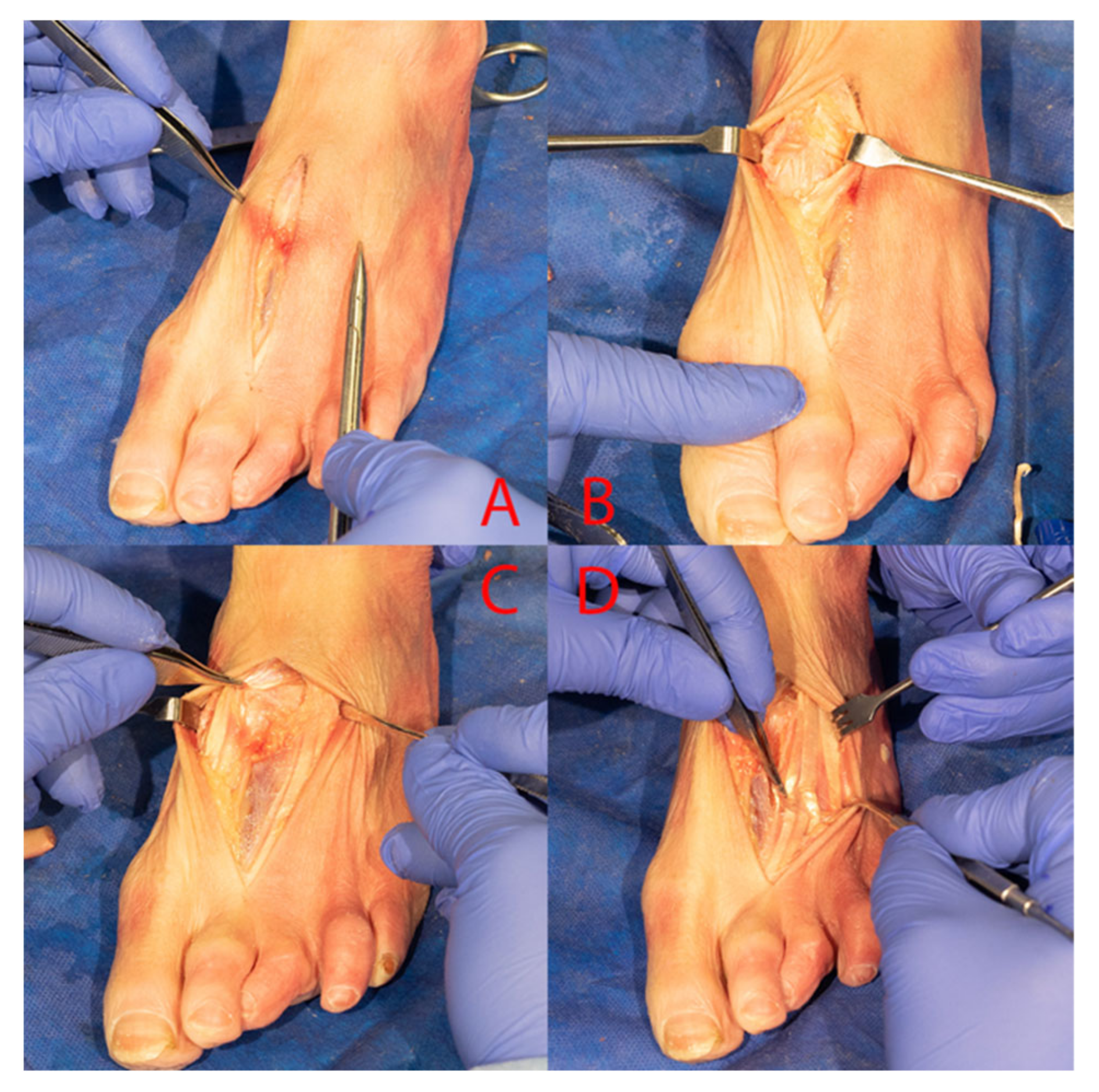
2.2. Modified Dorsal (Single Incision) Approach

A dorsal skin incision was made overlying the second metatarsal starting proximal to the TMT joint and extending distally to the MTP joint (Figure 2). If necessary, the 2nd metatarsal was localized under fluoroscopy to ensure proper placement of the incision. Three windows were created to assess the first, second, and third TMT joints. A medial window was made just medial to the EHL, which allowed for the visualization of the first TMT joint. For exploration of the second TMT joint, a middle window was created. As the neurovascular bundle lies closely in relation to this joint, this window was developed by starting on the MT shaft distally and dissecting proximally while staying close to the bone. The extensor digitorum brevis (EDB) muscle belly crosses from lateral to medial at the proximal end of the second TMT was retracted medially, thus protecting the neurovascular bundle without formal identification. For assessment of the third TMT, a lateral interval was developed between the third and fourth tendon slips of the extensor digitorum longus (EDL). The joint capsules for the first through third TMTs were incised sharply in a longitudinal manner, to bone and subperiosteally elevated to expose the cartilage of the respective joints. For visualization of the joint space, a Hintermann pin distractor was utilized. The joint and all visible cartilage was prepared for arthrodesis with curettes and osteotomes.

2.3. Articular Preparation, Imaging, and Statistical Analysis

Articular preparation was performed by two fellowship trained foot and ankle surgeons. When one surgeon completed joint preparation on a specimen, the other surgeon reviewed the preparation and only after agreement of both surgeons on the adequacy of joint preparation was the specimen disarticulated for analysis. Photographs of the articular surfaces were obtained with a high-resolution digital mirrorless camera. All photographs included a scientific ruler used for scale in the plane of the joint surface being captured. Digital photographs were imported into ImageJ software (Wayne Rasband, National Institutes of Health, Bethesda, MD, USA) for assessment of total joint surface area prepared. All image analysis was completed on 4 K resolution monitors by two blinded reviewers. Reviewers first measured the total articular area and then measured the area of the joint surface that was denuded for the base of the metatarsals and each cuneiform. (Figure 3). The results of the two reviewers were averaged for statistical analysis.

2.4. Statistical Analysis

Categorical variables (sex and laterality) were compared using Fisher’s exact tests, while Mann–Whitney U tests were used for non-parametric comparisons between groups (surface area prepared, age, and BMI). An intraclass correlation was used to assess for reliability of measurements between the two blinded ImageJ reviewers. Statistical analysis was run using IBM SPSS Statistics for Macintosh, version 27 (IBM Corp., Armonk, NY, USA). Statistical significance was designated at p < 0.05.

3. Results

A total of five male and five female specimens with an average age of 71.3  ±  10.1 were utilized for analysis. The specimens were randomly assigned to each approach. The intraclass correlation coefficient showed excellent agreement in joint preparation measurement between the two reviewers (k  =  0.961 p  =  < 0.001). There was no significant difference in the amount of joint prepared when comparing the standard versus modified dorsal approach for TMT joints one through three (p = 0.548, p = 0.310, p = 0.548). Patient variables such as age, sex, BMI, and laterality did not have a significant impact on the amount of joint prepared between the two approaches (p  =  0.548, p = 1.0. p = 0.206 and p = 0.421, respectively).
The percentage of joint preparation utilizing the standard dorsal approach versus the modified dorsal approach for TMT joints one through three is as follows (percentages utilized are listed as the median value with its correlating range): First TMT-67.6% (±13%) by the standard approach versus 71.7% (±4.5%) by the modified dorsal approach, second TMT-67.9% (±12%) versus 65.7% (±6%), and third TMT- 65.9% (±21%) versus 59.6% (±12%) (Table 1).

4. Discussion

In the setting of midfoot injuries, many patients suffer long-term consequences with ligamentous disruption and Lisfranc fracture dislocation and their repair, leading to chronic pain, arthritis, and lasting instability [9]. Many options exist for treatment, but the current literature seems to favor the open reduction internal fixation of Lisfranc injuries due to improved short- and medium-term follow-up pain scores [4,9,10]. Despite this, some studies have shown decreased re-operations with a primary arthrodesis, and in some patients, this may be the best option for patient treatment compared to the traditional ORIF [8,11]. We sought to understand the difference between the modified dorsal approach and the standard approach for the arthrodesis treatment of Lisfranc injuries. Based on this study, we did not find a significant difference between the cadaveric preparations of the first through third TMT joints via a traditional two-incision approach versus a dorsal single-incision approach. Both approaches provided similar exposure and similar visualization of TMT surface area with 69% average surface prepared in the standard approach and 66.1% in the modified dorsal approach.
Surgical management of Lisfranc injuries is challenging due of the small soft tissue space. A study in 1909, not performed by Dr. Lisfranc, was the first to describe these injuries [12]. A systematic review describes the relevant concepts for the treatment of Lisfranc joint injury including significant disability as a result of post-traumatic arthritis [4]. Current recommendations favor the ORIF approach to fixation of these injuries due to quicker recovery and better outcomes [13,14,15]. Due to the screw construct, however, there are fears over early mobilization as this may lead to loosening and loss of fixation in these patients [16]. Alberta et al. even suggests that this screw fixation may lead to further articular damage, potentiating future arthritic pain [16]. In response to this, there has been some research into dorsal plating and fusion of the TMT which provides better mechanical construct in one cadaveric model [17]. In the recent literature, there has also been a push for arthrodesis as the primary option rather than a secondary option for patients with continual pain after primary fixation [11]. In a study by Henning et al., it was shown that patients with a primary arthrodesis had similar mobility as ORIF but decreased rates of reoperation after arthrodesis [11].
Despite different options for fixation, the standard two-incision approach to the TMT joint has remained consistent until the introduction of a single-incision dorsal approach [7]. The two-incision approach is the current standard for both ORIF and for arthrodesis, but the modified dorsal approach in the management of Lisfranc injuries has been hypothesized to have similar exposure to the TMT joint space for preparation [7]. Compared to the dual-incision technique, the single-incision technique may help decrease damage to the soft tissue, decrease the risk for wound infection/dehiscence, and decrease the risk of avascular necrosis.
Though the initial work by Philipott et al. suggests the single-incision approach is viable, it is primarily focused on using this approach for an ORIF and as such did not evaluate surface area exposed, an important parameter when considering arthrodesis. A full literature review revealed no previous studies that measured the percent of joint space preparation at the TMT joint comparing a modified dorsal approach to a dual-incision approach. Previously some investigation on TMT joint preparation was conducted regarding the osteotome approach to a saw approach to the first TMT [18]. Another study also investigated radiographic assessment of first tarsometatarsal joint shape and orientation [19]. To continue our study of this important topic, we used this cadaver study to measure the precise amount of joint surface preparation using the modified dorsal approach in comparison to the standard dual-excision approach with skin bridges. This study further confirmed this notion, adding credibility backed by data to those set forth by Philipott et al.
With our findings, we demonstrate that a modified dorsal approach with a single incision did not limit the ability to prepare the joint space. We found high levels of joint space preparation using the modified dorsal approach supporting the use of this technique. Given this finding, we can see many benefits to the single-incision approach compared to the dual-incision. When making two incisions, soft-tissue complications can arise due to the narrow skin bridges created. Of the complications noted in the systematic review by Moracia et al., 0% to 9% of patients developed a superficial infection and 0% to 13% experienced a delay in the healing of the surgical wound [20,21]. Additionally, multiple cadaveric studies have demonstrated significant variability in the anatomy of the neurovascular bundle of the dorsal foot, increasing likelihood of injury with increasing number of incisions [22,23,24]. With a single-incision approach, some of this risk can be avoided by minimizing our dissections. With this, however, additional clinical studies can measure clinical outcomes including soft tissue complications, nonunion, malunion, and the need for reoperation to determine the clinical utility in varied settings.
There are some limitations that must be considered when looking at this study. First, this study measured joint surface preparation which could be affected by the surgeon skill and technique. To combat this, the same two surgeons preformed both groups. Additionally, being a cadaveric study, tissue structure and maneuverability may not be the same as living patient tissue. With this being a cadaveric model, we also lack important follow up to assess patient outcomes after procedure.
More research is needed to determine the effect of the modified dorsal approach in TMT fusion or fixation. Our study showed important findings related to the specific amount of joint surface preparation possible with two different approaches. Our findings support the hypothesis of Philpott et al. that a modified dorsal approach is a reasonable alternative to the classic two-incision surgical approach to the Lisfranc joint. Additional research will contribute to the ongoing question about safe and effective techniques for state-of-the-art surgery in the foot and ankle.

Author Contributions

Conceptualization, A.S., A.A. and N.A.A.; methodology, E.L. and N.A.A.; formal analysis, N.A.A. and Z.L.; investigation, N.A.A., Z.L., S.M. and S.Y.; data curation Z.L., S.M. and N.A.A.; writing—original draft preparation, Z.L. and S.M.; writing—review and editing, Z.L., S.M. and S.Y.; visualization, A.S. and N.A.A.; supervision, A.A. All authors have read and agreed to the published version of the manuscript.

Funding

This research received no external funding.

Institutional Review Board Statement

Our study, being a cadaver study, does not require prior IRB approval according to federal law. The study was confirmed with our institution’s own review board prior to commencement.

Informed Consent Statement

Not applicable.

Data Availability Statement

This study did not utilize a public database.

Conflicts of Interest

The authors declare no conflict of interest. The conduct of the study was supported by Zimmer Biomet and the Department of Orthopaedics Surgery at University of Alabama at Birmingham.

References

  1. Weatherford, B.M.; Anderson, J.G.; Bohay, D.R. Management of Tarsometatarsal Joint Injuries. J. Am. Acad. Orthop. Surg. 2017, 25, 469–479. [Google Scholar] [CrossRef] [PubMed]
  2. Ponkilainen, V.T.; Laine, H.J.; Mäenpää, H.M.; Mattila, V.M.; Haapasalo, H.H. Incidence and Characteristics of Midfoot Injuries. Foot Ankle Int. 2019, 40, 105–112. [Google Scholar] [CrossRef] [PubMed]
  3. Stødle, A.H.; Hvaal, K.H.; Enger, M.; Brøgger, H.; Madsen, J.E.; Ellingsen Husebye, E. Lisfranc injuries: Incidence, mechanisms of injury and predictors of instability. Foot Ankle Surg. 2020, 26, 535–540. [Google Scholar] [CrossRef] [PubMed]
  4. Watson, T.S.; Shurnas, P.S.; Denker, J. Treatment of Lisfranc joint injury: Current concepts. J. Am. Acad. Orthop. Surg. 2010, 18, 718–728. [Google Scholar] [CrossRef] [PubMed] [Green Version]
  5. Vertullo, C.J.; Easley, M.E.; Nunley, J.A. The Transverse Dorsal Approach to the Lisfranc Joint. Foot Ankle Int. 2002, 23, 420–426. [Google Scholar] [CrossRef] [PubMed]
  6. Treadwell, J.R.; Kahn, M.D. Lisfranc arthrodesis for chronic pain: A cannulated screw technique. J. Foot Ankle Surg. 1998, 37, 28–36. [Google Scholar] [CrossRef]
  7. Philpott, A.; Lawford, C.; Lau, S.C.; Chambers, S.; Bozin, M.; Oppy, A. Modified Dorsal Approach in the Management of Lisfranc Injuries. Foot Ankle Int. 2018, 39, 573–584. [Google Scholar] [CrossRef]
  8. Ly, T.V.; Coetzee, J.C. Treatment of primarily ligamentous Lisfranc joint injuries: Primary arthrodesis compared with open reduction and internal fixation. A prospective, randomized study. J. Bone Jt. Surg. Am. 2006, 88, 514–520. [Google Scholar] [CrossRef]
  9. Hu, S.J.; Chang, S.M.; Li, X.H.; Yu, G.R. Outcome comparison of Lisfranc injuries treated through dorsal plate fixation versus screw fixation. Acta Ortop. Bras. 2014, 22, 315–320. [Google Scholar] [CrossRef] [Green Version]
  10. Stavlas, P.; Roberts, C.S.; Xypnitos, F.N.; Giannoudis, P.V. The role of reduction and internal fixation of Lisfranc fracture-dislocations: A systematic review of the literature. Int. Orthop. 2010, 34, 1083–1091. [Google Scholar] [CrossRef] [Green Version]
  11. Henning, J.A.; Jones, C.B.; Sietsema, D.L.; Bohay, D.R.; Anderson, J.G. Open reduction internal fixation versus primary arthrodesis for lisfranc injuries: A prospective randomized study. Foot Ankle Int. 2009, 30, 913–922. [Google Scholar] [CrossRef] [PubMed]
  12. Quénu, E. Etude sur les luxations du metatarse (luxations metatarsotarsiennes) du diastasis entre 1 et le 2 metatarsien. Rev. Chir. 1909, 39, 1093–1134. [Google Scholar]
  13. Arntz, C.T.; Hansen, S.T., Jr. Dislocations and fracture dislocations of the tarsometatarsal joints. Orthop. Clin. N. Am. 1987, 18, 105–114. [Google Scholar] [CrossRef]
  14. Myerson, M.S.; Cerrato, R.A. Current management of tarsometatarsal injuries in the athlete. J. Bone Jt. Surg. Am. 2008, 90, 2522–2533. [Google Scholar]
  15. Arntz, C.T.; Veith, R.G.; Hansen, S.T., Jr. Fractures and fracture-dislocations of the tarsometatarsal joint. J. Bone Jt. Surg. Am. 1988, 70, 173–181. [Google Scholar] [CrossRef]
  16. Alberta, F.G.; Aronow, M.S.; Barrero, M.; Diaz-Doran, V.; Sullivan, R.J.; Adams, D.J. Ligamentous Lisfranc joint injuries: A biomechanical comparison of dorsal plate and transarticular screw fixation. Foot Ankle Int. 2005, 26, 462–473. [Google Scholar] [CrossRef]
  17. Marks, R.M.; Parks, B.G.; Schon, L.C. Midfoot fusion technique for neuroarthropathic feet: Biomechanical analysis and rationale. Foot Ankle Int. 1998, 19, 507–510. [Google Scholar] [CrossRef]
  18. Dahlgren, N.; Johnson, J.L.; Huntley, S.; McKissack, H.; Chinnakkannu, K.; Naranje, S.; Shah, A. First tarsometatarsal fusion using saw preparation vs. standard preparation of the joint: A cadaver study. Foot Ankle Surg. 2020, 26, 703–707. [Google Scholar] [CrossRef]
  19. Koury, K.; Staggers, J.R.; Pinto, M.C.; Godoy-Santos, A.L.; Smyth, N.A.; Shah, A.B.; de Cesar Netto, C. Radiographic Assessment of First Tarsometatarsal Joint Shape and Orientation. Foot Ankle Int. 2019, 40, 1438–1446. [Google Scholar] [CrossRef]
  20. Moracia-Ochagavía, I.; Rodríguez-Merchán, E.C. Lisfranc fracture-dislocations: Current management. EFORT Open Rev. 2019, 4, 430–444. [Google Scholar] [CrossRef]
  21. Sheibani-Rad, S.; Coetzee, J.C.; Giveans, M.R.; DiGiovanni, C. Arthrodesis versus ORIF for Lisfranc fractures. Orthopedics 2012, 35, e868–e873. [Google Scholar] [CrossRef] [PubMed] [Green Version]
  22. Parikh, S.; Dawe, E.; Lee, C.; Whitehead-Clarke, T.; Smith, C.; Bendall, S. A cadaveric study showing the anatomical variations in the branches of the dorsalis pedis artery at the level of the ankle joint and its clinical implication in ankle arthroscopy. Ann. R. Coll. Surg. Engl. 2017, 99, 286–288. [Google Scholar] [CrossRef] [PubMed] [Green Version]
  23. Ranade, A.V.; Rajanigandha, V.; Rai, R.; Ebenezer, D.A. Relationship between the deep peroneal nerve and dorsalis pedis artery in the foot: A cadaveric study. Clin. Anat. 2008, 21, 705–712. [Google Scholar] [CrossRef] [PubMed]
  24. Manjunatha, H.N. Variations in the origin, course and branching pattern of dorsalis pedis artery with clinical significance. Sci. Rep. 2021, 11, 1448. [Google Scholar] [CrossRef]
Figure 1. (A,B) Medial incision for standard approach. (C,D) Lateral incision for standard approach.
Osteology 02 00011 g001
Figure 2. (AC) Medial exposure for modified dorsal approach. (D) Lateral exposure for modified dorsal approach.
Osteology 02 00011 g002
Figure 3. Representative image of ImageJ analysis of the TMT joint area prepared (blue line represents total joint surface with yellow indicating area prepared) (for interpretation of the references to color in the figure legend, the reader is referred to the web version of this article).
Osteology 02 00011 g003
Table 1. Summarized results and comparison of operative approaches.
ApproachMedian Percent of the 1st TMT Joint Surface Preparedp-ValueMedian Percent of the 2nd TMT Joint Surface Preparedp-ValueMedian Percent of the 3rd TMT Joint Surface Preparedp-Value
Standard (two incision)67.60%0.54867.90%0.3165.90%0.548
Modified Dorsal
(one incision)
71.70% 65.70% 59.60%
Publisher’s Note: MDPI stays neutral with regard to jurisdictional claims in published maps and institutional affiliations.

