
Cell Viability Study of ZnCuInS/ZnS–TPPS4 Conjugates against Different Cell Lines as a Promising Fluorescent Probe
 , , , , and
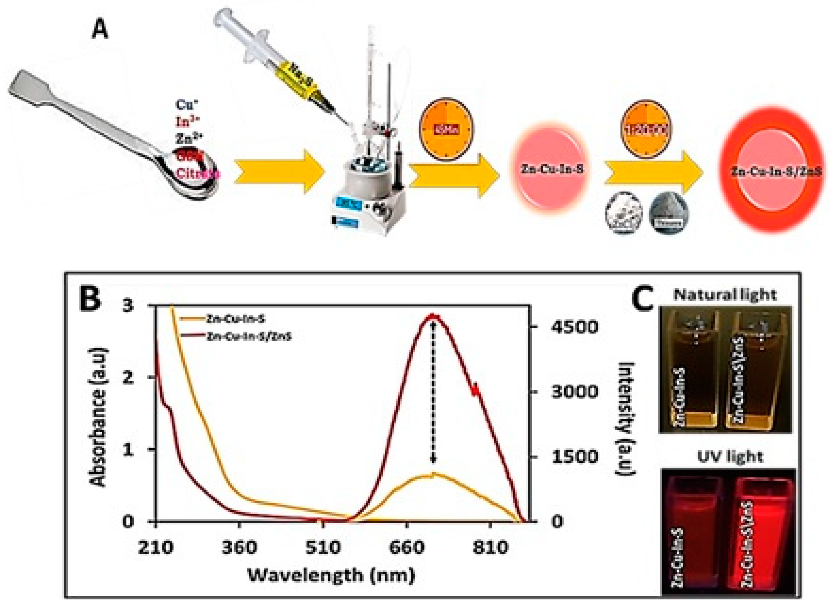
Abstract
:1. Introduction
2. Materials and Methods
2.1. Chemicals
2.2. Synthesis of Alloy ZnCuInS QDs and ZnCuInS/ZnS QDs
2.3. Synthesis and Purification of TPPH2
2.4. Synthesis of Meso-Tetra-(4-Sulfonatophenyl) Porphyrin (TPPS4)
2.5. Conjugation of ZnCuInS/ZnS QDs–TPPS4
2.6. In Vitro Cytotoxicity of TPPS4 and ZnCuInS/ZnS–TPPS4 Conjugate on BHK21, A549, Hek 293, and Hela Cell Lines
2.7. Characterization
3. Results
3.1. Characterization of ZnCuInS QDs and ZnCuInS/ZnS QDs
3.1.1. Optical Properties of ZnCuInS QDs and ZnCuInS/ZnS QDs
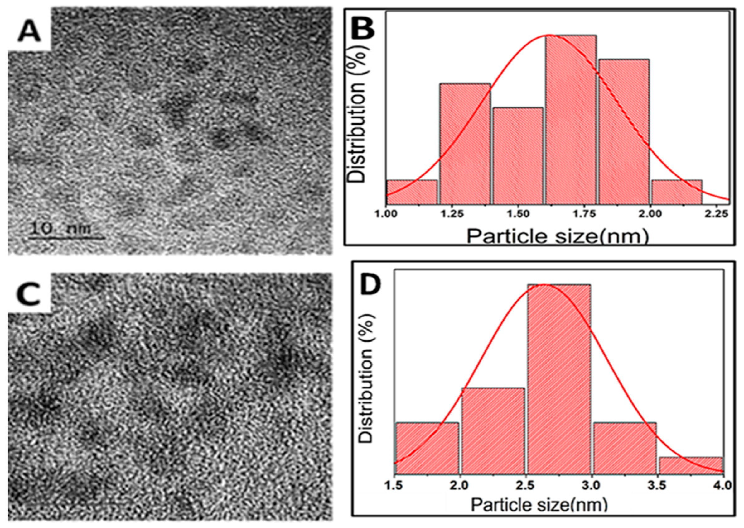
3.1.2. Morphology and Structure Characterization of ZnCuInS and ZnCuInS/ZnS QDs
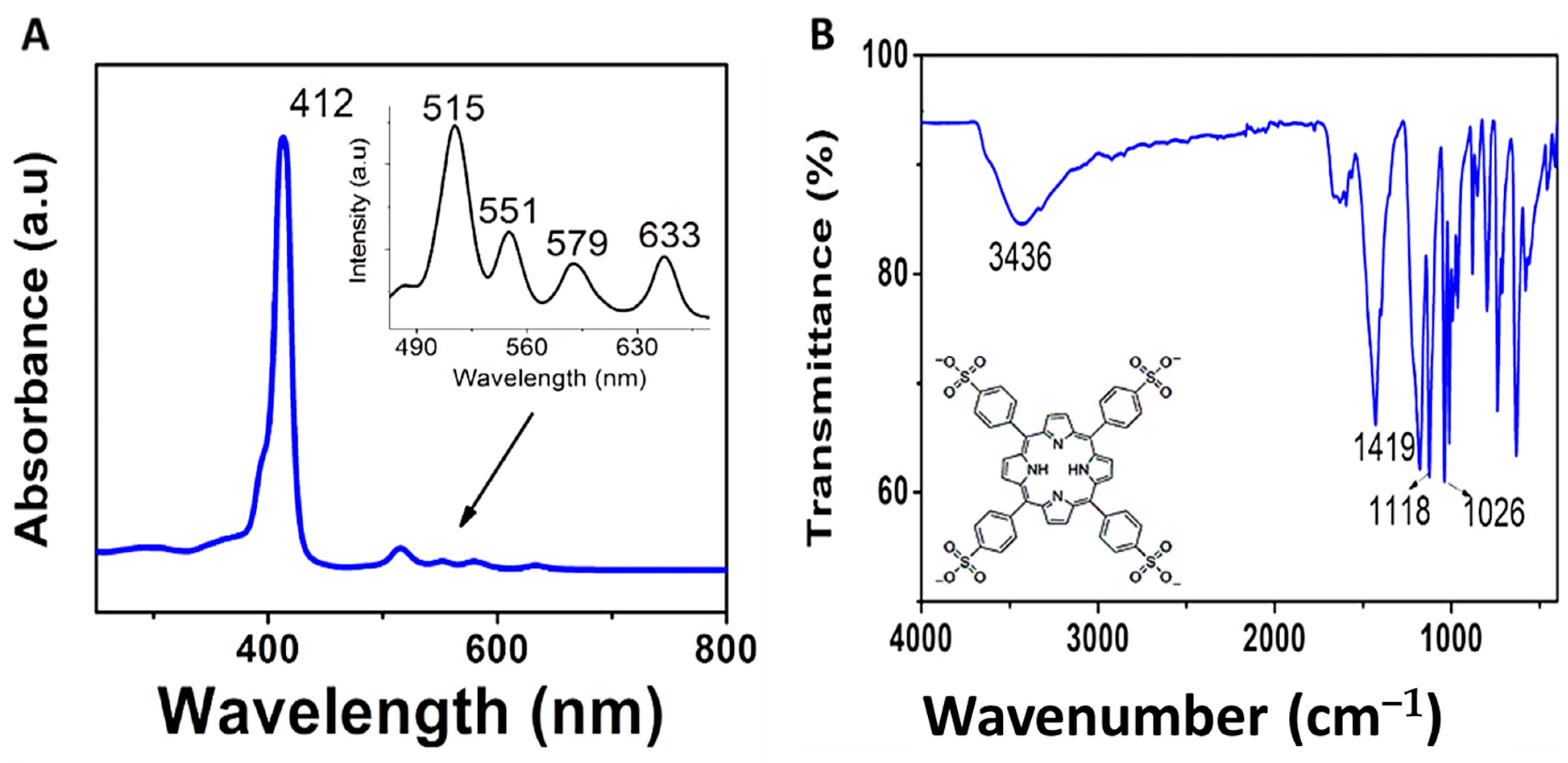
3.2. Characterization of TPPS4
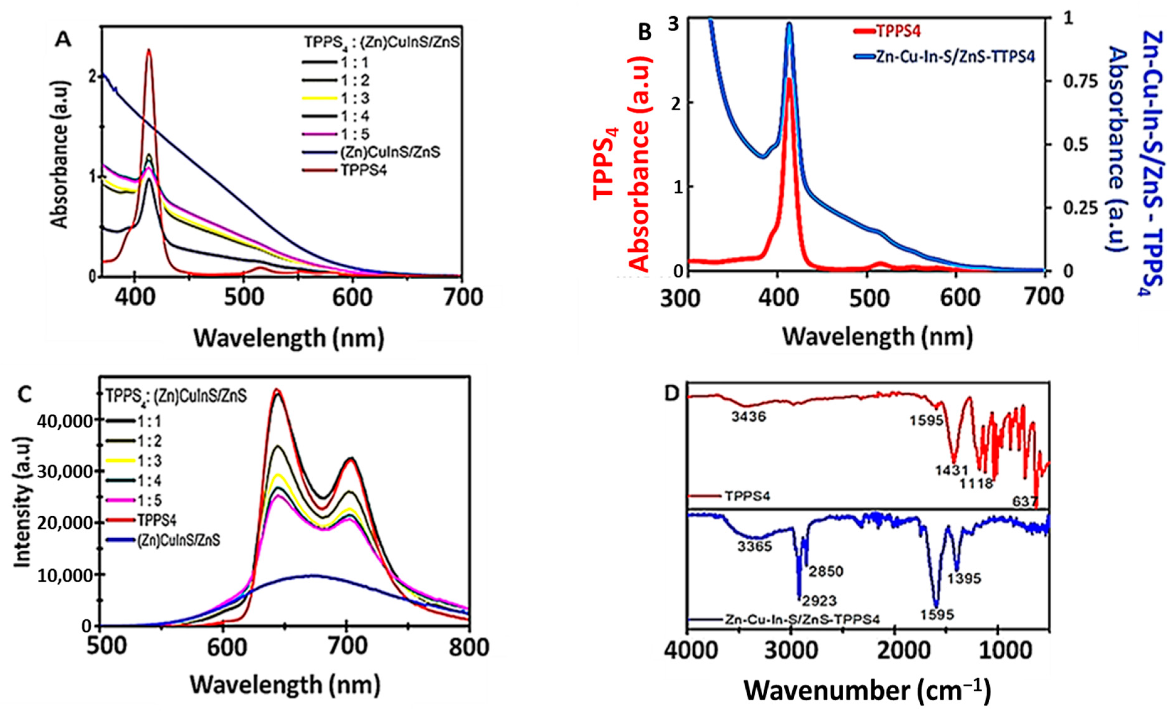
3.3. Characterization of ZnCuInS/ZnS–TPPS4 Conjugate
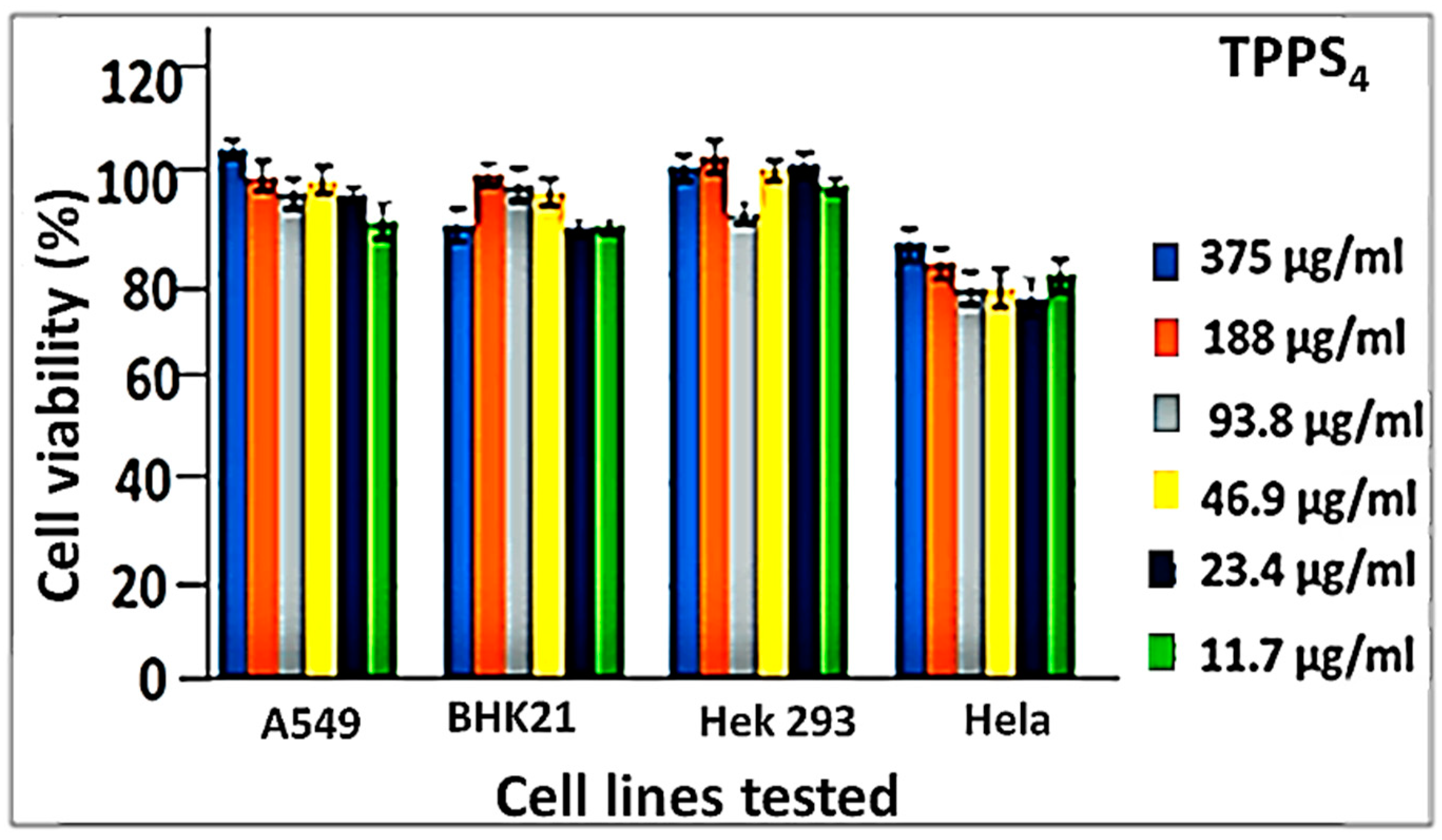
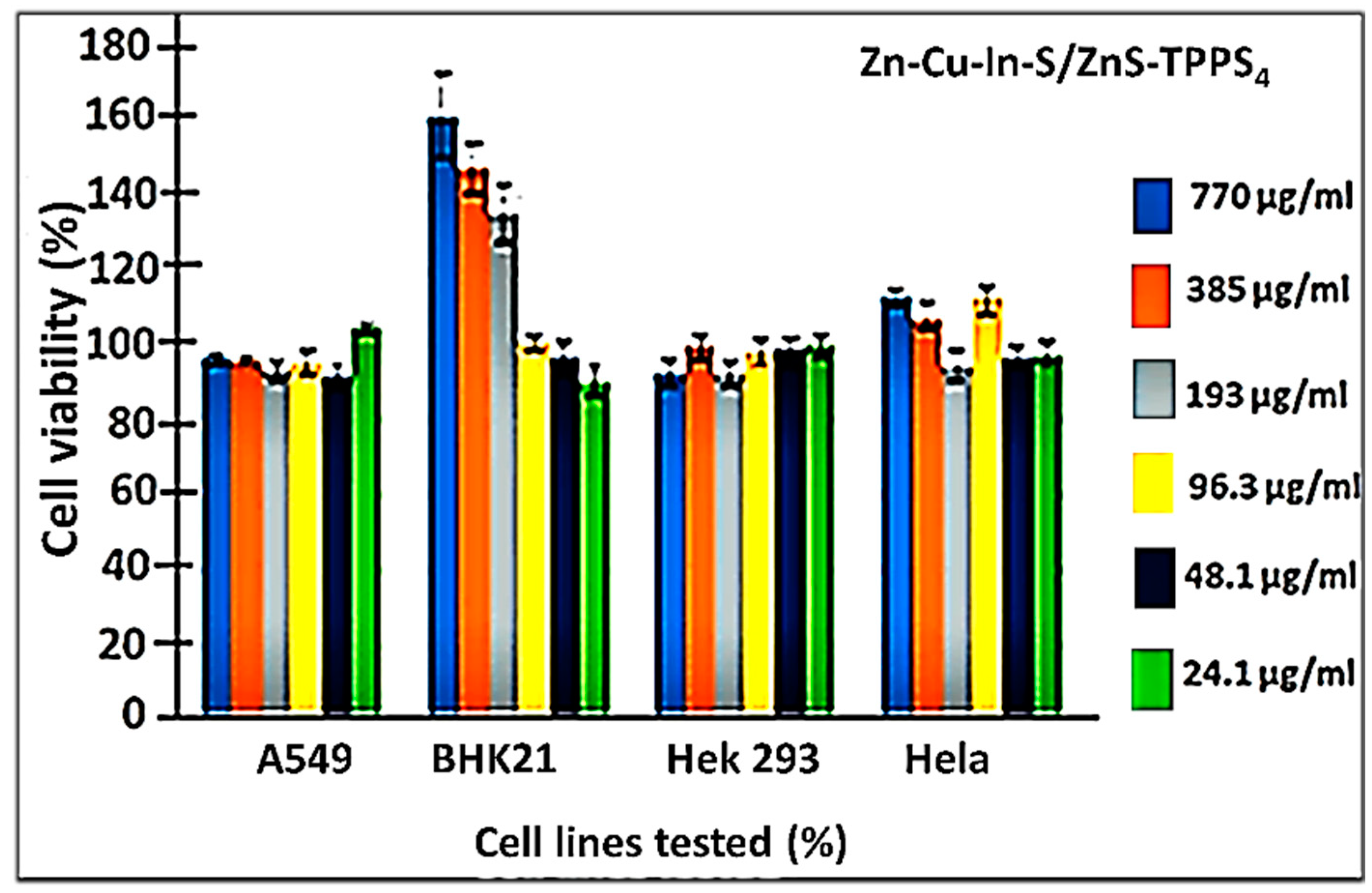
3.4. Cytotoxicity Study of TPPS4 and ZnCuInS/ZnS–TPPS4 Conjugate
4. Conclusions
Supplementary Materials
Author Contributions
Funding
Data Availability Statement
Acknowledgments
Conflicts of Interest
References
- Tsolekile, N.; Parani, S.; Matoetoe, M.C.; Songca, S.P.; Oluwafemi, O.S. Evolution of ternary I–III–VI QDs: Synthesis, characterization, and application. Nanostruct. Nano-Objects 2017, 12, 46–56. [Google Scholar] [CrossRef]
- Jia, Y.; Wang, H.; Xiang, L.; Liu, X.; Wei, W.; Ma, N.; Sun, D. Tunable emission properties of core-shell ZnCuInS-ZnS quantum dots with enhanced fluorescence intensity. J. Mater. Sci. Technol. 2017, 34, 942–948. [Google Scholar] [CrossRef]
- Ilaiyaraja, P.; Mocherla, P.S.V.; Srinivasan, T.K.; Sudakar, C. Synthesis of Cu-Deficient and Zn-Graded Cu-In-Zn-S Quantum Dots and Hybrid Inorganic-Organic Nanophosphor Composite for White Light Emission. ACS Appl. Mater. Interfaces 2016, 8, 12456–12465. [Google Scholar] [CrossRef] [PubMed]
- Yang, L.; Antanovich, A.; Prudnikau, A.; Taniya, O.S.; Grzhegorzhevskii, K.V.; Zelenovskiy, P.; Terpinskaya, T.; Tang, J.; Artemyev, M. Highly luminescent Zn–Cu–In–S/ZnS core/gradient shell quantum dots prepared from indium sulfide by cation exchange for cell labeling and polymer composites. Nanotechnology 2019, 30, 395603. [Google Scholar] [CrossRef] [PubMed]
- Nady, J.E.; Ali, M.; Kamel, O.A.; Ebrahim, S.; Soliman, M. Room temperature synthesis of aqueous ZnCuInS/ZnS quantum dots. J. Dispers Sci. Technol. 2019, 41, 1956–1962. [Google Scholar] [CrossRef]
- Zhang, B.; Wang, Y.; Yang, C.; Hu, S.; Gao, Y.; Zhang, Y.; Wang, Y.; Demir, H.V.; Liu, L.; Yong, K.-T. The Composition Effect on the Optical Properties of Aqueous Synthesized Cu–in–S and Zn–Cu–in–S Quantum Dot Nanocrystals. Phys. Chem. Chem. Phys. 2015, 17, 25133–25141. [Google Scholar] [CrossRef]
- Fang, Z.; Huang, Y.; Cheng, S.; Zhu, Q.; Zhang, W.; Zhao, F.; Huang, G.; Jiang, G.; Li, F. Quaternary alloyed quantum dots with a wide-ranging tunable emission for high color-rendering white light-emitting diodes. J. Alloys Compd. 2023, 932, 167608. [Google Scholar] [CrossRef]
- Abdulaeva, I.A.; Birin, K.P.; Bessmertnykh-Lemeune, A.; Tsivadze, A.Y.; Gorbunova, Y.G. Heterocycle-appended porphyrins: Synthesis and challenges. Coord. Chem. Rev. 2020, 407, 213108. [Google Scholar] [CrossRef]
- Moreira, X.; Santos, P.; Faustino, M.A.F.; Raposo, M.M.M.; Costa, S.P.G.; Moura, N.M.M.; Gomes, A.T.P.C.; Almeida, A.; Neves, M.G.P.M.S. An insight into the synthesis of cationic porphyrin-imidazole derivatives and their photodynamic inactivation efficiency against Escherichia coli. Dye. Pigment. 2020, 178, 108330. [Google Scholar] [CrossRef]
- Castriciano, M.A.; Zagami, R.; Casaletto, M.P.; Martel, B.; Trapani, M.; Romeo, A.; Villari, V.; Sciortino, M.T.; Grasso, L.; Guglielmino, S.; et al. Poly(carboxylic acid)-Cyclodextrin/Anionic Porphyrin Finished Fabrics as Photosensitizer Releasers for Antimicrobial Photodynamic Therapy. Biomacromolecules 2017, 18, 1134–1144. [Google Scholar] [CrossRef]
- Yang, L.; Zhou, J.; Wang, Z.; Li, H.; Wang, K.; Liu, H.; Wu, F. Biocompatible conjugated porphyrin nanoparticles with photodynamic/photothermal performances in cancer therapy. Dye. Pigment. 2020, 182, 108664. [Google Scholar] [CrossRef]
- Bera, K.; Maiti, S.; Maity, M.; Mandal, C.; Maiti, N.C. Porphyrin-Gold Nanomaterial for Efficient Drug Delivery to Cancerous Cells. ACS Omega 2018, 3, 4602–4619. [Google Scholar] [CrossRef] [Green Version]
- Kyropoulou, M.; DiLeone, S.; Lanzilotto, A.; Constable, E.C.; Housecroft, C.E.; Meier, W.P.; Cornelia, G. Porphyrin Containing Polymersomes with Enhanced ROS Generation Efficiency: In Vitro Evaluation. Macromol. Biosci. 2020, 20, 1900291–1900300. [Google Scholar] [CrossRef]
- Rojkiewicz, M.; Kuś, P.; Kozub, P.; Kempa, M. The synthesis of new potential photosensitizers. Dye. Pigment. 2013, 99, 627–635. [Google Scholar] [CrossRef]
- Chen, J.; Ma, Q.; Hu, X.; Gao, Y.; Yan, X.; Qin, D.; Lu, X. Design of a novel naked-eye and turn-on fluorescence sensor based on the 5,10,15,20-(4-sulphonatophenyl) porphyrin (TPPS4)-Hg2+ system: Monitoring of glutathione (GSH) in real samples and DFT calculation. Sens. Actuators B Chem. 2018, 254, 475–482. [Google Scholar] [CrossRef]
- Parra, G.G.; Ferreira, L.P.; Gonçalves, P.J.; Sizova, S.V.; Oleinikov, V.A.; Morozov, V.N.; Kuzmin, V.A.; Borissevitch, I.E. Stimulation of Cysteine-Coated CdSe/ZnS Quantum Dot Luminescence by meso-Tetrakis (p-sulfonato-phenyl) Porphyrin. Nanoscale Res. Lett. 2018, 13, 40. [Google Scholar] [CrossRef] [Green Version]
- Kou, J.; Dou, D.; Yang, L. Porphyrin photosensitizers in photodynamic therapy and its applications. Oncotarget 2017, 8, 81591–81603. [Google Scholar] [CrossRef] [PubMed] [Green Version]
- Managa, M.; Ngoy, B.P.; Nyokong, T. Photophysical properties and photodynamic therapy activity of a meso-tetra(4-carboxyphenyl)porphyrin tetramethyl ester-graphene quantum dot conjugate. New J. Chem. 2019, 43, 4518–4524. [Google Scholar] [CrossRef]
- Martínez, S.R.; Ibarra, L.E.; Ponzio, R.A.; Forcone, M.V.; Wendel, A.B.; Chesta, C.; Spesia, M.B.; Palacios, R.E. Photodynamic inactivation of ESKAPE group bacterial pathogens in planktonic and biofilm cultures using metallated porphyrin-doped conjugated polymer nanoparticles. ACS Infect. Dis. 2020, 6, 2202–2213. [Google Scholar] [CrossRef]
- Tsolekile, N.; Nahle, S.; Zikalala, N.; Parani, S.; Sakho, E.H.M.; Joubert, O.; Matoetoe, M.C.; Songca, S.P.; Oluwafemi, O.S. Cytotoxicity, fluorescence tagging and gene-expression study of CuInS/ZnS QDS-Meso (hydroxyphenyl) porphyrin conjugate against human monocytic leukemia cells. Sci. Rep. 2020, 10, 4936. [Google Scholar] [CrossRef] [Green Version]
- Yue, L.; Rao, H.; Du, J.; Pan, Z.; Yu, J.; Zhong, X. Comparative advantages of Zn-Cu-In-S alloy QDs in the construction of quantum dot-sensitized solar cells. RSC Adv. 2018, 8, 3637–3645. [Google Scholar] [CrossRef] [PubMed] [Green Version]
- Guo, W.; Tu, Y.; Dong, C.; Zhang, B.; Hu, C.; Chang, J. Synthesis of Zn-Cu-In-S/ZnS Core/Shell Quantum Dots with Inhibited Blue-Shift Photoluminescence and Applications for Tumor Targeted Bioimaging. Theranostics 2013, 3, 99–108. [Google Scholar] [CrossRef] [PubMed] [Green Version]
- Lin, W.; Huang, Y.; Lee, T.; Lin, P.; Chung, S. High color rendering index of ZCIS quantum dots-based white light-emitting diodes. NanoSci. + Eng. 2020, 104, 261106. [Google Scholar]
- Jia, Y.; Wang, H.; Yan, Z.; Deng, L.; Dong, H. RSC Advances A facile method for the synthesis of CuInS2–ZnS quantum dots with tunable photoluminescent. RSC Adv. 2016, 6, 93303–93308. [Google Scholar] [CrossRef]
- Park, J.; Kim, S.-W. CuInS2/ZnS core/shell quantum dots by cation exchange and their blue-shifted photoluminescence. J. Mater. Chem. 2011, 21, 3745–3750. [Google Scholar] [CrossRef]
- Jawhar, N.N.; Soheyli, E.; Yazici, A.F.; Mutlugun, E.; Sahraei, R. Preparation of highly emissive and reproducible Cu–In–S/ZnS core/shell quantum dots with a mid-gap emission character. J. Alloys Compd. 2020, 824, 153906. [Google Scholar] [CrossRef]


Disclaimer/Publisher’s Note: The statements, opinions and data contained in all publications are solely those of the individual author(s) and contributor(s) and not of MDPI and/or the editor(s). MDPI and/or the editor(s) disclaim responsibility for any injury to people or property resulting from any ideas, methods, instructions or products referred to in the content. |
© 2023 by the authors. Licensee MDPI, Basel, Switzerland. This article is an open access article distributed under the terms and conditions of the Creative Commons Attribution (CC BY) license (https://creativecommons.org/licenses/by/4.0/).
Share and Cite
Tsolekile, N.; Parani, S.; Lebepe, T.C.; Maluleke, R.; Ncapayi, V.; Matoetoe, M.C.; Songca, S.P.; Oluwafemi, O.S. Cell Viability Study of ZnCuInS/ZnS–TPPS4 Conjugates against Different Cell Lines as a Promising Fluorescent Probe. Organics 2023, 4, 126-136. https://doi.org/10.3390/org4010010
Tsolekile N, Parani S, Lebepe TC, Maluleke R, Ncapayi V, Matoetoe MC, Songca SP, Oluwafemi OS. Cell Viability Study of ZnCuInS/ZnS–TPPS4 Conjugates against Different Cell Lines as a Promising Fluorescent Probe. Organics. 2023; 4(1):126-136. https://doi.org/10.3390/org4010010
Chicago/Turabian StyleTsolekile, Ncediwe, Sundararajan Parani, Thabang Calvin Lebepe, Rodney Maluleke, Vuyelwa Ncapayi, Mangaka Clara Matoetoe, Sandile Phinda Songca, and Oluwatobi Samuel Oluwafemi. 2023. "Cell Viability Study of ZnCuInS/ZnS–TPPS4 Conjugates against Different Cell Lines as a Promising Fluorescent Probe" Organics 4, no. 1: 126-136. https://doi.org/10.3390/org4010010