Review

Insights into the Molecular and Hormonal Regulation of Complications of X-Linked Hypophosphatemia

1
Brigham and Women’s Hospital, Division of Endocrinology, Diabetes and Hypertension, Boston, MA 02115, USA
2
Harvard Medical School, Boston, MA 02115, USA
*
Author to whom correspondence should be addressed.
Endocrines 2023, 4(1), 151-168; https://doi.org/10.3390/endocrines4010014
Submission received: 1 February 2023 / Revised: 22 February 2023 / Accepted: 1 March 2023 / Published: 3 March 2023
(This article belongs to the Section Parathyroid Disorders, Mineral Metabolism and Bone Functions)

Abstract

:
X-linked hypophosphatemia (XLH) is characterized by mutations in the PHEX gene, leading to elevated serum levels of FGF23, decreased production of 1,25 dihydroxyvitamin D3 (1,25D), and hypophosphatemia. Those affected with XLH manifest impaired growth and skeletal and dentoalveolar mineralization as well as increased mineralization of the tendon–bone attachment site (enthesopathy), all of which lead to decreased quality of life. Many molecular and murine studies have detailed the role of mineral ions and hormones in regulating complications of XLH, including how they modulate growth and growth plate maturation, bone mineralization and structure, osteocyte-mediated mineral matrix resorption and canalicular organization, and enthesopathy development. While these studies have provided insight into the molecular underpinnings of these skeletal processes, current therapies available for XLH do not fully prevent or treat these complications. Therefore, further investigations are needed to determine the molecular pathophysiology underlying the complications of XLH.

1. Introduction

X-Linked Hypophosphatemia (XLH) is the most common inheritable form of rickets (1:20,000) caused by an X-linked dominant inactivating mutation of the Phosphate Regulating Endopeptidase Homolog, X-Linked (PHEX) gene [1,2,3,4]. PHEX-inactivating mutations lead to high circulating levels of fibroblast growth factor 23 (FGF23), which results in hypophosphatemia, impaired production of the active form of vitamin D, 1,25 dihydroxyvitamin D3 (1,25D) [5], and a mild elevation in serum parathryoid hormone (PTH) levels. Children with XLH manifest symptoms by 1–2 years of age, including rickets, bone deformities, and dental caries, while adults develop osteomalacia, osteoarthritis, and painful mineralizations of the bone–tendon attachment site (enthesis), which is called enthesopathy [6,7]. Both affected males and females present with similar biochemical characteristics and as children do not differ in height [8]. As adults, affected males have been reported as having more severe complications than females, which is possibly due to the X-chromosome inactivation seen in females. Affected males have been reported to have increased incidence of dental abscesses and more severe gait abnormalities and lower limb deformities [8,9]. Over 800 mutations in PHEX along the entire gene have been identified, including missense, nonsense, splicing, frameshift, and copy-number mutations [7]. These mutations have complete penetrance, but the disorder can manifest from mild to severe complications even among those who carry the same mutation in males and females and/or within the same family, suggesting that additional modifying genes or environmental factors impact the XLH phenotype. Although mutations in PHEX were identified as causing XLH, the direct targets of PHEX remain unknown, thus limiting the understanding of the variability in how complications manifest in those affected with XLH [3,10].
Mice with XLH (Hyp) carry a loss-of-function mutation within the PHEX gene [11,12], thus exhibiting a phenotype analogous to human XLH patients, including similar biochemical alterations leading to hypophosphatemic rickets, bone abnormalities, and enthesopathy [11,13,14]. Over the past several decades, many studies using Hyp mice and genetically altered mouse models have contributed to understanding the molecular and hormonal regulation of XLH complications, including impaired growth, skeletal mineralization, and osteocyte lacuno-canalicular remodeling as well as enthesopathy development. While there are review articles discussing the clinical phenotypes of XLH available, there are few manuscripts which review the molecular mechanisms underlying XLH comlications. To assure that the currently available basic and translational studies are included in this review, a search was performed on PubMed and Google Scholar using terms “XLH”, “Hyp” and/or the complication under discussion such as “growth”, “growth plate maturation”, “mineralization”, “biomechanics”, “microarchitecture” or “enthesopathy”.

1.1. Phosphate Regulating Hormones: 1,25 Dihydroxyvitamin D3 (1,25D) and FGF23

Vitamin D3 (cholecalciferol) is synthesized from 7-dehydroxycholesterol in the skin when exposed to UV-B light. In the liver, cholecalciferol is hydroxylated to form 25-hydroxyvitamin D3 (25(OH)D), which is the predominant circulating form of vitamin D. Bound to the vitamin D binding protein, 25(OH)D is transported to the kidney, and in the proximal renal tubule, 25(OH)D is further hydroxylated to form the biologically active form of vitamin D, 1,25D, by vitamin D 1-alpha-hydroxylase (CYP27B1) [15,16]. The biological effects of 1,25D are mediated through the vitamin D receptor (VDR), which is a nuclear steroid receptor. 1,25D binds to VDR, upon which VDR forms an obligate heterodimer with retinoic X receptor (RXR) and binds to target genes via two cysteine-rich zinc finger structures [15,17,18]. 1,25D acts to regulate calcium and phosphate homeostasis by promoting bone resorption as well as intestinal absorption and renal reabsorption of calcium and phosphate [15]. 1,25D also acts to decrease parathyroid hormone gene transcription and parathyroid cell proliferation, thus leading to decreased serum levels of PTH and increased renal reabsorption of phosphate [19,20].
FGF23 is a ligand hormone synthesized predominantly in osteocytes and osteoblasts to regulate phosphate homeostasis [21]. FGF23 decreases serum phosphate levels by directly increasing renal phosphate wasting and indirectly decreasing phosphate gastrointestinal absorption [22,23]. FGF23 increases phosphate renal excretion by downregulating the expression of sodium–phosphate co-transporters NPT2a and NPT2c in the renal proximal tubules [24,25]. This phosphate-regulating hormone also decreases the expression of CYP27B1 and enhances the expression of CYP24A1, which catabolizes 1,25D, resulting in decreased serum levels of 1,25D. This decrease in 1,25D synthesis in turn leads to the suppression of intestinal absorption and renal reabsorption of phosphate [26,27].

1.2. Treatment of XLH

Conventional therapy for XLH patients includes a combination of exogenous 1,25D (calcitriol) and phosphate supplements. Excessive phosphate supplementation leads to secondary or tertiary hyperparathyroidism; thus, 1,25D therapy is added to phosphate supplementation not only to increase serum phosphate levels but to also prevent secondary/tertiary hyperparathyroidism [6]. Recently, a humanized antibody targeting FGF23, burosumab (Crysvita), was approved for the treatment of children and adults with XLH. Burosumab increases and sustains serum phosphate levels in the low–normal range while maintaining normal serum and urine calcium and serum PTH levels [28,29,30]. Although one of the advantages of burosumab is its ability to block FGF23-specific actions in addition to improving phosphate homeostasis, it is not able to sustain increased serum 1,25D levels during long-term treatment [28,31,32]. Clinical trials have demonstrated that burosumab significantly improves rachitic growth plate changes and mildly improves growth compared to traditional supplementation treatment [32]. In adults with XLH, burosumab improved pseudofracture healing, decreased osteomalacia, and improved quality of life [28,33]. Data on the effects of burosumab on chronic complications of XLH like enthesopathy and osteoarthritis are unknown.

2. Impairment of Growth and Growth Plate Maturation

2.1. Growth in XLH

Individuals with XLH exhibit short stature and disproportionate growth characterized by a longer torso and shorter long bones [34,35]. The rickets observed in XLH is associated with delayed ambulation, deformities of the weight-bearing lower limbs (varus or valgus deformities), and waddling gait [36]. Pediatric patients with XLH demonstrate normal length/height at birth, but growth velocity decreases during infancy/early childhood by one year of age and then progressively decelerates to a nadir in early childhood and remains abnormally low thereafter [37,38,39]. The final height of adults with XLH is significantly shorter than unaffected individuals, where affected adults have an average z score of −1.9 [34].
Growth plate abnormalities are evident in rapidly growing long bones of children affected with XLH, including the distal femur, radius and ulna and proximal and distal tibia, and distal radius and ulna. Radiographs of long bones demonstrate widened growth plates with irregular metaphyseal margins. There may also be fraying and/or cupping of the metaphyses. The degree of growth retardation correlates with the severity of the growth plate abnormalities [40].

2.2. Growth Plate Abnormalities in XLH

Growth plate maturation is essential to longitudinal bone growth. During this process, proliferative chondrocytes differentiate into prehypertrophic chondrocytes and then finally hypertrophic chondrocytes [41]. Hypertrophic chondrocytes are characterized by the expression of type X collagen and a widespread increase in cell volume. They promote the vascularization of the cartilage template by secreting molecules such as vascular endothelial growth factor (VEGF), mineralization of the adjacent skeletal matrix, and osteoblast differentiation from nearby perichondrial cells [42]. Upon terminal differentiation, hypertrophic chondrocytes undergo apoptosis, allowing for osteoblasts to invade and promote mineralization of the primary spongiosa [43].
Like those affected with XLH, mice with XLH (Hyp) also exhibit a smaller size and weight, with decreased spine and long bone lengths [44]. The growth plates in Hyp mice are characterized by the impaired apoptosis of hypertrophic chondrocytes, leading to an expansion of the hypertrophic chondrocyte layer [45]. The height of the proliferative layer in Hyp mice remains normal. In unaffected individuals, chondrocytes in growth plates are organized in columns distributed in parallel, while growth plates in those affected with XLH are disorganized with a lack of columnar organization of hypertrophic chondrocytes, which are also unpolarized and abnormally oriented. The hypertrophic chondrocyte abnormalities lead to aberrant vascularization of chondro-osseous tissue and impaired mineralization of the primary spongiosa [46,47].

2.3. Molecular Mechanisms of Impaired Growth in XLH

Investigations into the pathophysiology of the growth plate abnormalities of XLH have demonstrated that hypophosphatemia leads to the impaired apoptosis of terminally differentiated hypertrophic chondrocytes, resulting in the expansion of the hypertrophic chondrocyte layer of the growth plate [45,48,49,50]. Normalization of serum calcium and phosphate levels in mice lacing the vitamin D receptor (VDR) prevented rickets, while Hyp mice and mice fed a low-phosphate diet (both of which have low serum phosphate) continue to have expansion of the hypertrophic chondrocyte layer and impaired hypertrophic chondrocyte apoptosis. Hypophosphatemia is present in all of these murine models of rickets, demonstrating that phosphate is critical for normal growth plate maturation and hypertrophic chondrocyte apoptosis [45].
In vitro and in vivo studies also showed that phosphate-mediated hypertrophic chondrocyte apoptosis is dependent on induction of ERK1/2 signaling and activation of the mitochondrial pro-apoptosis caspase pathway [45,51]. Inhibition of the ERK1/2 signaling by treatment with MEK inhibitors suppresses the phosphate-induced apoptosis of hypertrophic chondrocytes in vitro and in vivo [51]. These observations point toward a critical role of phosphate in the induction of ERK1/2 phosphorylation in hypertrophic chondrocyte apoptosis and growth plate maturation.
Raf kinases, including A-, B-, and C-Raf, activate MEK/ERK signaling. Proliferative chondrocytes primarily express A-Raf and B-Raf, where the ablation of either Raf isoform does not alter normal growth plate maturation [52]. Hypertrophic chondrocytes predominantly express C-Raf [53], where the deletion of C-Raf in chondrocytes leads to an expansion of the hypertrophic layer of the growth plate with an impaired induction of p-ERK1/2, hypertrophic chondrocyte apoptosis, and vascular invasion at the chondroosseous junction [54]. In cultured primary hypertrophic chondrocytes, the deletion of C-Raf alone is not sufficient to impair phosphate-induced ERK1/2 phosphorylation [55]. However, the ablation of all three Raf isoforms in primary chondrocytes led to impaired phosphate-induced p-ERK1/2 in vitro. Corresponding with this, the deletion of all three Raf isoforms in the chondrocytes of mice leads to a significant expansion of the hypertrophic chondrocyte layer accompanied by impairment of hypertrophic chondrocyte apoptosis, caspase activation, and vascular invasion [55]. These studies highlight the essential and partial redundant roles of the Raf kinase isoforms in phosphate-induced ERK1/2 phosphorylation in hypertrophic chondrocytes and growth plate maturation.
In the growth plate, periarticular chondrocytes secrete parathyroid hormone-related peptide (PTHrP), which acts through the PTH/PTHrP receptor (PPR) on proliferating chondrocytes to stimulate their proliferation and inhibit their differentiation [41,56,57]. Studies demonstrated that the impaired chondrocyte differentiation and decreased p-ERK1/2 observed in the chondrocytes of embryonic murine metatarsal treated with low phosphate media requires PTHrP action [58]. Furthermore, mice haploinsufficient for PTHrP fed a low-phosphate diet did not have an expansion of the hypertrophic chondrocyte layer and decreased p-ERK1/2 and hypertrophic chondrocyte apoptosis as seen in WT mice fed a low-phosphate diet [58]. These studies indicate that PTHrP action contributes to the impaired chondrocyte differentiation and growth plate abnormalities seen with hypophosphatemia and thus in mice and humans with XLH. In vitro studies in primary murine hypertrophic chondrocytes showed that PTH pretreatment impairs mitochondrial phosphate-induced ERK1/2 phosphorylation and the subcellular redistribution of apoptosis-regulating proteins p-BAD and BAD, supporting the hypothesis that PTH action impairs hypertrophic chondrocyte apoptosis [58]. Periarticular chondrocytes in Hyp long bones have an increased expression of PTHrP, and phosphate restriction increases PTHrP expression in murine metatarsals [44,51]; therefore, the results of this study indicate that the enhanced PTHrP action in Hyp mice likely plays a pathogenic role in the impaired hypertrophic chondrocyte apoptosis seen in growth plates of mice and humans with XLH.
In addition to PTHrP, 1,25D has also been shown to play a role in the growth plate abnormalities of Hyp mice. Deletion of the sodium-dependent phosphate transporter 2a (NPT2a), which enables phosphate resorption in the renal proximal tubules, in mice leads to hypophosphatemia, which in turn results in suppressed PTH levels and high serum 1,25D levels with hypercalcemia [59]. In contrast to Hyp mice, NPT2aKO mice develop an expansion of the hypertrophic chondrocyte layer of the growth plate at 2 weeks of age, where this growth plate expansion normalizes by 5 weeks of age despite persistent hypophosphatemia [50]. This improvement in growth plate maturation corresponds with increased serum levels of 1,25D. Ablation of the vitamin D receptor (VDR) in NPT2aKO mice resulted in persistent expansion of the hypertrophic chondrocyte layer, thus demonstrating that enhanced 1,25D action can compensate for hypophosphatemia during growth plate maturation and prevent rickets [50]. In further support for a role for 1,25D actions in growth plate maturation, the treatment of primary murine hypertrophic chondrocytes with 1,25D further enhanced phosphate-induced pERK1/2, indicating 1,25D and phosphate play roles in enabling hypertrophic chondrocyte apoptosis. The results of these studies imply that the impaired 1,25D action due to high circulating levels of FGF23 in Hyp mice combined with hypophosphatemia contribute to the impaired growth observed in these mice.
Treating Hyp mice with daily 1,25D or an anti-FGF23 blocking antibody (FGF23Ab) starting Day 2 of post-natal life in the absence of phosphate supplementation significantly increases serum phosphate, normalizes serum PTH levels, while maintaining normocalcemia [44]. Both 1,25D and FGF23Ab therapies improved growth (measured by weight, femur length, and vertebral length) and growth plate morphology, restoring the columnar organization of growth plate chondrocytes and improving metaphyseal organization. Treated growth plates had decreased expansion of the hypertrophic chondrocyte layer and increased apoptosis of hypertrophic chondrocytes. While both 1,25D and FGF23Ab improved serum phosphate levels to similar levels, the treatment of Hyp mice with 1,25D alone improved growth and growth plate morphology more than FGF23Ab, implicating the direct beneficial effects of 1,25D on growth and growth plate maturation in XLH, independent of its role in regulating phosphate homeostasis [44].
The importance of 1,25D action on growth and optimizing 1,25D action in XLH therapy were further corroborated by the study showing that treatment Hyp mice with the potent active vitamin D3 analog, eldecalcitol (1α,25-dihydroxy-2β-(3-hydroxypropyloxy) vitamin D3; ED71) without phosphate supplementation improved hypophosphatemia, growth, and growth plate structure [60]. Jehan et al. (2008) reported that the variation in growth observed in XLH patients may be linked to different vitamin D receptor promoter haplotypes, providing a possible explanation for some of the clinical variability observed in XLH and also supporting the importance of 1,25D action in growth [61]. In human studies, it was reported that adults who started phosphate and 1,25D therapy in infancy (prior to 1 year old) had better growth and radiographic rickets scores compared to those with XLH who were started on therapy after 1 year of age, despite both cohorts having the same level of hypophosphatemia [62,63]. Thus, like the results of murine studies discussed above, these clinical data underscore the importance of optimizing mineral ion and hormone homeostasis starting early in childhood.

3. Skeletal Mineralization, Microarchitecture, and Biomechanics in XLH

Mice and humans with XLH have poorly mineralized bones. Histomorphometric analyses show that both Hyp cortical and trabecular bone have a significant increase in osteoid volume with dramatically impaired bone formation rate and mineralization apposition rate [64]. Micro-CT analyses demonstrate that Hyp femurs have significantly decreased whole distal femur bone volume fraction (BV/TV) and impaired cortical microarchitecture, including decreased cortical thickness (Ct.Th) and cortical area fraction (Ct.Ar/Tt.Ar) as well as increased cortical porosity [44]. Moreover, Hyp femurs have an increase in circumference with increased total cross-sectional area (Tt.Ar) and medullary area (Ma.Ar) compared to WT [65]. Trabecular microarchitecture is also severely abnormal, with Hyp femurs having significantly reduced trabecular bone volume fraction (BV/TV), which is associated with decreased trabecular number (Tb.N) and increased trabecular spacing (Tb.Sp) [65]. Consistent with the micro-CT analyses, histomorphometric analyses demonstrate that Hyp femurs have very few trabeculae. Interestingly, mice lacking the sodium phosphate transporter 2a (NPT2a), which have hypophosphatemia, low serum FGF23 levels, and high 1,25D levels, have mild abnormalities in cortical microarchitecture but severe compromise in trabecular structure. Like Hyp bones, NPT2aKO bones have decreased trabecular BV/TV and increased trabecular spacing, suggesting that phosphate may play an important role in regulating trabecular structure. Micro-CT evaluation of inferred biomechanical parameters showed a significant decrease in polar moments of inertia (pMOI), Imin, and Imax) in Hyp bones. Consistent with this and the dramatically impaired skeletal mineralization, biomechanical testing demonstrated that Hyp femurs have increased elasticity with extremely decreased strength and toughness [44].
Studies have reported the beneficial effects of improving mineral ion and hormone homeostasis on mineralization, microarchitecture, and biomechanics in Hyp mice [44,66]. Early studies reported that infusing Hyp mice with increasing doses of 1,25D alone correlated with dose-dependent increases in mineral ash weight and improvement in osteoid volume as assessed by histomorphometry [67]. Likewise, treatment of Hyp mice with two different doses of antibodies targeting FGF23 from Kyowa Hakko Kirin, with both doses similarly increasing serum 1,25D levels, also resulted in improved skeletal mineralization, as measured by mineral ash weight [14]. Blocking FGF23 action in Hyp mice with a small molecule inhibitor also improved bone density and cortical thickness in a dose-dependent manner [68]. Another study reported that treating with daily 1,25D or an antibody targeting FGF23 (FGF23Ab, Amgen), which both similarly increased serum phosphate levels while normalizing serum PTH levels and maintaining normocalemia, significantly improved, but did not normalize, skeletal microarchitecture and biomechanical properties [44]. Although histomorphometric analyses demonstrated that 1,25D and FGF23Ab significantly improved osteoid volume and micro-CT analyses showed that distal whole femur BV/TV was improved, the increase in mineralized bone is largely localized to the secondary and primary ossification centers. 1,25D is also superior to FGF23Ab in improving trabecular structure, indicating a role for 1,25D in regulating trabecular organization. Similar to the results in this study, high-dose vitamin D3 treatment of Hyp mice also improved trabecular micro-CT parameters more than cortical microarchitecture, despite treated mice having persistent hypophosphatemia, thus underscoring the importance of vitamin D action in modulating trabecular mineralization and structure [66]. Moreover, studies have suggested that the inability of optimized 1,25D or FGF23Ab therapies to normalize skeletal mineralization indicate an intrinsic defect in the osteoblasts of Hyp mice even though Hyp bones have normal osteoblast surface (ObS/BS) as quantitated by histomorphometric analyses [44]. In support of this, Hyp bone nodules transplanted into wild-type mice continue to form osteoid and 1,25D is not able to stimulate an increase in alkaline phosphatase expression in osteoblasts isolated from Hyp bones despite normal phosphate levels in vitro [69,70]. Furthermore, the treatment of Hyp mice with antibodies blocking sclerostin, an inhibitor of bone formation, only modestly improved skeletal microarchitecture despite the normalization of serum phosphate levels [71].
The effects of 1,25D versus FGF23Ab monotherapies on dentoalveolar mineralization in Hyp mice has also been reported [72]. 1,25D increased both crown dentin volume and thickness and root dentin/cementum volume, while FGF23Ab only improved crown dentin volume. Additionally, 1,25D increased alveolar bone mineral density, bone volume fraction, and tissue mineral density in Hyp mice, whereas FGF23Ab did not affect these parameters. Both therapies improved but did not normalize predentin and cementum thickness and periodontal ligament organization [72]. Thus, the study showed that 1,25D and FGF23Ab therapies only partially improve dentoalveolar mineralization, suggesting that molecular factors independent of 1,25D or FGF23 such as those specifically modulated by PHEX may regulate dentoalveolar mineralization.
Although trials examining the effects of burosumab on pediatric and adults with XLH were not powered to specifically examine how burosumab impacts the development of dental complications in patients with XLH, they reported that both treated children and adults continued to develop dental abscesses despite burosumab therapy. In a phase III randomized control trial examining children age 1–12 treated with burosumab or conventional therapy for 64 weeks, children treated with burosumab had an increased incidence of dental caries compared with children treated with conventional therapy [32]. In a post hoc analysis, it was reported that while children younger than age 5 treated with burosumab had decreased dental abscesses compared with those treated with conventional therapy, children older than 5 treated with burosumab continued to develop dental abscesses while those older than 5 years treated with conventional therapy did not develop dental abscesses [73]. In contrast to these data, Gadion et al. examined a cohort of children with XLH younger than 18 treated with burosumab for one year and reported that there was a decrease in incidence in dental abcesses in children treated with burosumab compared with those treated with conventional therapy [74]. Moreover, adults affected with XLH who were treated with burosumab for 24 weeks in a phase III trial also had an increase in incidence of dental caries compared with adults treated with placebo [28]. All in all, the data currently available suggest that burosumab may not necessarily be protective of dental complications in those affected with XLH.
Although the mechanisms underlying the defective skeletal mineralization in XLH is not completely understood, studies have supported roles for the mineralization inhibitors pyrophosphate (PPi) and proteins of the SIBLING family (small integrin-binding ligand N-linked glycosylated proteins) which contain the ASARM motif (acidic serine- and aspartic acid-rich motif) in modulating the impaired mineralization seen in mice and patients with XLH. Murali et al. showed that PPi levels are enhanced in Hyp bones and Hyp osteocytes have decreased expression of tissue nonspecific alkaline phosphatase (Tnap), which normally cleaves PPi [75]. Both inhibition of FGF23 action in Hyp mice by treatment with anti-FGF23 blocking antibodies or deletion of FGF23 in Hyp bones improved Tnap expression in Hyp osteocytes, indicating that FGF23 regulates osteocytic Tnap expression. Spatial metabolomics studies have also demonstrated that there is an upregulation in the expression of molecules that regulate the production of PPi, including those that modulate the extracellular production of PPi and the intracellular release of PPi in Hyp cortical bone [76]. Thus, these studies suggest that increased levels of the mineralization inhibitor PPi in Hyp bones contribute to the decreased mineralization observed [75,76].
The expression of mineralization inhibitor ASARM peptides (MEPE) and osteopontin is upregulated in Hyp bones, with osteopontin expression also increased in the alveolar bone of Hyp mice [72,77]. Addison et al. reported that that the phosphorylation of ASARM peptides is required for the inhibition of mineralization [77]. PHEX prevented the p-ASARM-mediated inhibition of osteoblast culture mineralization, with p-ASARM acting as a substrate for PHEX cleavage, thus showing that ASARM peptides play a role in the poor skeletal mineralization seen in XLH. Phosphorylated osteopontin, a member of the SIBLING ASARM peptide family of proteins, was also shown to inhibit in vitro mineralization and be a substrate for cleavage by PHEX [78]. The deletion of osteoponin in Hyp mice moderately improved trabecular mineralization as assessed by osteoid volume, von Kossa staining, and micro-CT analyses, indicating that molecules in addition to osteopontin contribute to the osteomalacia seen in XLH [79].

4. Osteocyte Perilacunar and Canalicular Organization

4.1. Osteocytes

Osteocytes are the most abundant bone cell in the skeleton [80]. These bone cells not only act as mechanosensors for the skeleton [81,82], they also secrete sclerostin, which binds to low-density lipoprotein receptor-related protein (Lrp)5/6 in order to antagonize Wnt signaling and block bone formation [83]. Osteocytes also serve as endocrine cells by secreting FGF23 to regulate phosphate and 1,25D homeostasis [84].
Throughout the calcified bone matrix, osteocytes are embedded in cave-like structures called lacunae (15–20 µm) and are interconnected by long dendritic cell extensions termed canaliculi (approximately 250–300 nm in diameter) [85]. The lacunae, together with the canaliculi, form the lacuno-canalicular network (LCN). The interstitial fluid flows through the canaliculi in response to the bone matrix deformations resulting from mechanical loads on the bone [86]. The interconnected canaliculi and the gap junctions allow for communication with neighboring osteocytes and other adjacent cells by carrying oxygen, nutrients, and small molecules [87]. Thus, this network is essential in maintaining bone quality [88].

4.2. Osteocyte LCN Remodeling

Qing et al. demonstrated that osteocytes, like osteoclasts, can also remodel the mineralized extracellular matrix in a process called perilacunar remodeling [89]. This study reported that lactating mice have increase lacunar size, suggesting that the increased demand for calcium during lactation leads to enhanced perilacunar matrix resorption. Gene array and gene expression analysis demonstrated that osteocytes from lactating mice, as compared to those from virgin mice, have an increased expression of genes traditionally expressed by osteoclasts to enable bone resorption, including Tartrate Resistant Acid Phosphatase (TRAP), cathepsin K (CTSK), ATPase H+ transporting V0 subunit D2 (ATP6v0d2), ATPase H+ transporting V1 subunit G1 (ATP6v1g1), carbonic anhydrase (CAR) 1 and 2, and Na+/H+ exchanger domain containing 2 (NHDEC2). Corresponding with the restoration of lacunar size and the decrease in calcium demand during post-weaning, the expression of these matrix resorption genes returned to virgin levels after lactation stopped [89]. Deletion of the PTH receptor (PTHR1) in mice prevented the increase in lacunar size and enhanced osteocyte staining for TRAP and cathepsin K during lactation [89]. Consistent with this, enlarged osteocyte lacunae were observed in rats treated with PTHrP [90]. These studies support a role for PTH/PTHrP in regulating osteocyte LCN remodeling.
Metalloproteinases (MMPs) are important for canalicular network organization. Metalloproteinase 13 (MMP13: a bone matrix protein that cleaves type I collagen and maintains bone quality), membrane type 1-matrix metalloproteinase 1 (MT1-MMP), and metalloproteinase 2 (MMP2) are required for normal osteocyte canalicular network structure, suggesting that MMP-mediated extracellular matrix degradation is required for canalicular network formation [91,92,93]. Moreover, bones from Col1a1r/r mice, which express a mutant type 1 collagen that is unable to be cleaved by MMPs, have impaired canalicular structure [93].

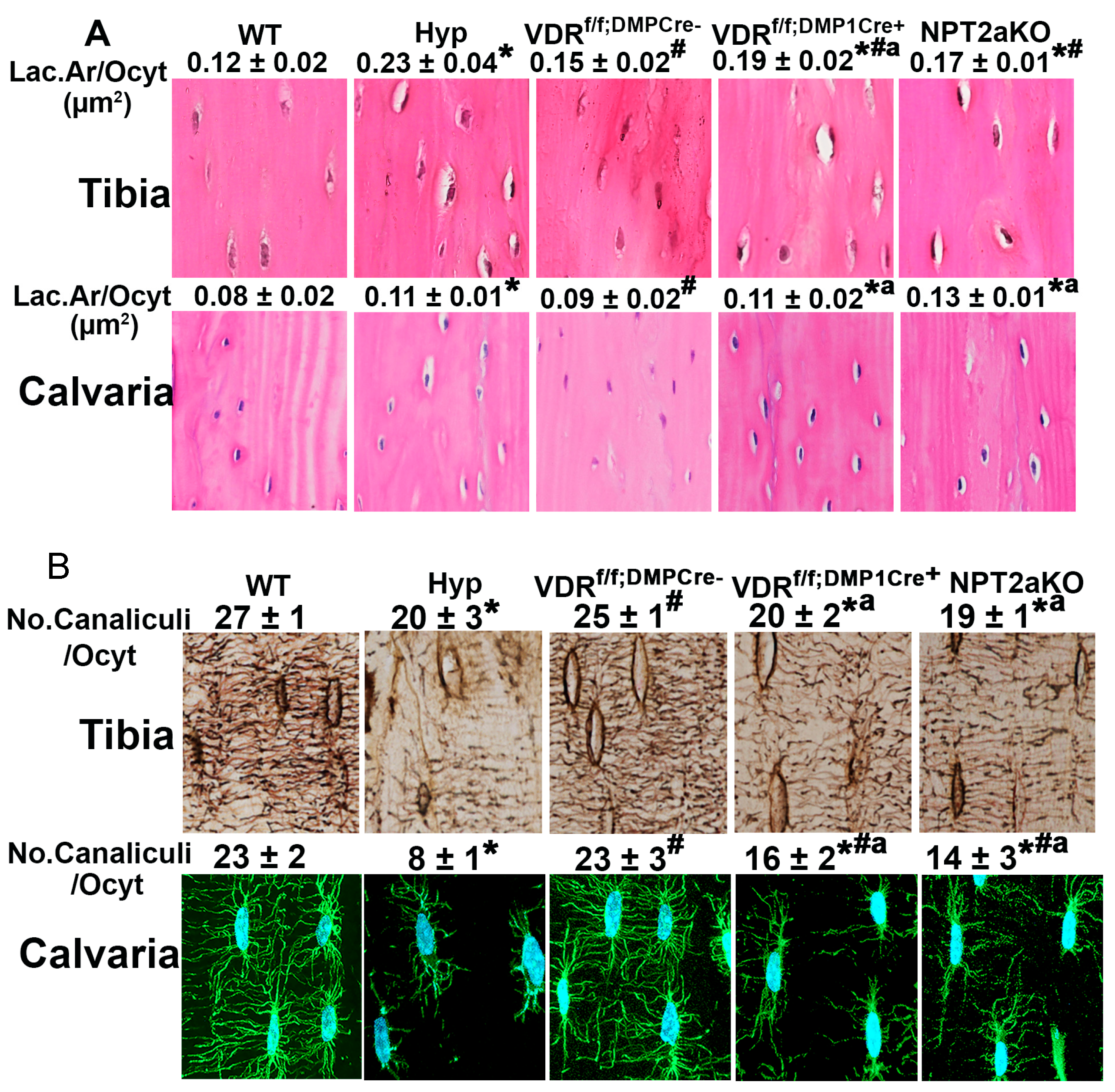
4.3. Regulation of Osteocyte LCN Organization in Hyp Mice

It has long been shown that there are perilacunar halos of osteoid in the bones of Hyp mice [94]. More recently, it was reported that Hyp bones also have decreased osteocyte number and increased osteocyte apoptosis, corresponding with dramatically impaired whole-bone biomechanics [44]. The calvaria and tibiae of Hyp mice have enlarged lacunae and impaired canalicular organization, with Hyp bones being characterized by very sparse and few canaliculi with decreased canalicular branching and connectivity compared to WT control (Figure 1) [88]. Similar to lactating mice, the enlarged lacunae in Hyp mice are associated with the enhanced expression of LCN regulating genes such as CTSK, TRAP, and MMP13 [88]. The administration of daily 1,25D or the FGF23Ab to Hyp mice suppresses osteocyte cell death, restores lacunar size, and improves canalicular morphology. 1,25D treatment of Hyp mice normalizes osteocyte expression of MMP13 and classic osteoclast markers, while FGF23Ab decreases the expression of MMP13 and selected osteoclast markers [88]. These data supported roles for phosphate or 1,25D in regulating perilacunar and canalicular remodeling [88].
The role of 1,25D in maintaining LCN morphology Is corroborated by the increased osteocyte lacunar volume [95] and poor canalicular structure [96] seen in vitamin D-deficient human cortical bone as compared to vitamin D sufficient controls. Moreover, mice deficient in vitamin D receptor (VDR) fed with a normal diet have larger lacunar areas than WT. However, bones from vitamin D-deficient human and murine models are characterized by osteomalacia; therefore, it is not clear whether the LCN abnormalities are due to impaired mineralization or decreased 1,25D signaling.
In order to study the role of 1,25D in regulating osteocyte-mediated perilacunar remodeling and canalicular organization, LCN organization was analyzed in bones from mice lacking the VDR in osteocytes (VDRf/f;DMP1Cre+) [65]. These mice demonstrate normal serum mineral ion and hormone levels and also have normal skeletal microarchitecture, thus allowing for the determination of 1,25D-specific effects on LCN remodeling. Mice null for sodium phosphate transporter 2a (NPT2aKO) have hypophosphatemia and high serum 1,25D levels with low FGF23 levels [50]; therefore, LCN remodeling was analyzed in the mice to determine if a physiological increase in 1,25D can compensate for low serum phosphate in regulating LCN remodeling [65] (Figure 1). No significant alterations in cortical microarchitecture were observed in NPT2aKO, with bones from these mice having a mild decrease in cortical thickness and tissue mineral density. Like Hyp mice, tibial and calvarial osteocytes from VDRf/f;DMP1Cre+ and NPT2aKO mice have enlarged osteocyte lacunae and impaired canalicular organization compared to respective controls. In addition, like Hyp osteocytes, the osteocytes isolated from tibiae in VDRf/f;DMP1Cre+ and NPT2aKO mice have enhanced expression of osteoclast expressed matrix resorption genes (Ctsk, Acp5, Atp6v0d2, Nhedc2). The treatment of Ocy454 osteocytes with 1,25D or phosphate inhibits the expression of these genes, demonstrating that 1,25D and phosphate each act directly on osteocytes to regulate the expression of LCN-regulating genes. Taken together, these studies show that 1,25D acts directly on osteocytes to modulate LCN organization. They also demonstrate that the high 1,25D levels in NPT2aKO mice cannot compensate for the low serum phosphate levels in modulating LCN organization, suggesting that hypophosphatemia plays a role in regulating LCN remodeling. It implies that the low-phosphate state stimulates osteocyte lacunar matrix resorption in order to help maintain phosphate homeostasis [65].

5. Enthesopathy

5.1. The Enthesis

The region where the tendon inserts into bone, known as the enthesis, is a specialized tissue that is critical for joint movement [97]. The enthesis allows for the transmission of contractile forces from muscle to bone [98,99]. Fibrocartilaginous entheses attach to bone via a transitionary layer of fibrocartilage [100] and consist of four different zones: the bony eminence, mineralized fibrocartilage, unmineralized fibrocartilage and tendon [100,101]. This region has a characteristic gradation in mineral concentration and collagen orientation [102,103]. There is a linear increase in mineral volume fraction between tendon and bone as well as a decrease in the alignment of collagen fibers between tendon and bone [103].
Scleraxis (Scx) is a bHLH transcription factor that is a marker for tendon and ligament progenitors [104]. The deletion of scleraxis in cells expressing Prx1 (limb progenitor cells) resulted in abnormal morphology, impaired biomechanical properties, and disorganized collagen fiber orientation in supraspinatus entheses [104], thus demonstrating that Scx is necessary for normal enthesis organization. In addition, regulators of chondrogenesis including bone morphogenic proteins (BMPs) and Sox9 have been implicated in enthesis development [13,105,106]. When BMP4 is deleted in Scx-expressing cells, formation of the bony ridge onto which the deltoid tendon inserts into is impaired [106]. SOX9, a BMP target gene, is necessary for chondrogenesis and is expressed in chondroprogenitor cells as well as in enthesis cells [13,107] and in the bony eminences onto which the entheses insert [108,109]. Lineage tracing studies demonstrated that enthesis cells are descendants of both Scx+ and Sox9+ progenitor cells [13,110]. When SOX9 was deleted in Scx+ cells, enthesis development and organization were compromised [107], indicating that Sox9 plays a role in enthesis formation [109].
Other regulators of chondrogenesis including Parathyroid hormone related protein (PTHrP), Indian Hedgehog (IHH), and the IHH receptor, Patched (PTCH), are also expressed in entheses [105,111]. Gli1, a transcriptional effector and target of IHH, also plays a role in enthesis development [112]. Enthesis cells are descendants of Gli1 expressing progenitor cells, and the deletion of Gli1+ cells in entheses resulted in a loss of enthesis fibrocartilage organization [112].

5.2. Enthesopathy in XLH

Enthesopathy is an abnormal mineralization of the tendon–bone insertion, which results in pain, impaired movement, and altered gait. This complication is observed in a majority of adults affected with XLH [113] and can lead to a significant impairment of quality of life [113,114,115]. In a survey of 39 patients with XLH, 49% of patients exhibited enthesopathy at the pelvis, 56% at the knees, 74% at the ankles, and 41% at the spine [113]. A survey of 114 adult XLH patients attending a specialized center, with an average age of 42 years, found that age and severe dental disease were associated with enthesopathy, and 67% of participants had at least one enthesopathy site at the spine or pelvis [116]. Those affected with XLH enthesopathy often express concern for fear of falling, needing assist devices for walking, and lacking credence from their health-care providers [115]. The chronic pain XLH patients suffer can impact their social activity and choice of career [115].
The conventional treatment of XLH consists of daily doses of oral phosphate and active vitamin D analogss such as calcitriol [62,114]. While there are limited clinical data examining the effects of conventional therapy on XLH enthesopathy development, Gjorup et al. demonstrated that those affected with XLH who were treated consistently with conventional therapy during childhood had a decreased incidence of vertebral enthesopathy compared to XLH patients who received intermittent or no therapy as a child [117]. In addition, a retrospective study on adult XLH subjects showed that adults without enthesopathy had a higher percent of subjects who were treated as a child with conventional therapy than adults with enthesopathy [116]. On the other hand, an observational cross-sectional study of adult XLH patients found no relationship between proportion of adult life treated with conventional therapy and number of enthesopathy sites; however, age, BMI and sex were found to be predictors of the number of enthesopathy sites [118]. This study did not address the dose of treatment or severity of enthesopathy [118].

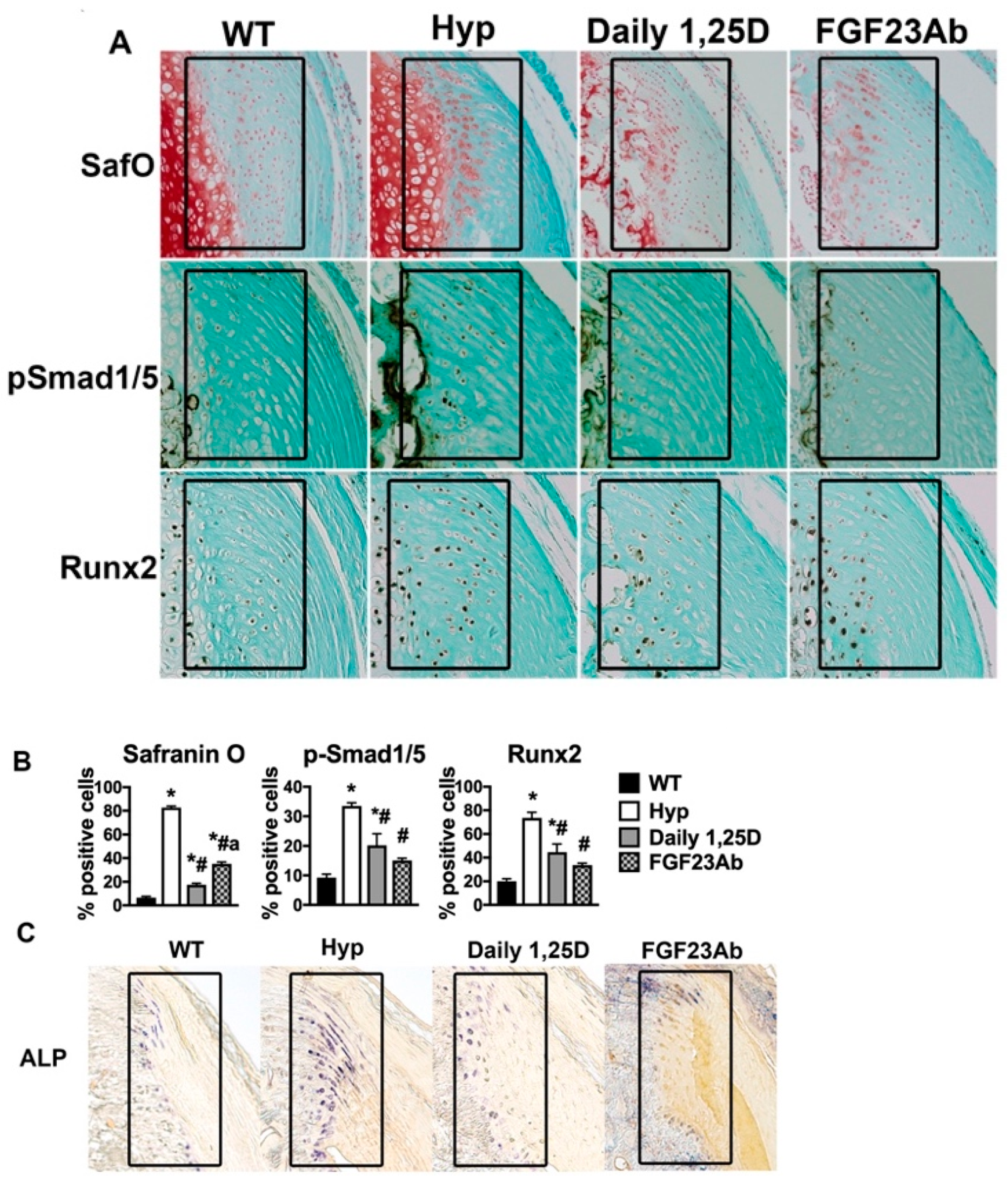
5.3. Molecular Pathogenesis of XLH Enthesopathy

The molecular regulation of enthesopathy development in XLH is poorly understood. The Hyp mouse model of the XLH mutation has been used to investigate the pathogenesis of enthesopathy [113]. Achilles entheses in Hyp mice demonstrate an expansion of hypertrophic-appearing chondrogenic cells that are positive for Safranin O (SafO, stain for cartilage proteoglycans) and alkaline phosphatase activity (ALP, marker of mineralization) [13,113,116]. Lineage-tracing studies showed that post-natal enthesis cells of both wild-type and Hyp mice originate from Scx and Sox9-expressing progenitors [13], with Hyp entheses having an expansion of SafO/ALP+ cells that express Sox9 by P14 [13]. Corresponding with the chondrogenic characteristics of the enthesopathy cells, the hypertrophic-appearing cells in Hyp entheses also demonstrate an increased expression of BMP signaling marker p-Smad 1/5/8, BMP signaling target IHH, and IHH signaling targets PTCH and Runx2 by P14 [13]. These data support a pathogenic role for BMP/IHH signaling in XLH enthesopathy development.
Achilles entheses from Hyp mice treated with either daily 1,25D or an anti-FGF23 targeting antibody, both of which increase 1,25D action, starting P2 (prior to enthesopathy development in Hyp mice) attenuated enthesopathy development, with treated Hyp entheses having a decreased expansion of SafO/ALP+ cells and decreased BMP/IHH signaling compared to untreated Hyp entheses (Figure 2) [13]. In contrast, treatment of Hyp mice with phosphate and calcitriol starting P30 (after enthesopathy has developed) did not attenuate the expansion of ALP+ cells observed in Hyp entheses [119]. Likewise, Cauliez et al. treated Hyp mice with combination phosphate and calcitriol therapy starting P2 or P30, showing that Hyp enthesopathy, as assessed by ALP activity, is partially prevented with early therapy but not altered with therapy starting in mature entheses at P30 [120]. Taken together with the clinical data available, these studies suggest that enthesopathy development may be attenuated with early and consistent therapy, but it may not be modifiable once it has developed. Since both 1,25D and the FGF23Ab prevent Hyp enthesopathy, increased circulating FGF23, leading to impaired 1,25D action, may play a role in enthesopathy development and the increase in BMP/IHH signaling observed in entheses. In support of this, mice that overexpress FGF23 develop a similar enthesopathy as observed in Hyp mice [119].

6. Future Directions

In 2018, burosumab, a human monoclonal anti-FGF23 antibody, was approved to treat XLH in the European Union and USA. Both human and murine studies have supported that the early diagnosis and initiation of therapy leads to better attenuation of XLH complications. While studies have examined the beneficial effects of burosumab on growth velocity in children and osteomalacia in adults, it remains unknown if growth and mineralization, which increase but do not normalize despite normal serum phosphate, will further improve with long-term treatment. Moreover, no data have been reported on the effectiveness of burosumab in preventing or treating enthesopathy [28,121,122]. Based on studies that 1,25D may play an important role in regulating complications of XLH such as decreased growth velocity, trabecular structure, osteocyte lacuno-canalicular remodeling, and enthesopathy development, it remains to be determined if the gradual decline in serum 1,25D levels during long-term burosumab treatment will have any impact on the prevention or treatment of these complications. Although burosumab has been shown to be effective in attenuating select complications of XLH, studies demonstrated that the molecular mechanisms underlying these complications such as impaired mineralization and growth are not completely dependent on the abnormalities in mineral ion and hormone homeostasis see in XLH. While studies have dissected the molecular mechanisms of these complications, it remains to be seen if improving those molecular pathways would serve as effective therapies for XLH. Further insight into the phenotypic variation seen in XLH could be provided by identifying additional direct targets of PHEX. Single-cell transcriptomics of human tissue has revealed the tissues and cells that are enriched for PHEX expression. For instance, these studies demonstrated that lung and prostate tissue and granulosa cells in the ovary are enriched for PHEX expression. However, proteomic data that potentially could reveal the targets of PHEX are still lacking [123]. Further research is necessary to improve the understanding of the pathophysiology of XLH complications.

Author Contributions

Conceptualization, E.S.L.; writing—original draft preparation, S.J., S.V., M.S. and E.S.L.; writing—review and editing, S.J. and E.S.L. and supervision, E.S.L. All authors have read and agreed to the published version of the manuscript.

Funding

This work was supported by a grant from the National Institute of Health: R01 AR076973 (to E.S.L.).

Institutional Review Board Statement

The animal study protocol was approved by the Institutional Review Board (or Ethics Committee) of Brigham and Women’s Hospital (protocol code 2018N000044 and date of approval is 18 January 2023).

Informed Consent Statement

Not applicable.

Data Availability Statement

No new data was generated in this manuscript. All data represented in the figures were previously published and there are no issues with copyright.

Conflicts of Interest

The authors declare no conflict of interest.

References

  1. Jonsson, K.B.; Zahradnik, R.; Larsson, T.; White, K.E.; Sugimoto, T.; Imanishi, Y.; Yamamoto, T.; Hampson, G.; Koshiyama, H.; Ljunggren, Ö.; et al. Fibroblast Growth Factor 23 in Oncogenic Osteomalacia and X-Linked Hypophosphatemia. N. Engl. J. Med. 2003, 348, 1656–1663. [Google Scholar] [CrossRef]
  2. Liu, S.; Zhou, J.; Tang, W.; Jiang, X.; Rowe, D.W.; Quarles, L.D. Pathogenic role of Fgf23 in Hyp mice. Am. J. Physiol. Endocrinol. Metab. 2006, 291, E38–E49. [Google Scholar] [CrossRef]
  3. Holm, I.A.; Nelson, A.E.; Robinson, B.G.; Mason, R.S.; Marsh, D.J.; Cowell, C.T.; Carpenter, T.O. Mutational analysis and genotype-phenotype correlation of the PHEX gene in X-linked hypophosphatemic rickets. J. Clin. Endocrinol. Metab. 2001, 86, 3889–3899. [Google Scholar] [CrossRef]
  4. Liu, E.S.; Carpenter, T.O.; Gundberg, C.M.; Simpson, C.A.; Insogna, K.L. Calcitonin Administration in X-Linked Hypophosphatemia. N. Engl. J. Med. 2011, 364, 1678–1680. [Google Scholar] [CrossRef] [Green Version]
  5. Farrow, E.G.; White, K.E. Recent advances in renal phosphate handling. Nat. Rev. Nephrol. 2010, 6, 207–217. [Google Scholar] [CrossRef] [PubMed]
  6. Carpenter, T.O.; Imel, E.A.; Holm, I.A.; de Beur, S.M.J.; Insogna, K.L. A clinician’s guide to X-linked hypophosphatemia. J. Bone Miner. Res. 2011, 26, 1381–1388. [Google Scholar] [CrossRef] [Green Version]
  7. Sarafrazi, S.; Daugherty, S.C.; Miller, N.; Boada, P.; Carpenter, T.O.; Chunn, L.; Dill, K.; Econs, M.J.; Eisenbeis, S.; Imel, E.A.; et al. Novel PHEX gene locus-specific database: Comprehensive characterization of vast number of variants associated with X-linked hypophosphatemia (XLH). Hum. Mutat. 2021, 43, 143–157. [Google Scholar] [CrossRef] [PubMed]
  8. Whyte, M.P.; Schranck, F.W.; Armamento-Villareal, R. X-linked hypophosphatemia: A search for gender, race, anticipation, or parent of origin effects on disease expression in children. J. Clin. Endocrinol. Metab. 1996, 81, 4075–4080. [Google Scholar] [CrossRef] [PubMed]
  9. Dahir, K.M.; Black, M.; Gottesman, G.S.; Imel, E.A.; Mumm, S.; Nichols, C.M.; Whyte, M.P. X-Linked Hypophosphatemia Caused by the Prevailing North American PHEX Variant c.*231A>G; Exon 13–15 Duplication Is Often Misdiagnosed as Ankylosing Spondylitis and Manifests in Both Men and Women. JBMR Plus 2022, 6, e10692. [Google Scholar] [CrossRef] [PubMed]
  10. Song, H.-R.; Park, J.-W.; Cho, D.-Y.; Yang, J.H.; Yoon, H.-R.; Jung, S.-C. PHEX Gene Mutations and Genotype-Phenotype Analysis of Korean Patients with Hypophosphatemic Rickets. J. Korean Med. Sci. 2007, 22, 981–986. [Google Scholar] [CrossRef] [Green Version]
  11. Eicher, E.M.; Southard, J.L.; Scriver, C.R.; Glorieux, F.H. Hypophosphatemia: Mouse model for human familial hypophosphatemic (vitamin D-resistant) rickets. Proc. Natl. Acad. Sci. USA 1976, 73, 4667–4671. [Google Scholar] [CrossRef] [PubMed] [Green Version]
  12. Strom, T.M.; Francis, F.; Lorenz, B.; Böddrich, A.; Lehrach, H.; Econs, M.J.; Meitinger, T. Pex Gene Deletions in Gy and Hyp Mice Provide Mouse Models for X-Linked Hypophosphatemia. Hum. Mol. Genet. 1997, 6, 165–171. [Google Scholar] [CrossRef] [PubMed] [Green Version]
  13. Liu, E.S.; Martins, J.S.; Zhang, W.; Demay, M.B. Molecular analysis of enthesopathy in a mouse model of hypophosphatemic rickets. Development 2018, 145, dev163519. [Google Scholar] [CrossRef] [Green Version]
  14. Aono, Y.; Yamazaki, Y.; Yasutake, J.; Kawata, T.; Hasegawa, H.; Urakawa, I.; Fujita, T.; Wada, M.; Yamashita, T.; Fukumoto, S.; et al. Therapeutic effects of anti-FGF23 antibodies in hypophosphatemic rickets/osteomalacia. J. Bone Miner. Res. 2009, 24, 1879–1888. [Google Scholar] [CrossRef]
  15. Christakos, S.; Dhawan, P.; Verstuyf, A.; Verlinden, L.; Carmeliet, G. Vitamin D: Metabolism, Molecular Mechanism of Action, and Pleiotropic Effects. Physiol. Rev. 2016, 96, 365–408. [Google Scholar] [CrossRef] [Green Version]
  16. Bikle, D.; Christakos, S. New aspects of vitamin D metabolism and action—Addressing the skin as source and target. Nat. Rev. Endocrinol. 2020, 16, 234–252. [Google Scholar] [CrossRef]
  17. Sone, T.; Kerner, S.; Pike, J. Vitamin D receptor interaction with specific DNA. Association as a 1,25-dihydroxyvitamin D3-modulated heterodimer. J. Biol. Chem. 1991, 266, 23296–23305. [Google Scholar] [CrossRef]
  18. Sone, T.; Ozono, K.; Pike, J.W. A 55-Kilodalton Accessory Factor Facilitates Vitamin D Receptor DNA Binding. Mol. Endocrinol. 1991, 5, 1578–1586. [Google Scholar] [CrossRef] [Green Version]
  19. Okazaki, T.; Igarashi, T.; Kronenberg, H.M. 5’-flanking region of the parathyroid hormone gene mediates negative regulation by 1,25-(OH)2 vitamin D3. J. Biol. Chem. 1988, 263, 2203–2208. [Google Scholar] [CrossRef]
  20. Canalejo, A.; Almadén, Y.; Torregrosa, V.; Gomez-Villamandos, J.C.; Ramos, B.; Campistol, J.M.; Felsenfeld, A.J.; Rodríguez, M. The In Vitro Effect of Calcitriol on Parathyroid Cell Proliferation and Apoptosis. J. Am. Soc. Nephrol. 2000, 11, 1865–1872. [Google Scholar] [CrossRef]
  21. Bonewald, L.F.; Wacker, M.J. FGF23 production by osteocytes. Pediatr. Nephrol. 2013, 28, 563–568. [Google Scholar] [CrossRef] [PubMed] [Green Version]
  22. Saito, H.; Kusano, K.; Kinosaki, M.; Ito, H.; Hirata, M.; Segawa, H.; Miyamoto, K.; Fukushima, N. Human fibroblast growth factor-23 mutants suppress Na+-dependent phosphate co-transport activity and 1alpha,25-dihydroxyvitamin D3 production. J. Biol. Chem. 2003, 278, 2206–2211. [Google Scholar] [CrossRef] [PubMed] [Green Version]
  23. Bai, X.-Y.; Miao, D.; Goltzman, D.; Karaplis, A.C. The Autosomal Dominant Hypophosphatemic Rickets R176Q Mutation in Fibroblast Growth Factor 23 Resists Proteolytic Cleavage and Enhances in Vivo Biological Potency. J. Biol. Chem. 2003, 278, 9843–9849. [Google Scholar] [CrossRef] [PubMed] [Green Version]
  24. Shimada, T.; Urakawa, I.; Yamazaki, Y.; Hasegawa, H.; Hino, R.; Yoneya, T.; Takeuchi, Y.; Fujita, T.; Fukumoto, S.; Yamashita, T. FGF-23 transgenic mice demonstrate hypophosphatemic rickets with reduced expression of sodium phosphate cotransporter type IIa. Biochem. Biophys. Res. Commun. 2004, 314, 409–414. [Google Scholar] [CrossRef]
  25. Larsson, T.; Marsell, R.; Schipani, E.; Ohlsson, C.; Ljunggren, O.; Tenenhouse, H.S.; Juppner, H.; Jonsson, K.B. Transgenic mice expressing fibroblast growth factor 23 under the control of the alpha1(I) collagen promoter exhibit growth retardation, osteomalacia, and disturbed phosphate homeostasis. Endocrinology 2004, 145, 3087–3094. [Google Scholar] [CrossRef] [Green Version]
  26. Chanakul, A.; Zhang, M.Y.H.; Louw, A.; Armbrecht, H.J.; Miller, W.L.; Portale, A.A.; Perwad, F. FGF-23 Regulates CYP27B1 Transcription in the Kidney and in Extra-Renal Tissues. PLoS ONE 2013, 8, e72816. [Google Scholar] [CrossRef] [Green Version]
  27. Bai, X.; Miao, D.; Xiao, S.; Qiu, D.; St-Arnaud, R.; Petkovich, M.; Gupta, A.; Goltzman, D.; Karaplis, A.C. CYP24 inhibition as a therapeutic target in FGF23-mediated renal phosphate wasting disorders. J. Clin. Investig. 2016, 126, 667–680. [Google Scholar] [CrossRef] [Green Version]
  28. Insogna, K.L.; Briot, K.; Imel, E.A.; Kamenický, P.; Ruppe, M.D.; Portale, A.A.; Weber, T.; Pitukcheewanont, P.; Cheong, H.I.; de Beur, S.J.; et al. A Randomized, Double-Blind, Placebo-Controlled, Phase 3 Trial Evaluating the Efficacy of Burosumab, an Anti-FGF23 Antibody, in Adults With X-Linked Hypophosphatemia: Week 24 Primary Analysis. J. Bone Miner. Res. 2018, 33, 1383–1393. [Google Scholar] [CrossRef] [Green Version]
  29. Carpenter, T.O.; Imel, E.A.; Ruppe, M.D.; Weber, T.J.; Klausner, M.A.; Wooddell, M.M.; Kawakami, T.; Ito, T.; Zhang, X.; Humphrey, J.; et al. Randomized trial of the anti-FGF23 antibody KRN23 in X-linked hypophosphatemia. J. Clin. Investig. 2014, 124, 1587–1597. [Google Scholar] [CrossRef] [Green Version]
  30. Portale, A.A.; Carpenter, T.O.; Brandi, M.L.; Briot, K.; Cheong, H.I.; Cohen-Solal, M.; Crowley, R.; De Beur, S.J.; Eastell, R.; Imanishi, Y.; et al. Continued Beneficial Effects of Burosumab in Adults with X-Linked Hypophosphatemia: Results from a 24-Week Treatment Continuation Period After a 24-Week Double-Blind Placebo-Controlled Period. Calcif. Tissue Int. 2019, 105, 271–284. [Google Scholar] [CrossRef] [Green Version]
  31. Imel, E.A.; Zhang, X.; Ruppe, M.D.; Weber, T.J.; Klausner, M.A.; Ito, T.; Vergeire, M.; Humphrey, J.S.; Glorieux, F.H.; Portale, A.A.; et al. Prolonged Correction of Serum Phosphorus in Adults With X-Linked Hypophosphatemia Using Monthly Doses of KRN23. J. Clin. Endocrinol. Metab. 2015, 100, 2565–2573. [Google Scholar] [CrossRef] [PubMed] [Green Version]
  32. Imel, E.A.; Glorieux, F.H.; Whyte, M.P.; Munns, C.F.; Ward, L.M.; Nilsson, O.; Simmons, J.; Padidela, R.; Namba, N.; Cheong, H.I.; et al. Burosumab versus conventional therapy in children with X-linked hypophosphataemia: A randomised, active-controlled, open-label, phase 3 trial. Lancet 2019, 393, 2416–2427. [Google Scholar] [CrossRef] [PubMed]
  33. Insogna, K.L.; Rauch, F.; Kamenický, P.; Ito, N.; Kubota, T.; Nakamura, A.; Zhang, L.; Mealiffe, M.; Martin, J.S.; Portale, A.A. Burosumab Improved Histomorphometric Measures of Osteomalacia in Adults with X-Linked Hypophosphatemia: A Phase 3, Single-Arm, International Trial. J. Bone Miner. Res. 2019, 34, 2183–2191. [Google Scholar] [CrossRef] [PubMed] [Green Version]
  34. Beck-Nielsen, S.S.; Brusgaard, K.; Rasmussen, L.M.; Brixen, K.; Brock-Jacobsen, B.; Poulsen, M.R.; Vestergaard, P.; Ralston, S.H.; Albagha, O.M.E.; Poulsen, S.; et al. Phenotype Presentation of Hypophosphatemic Rickets in Adults. Calcif. Tissue Int. 2010, 87, 108–119. [Google Scholar] [CrossRef]
  35. Živičnjak, M.; Schnabel, D.; Billing, H.; Staude, H.; Filler, G.; Querfeld, U.; Schumacher, M.; Pyper, A.; Schröder, C.; Bramswig, J.; et al. Age-related stature and linear body segments in children with X-linked hypophosphatemic rickets. Pediatr. Nephrol. 2010, 26, 223–231. [Google Scholar] [CrossRef] [PubMed]
  36. Rodríguez-Rubio, E.; Gil-Peña, H.; Chocron, S.; Madariaga, L.; de la Cerda-Ojeda, F.; Fernández-Fernández, M.; de Lucas-Collantes, C.; Gil, M.; Luis-Yanes, M.I.; Vergara, I.; et al. Phenotypic characterization of X-linked hypophosphatemia in pediatric Spanish population. Orphanet J. Rare Dis. 2021, 16, 104. [Google Scholar] [CrossRef] [PubMed]
  37. Schutt, S.; Schumacher, M.; Holterhus, P.; Felgenhauer, S.; Hiort, O. Effect of GH replacement therapy in two male siblings with combined X-linked hypophosphatemia and partial GH deficiency. Eur. J. Endocrinol. 2003, 149, 317–321. [Google Scholar] [CrossRef] [Green Version]
  38. Cagnoli, M.; Richter, R.; Bohm, P.; Knye, K.; Empting, S.; Mohnike, K. Spontaneous Growth and Effect of Early Therapy with Calcitriol and Phosphate in X-linked Hypophosphatemic Rickets. Pediatr. Endocrinol. Rev. 2017, 15 (Suppl. S1), 119–122. [Google Scholar] [CrossRef]
  39. Mao, M.; Carpenter, T.O.; Whyte, M.P.; Skrinar, A.; Chen, C.-Y.; Martin, J.S.; Rogol, A.D. Growth Curves for Children with X-linked Hypophosphatemia. J. Clin. Endocrinol. Metab. 2020, 105, 3243–3249. [Google Scholar] [CrossRef]
  40. Thacher, T.D.; Pettifor, J.M.; Tebben, P.J.; Creo, A.L.; Skrinar, A.; Mao, M.; Chen, C.-Y.; Chang, T.; Martin, J.S.; Carpenter, T.O. Rickets severity predicts clinical outcomes in children with X-linked hypophosphatemia: Utility of the radiographic Rickets Severity Score. Bone 2019, 122, 76–81. [Google Scholar] [CrossRef]
  41. Kronenberg, H.M. Developmental regulation of the growth plate. Nature 2003, 423, 332–336. [Google Scholar] [CrossRef] [PubMed]
  42. Street, J.; Bao, M.; DeGuzman, L.; Bunting, S.; Peale, F.V., Jr.; Ferrara, N.; Steinmetz, H.; Hoeffel, J.; Cleland, J.L.; Daugherty, A.; et al. Vascular endothelial growth factor stimulates bone repair by promoting angiogenesis and bone turnover. Proc. Natl. Acad. Sci. USA 2002, 99, 9656–9661. [Google Scholar] [CrossRef] [PubMed] [Green Version]
  43. Yang, L.; Tsang, K.Y.; Tang, H.C.; Chan, D.; Cheah, K.S.E. Hypertrophic chondrocytes can become osteoblasts and osteocytes in endochondral bone formation. Proc. Natl. Acad. Sci. USA 2014, 111, 12097–12102. [Google Scholar] [CrossRef] [Green Version]
  44. Liu, E.S.; Martins, J.S.; Raimann, A.; Chae, B.T.; Brooks, D.J.; Jorgetti, V.; Bouxsein, M.L.; Demay, M.B. 1,25-Dihydroxyvitamin D Alone Improves Skeletal Growth, Microarchitecture and Strength in a Murine Model of XLH, Despite Enhanced FGF23 Expression. J. Bone Miner. Res. 2016, 31, 929–939. [Google Scholar] [CrossRef] [Green Version]
  45. Sabbagh, Y.; Carpenter, T.O.; Demay, M.B. Hypophosphatemia leads to rickets by impairing caspase-mediated apoptosis of hypertrophic chondrocytes. Proc. Natl. Acad. Sci. USA 2005, 102, 9637–9642. [Google Scholar] [CrossRef] [Green Version]
  46. Fuente, R.; Garcia-Bengoa, M.; Fernandez-Iglesias, A.; Gil-Pena, H.; Santos, F.; Lopez, J.M. Cellular and Molecular Alterations Underlying Abnormal Bone Growth in X-Linked Hypophosphatemia. Int. J. Mol. Sci. 2022, 23, 934. [Google Scholar] [CrossRef]
  47. Fuente, R.; Gil-Peña, H.; Claramunt-Taberner, D.; Hernández-Frías, O.; Fernández-Iglesias, Á.; Hermida-Prado, F.; Anes-González, G.; Rubio-Aliaga, I.; Lopez, J.M.; Santos, F. Marked alterations in the structure, dynamics and maturation of growth plate likely explain growth retardation and bone deformities of young Hyp mice. Bone 2018, 116, 187–195. [Google Scholar] [CrossRef] [PubMed]
  48. Donohue, M.M.; Demay, M.B. Rickets in VDR Null Mice Is Secondary to Decreased Apoptosis of Hypertrophic Chondrocytes. Endocrinology 2002, 143, 3691–3694. [Google Scholar] [CrossRef] [Green Version]
  49. Bergwitz, C.; Roslin, N.M.; Tieder, M.; Loredo-Osti, J.C.; Bastepe, M.; Abu-Zahra, H.; Frappier, D.; Burkett, K.; Carpenter, T.O.; Anderson, D.; et al. SLC34A3 Mutations in Patients with Hereditary Hypophosphatemic Rickets with Hypercalciuria Predict a Key Role for the Sodium-Phosphate Cotransporter NaPi-IIc in Maintaining Phosphate Homeostasis. Am. J. Hum. Genet. 2006, 78, 179–192. [Google Scholar] [CrossRef] [Green Version]
  50. Miedlich, S.U.; Zhu, E.D.; Sabbagh, Y.; DeMay, M.B. The Receptor-Dependent Actions of 1,25-Dihydroxyvitamin D Are Required for Normal Growth Plate Maturation in NPt2a Knockout Mice. Endocrinology 2010, 151, 4607–4612. [Google Scholar] [CrossRef]
  51. Miedlich, S.U.; Zalutskaya, A.; Zhu, E.D.; Demay, M.B. Phosphate-induced Apoptosis of Hypertrophic Chondrocytes Is Associated with a Decrease in Mitochondrial Membrane Potential and Is Dependent upon Erk1/2 Phosphorylation. J. Biol. Chem. 2010, 285, 18270–18275. [Google Scholar] [CrossRef] [PubMed] [Green Version]
  52. Provot, S.; Nachtrab, G.; Paruch, J.; Chen, A.P.; Silva, A.; Kronenberg, H.M. A-Raf and B-Raf Are Dispensable for Normal Endochondral Bone Development, and Parathyroid Hormone-Related Peptide Suppresses Extracellular Signal-Regulated Kinase Activation in Hypertrophic Chondrocytes. Mol. Cell. Biol. 2008, 28, 344–357. [Google Scholar] [CrossRef] [PubMed] [Green Version]
  53. Kaneko, Y.; Tanzawa, H.; Sato, K. The proto-oncogene C-raf-1 is highly expressed only in the hypertrophic zone of the growth plate. Calcif. Tissue Int. 1994, 54, 426–430. [Google Scholar] [CrossRef]
  54. Liu, E.S.; Raimann, A.; Chae, B.T.; Martins, J.S.; Baccarini, M.; Demay, M.B. C-Raf promotes Angiogenesis during Normal Growth Plate Maturation. Development 2015, 143, 348–355. [Google Scholar] [CrossRef] [Green Version]
  55. Papaioannou, G.; Petit, E.T.; Liu, E.S.; Baccarini, M.; Pritchard, C.; Demay, M.B. Raf Kinases Are Essential for Phosphate Induction of ERK1/2 Phosphorylation in Hypertrophic Chondrocytes and Normal Endochondral Bone Development. J. Biol. Chem. 2017, 292, 3164–3171. [Google Scholar] [CrossRef] [Green Version]
  56. Lanske, B.; Karaplis, A.C.; Lee, K.; Luz, A.; Vortkamp, A.; Pirro, A.; Karperien, M.; Defize, L.H.K.; Ho, C.; Mulligan, R.C.; et al. PTH/PTHrP Receptor in Early Development and Indian Hedgehog—Regulated Bone Growth. Science 1996, 273, 663–666. [Google Scholar] [CrossRef] [PubMed]
  57. Schipani, E.; Lanske, B.; Hunzelman, J.; Luz, A.; Kovacs, C.S.; Lee, K.; Pirro, A.; Kronenberg, H.M.; Jüppner, H. Targeted expression of constitutively active receptors for parathyroid hormone and parathyroid hormone-related peptide delays endochondral bone formation and rescues mice that lack parathyroid hormone-related peptide. Proc. Natl. Acad. Sci. USA 1997, 94, 13689–13694. [Google Scholar] [CrossRef] [Green Version]
  58. Liu, E.S.; Zalutskaya, A.; Chae, B.T.; Zhu, E.D.; Gori, F.; Demay, M.B. Phosphate Interacts With PTHrP to Regulate Endochondral Bone Formation. Endocrinology 2014, 155, 3750–3756. [Google Scholar] [CrossRef] [Green Version]
  59. Beck, L.; Karaplis, A.C.; Amizuka, N.; Hewson, A.S.; Ozawa, H.; Tenenhouse, H.S. Targeted inactivation of Npt2 in mice leads to severe renal phosphate wasting, hypercalciuria, and skeletal abnormalities. Proc. Natl. Acad. Sci. USA 1998, 95, 5372–5377. [Google Scholar] [CrossRef] [Green Version]
  60. Kaneko, I.; Segawa, H.; Ikuta, K.; Hanazaki, A.; Fujii, T.; Tatsumi, S.; Kido, S.; Hasegawa, T.; Amizuka, N.; Saito, H.; et al. Eldecalcitol Causes FGF23 Resistance for Pi Reabsorption and Improves Rachitic Bone Phenotypes in the Male Hyp Mouse. Endocrinology 2018, 159, 2741–2758. [Google Scholar] [CrossRef]
  61. Jehan, F.; Gaucher, C.; Nguyen, T.M.; Walrant-Debray, O.; Lahlou, N.; Sinding, C.; Déchaux, M.; Garabédian, M. Vitamin D Receptor Genotype in Hypophosphatemic Rickets as a Predictor of Growth and Response to Treatment. J. Clin. Endocrinol. Metab. 2008, 93, 4672–4682. [Google Scholar] [CrossRef] [PubMed] [Green Version]
  62. Linglart, A.; Duplan, M.B.; Briot, K.; Chaussain, C.; Esterle, L.; Guillaume-Czitrom, S.; Kamenicky, P.; Nevoux, J.; Prié, D.; Rothenbuhler, A.; et al. Therapeutic management of hypophosphatemic rickets from infancy to adulthood. Endocr. Connect. 2014, 3, R13–R30. [Google Scholar] [CrossRef] [PubMed]
  63. Mäkitie, O.; Doria, A.; Kooh, S.W.; Cole, W.G.; Daneman, A.; Sochett, E. Early Treatment Improves Growth and Biochemical and Radiographic Outcome in X-Linked Hypophosphatemic Rickets. J. Clin. Endocrinol. Metab. 2003, 88, 3591–3597. [Google Scholar] [CrossRef] [PubMed] [Green Version]
  64. Brownstein, C.; Zhang, J.; Stillman, A.; Ellis, B.; Troiano, N.; Adams, D.J.; Gundberg, C.M.; Lifton, R.P.; Carpenter, T.O. Increased Bone Volume and Correction of HYP Mouse Hypophosphatemia in the Klotho/HYP Mouse. Endocrinology 2010, 151, 492–501. [Google Scholar] [CrossRef] [PubMed]
  65. Yuan, Y.; Jagga, S.; Martins, J.S.; Rana, R.; Pajevic, P.D.; Liu, E.S. Impaired 1,25 dihydroxyvitamin D3 action and hypophosphatemia underlie the altered lacuno-canalicular remodeling observed in the Hyp mouse model of XLH. PLoS ONE 2021, 16, e0252348. [Google Scholar] [CrossRef]
  66. Barratt, K.R.; Sawyer, R.K.; Atkins, G.J.; St-Arnaud, R.; Anderson, P.H. Vitamin D supplementation improves bone mineralisation independent of dietary phosphate in male X-linked hypophosphatemic (Hyp) mice. Bone 2021, 143, 115767. [Google Scholar] [CrossRef]
  67. Marie, P.J.; Travers, R.; Glorieux, F.H. Healing of Bone Lesions with 1,25-Dihydroxyvitamin D 3 in the Young X-Linked Hypophosphatemic Male Mouse. Endocrinology 1982, 111, 904–911. [Google Scholar] [CrossRef]
  68. Xiao, Z.; Liu, J.; Liu, S.-H.; Petridis, L.; Cai, C.; Cao, L.; Wang, G.; Chin, A.L.; Cleveland, J.W.; Ikedionwu, M.O.; et al. Novel Small Molecule Fibroblast Growth Factor 23 Inhibitors Increase Serum Phosphate and Improve Skeletal Abnormalities in Hyp Mice. Mol. Pharmacol. 2022, 101, 408–421. [Google Scholar] [CrossRef]
  69. Ecarot, B.; Glorieux, F.; Desbarats, M.; Travers, R.; Labelle, L. Effect of 1,25-dihydroxyvitamin D3 treatment on bone formation by transplanted cells from normal and X-linked hypophosphatemic mice. J. Bone Miner. Res. 1995, 10, 424–431. [Google Scholar] [CrossRef]
  70. Yamamoto, T.; Ecarot, B.; Glorieux, F. Abnormal response of osteoblasts from Hyp mice to 1,25-dihydroxyvitamin D3. Bone 1992, 13, 209–215. [Google Scholar] [CrossRef]
  71. Carpenter, K.A.; Ross, R.D. Sclerostin Antibody Treatment Increases Bone Mass and Normalizes Circulating Phosphate Levels in Growing Hyp Mice. J. Bone Miner. Res. 2020, 35, 596–607. [Google Scholar] [CrossRef]
  72. Dos Santos, E.L.; Chavez, M.; Tan, M.; Mohamed, F.; Kolli, T.; Foster, B.; Liu, E. Effects of Active Vitamin D or FGF23 Antibody on Hyp Mice Dentoalveolar Tissues. J. Dent. Res. 2021, 100, 1482–1491. [Google Scholar] [CrossRef]
  73. Ward, L.M.; Glorieux, F.H.; Whyte, M.P.; Munns, C.F.; Portale, A.A.; Högler, W.; Simmons, J.H.; Gottesman, G.S.; Padidela, R.; Namba, N.; et al. Effect of Burosumab Compared with Conventional Therapy on Younger vs Older Children with X-linked Hypophosphatemia. J. Clin. Endocrinol. Metab. 2022, 107, e3241–e3253. [Google Scholar] [CrossRef] [PubMed]
  74. Gadion, M.; Hervé, A.; Herrou, J.; Rothenbuhler, A.; Smail-Faugeron, V.; Courson, F.; Linglart, A.; Chaussain, C.; Duplan, M.B. Burosumab and Dental Abscesses in Children with X-Linked Hypophosphatemia. JBMR Plus 2022, 6, e10672. [Google Scholar] [CrossRef]
  75. Murali, S.K.; Andrukhova, O.; Clinkenbeard, E.L.; White, K.E.; Erben, R.G. Excessive Osteocytic Fgf23 Secretion Contributes to Pyrophosphate Accumulation and Mineralization Defect in Hyp Mice. PLoS Biol. 2016, 14, e1002427. [Google Scholar] [CrossRef] [PubMed] [Green Version]
  76. Buck, A.; Prade, V.M.; Kunzke, T.; Erben, R.G.; Walch, A. Spatial metabolomics reveals upregulation of several pyrophosphate-producing pathways in cortical bone of Hyp mice. J. Clin. Investig. 2022, 7. [Google Scholar] [CrossRef] [PubMed]
  77. Addison, W.N.; Nakano, Y.; Loisel, T.; Crine, P.; McKee, M.D. MEPE-ASARM Peptides Control Extracellular Matrix Mineralization by Binding to Hydroxyapatite: An Inhibition Regulated by PHEX Cleavage of ASARM. J. Bone Miner. Res. 2008, 23, 1638–1649. [Google Scholar] [CrossRef] [PubMed]
  78. Addison, W.N.; Masica, D.L.; Gray, J.J.; McKee, M.D. Phosphorylation-Dependent Inhibition of Mineralization by Osteopontin ASARM Peptides is Regulated by PHEX Cleavage. J. Bone Miner. Res. 2010, 25, 695–705. [Google Scholar] [CrossRef]
  79. Hoac, B.; Østergaard, M.; Wittig, N.K.; Boukpessi, T.; Buss, D.J.; Chaussain, C.; Birkedal, H.; Murshed, M.; McKee, M.D. Genetic Ablation of Osteopontin in Osteomalacic Hyp Mice Partially Rescues the Deficient Mineralization Without Correcting Hypophosphatemia. J. Bone Miner. Res. 2020, 35, 2032–2048. [Google Scholar] [CrossRef]
  80. Parfitt, A.M. The cellular basis of bone turnover and bone loss: A rebuttal of the osteocytic resorption—Bone flow theory. Clin. Orthop. Relat. Res. 1977, 127, 236–247. [Google Scholar] [CrossRef]
  81. Klein-Nulend, J.; Van Der Plas, A.; Semeins, C.M.; Ajubi, N.E.; Erangos, J.A.; Nijweide, P.J.; Burger, E.H. Sensitivity of osteocytes to biomechanical stress in vitro. FASEB J. 1995, 9, 441–445. [Google Scholar] [CrossRef]
  82. McGarry, J.G.; Klein-Nulend, J.; Mullender, M.G.; Prendergast, P.J. A comparison of strain and fluid shear stress in stimulating bone cell responses—A computational and experimental study. FASEB J. 2005, 19, 482–484. [Google Scholar] [CrossRef] [PubMed]
  83. Agoro, R.; Ni, P.; Noonan, M.L.; White, K.E. Osteocytic FGF23 and Its Kidney Function. Front. Endocrinol. 2020, 11, 592. [Google Scholar] [CrossRef] [PubMed]
  84. Beck-Nielsen, S.S.; Mughal, Z.; Haffner, D.; Nilsson, O.; Levtchenko, E.; Ariceta, G.; de Lucas Collantes, C.; Schnabel, D.; Jandhyala, R.; Mäkitie, O. FGF23 and its role in X-linked hypophosphatemia-related morbidity. Orphanet J. Rare Dis. 2019, 14, 58. [Google Scholar] [CrossRef]
  85. Beno, T.; Yoon, Y.-J.; Cowin, S.C.; Fritton, S.P. Estimation of bone permeability using accurate microstructural measurements. J. Biomech. 2006, 39, 2378–2387. [Google Scholar] [CrossRef] [PubMed]
  86. Hemmatian, H.; Bakker, A.D.; Klein-Nulend, J.; Van Lenthe, G.H. Aging, Osteocytes, and Mechanotransduction. Curr. Osteoporos. Rep. 2017, 15, 401–411. [Google Scholar] [CrossRef] [Green Version]
  87. Wittig, N.K.; Laugesen, M.; Birkbak, M.E.; Bach-Gansmo, F.L.; Pacureanu, A.; Bruns, S.; Wendelboe, M.H.; Brüel, A.; Sørensen, H.O.; Thomsen, J.S.; et al. Canalicular Junctions in the Osteocyte Lacuno-Canalicular Network of Cortical Bone. ACS Nano 2019, 13, 6421–6430. [Google Scholar] [CrossRef]
  88. Tokarz, D.; Martins, J.S.; Petit, E.T.; Lin, C.P.; Demay, M.B.; Liu, E.S. Hormonal Regulation of Osteocyte Perilacunar and Canalicular Remodeling in the Hyp Mouse Model of X-Linked Hypophosphatemia. J. Bone Miner. Res. 2017, 33, 499–509. [Google Scholar] [CrossRef] [Green Version]
  89. Qing, H.; Ardeshirpour, L.; Pajevic, P.D.; Dusevich, V.; Jähn, K.; Kato, S.; Wysolmerski, J.; Bonewald, L.F. Demonstration of osteocytic perilacunar/canalicular remodeling in mice during lactation. J. Bone Miner. Res. 2012, 27, 1018–1029. [Google Scholar] [CrossRef] [Green Version]
  90. Tazawa, K.; Hoshi, K.; Kawamoto, S.; Tanaka, M.; Ejiri, S.; Ozawa, H. Osteocytic osteolysis observed in rats to which parathyroid hormone was continuously administered. J. Bone Miner. Metab. 2004, 22, 524–529. [Google Scholar] [CrossRef]
  91. Tang, S.Y.; Herber, R.-P.; Ho, S.P.; Alliston, T. Matrix metalloproteinase-13 is required for osteocytic perilacunar remodeling and maintains bone fracture resistance. J. Bone Miner. Res. 2012, 27, 1936–1950. [Google Scholar] [CrossRef] [PubMed] [Green Version]
  92. Holmbeck, K.; Bianco, P.; Pidoux, I.; Inoue, S.; Billinghurst, R.C.; Wu, W.; Chrysovergis, K.; Yamada, S.; Birkedal-Hansen, H.; Poole, A.R. The metalloproteinase MT1-MMP is required for normal development and maintenance of osteocyte processes in bone. J. Cell Sci. 2005, 118, 147–156. [Google Scholar] [CrossRef] [PubMed] [Green Version]
  93. Inoue, K.; Mikuni-Takagaki, Y.; Oikawa, K.; Itoh, T.; Inada, M.; Noguchi, T.; Park, J.-S.; Onodera, T.; Krane, S.M.; Noda, M.; et al. A Crucial Role for Matrix Metalloproteinase 2 in Osteocytic Canalicular Formation and Bone Metabolism. J. Biol. Chem. 2006, 281, 33814–33824. [Google Scholar] [CrossRef] [PubMed] [Green Version]
  94. Marie, P.J.; Glorieux, F.H. Relation between hypomineralized periosteocytic lesions and bone mineralization in vitamin D-resistant rickets. Calcif. Tissue Int. 1983, 35, 443–448. [Google Scholar] [CrossRef]
  95. Busse, B.; Bale, H.A.; Zimmermann, E.A.; Panganiban, B.; Barth, H.D.; Carriero, A.; Vettorazzi, E.; Zustin, J.; Hahn, M.; Ager, J.W., 3rd; et al. Vitamin D Deficiency Induces Early Signs of Aging in Human Bone, Increasing the Risk of Fracture. Sci. Transl. Med. 2013, 5, 193ra188. [Google Scholar] [CrossRef]
  96. Rolvien, T.; Krause, M.; Jeschke, A.; Yorgan, T.; Püschel, K.; Schinke, T.; Busse, B.; Demay, M.B.; Amling, M. Vitamin D regulates osteocyte survival and perilacunar remodeling in human and murine bone. Bone 2017, 103, 78–87. [Google Scholar] [CrossRef]
  97. Benjamin, M.; Ralphs, J. Entheses--the bony attachments of tendons and ligaments. Ital. J. Anat. Embryol. 2001, 106 (Suppl. S1), 151–157. [Google Scholar]
  98. Lu, H.H.; Thomopoulos, S. Functional Attachment of Soft Tissues to Bone: Development, Healing, and Tissue Engineering. Annu. Rev. Biomed. Eng. 2013, 15, 201–226. [Google Scholar] [CrossRef] [Green Version]
  99. Shaw, H.M.; Benjamin, M. Structure-function relationships of entheses in relation to mechanical load and exercise. Scand. J. Med. Sci. Sports 2007, 17, 303–315. [Google Scholar] [CrossRef]
  100. Woo, S.-Y. Ligament, tendon, and joint capsule insertions to bone. Inj. Repair Musculoskelet. Soft Tissues 1988, 133–166. [Google Scholar]
  101. Benjamin, M.; Toumi, H.; Ralphs, J.; Bydder, G.; Best, T.M.; Milz, S. Where tendons and ligaments meet bone: Attachment sites (‘entheses’) in relation to exercise and/or mechanical load. J. Anat. 2006, 208, 471–490. [Google Scholar] [CrossRef] [PubMed]
  102. Genin, G.M.; Kent, A.; Birman, V.; Wopenka, B.; Pasteris, J.D.; Marquez, P.J.; Thomopoulos, S. Functional Grading of Mineral and Collagen in the Attachment of Tendon to Bone. Biophys. J. 2009, 97, 976–985. [Google Scholar] [CrossRef] [PubMed] [Green Version]
  103. Schwartz, A.G.; Pasteris, J.; Genin, G.M.; Daulton, T.L.; Thomopoulos, S. Mineral Distributions at the Developing Tendon Enthesis. PLoS ONE 2012, 7, e48630. [Google Scholar] [CrossRef] [PubMed] [Green Version]
  104. Killian, M.L.; Thomopoulos, S. Scleraxis is required for the development of a functional tendon enthesis. FASEB J. 2015, 30, 301–311. [Google Scholar] [CrossRef] [Green Version]
  105. Minina, E.; Wenzel, H.M.; Kreschel, C.; Karp, S.; Gaffield, W.; McMahon, A.P.; Vortkamp, A. BMP and Ihh/PTHrP signaling interact to coordinate chondrocyte proliferation and differentiation. Development 2001, 128, 4523–4534. [Google Scholar] [CrossRef]
  106. Blitz, E.; Viukov, S.; Sharir, A.; Shwartz, Y.; Galloway, J.L.; Pryce, B.A.; Johnson, R.L.; Tabin, C.J.; Schweitzer, R.; Zelzer, E. Bone Ridge Patterning during Musculoskeletal Assembly Is Mediated through SCX Regulation of Bmp4 at the Tendon-Skeleton Junction. Dev. Cell 2009, 17, 861–873. [Google Scholar] [CrossRef] [Green Version]
  107. Sugimoto, Y.; Takimoto, A.; Akiyama, H.; Kist, R.; Scherer, G.; Nakamura, T.; Hiraki, Y.; Shukunami, C. Scx+/Sox9+ progenitors contribute to the establishment of the junction between cartilage and tendon/ligament. Development 2013, 140, 2280–2288. [Google Scholar] [CrossRef] [Green Version]
  108. Bi, W.M.; Deng, J.M.; Zhang, Z.P.; Behringer, R.R.; De Crombrugghe, B. Sox9 is required for cartilage formation. Nat. Genet. 1999, 22, 85–89. [Google Scholar] [CrossRef]
  109. Blitz, E.; Sharir, A.; Akiyama, H.; Zelzer, E. Tendon-bone attachment unit is formed modularly by a distinct pool of Scx- and Sox9-positive progenitors. Development 2013, 140, 2680–2690. [Google Scholar] [CrossRef] [Green Version]
  110. Felsenthal, N.; Rubin, S.; Stern, T.; Krief, S.; Pal, D.; Pryce, B.A.; Schweitzer, R.; Zelzer, E. Development of migrating tendon-bone attachments involves replacement of progenitor populations. Development 2018, 145, dev165381. [Google Scholar] [CrossRef] [Green Version]
  111. Chen, X.; Macica, C.M.; Dreyer, B.E.; Hammond, V.E.; Hens, J.R.; Philbrick, W.M.; Broadus, A.E. Initial Characterization of PTH-Related Protein Gene-Driven lacZ Expression in the Mouse. J. Bone Miner. Res. 2005, 21, 113–123. [Google Scholar] [CrossRef] [PubMed]
  112. Schwartz, A.G.; Long, F.; Thomopoulos, S. Enthesis fibrocartilage cells originate from a population of Hedgehog-responsive cells modulated by the loading environment. Development 2015, 142, 196–206. [Google Scholar] [CrossRef] [Green Version]
  113. Liang, G.; Katz, L.D.; Insogna, K.L.; Carpenter, T.O.; Macica, C.M. Survey of the Enthesopathy of X-Linked Hypophosphatemia and Its Characterization in Hyp Mice. Calcif. Tissue Int. 2009, 85, 235–246. [Google Scholar] [CrossRef] [PubMed] [Green Version]
  114. Skrinar, A.; Dvorak-Ewell, M.; Evins, A.; Macica, C.; Linglart, A.; Imel, E.A.; Theodore-Oklota, C.; San Martin, J. The Lifelong Impact of X-Linked Hypophosphatemia: Results From a Burden of Disease Survey. J. Endocr. Soc. 2019, 3, 1321–1334. [Google Scholar] [CrossRef] [Green Version]
  115. Hughes, M.; Macica, C.; Meriano, C.; Doyle, M. Giving Credence to the Experience of X-Linked Hypophosphatemia in Adulthood: An Interprofessional Mixed-Methods Study. J. Patient-Centered Res. Rev. 2020, 7, 176–188. [Google Scholar] [CrossRef]
  116. Herrou, J.; Picaud, A.S.; Lassalle, L.; Pacot, L.; Chaussain, C.; Merzoug, V.; Hervé, A.; Gadion, M.; Rothenbuhler, A.; Kamenický, P.; et al. Prevalence of Enthesopathies in Adults With X-linked Hypophosphatemia: Analysis of Risk Factors. J. Clin. Endocrinol. Metab. 2021, 107, e224–e235. [Google Scholar] [CrossRef]
  117. Gjørup, H.; Kjaer, I.; Beck-Nielsen, S.S.; Poulsen, M.R.; Haubek, D. A radiological study on intra- and extra-cranial calcifications in adults with X-linked hypophosphatemia and associations with other mineralizing enthesopathies and childhood medical treatment. Orthod. Craniofacial Res. 2016, 19, 114–125. [Google Scholar] [CrossRef]
  118. Connor, J.; Olear, E.A.; Insogna, K.; Katz, L.; Baker, S.; Kaur, R.; Simpson, C.A.; Sterpka, J.; Dubrow, R.; Zhang, J.H.; et al. Conventional Therapy in Adults With X-Linked Hypophosphatemia: Effects on Enthesopathy and Dental Disease. J. Clin. Endocrinol. Metab. 2015, 100, 3625–3632. [Google Scholar] [CrossRef] [PubMed]
  119. Karaplis, A.C.; Bai, X.; Falet, J.-P.R.; Macica, C.M. Mineralizing Enthesopathy Is a Common Feature of Renal Phosphate-Wasting Disorders Attributed to FGF23 and Is Exacerbated by Standard Therapy in Hyp Mice. Endocrinology 2012, 153, 5906–5917. [Google Scholar] [CrossRef] [PubMed] [Green Version]
  120. Cauliez, A.; Zhukouskaya, V.V.; Hilliquin, S.; Sadoine, J.; Slimani, L.; Miceli-Richard, C.; Briot, K.; Linglart, A.; Chaussain, C.; Bardet, C. Impact of Early Conventional Treatment on Adult Bone and Joints in a Murine Model of X-Linked Hypophosphatemia. Front. Cell Dev. Biol. 2020, 8, 591417. [Google Scholar] [CrossRef]
  121. Lafage-Proust, M.-H. What are the benefits of the anti-FGF23 antibody burosumab on the manifestations of X-linked hypophosphatemia in adults in comparison with conventional therapy? A review. Ther. Adv. Rare Dis. 2022, 3, 263300402210747. [Google Scholar] [CrossRef]
  122. Lamb, Y.N. Burosumab: First Global Approval. Drugs 2018, 78, 707–714. [Google Scholar] [CrossRef] [PubMed]
  123. Karlsson, M.; Zhang, C.; Méar, L.; Zhong, W.; Digre, A.; Katona, B.; Sjöstedt, E.; Butler, L.; Odeberg, J.; Dusart, P.; et al. A single–cell type transcriptomics map of human tissues. Sci. Adv. 2021, 7, eabh2169. [Google Scholar] [CrossRef] [PubMed]
Figure 1. Decreased 1,25D action and hypophosphatemia contribute to the impaired lacuno-canalicular (LCN) organization seen in XLH: (A) H&E stain of post-natal day (P) 30 tibiae and P75 calvariae from wild-type (WT) and Hyp mice and mice lacking the vitamin D receptor in osteocytes (VDRf/f;DMP1Cre+) and mice lacking the sodium phosphate transporter 2a (NPT2aKO) with histomorphometric quantitation of the lacunar area per osteocyte (Lac.Ar/Ocyt). (B) Silver stain of P75 tibial canaliculi and phalloidin labeling of P30 calvarial canaliculi with number of canaliculi/ocyt (No. canaliculi/ocyt). Like Hyp mice, VDRf/f;DMP1Cre+ and NPT2aKO mice have enlarged osteocyte lacunae and abnormal canalicular structure in both tibia and calvariae, indicating that the impaired 1,25D and phosphate actions in Hyp mice underlie the abnormal LCN structure seen in XLH. All data shown were previously published [65]. * = p < 0.05 vs. WT, # = p < 0.05 vs. Hyp, a = p < 0.05 vs. VDRf/f;DMP1Cre−. The figure is modified from Yuan et al. [65] and there are no issues with copyright.
Endocrines 04 00014 g001
Figure 2. Treatment of Hyp mice with 1,25D or FGF23Ab starting early post-natally (P2) prevents enthesopathy: Hyp mice were treated with daily 1,25D or FGF23Ab 3x per week from post-natal day (P) 2 to P30 and Achilles entheses harvested. Hyp enthesopathy is characterized by increased bone morphogenic protein (BMP) and Indian hedgehog (IHH) signaling and alkaline phosphatase (ALP) activity in Achilles entheses, where either 1,25D or FGF23Ab treatment of Hyp mice is able to prevent development of enthesopathy. (A) Safranin O (SafO) stain, immunohistochemistry (IHC) for BMP marker pSmad1/5/8 and IHH signaling target Runx2 (B) quantification of % positive cells (C) ALP activity of entheses from P30 WT, control Hyp, and Hyp mice treated with 1,25D or FGF23Ab. * p < 0.05 vs. WT, # p < 0.05 vs. Hyp, a p < 0.05 vs. Daily 1,25D. All data shown were previously published in [13], and there are no issues with copyright.
Endocrines 04 00014 g002
Disclaimer/Publisher’s Note: The statements, opinions and data contained in all publications are solely those of the individual author(s) and contributor(s) and not of MDPI and/or the editor(s). MDPI and/or the editor(s) disclaim responsibility for any injury to people or property resulting from any ideas, methods, instructions or products referred to in the content.

