Review

Genomic Insults and their Redressal in the Eutopic Endometrium of Women with Endometriosis

Cell Physiology and Pathology Laboratory, Indian Council of Medical Research-National Institute for Research in Reproductive and Child Health (ICMR-NIRRCH), Mumbai 400012, India
*
Author to whom correspondence should be addressed.
Reprod. Med. 2023, 4(2), 74-88; https://doi.org/10.3390/reprodmed4020009
Submission received: 1 January 2023 / Revised: 18 March 2023 / Accepted: 4 April 2023 / Published: 15 April 2023
(This article belongs to the Special Issue Endometrial Physiology and Pregnancy Success)

Abstract

:
Endometrium, a highly dynamic tissue, is known for its remarkable ability to regenerate, differentiate, and degenerate in a non-conception cycle and transform into a specialized tissue to nurture and protect the embryo in a conception cycle. This plasticity of the endometrium endows the uterus to execute its major function, i.e., embryo implantation. However, this boon becomes a bane, when endometrium- or endometrium-like cells adhere, grow, and invade extrauterine sites, leading to endometriosis. Endometrial deposits at the extrauterine site lead to severe pelvic pain, painful menstruation, and infertility in endometriosis. Although benign, endometriotic lesions share several traits with cancerous cells, excessive proliferation, adhesion, invasion, and angiogenesis make endometriotic lesions analogous to cancer cells in certain aspects. There exists evidence to support that, akin to the cancer cell, endometriotic lesions harbor somatic mutations. These lesions are known to experience higher proliferative stress, oxidative stress, and inflammation, which may contribute to somatic mutations. However, it would be of more interest to establish whether in the eutopic endometriosis also, the mutational burden is higher or whether the DNA Damage Response (DDR) is compromised in the eutopic endometrium, in endometriosis. Such investigations may provide more insights into the pathobiology of endometriosis and may also unravel cellular events associated with the origin of the disease. This review compiles inferences from the studies conducted to assess DNA damage and DDR in endometriosis.

1. Introduction

The human body is a conglomeration of approximately 30 trillion cells; several billion new cells are formed every day from preexisting cells. These new cells inherit a replica of the parental genome that ensures the preservation of the lineage, developmental and differentiation programmatic memory, and functional competence of newly formed cells. This is not an ordinary feat, considering that parental cells routinely experience several extraneous and endogenous assaults on their genomes. These assaults result in double/single-stranded DNA breaks and modifications, such as oxidation, alkylation, deamination of nitrogenous bases, DNA adduct formation, or inter- and intra-strand crosslinks. Endogenous insults result from intrinsic biochemical and molecular reactions going astray. These errors manifest as mismatches during DNA replication. DNA strands break due to aberrant topoisomerase I and topoisomerase II function, hydrolytic, and base pair lesions due to oxidative and methylation reactions, etc. Genomic integrity also gets breached due to exposure to extrinsic factors, such as environmental toxins, radiation, and various pathologies, such as cancers, infections, and also therapies in certain cases. However, these genomic assaults, which can compromise cellular survival or function if left unrepaired, are efficiently handled by healthy cells. Healthy cells are equipped with several safeguard mechanisms to detect and repair different kinds of DNA damage. These repair mechanisms include mismatch repair, base excision repair, nucleotide excision repair, homologous recombination, and non-homologous end-joining repair pathway [1].
DNA damage activates the DNA Damage Response (DDR)—a group of signaling pathways mediated by damage sensors, signal transducers, repair effectors, and arrest or death effectors [2]. DDR results in either cell cycle arrest, apoptosis, or senescence, depending upon the extent of damage, the time taken to repair DNA, the stage of the cell cycle, and also the cell context [3]. Emerging data suggest that although all cell types are equipped with evolutionarily conserved DDR and repair mechanisms, cells in different tissues respond differently to DNA damage. For example, skin cells, compared with blood cells, are known to be more resistant to radiation-induced DNA damage [4]. Irradiated human umbilical cord blood (CB) derived from fetal hematopoietic stem cells (HSCs) show a slower rate of double-stranded break repair and undergo apoptosis whereas irradiated adult mouse quiescent HSCs escape cell death [5]. Thus, fetal and adult HSCs employ different DDR pathways to the same genomic insult and have different fates. Evidence exists to suggest that some cells, despite having damaged DNA, continue to proliferate instead of undergoing cell-cycle arrest, apoptosis, or senescence [6,7]. One such example is of the estrogen receptor-positive ovarian carcinoma (BG-1) cell line treated with higher concentrations of estradiol [8]. Interestingly, DDR is also implicated in maintaining the balance between proliferation and differentiation. It was demonstrated that the DNA damage induced by oncogenic activation or UV irradiation or blockade of mitosis prompts keratinocytes to differentiate into squamous cells [9,10]. It may be added here that DDR activation in proliferating human epidermal keratinocytes is an important component of their differentiation [11]. Similarly, DNA damage influences the differentiation of stem cells [12]. Overall, it appears that DNA damage and DDR may have roles not only in disease, but also in physiology. Further, DNA damage and DDR outcomes seem to be cell-context dependent. Thus, more studies are warranted to understand the mechanisms underlying the responses of different cell types to basal DNA damage and also to DNA damage induced by extrinsic factors. Information gathered from such investigations may help us devise strategies against various cellular dysfunctions, especially those related to excessive proliferation, degeneration, differentiation, and aging.

2. DNA Damage and Its Repair in the Endometrium

The endometrium, the inner lining of the uterus, is a very specialized tissue with the remarkable ability to regenerate in every menstrual cycle or transform during pregnancy. In a healthy woman, the endometrium undergoes approximately 400 cycles of growth, differentiation, and shedding during her reproductive life span. While the role of hormones, especially progesterone, in endometrial differentiation, is well established, more investigations are needed to understand whether DNA damage also contributes to the endometrial differentiation process. Our recent studies demonstrated that like all other cells, human endometrial cells are equipped with DDR and DNA repair machinery. Further, the expression of factors mediating these pathways was found to differ in proliferating and differentiated human endometrium [13], indicating hormonal regulation of these pathways. There also exists data to suggest that the factors involved in various repair machinery i.e., MisMatch Repair (MMR) [14,15], Homologous Repair (HR) [16], Non-Homologous End Joining (NHEJ) [17], and Base Excision Repair (BER) [13,18] are expressed in human endometrium. Further, our unpublished data revealed the ability of endometrial epithelial and stromal cells to counteract DNA damage induced either through oxidative or toxic stress by upregulating the expression of Growth Arrest and DNA Damage Inducible (GADD45) proteins. It may be inferred from these observations that human endometrial cells have the competence to recognize and repair DNA damage. Paradoxically, emerging data demonstrates that cancer-driver somatic mutations are present even in histologically normal endometrium [19,20]. Histologically normal epithelial glands were found to harbor several somatic mutations in cancer-driver genes, such as PhosphatidylInositol-4,5-bisphosphate 3-Kinase Catalytic subunit Alpha (PIK3CA), Phosphatase, Tensin homolog (PTEN), and Kirsten Rat Sarcoma virus (KRAS) [19,21,22,23,24]. Interestingly, individual epithelial glands were found to have clones with distinct somatic mutations. This not only reflects the genetic mosaicism of the endometrial epithelial compartment, but also hints at the permissiveness of normal endometrial epithelium to harbor cells with mutated DNA. More research, especially longitudinal studies, is needed to estimate a cost-benefit ratio of having a proportion of cells with mutated DNA in epithelial glands. It would also be of interest to know whether human endometrium evolved different adaptive mechanisms to repair DNA errors/lesions, compared with other highly proliferative tissues, such as the skin or liver. It is also not yet known how a proliferative endometrium responds in the wake of genotoxic assaults, whether its response is governed by the need to continue proliferating to generate a sufficient pool of cells for subsequent differentiation and implantation or it is dictated by the need to maintain genomic integrity for achieving functional embryo receptivity with high transcriptional fidelity. Investigations in this direction would help us gain insights into the role of basal DNA damage and DDR in endometrial functions. Further, there also exists opportunities to investigate the role of DNA damage and DDR in various endometrial pathologies, such as endometrial cancers, endometrial hyperplasia, adenomyosis, and endometriosis. This review compiles the inferences drawn from existing reports on the endometrial DNA damage response in endometriosis. For this, keywords used for the literature search on PubMed were ‘(DNA damage) AND (endometriosis)’, ‘(DNA repair) AND (endometriosis)’, ‘(Oxidative stress) AND (endometriosis)’, and ‘(Inflammation) AND (endometriosis). Original reports were reviewed in this article.

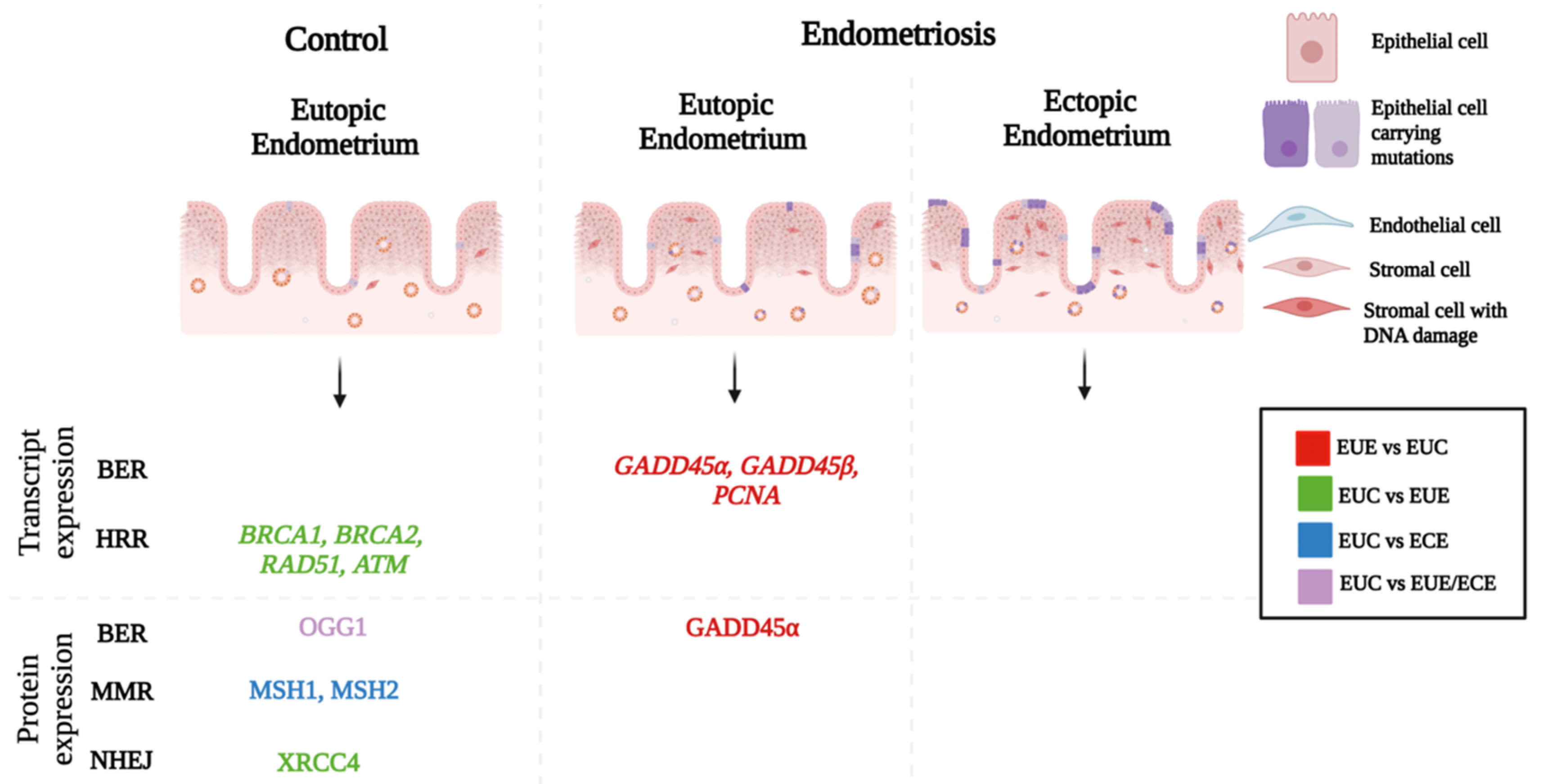
3. Endometrial DNA Damage in Endometriosis

Endometriosis is characterized by the presence of endometrium-like cells or endometriotic ectopic lesions outside the uterus. Lesion characteristics, such as size, depth, number, and the extent of adhesions to adjoining regions, are used to classify endometriosis into minimal, mild, moderate, and severe stages by the ASRM criteria [25]. Endometriotic lesions are predominantly detected on the surface of the ovary (endometrioma; OMA), peritoneum cavity wall (superficial peritoneal lesion; SUP), and also on the intestine, rectum, and the pouch of Douglas as Deep Infiltrating Endometriosis (DIE). The presence of ectopic lesions in the liver, kidney, pleural cavity, and even the brain has been reported. Some asymptomatic women with endometriosis may remain undiagnosed. However, a majority of women with the disease exhibit an array of symptoms, predominantly chronic pelvic pain, dysmenorrhea, dysuria, dyschezia, dyspareunia, and infertility. These morbidities adversely affect the quality of life of approximately 247 million women worldwide and 42 million women in India [26].
Several theories have been put forth to explain the origin of endometriosis. These include retrograde menstruation, lymphatic dissemination, stem cell induction, coelomic metaplasia, and the presence of mullerian remnants [27]. Among these, the most accepted is the retrograde menstruation theory. More support for the retrograde theory has come from recent reports indicating the clonal expansion of epithelial cells harboring cancer-driver mutations in genes, such as oncogenic PIK3CA and KRAS, in endometriotic lesions. On the other hand, these mutations remain in a subclonal state in healthy endometrium [19]. This clonal relationship, as indicated by shared somatic mutations in normal and endometriotic endometrium, supports the origin of endometriosis from the possible dissemination and growth of the menstrual phase endometrium into the peritoneal cavity. Further, endometriosis is known to be one of the risk factors for clear cell and endometrioid ovarian carcinoma [28,29]. ARID1A, PIK3CA, and KRAS genes are reported to be frequently mutated in endometriosis and ovarian carcinoma [19,21,22,23,24,30]. In 2020, Suda et al., using whole exome sequencing data from histologically normal endometrial tissues, endometriotic tissues excised from distant and adjacent sites from ovarian clear cell carcinoma (OCCC), and primary OCCC tissue, concluded that the precursors of both ovarian endometriosis and OCCC were common and present in normal endometrium. It was postulated that the accumulation of cancer-associated mutations in normal endometrium and their subsequent genomic evolution contributed to endometriosis and endometriosis-related ovarian cancer [19]. While these recent investigations on the mutational landscape of endometrium have introduced more insights to the genetic relationship between the uterine and extrauterine endometrial samples, these reports fail to explain how a normal trait, “accumulation of somatic mutations,” in eutopic endometrial epithelial cells (that are shed off every month) contributes to endometriosis in some women. Our previous study demonstrated a higher expression of DDR-associated genes in the proliferative phase in the eutopic endometrium of women with endometriosis compared with those without the disease [13]. While this may be due to higher proliferative stress in the endometrium of women with endometriosis, it remains to be established whether altered DDR in the proliferative phase modulates the endometrial differentiation program in endometriosis. Indeed, the differentiation of endometrial mesenchymal stromal cells (eMSCs) and stromal fibroblasts in women with endometriosis was found to be dysregulated [31]. It was postulated by authors that mesenchymal differentiation takes a different route in some women with endometriosis and results in a subset of senescent fibroblasts [31]. More research is needed to answer whether it is (a) the niche at extrauterine sites that is more conducive for the expansion of endometrial epithelial clones harboring mutations in cancer-driver genes or (b) the frequency of menstruation or early menarche or volume of menstrual efflux or (c) the shedding of a part of the basalis compartment along with the functionalis during menstruation in women with endometriosis.
Interestingly, 3D imaging analyses of the human endometrium have revealed the presence of rhizome-like structures in which several epithelial vertical glands are connected to horizontal glands in the basalis compartment of the endometrium [32]. Yamaguchi et al. (2022) reported the monoclonal origin of the continuum of rhizome and vertical glands [33]. Authors further proposed that after menstruation, residual glands in the basalis extend horizontally along the myometrium to form monoclonal rhizomes and each monoclonal rhizome gives rise to several vertical glands. It was further hypothesized that some rhizomes are long-lived, and during this period, some cells, including stem cell/progenitor cells in these rhizomes, are likely to acquire cancer-driver mutations while undergoing several cycles of repair and regeneration. These cells or clones with somatic mutations are likely to have a proliferative advantage and thus may contribute to endometrial regeneration by expanding the rhizome structure. However, when lodged at ectopic or other sites, these clones may contribute to pathologies, such as endometriosis. At extrauterine sites, the clones are likely to be released from anatomical constraints or these may experience paracrine influences from ectopic sites and thereby proliferate excessively. Considering the extent of heterogeneity of the genetic makeup of endometrial glands, causal events leading to the growth of endometrium at ectopic sites are likely to be stochastic and this may explain the differential susceptibility of women to endometriosis.
Few strides have been made to investigate whether endometriosis is associated with higher endometrial DNA damage. Lymphocytes isolated from women with endometriosis, following exposure to bleomycin, were found to have a higher number of chromatid breaks compared with those from women without the disease [34]. This shows that the blood cells of women with endometriosis are more susceptible to DNA damage. The eutopic endometrium in women with endometriosis also shows higher DNA damage. The uutopic endometrial epithelial as well as stromal cells show a higher number of foci of the γH2AX—a DNA damage marker in women with endometriosis, compared with their counterparts of control women [13,16]. In both investigations, women with endometrioma (stage III–IV) were included as study participants. These observations were in contrast to the conclusion drawn by Hapangama et al. [35].
This discordance in results may be due to the investigations of endometrial samples from different subtypes of endometriosis. In addition to genomic DNA, mitochondrial DNA damage has been investigated for its association with endometriosis. A 4 kbp deletion was found in the mitochondrial genome of eutopic endometrium from women with endometriosis. The frequency of this deletion was found to be higher in the tissues from chocolate cysts (endometrioma) compared with myoma, adenomyoma, and normal endometrium [36]. In the mitochondrial genome, 1.2 and 3.7 Kb deletions are reported to have the potential to serve as a promising biomarker of endometriosis [37]. Mutations in the mitochondrial DNA of eutopic and ectopic endometrium from women with endometriosis have also been reported [38]. Seventeen somatic mutations identified in mitochondrial DNA were predicted to cause defects in the oxidative phosphorylation system. Evidence also suggests that mitochondrial energy metabolism is reduced in the ectopic and eutopic endometrium from non-human primates (Macaca fascicularis and Macaca mulatta) with endometriosis [39]. Altogether, these reports suggest higher DNA damage in the eutopic endometrium of women with endometriosis.

4. Potential Causes of DNA Damage

Although the data on endometrial DNA damage and mechanisms that counteract DNA damage in the context of endometriosis are limited, it is apparent that the extent of DNA damage in the endometrium in women with endometriosis is higher compared with those without the disease. The higher endometrial DNA damage seen in endometriosis could be because of replicative stress, oxidative stress, inflammation, or environmental toxicants, as there exists a significant amount of data to suggest that the eutopic as well as ectopic endometrium in women with endometriosis experience replicative stress, an imbalance in oxidant and antioxidant factors, and also inflammation.

4.1. Proliferative Stress

There exists sufficient data to suggest that the eutopic endometrium in women with endometriosis shows a higher proliferative index [40,41]. In addition, a higher expression of telomerase was reported in the epithelial cells of the proliferative and secretory phases and the stromal cells of the secretory phase eutopic endometrium in women with peritoneal endometriosis [35]. Other reports have also demonstrated an increase in telomerase [42] and telomerase-associated gene [43] expression and activity [42] in the secretory phase eutopic endometrium in women with endometriosis. A recent study showed higher levels of DNA replication of ATP-dependent helicase/nuclease 2 (DNA2) in the eutopic and ectopic endometrium of women with endometriosis [44]. Similar results were reported for eMSCs isolated from the eutopic and ectopic endometrium of women with endometriosis [44]. Expression of Ki67, a proliferation marker, was found to be higher in the proliferative phase eutopic endometrium in women with endometriosis, compared with women with uterine polyps [45]. In addition, the secretory phase endometrium in women with peritoneal endometriosis was found to have a higher expression of proliferating cell nuclear antigen (PCNA) [35]. Nucleolin expression was also found to be higher in proliferative and secretory phase eutopic endometrium in women with endometriosis [35]. Investigations from our laboratory and other groups also indicate a higher proliferative index in the eutopic endometrium of women with endometriosis as revealed by a higher number of stromal cells per unit area and a significantly higher PCNA expression in the proliferative phase endometrium in women with endometriosis, compared with the phase-matched endometrial samples from control women [13].

4.2. Oxidative Stress

A relationship between oxidative stress and endometriosis has been extensively studied. Immunolocalization of an oxidized derivative of Guanosine (a DNA base)-8-hydroxy-2′-deoxyguanosine (8-OHdG), an oxidative stress marker, was found to increase with the severity of endometriosis [18]. In addition, significantly higher levels of high-density lipoprotein (HDL) and low-density lipoprotein (LDL) were found in women with moderate and severe endometriosis, compared with mild and minimal endometriosis [46]. Higher levels of thiols, advanced oxidation protein products (AOPP), protein carbonyl, and nitrates/nitrites were found in the peritoneal fluid of women with deep infiltrating endometriosis [47]. Eutopic and ectopic endometrial stromal and epithelial cells in women with endometriosis were found to produce more oxygen anion (O2−) and demonstrated higher proliferative capacity in response to oxidative stress [48]. Higher levels of lipid peroxides were also found in the peritoneal fluid of women with endometriosis [49]. Total oxidant capacity and lipid peroxide levels were higher in the serum and follicular fluid samples from women with endometriosis [50,51]. Follicular fluid from infertile women with endometriosis also had higher nitrate/nitrite levels, compared with infertile women [52]. Higher levels of vitamin E, an antioxidant, were found in women with endometriosis [53]. Vitamin C levels were lower in the follicular fluid of women with endometriosis [51]. Levels of antioxidants, such as glutathione peroxidase and superoxide dismutase, were found to be reduced in the peritoneal fluid of women with endometriosis [49,51]. Total antioxidant levels were also lower in the peritoneal fluid of women with endometriosis [49].
Higher oxidative stress in endometriosis has been attributed to dysregulated iron metabolism. Iron released from hemorrhage or hemolysis was found to be accumulated in endometriotic lesions/peritoneal fluid [54]. A higher menstrual influx via retrograde menstruation can contribute to iron overload. Higher levels of iron, ferritin, and hemoglobin have been found in the peritoneal fluid of women with endometriosis [54,55,56,57,58]. Further, lower levels of hemopexin (a scavenger of heme) are reported in the peritoneal fluid of women with endometriosis [59]. Lesions were also found to have higher iron deposits in endometriosis [60]. Iron conglomerates were detected in endometriotic lesions contributing to oxidative stress [61]. Interestingly, endometriotic epithelial cell proliferation was found to be higher in response to human menstrual debris supplemented with erythrocytes injected in nude mice [62]. This proliferation subsided in response to desferrioxamine, an iron chelator.

4.3. Inflammation

There is ample evidence to implicate endometriosis as an inflammatory disease. Inflammation is reported to be linked with the production of reactive oxygen species (ROS) and reactive nitrogen species (RNS), which contribute to DNA adducts, including 8-oxo-7,8-dihydro-2′-deoxyguanosine (8-OxodG) and 8-nitroguanidine [63]. NO forms peroxynitrite (ONOO-) upon reaction with superoxide (O2−). NO and O2− are also generated by neutrophils and macrophages in the inflammatory microenvironment. These DNA adducts lead to the formation of apurinic sites [64]. Furthermore, inflammatory cytokines, such as Tumor Necrosis Factor (TNF), released by macrophages, generate O2− and contribute to genomic instability [65]. Higher levels of TNF have been detected in the peritoneal fluid [66] and serum [67] of women with endometriosis.
Neutrophil numbers were found to be higher in the peritoneal fluid of women with endometriosis [68,69]. Higher levels of IL-8, a chemoattractant of neutrophils, were detected in the plasma [70] and peritoneal fluid [69,70,71] of women with endometriosis. Neutrophils showed decreased apoptosis when incubated with peritoneal fluid from women with endometriosis [72]. The levels of human neutrophil peptides (HNP) released by activated neutrophils, α-defensins, are also reported to be elevated in the peritoneal fluid of women with endometriosis and their levels correlated with the severity of the disease [70]. α-defensin acts as a chemoattractant for T cells, dendritic cells, and monocytes/macrophages. Macrophage numbers were found to be higher in all phases [72], including the proliferative phase [73] of the menstrual cycle in the eutopic endometrium of women with endometriosis. In another study, the endometrial macrophage population was found to be lower throughout the menstrual cycle in endometriosis [74]. The frequency of CD163+ macrophages/M2 macrophages (anti-inflammatory macrophages) was lower, whereas that of CD68+ macrophages/M1 macrophages (pro-inflammatory) was higher in the eutopic endometrium of women with endometriosis [73,75]. Another macrophage chemoattractant, monocyte chemotactic protein-1 (MCP-1), was found to be elevated in the eutopic endometrium of women with endometriosis [76]. Monocytes in-vitro differentiate into macrophages, rather than into dendritic cells, in response to the peritoneal fluid from women with advanced stage endometriosis [77]. In addition, peritoneal macrophages demonstrate a reduced phagocytotic potential [78] and release pro-inflammatory cytokines TNF, IL-6, and IL-1β in endometriosis [79]. A higher proliferation and invasion of the stromal cells of the endometriotic lesions were observed, when primed with macrophages from healthy women [80]. Macrophages from women with endometriosis led to the increased clonogenicity and self-renewal capacity of stromal and epithelial cells [81]. Collectively, these reports suggest that macrophages in women with endometriosis create an inflammatory microenvironment.
Alarmins or damage-associated molecular patterns (DAMPs), such as high-mobility group box 1 (HMGB1), are reported to be highly abundant in the menstrual fluid of women with endometriosis compared with control women with other gynecological conditions [82]. Endometriotic stromal cells stimulated with HMGB1 expressed higher levels of vascular endothelial growth factor (VGEF) [82]. In addition, HMGB1-treated eutopic endometrial stromal cells from women with endometriosis showed higher proliferation [83]. IL-33, another alarmin, was found to have increased expression in women with endometriosis [84,85]. Intraperitoneal injections of IL-33 stimulated the growth and vascularization of the lesion in mice [85].

4.4. Environmental Toxicants

DNA in the eutopic and ectopic endometrium may be more susceptible to damage in women with endometriosis because of higher replicative stress, oxidative stress, and inflammation. It is also likely that endometrial DNA gets exposed to more environmental toxicants in women with endometriosis. Studies have shown a correlation between dioxin exposure and peritoneal endometriosis [86] or stage III/IV endometriosis [87]. Evidence also exists to suggest a relationship between exposure to 2,3,7,8-Tetrachlorodibenzo—p-dioxin (TCDD) and endometriosis development in rhesus monkeys [88]. Polychlorinated biphenyl (PCB) and 1,1-dichloro-2,2,-bis (4-chlorophenyl)-ethene (DDE) [88] and phthalate [89,90,91,92] have been associated with risk of endometriosis. However, other studies could not establish an association between TCDD [93], Dioxin [94], PCBs [94] exposure, and endometriosis. These discrepant results can be due to the different ethnicities of the populations investigated and the small cohort sizes [94].

4.5. Estrogenic Toxicity

Endometriotic lesions are capable of synthesizing estrogen in-situ. Elevated levels of estrogen have been attributed to the higher expression of aromatase in the ectopic endometrium of women with endometriosis [93,94,95]. Estradiol stimulates the production of prostaglandins, specifically prostaglandin E2, which further stimulate the activity of aromatase [96]. Estrogen signaling is mediated by nuclear estrogen receptors. Higher expression of the estrogen receptor β (ERβ) is reported in women with endometriosis [95,96,97,98]. ERα is reported to be of significance in initiating lesion development mediated by estrogen. Further, IL-6 was found to be attenuated in ERα knockdown mice [99], demonstrating estrogen/ERα/IL-6-mediated cross-talk in endometriosis development. Another study showed the importance of ERβ in cell survival via higher IL-1β production [100].
The eutopic endometrium in women with endometriosis is also reported to have higher levels of estradiol [101]. Higher levels of estradiol cause genotoxicity through adduct formation. Estrogen hydroxylation at C4 produces metabolites, such as 4-hydroxy-estrone (4-OHE1). 4-OHE1 forms quinones catalyzed by peroxidases/CYP450. Quinones interact with DNA and form adducts, such as 4-OHE1(E2)-1-N3Ade and 4-OHE1(E2)-1-N7Gua. Higher levels of estrogen metabolite 4-OHE1 are reported in the eutopic endometrium in women with endometriosis [102].

5. DNA Repair in Women with Endometriosis

Attempts have been made to investigate whether genetic variations in DNA repair genes make women more or less susceptible to endometriosis. X-ray repair cross-complementing 4 (XRCC4), a core player involved in the NHEJ pathway, was found to have c.1394G > T polymorphism in Iranian and Taiwanese women with endometriosis [103,104]. This polymorphism in the promoter region might affect the expression of the XRCC4 protein and consequently, DNA repair. Incidentally, studies have demonstrated a reduced expression of XRCC4 in the eutopic endometrium of women with endometriosis, compared with healthy women [17]. XRCC3 polymorphism at the p.Met241Thr genotype in Turkish women has been reported to be associated with endometriosis [105]. PPARγ c.161T > C and PPARγ p.Pro12ALA polymorphisms were found to be associated with endometriosis in Japanese and German populations, respectively [106,107]. p.Pro72Arg polymorphism in the p53 gene was found to be associated with the risk of endometriosis in Italian women [108,109], but not in Mexican [110], Chinese [111], Taiwanese [112], Iranian [113], and Pakistani [114] populations. This polymorphism might influence mRNA splicing and influence p53 gene expression and DNA-protein interaction. In addition, women with XRCC1 codon p.Trp194Arg, p.Gln399Arg, and XRCC3 codon p.Met241Thr polymorphism were found to have higher damage in the DNA of their lymphocytes treated with bleomycin [115]. Another study showed an association between XRCC1 polymorphism p.Arg399Gln with the severity of endometriosis in the Taiwanese population [116]. Polymorphisms in NER proteins, such as ERCC1 (rs11615 TT), ERCC2 (rs1799793 AA), and ERCC6 (rs2228528 AA), were also associated with a risk of endometriosis [117].
Investigations have been undertaken to assess the expression of various DNA repair genes in the endometrium in women with endometriosis. BRCA1, BRCA2, RAD51, and ATM mRNA levels were found to be reduced in the eutopic endometrium in women with stage III-IV endometriosis, compared with women with neoplasm [16]. However, it remains to be ascertained whether the reduced levels of these DNA repair proteins contribute to unrepaired double-stranded DNA breaks or whether the endometrium adopts other mechanisms to ensure genomic integrity. On the other hand, expression of MSH2, a mismatch repair protein, was found to be higher in the eutopic stromal cells and endometriotic lesions of women with endometriosis, compared with the eutopic endometrium in women with leiomyoma [14]. In contrast, Fuseya et al. reported reduced levels of MSH1 and MSH2 in eutopic endometrium in women with ovarian endometriosis [15]. MMR protein levels (MSH1 and MSH2) were lower in the ectopic endometrium compared with the paired eutopic endometrium. Endometriotic lesions were found to have a hypermethylation of the MLH1 promoter. The methylation pattern correlated with low expression of MLH1 protein expression [118]. Matta et al. reported an increased DNA repair capacity in the lymphocytes of women with endometriosis, compared with women without endometriosis. However, in this study, the control group included women with breast cancer [119]. Our studies demonstrated a reduced endometrial expression of XRCC4, a core protein involved in the NHEJ repair pathway, in women with endometriosis, compared with women without endometriosis [17]. Expression of OGG1, another oxidative stress response gene, was found to be reduced in the ectopic endometrium of women with endometriosis [19].
Poli-Neto et al. analyzed five eutopic endometrial gene expression datasets, namely, GSE4888 [120], GSE6364 [121], GSE7305 [122], GSE7307 (GEO repository), and GSE51981 [123] to compare endometrial transcriptomes in women with and without endometriosis. The meta-analysis revealed downregulation of DNA repair genes (ATRX, BRIP1, EXO1, FANCI, FANCL, FEN1, MSH2, MSH6, NEIL3, PARPBP, PACIP1, PCNA, PDS5B, POLA1, POLE2, POL1, PRKDC, RPA1, SMC6, USP1) in the proliferative phase eutopic endometrium of women with endometriosis compared with control women [124]. However, RNA Seq analysis carried out by our group revealed a trend towards upregulation in the endometrial expression of DNA damage repair genes in women with endometriosis during their proliferative phase, compared with control women without endometriosis. This aligned with our observations indicating a higher number of γH2FX loci in the eutopic endometrium of women with endometriosis. Endometrial expression of DNA repair genes is upregulated probably to counteract higher DNA damage in endometriosis. We further observed that higher DNA damage in the endometrium persists in the mid-secretory phase of the menstrual cycle in women with endometriosis. However, a trend towards downregulation in the endometrial expression of DNA repair genes was apparent during the mid-secretory phase in women with endometriosis, compared with phase-matched samples from healthy women [13]. Several factors reported to be involved in DDR in proliferating cells are downregulated in differentiated cells. This may render differentiated cells resistant to genotoxic stimuli. In the context of eutopic endometrium in women with endometriosis, it is likely that persistent DNA damage due to oxidative stress or inflammation may lead to accumulation of unrepaired DNA lesions; eutopic endometrium with damaged DNA is likely to form lesions at conducive ectopic sites.

6. Conclusions

A large body of data links DNA damage, impaired DNA damage response (DDR), and aberrant DNA repair capacity with various pathologies, such as cancers, neurodegenerative diseases, premature aging, and cardiovascular diseases. Additionally, the role of DNA damage, DDR, and DNA repair in various physiological settings, such as the generation of immunoglobulin and T cell receptor diversity, telomere homeostasis, cell proliferation, and differentiation, the protection against pathogens is unequivocally established. However, DNA damage, DDR, and DNA repair capacity of human endometrium in health and disease, especially in endometriosis, have not received much attention. This review compiles the major inferences drawn from the investigations undertaken in this direction (Figure 1). A majority of these reports have focused on the expression/level of various components of DDR pathways and their dysregulation in the eutopic or ectopic endometrium of women with endometriosis. Some strides have also been made to assess whether genetic variations in DDR or DNA repair genes are associated with endometriosis risk. Collectively, these investigations highlight aberrations in the expression or levels of some DDR factors in the eutopic as well as ectopic endometrium of women with endometriosis, compared with women without endometriosis. More investigations are needed to resolve queries, including (I) whether these aberrations in DDR and DNA repair gene expression play a causal role in endometriosis or if these derangements appear after the onset of endometriosis; (II) how the activities of various DDR proteins are controlled in different phases of the menstrual cycle; (III) whether ectopic tissues preferentially employ a specific DDR pathway. Further, in the wake of emerging reports indicating an association between DDR and metabolic reprogramming, it would be of interest to investigate the potential effects of modulated DDR on endometrial metabolism and other functions in endometriosis. More knowledge about the DDR and DNA repair mechanisms that operate in the endometrium may open novel avenues for treating or managing endometriosis in the future.

Author Contributions

Conceptualization, G.S.; writing—review and editing, G.S. and I.M.; writing—original draft, I.M. All authors have read and agreed to the published version of the manuscript.

Funding

G.S. thanks the Indian Council for Medical Research-NIRRCH and the Department of Biotechnology (BT/PR30794/MED/97/439/2018) for the support extended to her group for investigations on endometriosis. I.M. thanks Lady TATA Memorial Trust (LTMT) for their Junior and Senior research fellowships.

Informed Consent Statement

Not applicable.

Conflicts of Interest

The authors declare no conflict of interest.

References

  1. Friedberg, E.C. DNA damage and repair. Nature 2003, 421, 436–440. [Google Scholar] [CrossRef] [PubMed]
  2. Sancar, A.; Lindsey-Boltz, L.A.; Ünsal-Kaçmaz, K.; Linn, S. Molecular Mechanisms of Mammalian DNA Repair and the DNA Damage Checkpoints. Annu. Rev. Biochem. 2004, 73, 39–85. [Google Scholar] [CrossRef] [PubMed]
  3. Blanpain, C.; Mohrin, M.; Sotiropoulou, P.A.; Passegué, E. DNA-Damage Response in Tissue-Specific and Cancer Stem Cells. Cell Stem Cell 2011, 8, 16–29. [Google Scholar] [CrossRef] [PubMed]
  4. Shin, E.; Lee, S.; Kang, H.; Kim, J.; Kim, K.; Youn, H.S.; Jin, Y.W.; Seo, S.; Youn, B.H. Organ-Specific Effects of Low Dose Radiation Exposure: A Comprehensive Review. Front. Genet. 2020, 11, 1178. [Google Scholar] [CrossRef] [PubMed]
  5. Mohrin, M.; Bourke, E.; Alexander, D.; Warr, M.R.; Barry-Holson, K.; Le Beau, M.M.; Morrison, C.G.; Passegué, E. Hematopoietic stem cell quiescence promotes error-prone DNA repair and mutagenesis. Cell Stem Cell 2010, 7, 174–185. [Google Scholar] [CrossRef]
  6. Harding, S.M.; Benci, J.L.; Irianto, J.; Discher, D.E.; Minn, A.J.; Greenberg, R.A. Mitotic progression following DNA damage enables pattern recognition within micronuclei. Nature 2017, 548, 466–470. [Google Scholar] [CrossRef]
  7. Kiraly, O.; Gong, G.; Olipitz, W.; Muthupalani, S.; Engelward, B.P. Inflammation-Induced Cell Proliferation Potentiates DNA Damage-Induced Mutations In Vivo. PLoS Genet. 2015, 11, e1004901. [Google Scholar] [CrossRef]
  8. Stopper, H.; Schmitt, E.; Gregor, C.; Mueller, S.O.; Fischer, W.H. Increased cell proliferation is associated with genomic instability: Elevated micronuclei frequencies in estradiol-treated human ovarian cancer cells. Mutagenesis 2003, 18, 243–247. [Google Scholar] [CrossRef]
  9. de Pedro, I.; Alonso-Lecue, P.; Sanz-Gómez, N.; Freije, A.; Gandarillas, A. Sublethal UV irradiation induces squamous differentiation via a p53-independent, DNA damage-mitosis checkpoint. Cell Death Dis. 2018, 9, 1094. [Google Scholar] [CrossRef]
  10. Sanz-Gómez, N.; Freije, A.; Ceballos, L.; Obeso, S.; Sanz, J.R.; García-Reija, F.; Morales-Angulo, C.; Gandarillas, A. Response of head and neck epithelial cells to a DNA damage-differentiation checkpoint involving polyploidization. Head Neck 2018, 40, 2487–2497. [Google Scholar] [CrossRef]
  11. Bredemeyer, A.L.; Helmink, B.A.; Innes, C.L.; Calderon, B.; McGinnis, L.M.; Mahowald, G.K.; Gapud, E.J.; Walker, L.M.; Collins, J.B.; Weaver, B.K.; et al. DNA double-strand breaks activate a multi-functional genetic program in developing lymphocytes. Nature 2008, 456, 819–823. [Google Scholar] [CrossRef] [PubMed]
  12. Inomata, K.; Aoto, T.; Binh, N.T.; Okamoto, N.; Tanimura, S.; Wakayama, T.; Iseki, S.; Hara, E.; Masunaga, T.; Shimizu, H.; et al. Genotoxic Stress Abrogates Renewal of Melanocyte Stem Cells by Triggering Their Differentiation. Cell 2009, 137, 1088–1099. [Google Scholar] [CrossRef] [PubMed]
  13. Bane, K.; Desouza, J.; Shetty, D.; Choudhary, P.; Kadam, S.; Katkam, R.R.; Fernandes, G.; Sawant, R.; Dudhedia, U.; Warty, N.; et al. Endometrial DNA damage response is modulated in endometriosis. Hum. Reprod. 2021, 36, 160–174. [Google Scholar] [CrossRef] [PubMed]
  14. Grassi, T.; Calcagno, A.; Marzinotto, S.; Londero, A.P.; Orsaria, M.; Canciani, G.N.; Beltrami, C.A.; Marchesoni, D.; Mariuzzi, L. Mismatch repair system in endometriotic tissue and eutopic endometrium of unaffected women. Int. J. Clin. Exp. Pathol. 2015, 8, 1867–1877. [Google Scholar]
  15. Fuseya, C.; Horiuchi, A.; Hayashi, A.; Suzuki, A.; Miyamoto, T.; Hayashi, T.; Shiozawa, T. Involvement of pelvic inflammation-related mismatch repair abnormalities and microsatellite instability in the malignant transformation of ovarian endometriosis. Hum. Pathol. 2012, 43, 1964–1972. [Google Scholar] [CrossRef]
  16. Choi, Y.S.; Park, J.H.; Lee, J.H.; Yoon, J.K.; Yun, B.H.; Park, J.H.; Seo, S.K.; Sung, H.-J.; Kim, H.-S.; Cho, S.H.; et al. Association Between Impairment of DNA Double Strand Break Repair and Decreased Ovarian Reserve in Patients With Endometriosis. Front. Endocrinol. 2018, 9, 772. [Google Scholar] [CrossRef]
  17. Bane, K.; Desouza, J.; Rojewale, A.; Katkam, R.R.; Fernandes, G.; Sawant, R.; Dudhedia, U.; Warty, N.; Chauhan, A.; Chaudhari, U.; et al. Dysregulation of X-ray repair cross-complementing 4 expression in the eutopic endometrium of women with endometriosis. Reproduction 2022, 163, 95–105. [Google Scholar] [CrossRef]
  18. Carvalho, L.F.P.; Abrão, M.S.; Biscotti, C.; Sharma, R.; Nutter, B.; Falcone, T. Oxidative cell injury as a predictor of endometriosis progression. Reprod. Sci. 2013, 20, 688–698. [Google Scholar] [CrossRef]
  19. Suda, K.; Nakaoka, H.; Yoshihara, K.; Ishiguro, T.; Tamura, R.; Mori, Y.; Yamawaki, K.; Adachi, S.; Takahashi, T.; Kase, H.; et al. Clonal Expansion and Diversification of Cancer-Associated Mutations in Endometriosis and Normal Endometrium. Cell Rep. 2018, 24, 1777–1789. [Google Scholar] [CrossRef]
  20. Lac, V.; Verhoef, L.; Aguirre-Hernandez, R.; Nazeran, T.M.; Tessier-Cloutier, B.; Praetorius, T.; Orr, N.L.; Noga, H.; Lum, A.; Khattra, J.; et al. Iatrogenic endometriosis harbors somatic cancer-driver mutations. Hum. Reprod. 2019, 34, 69–78. [Google Scholar] [CrossRef]
  21. Wiegand, K.C.; Shah, S.P.; Al-Agha, O.M.; Zhao, Y.; Tse, K.; Zeng, T.; Senz, J.; McConechy, M.K.; Anglesio, M.S.; Kalloger, S.E.; et al. ARID1A mutations in endometriosis-associated ovarian carcinomas. N. Engl. J. Med. 2010, 363, 1532–1543. [Google Scholar] [CrossRef]
  22. Chandler, R.L.; Damrauer, J.S.; Raab, J.R.; Schisler, J.C.; Wilkerson, M.D.; Didion, J.P.; Starmer, J.; Serber, D.; Yee, D.; Xiong, J.; et al. Coexistent ARID1A-PIK3CA mutations promote ovarian clear-cell tumorigenesis through pro-tumorigenic inflammatory cytokine signalling. Nat. Commun. 2015, 6, 6118. [Google Scholar] [CrossRef] [PubMed]
  23. Obata, K.; Hoshiai, H. Common genetic changes between endometriosis and ovarian cancer. Gynecol. Obstet. Invest. 2000, 50 (Suppl. 1), 39–43. [Google Scholar] [CrossRef] [PubMed]
  24. Anglesio, M.S.; Papadopoulos, N.; Ayhan, A.; Nazeran, T.M.; Noë, M.; Horlings, H.M.; Lum, A.; Jones, S.; Senz, J.; Seckin, T.; et al. Cancer-Associated Mutations in Endometriosis without Cancer. N. Engl. J. Med. 2017, 376, 1835–1848. [Google Scholar] [CrossRef]
  25. ”Endometriosis|ASRM”. Available online: https://www.asrm.org/topics/topics-index/endometriosis/ (accessed on 30 December 2022).
  26. Gajbhiye, R.K.; Montgomery, G.; Pai, M.V.; Phukan, P.; Shekhar, S.; Padte, K.; Dasmahapatra, P.; John, B.M.; Shembekar, C.; Bhurke, A.V.; et al. Protocol for a case–control study investigating the clinical phenotypes and genetic regulation of endometriosis in Indian women: The ECGRI study. BMJ Open 2021, 11, e050844. [Google Scholar] [CrossRef] [PubMed]
  27. Sourial, S.; Tempest, N.; Hapangama, D.K. Theories on the Pathogenesis of Endometriosis. Int. J. Reprod. Med. 2014, 2014, 179515. [Google Scholar] [CrossRef] [PubMed]
  28. Pearce, C.L.; Templeman, C.; Rossing, M.A.; Lee, A.; Near, A.M.; Webb, P.M.; Nagle, C.M.; Doherty, J.A.; Cushing-Haugen, K.L.; Wicklund, K.G.; et al. Association between endometriosis and risk of histological subtypes of ovarian cancer: A pooled analysis of case-control studies. Lancet. Oncol. 2012, 13, 385–394. [Google Scholar] [CrossRef]
  29. Kim, H.S.; Kim, T.H.; Chung, H.H.; Song, Y.S. Risk and prognosis of ovarian cancer in women with endometriosis: A meta-analysis. Br. J. Cancer 2014, 110, 1878–1890. [Google Scholar] [CrossRef]
  30. Koppolu, A.; Maksym, R.B.; Paskal, W.; Machnicki, M.; Rak, B.; Pępek, M.; Garbicz, F.; Pełka, K.; Kuśmierczyk, Z.; Jacko, J.; et al. Epithelial cells of deep infiltrating endometriosis harbor mutations in cancer driver genes. Cells 2021, 10, 749. [Google Scholar] [CrossRef]
  31. McKinnon, B.D.; Lukowski, S.W.; Mortlock, S.; Crawford, J.; Atluri, S.; Subramaniam, S.; Johnston, R.L.; Nirgianakis, K.; Tanaka, K.; Amoako, A.; et al. Altered differentiation of endometrial mesenchymal stromal fibroblasts is associated with endometriosis susceptibility. Commun. Biol. 2022, 5, 600. [Google Scholar] [CrossRef]
  32. Yamaguchi, M.; Yoshihara, K.; Suda, K.; Nakaoka, H.; Yachida, N.; Ueda, H.; Sugino, K.; Mori, Y.; Yamawaki, K.; Tamura, R.; et al. Three-dimensional understanding of the morphological complexity of the human uterine endometrium. iScience 2021, 24, 102258. [Google Scholar] [CrossRef] [PubMed]
  33. Yamaguchi, M.; Nakaoka, H.; Suda, K.; Yoshihara, K.; Ishiguro, T.; Yachida, N.; Saito, K.; Ueda, H.; Sugino, K.; Mori, Y.; et al. Spatiotemporal dynamics of clonal selection and diversification in normal endometrial epithelium. Nat. Commun. 2022, 13, 943. [Google Scholar] [CrossRef] [PubMed]
  34. Lin, J.; Zhang, X.; Chen, Y. Mutagen sensitivity as a susceptibility marker for endometriosis. Hum. Reprod. 2003, 18, 2052–2057. [Google Scholar] [CrossRef]
  35. Hapangama, D.K.; Turner, M.A.; Drury, J.A.; Quenby, S.; Hart, A.; Maddick, M.; Martin-Ruiz, C.; Von Zglinicki, T. Sustained replication in endometrium of women with endometriosis occurs without evoking a DNA damage response. Hum. Reprod. 2009, 24, 687–696. [Google Scholar] [CrossRef] [PubMed]
  36. Kao, S.H.; Huang, H.C.; Hsieh, R.H.; Chen, S.C.; Tsai, M.C.; Tzeng, C.R. Oxidative Damage and Mitochondrial DNA Mutations with Endometriosis. Ann. N. Y. Acad. Sci. 2005, 1042, 186–194. [Google Scholar] [CrossRef]
  37. Creed, J.; Maggrah, A.; Reguly, B.; Harbottle, A. Mitochondrial DNA deletions accurately detect endometriosis in symptomatic females of child-bearing age. Biomark. Med. 2019, 13, 291–306. [Google Scholar] [CrossRef]
  38. Govatati, S.; Tipirisetti, N.R.; Perugu, S.; Kodati, V.L.; Deenadayal, M.; Satti, V.; Bhanoori, M.; Shivaji, S. Mitochondrial genome variations in advanced stage endometriosis: A study in South Indian population. PLoS ONE 2012, 7, e40668. [Google Scholar] [CrossRef]
  39. Atkins, H.M.; Bharadwaj, M.S.; O’Brien Cox, A.; Furdui, C.M.; Appt, S.E.; Caudell, D.L. Endometrium and endometriosis tissue mitochondrial energy metabolism in a nonhuman primate model. Reprod. Biol. Endocrinol. 2019, 17, 70. [Google Scholar] [CrossRef]
  40. Kahyaoglu, I.; Kahyaoglu, S.; Moraloglu, O.; Zergeroglu, S.; Sut, N.; Batioglu, S. Comparison of Ki-67 proliferative index between eutopic and ectopic endometrium: A case control study. Taiwan. J. Obstet. Gynecol. 2012, 51, 393–396. [Google Scholar] [CrossRef]
  41. Eris Yalcin, S.; Ocal, I.; Yalcin, Y.; Sen Selim, H.; Demir Caltekin, M.; Aydogmus, H.; Kelekci, S. Evaluation of the Ki-67 Proliferation Index and Urocortin Expression in Women with Ovarian Endometriomas. Eurasian J. Med. 2017, 49, 107. [Google Scholar] [CrossRef] [PubMed]
  42. Kim, C.M.; Oh, Y.J.; Cho, S.H.; Chung, D.J.; Hwang, J.Y.; Park, K.H.; Cho, D.J.; Choi, Y.M.; Lee, B.S. Increased telomerase activity and human telomerase reverse transcriptase mRNA expression in the endometrium of patients with endometriosis. Hum. Reprod. 2007, 22, 843–849. [Google Scholar] [CrossRef] [PubMed]
  43. Alnafakh, R.; Choi, F.; Bradfield, A.; Adishesh, M.; Saretzki, G.; Hapangama, D.K. Endometriosis Is Associated with a Significant Increase in hTERC and Altered Telomere/Telomerase Associated Genes in the Eutopic Endometrium, an Ex-Vivo and In Silico Study. Biomedicines 2020, 8, 588. [Google Scholar] [CrossRef] [PubMed]
  44. Wang, X.; Zeng, W.; Xu, S.; Nie, J.; Huang, L.; Lai, Y.; Yu, Y. Up-regulation of DNA2 results in cell proliferation and migration in endometriosis. J. Mol. Histol. 2021, 52, 741–749. [Google Scholar] [CrossRef] [PubMed]
  45. Park, J.S.; Lee, J.H.; Kim, M.; Chang, H.J.; Hwang, K.J.; Chang, K.H. Endometrium from women with endometriosis shows increased proliferation activity. Fertil. Steril. 2009, 92, 1246–1249. [Google Scholar] [CrossRef] [PubMed]
  46. Ferda Verit, F.; Erel, O.; Celik, N. Serum paraoxonase-1 activity in women with endometriosis and its relationship with the stage of the disease. Hum. Reprod. 2008, 23, 100–104. [Google Scholar] [CrossRef] [PubMed]
  47. Santulli, P.; Chouzenoux, S.; Fiorese, M.; Marcellin, L.; Lemarechal, H.; Millischer, A.E.; Batteux, F.; Borderie, D.; Chapron, C. Protein oxidative stress markers in peritoneal fluids of women with deep infiltrating endometriosis are increased. Hum. Reprod. 2015, 30, 49–60. [Google Scholar] [CrossRef] [PubMed]
  48. Ngô, C.; Chéreau, C.; Nicco, C.; Weill, B.; Chapron, C.; Batteux, F. Reactive oxygen species controls endometriosis progression. Am. J. Pathol. 2009, 175, 225–234. [Google Scholar] [CrossRef]
  49. Szczepańska, M.; Koźlik, J.; Skrzypczak, J.; Mikołajczyk, M. Oxidative stress may be a piece in the endometriosis puzzle. Fertil. Steril. 2003, 79, 1288–1293. [Google Scholar] [CrossRef]
  50. Nasiri, N.; Moini, A.; Eftekhari-Yazdi, P.; Karimian, L.; Salman-Yazdi, R.; Arabipoor, A. Oxidative Stress Statues in Serum and Follicular Fluidof Women with Endometriosis. Cell J. 2017, 18, 582. [Google Scholar] [CrossRef]
  51. Prieto, L.; Quesada, J.F.; Cambero, O.; Pacheco, A.; Pellicer, A.; Codoceo, R.; Garcia-Velasco, J.A. Analysis of follicular fluid and serum markers of oxidative stress in women with infertility related to endometriosis. Fertil. Steril. 2012, 98, 126–130. [Google Scholar] [CrossRef]
  52. Goud, P.T.; Goud, A.P.; Joshi, N.; Puscheck, E.; Diamond, M.P.; Abu-Soud, H.M. Dynamics of nitric oxide, altered follicular microenvironment, and oocyte quality in women with endometriosis. Fertil. Steril. 2014, 102, 151–159. [Google Scholar] [CrossRef] [PubMed]
  53. Jackson, L.W.; Schisterman, E.F.; Dey-Rao, R.; Browne, R.; Armstrong, D. Oxidative stress and endometriosis. Hum. Reprod. 2005, 20, 2014–2020. [Google Scholar] [CrossRef] [PubMed]
  54. McCord, J.M. Iron, Free Radicals, and Oxidative Injury. J. Nutr. 2004, 134, 3171S–3172S. [Google Scholar] [CrossRef] [PubMed]
  55. Defrère, S.; Lousse, J.C.; González-Ramos, R.; Colette, S.; Donnez, J.; Van Langendonckt, A. Potential involvement of iron in the pathogenesis of peritoneal endometriosis. Mol. Hum. Reprod. 2008, 14, 377–385. [Google Scholar] [CrossRef]
  56. Van Langendonckt, A.; Casanas-Roux, F.; Donnez, J. Iron overload in the peritoneal cavity of women with pelvic endometriosis. Fertil. Steril. 2002, 78, 712–718. [Google Scholar] [CrossRef]
  57. Lousse, J.C.; Defrère, S.; Van Langendonckt, A.; Gras, J.; González-Ramos, R.; Colette, S.; Donnez, J. Iron storage is significantly increased in peritoneal macrophages of endometriosis patients and correlates with iron overload in peritoneal fluid. Fertil. Steril. 2009, 91, 1668–1675. [Google Scholar] [CrossRef]
  58. Arumugam, K. Endometriosis: Endometriosis and infertility: Raised iron concentration in the peritoneal fluid and its effect on the acrosome reaction. Hum. Reprod. 1994, 9, 1153–1157. [Google Scholar] [CrossRef]
  59. Wölfler, M.M.; Meinhold-Heerlein, I.M.; Henkel, C.; Rath, W.; Neulen, J.; Maass, N.; Bräutigam, K. Reduced hemopexin levels in peritoneal fluid of patients with endometriosis. Fertil. Steril. 2013, 100, 777–781.e2. [Google Scholar] [CrossRef]
  60. Van Langendonckt, A.; Casanas-Roux, F.; Dolmans, M.M.; Donnez, J. Potential involvement of hemoglobin and heme in the pathogenesis of peritoneal endometriosis. Fertil. Steril. 2002, 77, 561–570. [Google Scholar] [CrossRef]
  61. Van Langendonckt, A.; Casanas-Roux, F.; Eggermont, J.; Donnez, J. Characterization of iron deposition in endometriotic lesions induced in the nude mouse model. Hum. Reprod. 2004, 19, 1265–1271. [Google Scholar] [CrossRef]
  62. Defrère, S.; Van Langendonckt, A.; Vaesen, S.; Jouret, M.; Ramos, R.G.; Gonzalez, D.; Donnez, J. Iron overload enhances epithelial cell proliferation in endometriotic lesions induced in a murine model. Hum. Reprod. 2006, 21, 2810–2816. [Google Scholar] [CrossRef] [PubMed]
  63. Wiseman, H.; Halliwell, B. Damage to DNA by reactive oxygen and nitrogen species: Role in inflammatory disease and progression to cancer. Biochem. J. 1996, 313 Pt 1, 17–29. [Google Scholar] [CrossRef] [PubMed]
  64. Yermilov, V.; Rubio, J.; Ohshima, H. Formation of 8-nitroguanine in DNA treated with peroxynitrite in vitro and its rapid removal from DNA by depurination. FEBS Lett. 1995, 376, 207–210. [Google Scholar] [CrossRef] [PubMed]
  65. Canton, M.; Sánchez-Rodríguez, R.; Spera, I.; Venegas, F.C.; Favia, M.; Viola, A.; Castegna, A. Reactive Oxygen Species in Macrophages: Sources and Targets. Front. Immunol. 2021, 12, 4077. [Google Scholar] [CrossRef]
  66. Ragab, D.; Abbas, A.; Salem, R. Increased expression of IL-37 correlates with TNF-α levels and disease stage in endometriosis patients. Egypt. J. Med. Hum. Genet. 2022, 23, 72. [Google Scholar] [CrossRef]
  67. Senapati, S.; Clauw, D.; As-Sanie, S. The relationship between serum tumor necrosis factor-alpha (TNF-α) and interleukin-6 (IL-6) with pelvic pain symptoms in women with endometriosis. Fertil. Steril. 2009, 92, S112. [Google Scholar] [CrossRef]
  68. Milewski, Ł.; Dziunycz, P.; Barcz, E.; Radomski, D.; Roszkowski, P.I.; Korczak-Kowalska, G.; Kamiński, P.; Malejczyk, J. Increased levels of human neutrophil peptides 1, 2, and 3 in peritoneal fluid of patients with endometriosis: Association with neutrophils, T cells and IL-8. J. Reprod. Immunol. 2011, 91, 64–70. [Google Scholar] [CrossRef]
  69. Tariverdian, N.; Siedentopf, F.; Rücke, M.; Blois, S.M.; Klapp, B.F.; Kentenich, H.; Arck, P.C. Intraperitoneal immune cell status in infertile women with and without endometriosis. J. Reprod. Immunol. 2009, 80, 80–90. [Google Scholar] [CrossRef]
  70. Kwak, J.Y.; Park, S.W.; Kim, K.H.; Na, Y.J.; Lee, K.S. Modulation of neutrophil apoptosis by plasma and peritoneal fluid from patients with advanced endometriosis. Hum. Reprod. 2002, 17, 595–600. [Google Scholar] [CrossRef]
  71. Arici, A.; Seli, E.; Zeyneloglu, H.B.; Senturk, L.M.; Oral, E.; Olive, D.L. Interleukin-8 induces proliferation of endometrial stromal cells: A potential autocrine growth factor. J. Clin. Endocrinol. Metab. 1998, 83, 1201–1205. [Google Scholar] [CrossRef]
  72. Khan, K.N.; Masuzaki, H.; Fujishita, A.; Kitajima, M.; Sekine, I.; Ishimaru, T. Differential macrophage infiltration in early and advanced endometriosis and adjacent peritoneum. Fertil. Steril. 2004, 81, 652–661. [Google Scholar] [CrossRef] [PubMed]
  73. Berbic, M.; Schulke, L.; Markham, R.; Tokushige, N.; Russell, P.; Fraser, I.S. Macrophage expression in endometrium of women with and without endometriosis. Hum. Reprod. 2009, 24, 325–332. [Google Scholar] [CrossRef] [PubMed]
  74. Braun, P.D.; Ding, J.; Shen, J.; Rana, N.; Fernandez, B.B.; Dmowski, W.P. Relationship between apoptosis and the number of macrophages in eutopic endometrium from women with and without endometriosis. Fertil. Steril. 2002, 78, 830–835. [Google Scholar] [CrossRef] [PubMed]
  75. Takebayashi, A.; Kimura, F.; Kishi, Y.; Ishida, M.; Takahashi, A.; Yamanaka, A.; Wu, D.; Zheng, L.; Takahashi, K.; Suginami, H.; et al. Subpopulations of macrophages within eutopic endometrium of endometriosis patients. Am. J. Reprod. Immunol. 2015, 73, 221–231. [Google Scholar] [CrossRef] [PubMed]
  76. Jolicoeur, C.; Boutouil, M.; Drouin, R.; Paradis, I.; Lemay, A.; Akoum, A. Increased expression of monocyte chemotactic protein-1 in the endometrium of women with endometriosis. Am. J. Pathol. 1998, 152, 125. [Google Scholar] [CrossRef]
  77. Na, Y.J.; Jin, J.O.; Lee, M.S.; Song, M.G.; Lee, K.S.; Kwak, J.Y. Peritoneal fluid from endometriosis patients switches differentiation of monocytes from dendritic cells to macrophages. J. Reprod. Immunol. 2008, 77, 63–74. [Google Scholar] [CrossRef]
  78. Chuang, P.C.; Wu, M.H.; Shoji, Y.; Tsai, S.J. Downregulation of CD36 results in reduced phagocytic ability of peritoneal macrophages of women with endometriosis. J. Pathol. 2009, 219, 232–241. [Google Scholar] [CrossRef]
  79. Montagna, P.; Capellino, S.; Villaggio, B.; Remorgida, V.; Ragni, N.; Cutolo, M.; Ferrero, S. Peritoneal fluid macrophages in endometriosis: Correlation between the expression of estrogen receptors and inflammation. Fertil. Steril. 2008, 90, 156–164. [Google Scholar] [CrossRef]
  80. Shao, J.; Zhang, B.; Yu, J.J.; Wei, C.Y.; Zhou, W.J.; Chang, K.K.; Yang, H.L.; Jin, L.P.; Zhu, X.Y.; Li, M.Q. Macrophages promote the growth and invasion of endometrial stromal cells by downregulating IL-24 in endometriosis. Reproduction 2016, 152, 673–682. [Google Scholar] [CrossRef]
  81. Chan, R.W.S.; Lee, C.L.; Ng, E.H.Y.; Yeung, W.S.B. Co-culture with macrophages enhances the clonogenic and invasion activity of endometriotic stromal cells. Cell Prolif. 2017, 50, e12330. [Google Scholar] [CrossRef]
  82. Shimizu, K.; Kamada, Y.; Sakamoto, A.; Matsuda, M.; Nakatsuka, M.; Hiramatsu, Y. High Expression of High-Mobility Group Box 1 in Menstrual Blood: Implications for Endometriosis. Reprod. Sci. 2017, 24, 1532–1537. [Google Scholar] [CrossRef] [PubMed]
  83. Yun, B.H.; Chon, S.J.; Choi, Y.S.; Cho, S.; Lee, B.S.; Seo, S.K. Pathophysiology of Endometriosis: Role of High Mobility Group Box-1 and Toll-Like Receptor 4 Developing Inflammation in Endometrium. PLoS ONE 2016, 11, e0148165. [Google Scholar] [CrossRef] [PubMed]
  84. Santulli, P.; Borghese, B.; Chouzenoux, S.; Vaiman, D.; Borderie, D.; Streuli, I.; Goffinet, F.; De Ziegler, D.; Weill, B.; Batteux, F.; et al. Serum and peritoneal interleukin-33 levels are elevated in deeply infiltrating endometriosis. Hum. Reprod. 2012, 27, 2001–2009. [Google Scholar] [CrossRef] [PubMed]
  85. Miller, J.E.; Monsanto, S.P.; Ahn, S.H.; Khalaj, K.; Fazleabas, A.T.; Young, S.L.; Lessey, B.A.; Koti, M.; Tayade, C. Interleukin-33 modulates inflammation in endometriosis. Sci. Rep. 2017, 7, 17903. [Google Scholar] [CrossRef]
  86. Heilier, J.F.; Nackers, F.; Verougstraete, V.; Tonglet, R.; Lison, D.; Donnez, J. Increased dioxin-like compounds in the serum of women with peritoneal endometriosis and deep endometriotic (adenomyotic) nodules. Fertil. Steril. 2005, 84, 305–312. [Google Scholar] [CrossRef]
  87. Simsa, P.; Mihalyi, A.; Schoeters, G.; Koppen, G.; Kyama, C.M.; Den Hond, E.M.; Fülöp, V.; D’Hooghe, T.M. Increased exposure to dioxin-like compounds is associated with endometriosis in a case-control study in women. Reprod. Biomed. Online 2010, 20, 681–688. [Google Scholar] [CrossRef]
  88. Hoon Kim, S.; Chun, S.; Yeon Jang, J.M.; Dong Chae, H.; Kim, C.-H.; Moon Kang, B. Increased plasma levels of phthalate esters in women with advanced-stage endometriosis: A prospective case-control study. Fertil. Steril. 2011, 95, 357–359. [Google Scholar] [CrossRef]
  89. Rier, S.E.; Turner, W.E.; Martin, D.C.; Morris, R.; Lucier, G.W.; Clark, G.C. Serum Levels of TCDD and Dioxin-like Chemicals in Rhesus Monkeys Chronically Exposed to Dioxin: Correlation of Increased Serum PCB Levels with Endometriosis. Toxicol. Sci. 2001, 59, 147–159. [Google Scholar] [CrossRef]
  90. Porpora, M.G.; Medda, E.; Abballe, A.; Bolli, S.; De Angelis, I.; di Domenico, A.; Ferro, A.; Ingelido, A.M.; Maggi, A.; Panici, P.B.; et al. Endometriosis and organochlorinated environmental pollutants: A case-control study on Italian women of reproductive age. Environ. Health Perspect. 2009, 117, 1070–1075. [Google Scholar] [CrossRef]
  91. Huang, P.-C.; Tsai, E.-M.; Li, W.-F.; Liao, P.-C.; Chung, M.-C.; Wang, Y.-H.; Wang, S.-L. Association between phthalate exposure and glutathione S-transferase M1 polymorphism in adenomyosis, leiomyoma and endometriosis. Hum. Reprod. 2010, 25, 986–994. [Google Scholar] [CrossRef]
  92. Buck Louis, G.M.; Sundaram, R.; Sweeney, A.M.; Schisterman, E.F.; Maisog, J.; Kannan, K. Urinary bisphenol A, phthalates, and couple fecundity: The Longitudinal Investigation of Fertility and the Environment (LIFE) Study. Fertil. Steril. 2014, 101, 1359–1366. [Google Scholar] [CrossRef] [PubMed]
  93. Eskenazi, B.; Mocarelli, P.; Warner, M.; Samuels, S.; Vercellini, P.; Olive, D.; Needham, L.L.; Patterson, D.G.; Brambilla, P.; Gavoni, N.; et al. Serum dioxin concentrations and endometriosis: A cohort study in Seveso, Italy. Environ. Health Perspect. 2002, 110, 629–634. [Google Scholar] [CrossRef]
  94. Fierens, S.; Mairesse, H.; Heilier, J.F.; de Burbure, C.; Focant, J.F.; Eppe, G.; de Pauw, E.; Bernard, A. Dioxin/polychlorinated biphenyl body burden, diabetes and endometriosis: Findings in a population-based study in Belgium. Biomarkers 2003, 8, 529–534. [Google Scholar] [CrossRef] [PubMed]
  95. Zeitoun, K.M.; Bulun, S.E. Aromatase: A key molecule in the pathophysiology of endometriosis and a therapeutic target. Fertil. Steril. 1999, 72, 961–969. [Google Scholar] [CrossRef] [PubMed]
  96. Noble, L.S.; Takayama, K.; Zeitoun, K.M.; Putman, J.M.; Johns, D.A.; Hinshelwood, M.M.; Agarwal, V.R.; Zhao, Y.; Carr, B.R.; Bulun, S.E. Prostaglandin E2 stimulates aromatase expression in endometriosis-derived stromal cells. J. Clin. Endocrinol. Metab. 1997, 82, 600–606. [Google Scholar] [CrossRef]
  97. Mori, T.; Ito, F.; Koshiba, A.; Kataoka, H.; Takaoka, O.; Okimura, H.; Khan, K.N.; Kitawaki, J. Local estrogen formation and its regulation in endometriosis. Reprod. Med. Biol. 2019, 18, 305–311. [Google Scholar] [CrossRef]
  98. Monsivais, D.; Dyson, M.T.; Yin, P.; Coon, J.S.; Navarro, A.; Feng, G.; Malpani, S.S.; Ono, M.; Ercan, C.M.; Wei, J.J.; et al. ERβ- and prostaglandin E2-regulated pathways integrate cell proliferation via Ras-like and estrogen-regulated growth inhibitor in endometriosis. Mol. Endocrinol. 2014, 28, 1304–1315. [Google Scholar] [CrossRef]
  99. Burns, K.A.; Thomas, S.Y.; Hamilton, K.J.; Young, S.L.; Cook, D.N.; Korach, K.S. Early Endometriosis in Females Is Directed by Immune-Mediated Estrogen Receptor α and IL-6 Cross-Talk. Endocrinology 2018, 159, 103–118. [Google Scholar] [CrossRef]
  100. Han, S.J.; Jung, S.Y.; Wu, S.P.; Hawkins, S.M.; Park, M.J.; Kyo, S.; Qin, J.; Lydon, J.P.; Tsai, S.Y.; Tsai, M.J.; et al. Estrogen Receptor β Modulates Apoptosis Complexes and the Inflammasome to Drive the Pathogenesis of Endometriosis. Cell 2015, 163, 960–974. [Google Scholar] [CrossRef]
  101. Huhtinen, K.; Desai, R.; Ståhle, M.; Salminen, A.; Handelsman, D.J.; Perheentupa, A.; Poutanen, M. Endometrial and Endometriotic Concentrations of Estrone and Estradiol Are Determined by Local Metabolism Rather than Circulating Levels. J. Clin. Endocrinol. Metab. 2012, 97, 4228. [Google Scholar] [CrossRef]
  102. Othman, E.R.; Markeb, A.A.; Khashbah, M.Y.; Abdelaal, I.I.; ElMelegy, T.T.; Fetih, A.N.; Van der Houwen, L.E.; Lambalk, C.B.; Mijatovic, V. Markers of Local and Systemic Estrogen Metabolism in Endometriosis. Reprod. Sci. 2021, 28, 1001–1011. [Google Scholar] [CrossRef]
  103. Saliminejad, K.; Saket, M.; Kamali, K.; Memariani, T.; Khorram Khorshid, H.R. DNA Repair Gene XRCC1 and XRCC4 Variations and Risk of Endometriosis: An Association Study. Gynecol. Obstet. Invest. 2015, 80, 85–88. [Google Scholar] [CrossRef] [PubMed]
  104. Hsieh, Y.Y.; Bau, D.T.; Chang, C.C.; Tsai, C.H.; Chen, C.P.; Tsai, F.J. XRCC4 codon 247*A and XRCC4 promoter-1394*T related genotypes but not XRCC4 intron 3 gene polymorphism are associated with higher susceptibility for endometriosis. Mol. Reprod. Dev. 2008, 75, 946–951. [Google Scholar] [CrossRef] [PubMed]
  105. Attar, R.; Cacina, C.; Sozen, S.; Attar, E.; Agachan, B. DNA repair genes in endometriosis. Genet. Mol. Res. 2010, 9, 629–636. [Google Scholar] [CrossRef] [PubMed]
  106. Kiyomizu, M.; Kitawaki, J.; Obayashi, H.; Ohta, M.; Koshiba, H.; Ishihara, H.; Honjo, H. Association of Two Polymorphisms in the Peroxisome Proliferator-Acativated Receptor-γ Gene With Adenomyosis, Endometriosis, and Leiomyomata in Japanese Women. J. Soc. Gynecol. Investig. 2006, 13, 372–377. [Google Scholar] [CrossRef] [PubMed]
  107. Dogan, S.; Machicao, F.; Wallwiener, D.; Haering, H.U.; Diedrich, K.; Hornung, D. Association of peroxisome proliferator-activated receptor gamma 2 Pro-12-Ala polymorphism with endometriosis. Fertil. Steril. 2004, 81, 1411–1413. [Google Scholar] [CrossRef]
  108. Vietri, M.T.; Molinari, A.M.; Iannella, I.; Cioffi, M.; Bontempo, P.; Ardovino, M.; Scaffa, C.; Colacurci, N.; Cobellis, L. Arg72Pro p53 polymorphism in Italian women: No association with endometriosis. Fertil. Steril. 2007, 88, 1468–1469. [Google Scholar] [CrossRef]
  109. Lattuada, D.; Viganò, P.; Somigliana, E.; Abbiati, A.; Candiani, M.; Di Blasio, A.M. Analysis of the codon 72 polymorphism of the TP53 gene in patients with endometriosis. Mol. Hum. Reprod. 2004, 10, 651–654. [Google Scholar] [CrossRef]
  110. Gallegos-Arreola, M.P.; Figuera-Villanueva, L.E.; Puebla-Pérez, A.M.; Montoya-Fuentes, H.; Suarez-Rincon, A.E.; Zúñiga-González, G.M. Association of TP53 gene codon 72 polymorphism with endometriosis in Mexican women. Genet. Mol. Res. 2012, 11, 1401–1408. [Google Scholar] [CrossRef]
  111. Chang, C.C.; Hsieh, Y.Y.; Tsai, F.J.; Tsai, C.H.; Tsai, H.D.; Lin, C.C. The proline form of p53 codon 72 polymorphism is associated with endometriosis. Fertil. Steril. 2002, 77, 43–45. [Google Scholar] [CrossRef]
  112. Hsieh, Y.Y.; Lin, C.S. P53 codon 11, 72, and 248 gene polymorphisms in endometriosis. Int. J. Biol. Sci. 2006, 2, 188–193. [Google Scholar] [CrossRef] [PubMed]
  113. Nikbakht Dastjerdi, M.; Aboutorabi, R.; Eslami Farsani, B. Association of TP53 gene codon 72 polymorphism with endometriosis risk in Isfahan. Iran. J. Reprod. Med. 2013, 11, 473–478. [Google Scholar] [PubMed]
  114. Hussain, R.; Khaliq, S.; Raza, S.M.; Khaliq, S.; Lone, K.P. Association of TP53 codon 72 polymorphism in women suffering from endometriosis from Lahore, Pakistan. J. Pak. Med. Assoc. 2018, 68, 224–230. [Google Scholar] [PubMed]
  115. Monteiro, M.; Vilas Boas, D.; Gigliotti, C.; Salvadori, D. Association among XRCC1, XRCC3, and BLHX gene polymorphisms and chromosome instability in lymphocytes from patients with endometriosis and ovarian cancer. Genet. Mol. Res. 2014, 13, 636–648. [Google Scholar] [CrossRef]
  116. Hsieh, Y.Y.; Chang, C.C.; Chen, S.Y.; Chen, C.P.; Lin, W.H.; Tsai, F.J. XRCC1 399*Arg-related genotype and allele, but not XRCC1 His107Arg, XRCC1 Trp194Arg, KCNQ2, AT1R, and hOGG1 polymorphisms, are associated with higher susceptibility of endometriosis. Gynecol. Endocrinol. 2012, 28, 305–309. [Google Scholar] [CrossRef]
  117. Shen, T.C.; Tsai, C.W.; Chang, W.S.; Wang, Y.C.; Hsu, H.M.; Li, H.T.; Gu, J.; Bau, D.T. Genetic variants in the nucleotide excision repair genes are associated with the risk of developing endometriosis. Biol. Reprod. 2019, 101, 928–937. [Google Scholar] [CrossRef]
  118. Martini, M.; Ciccarone, M.; Garganese, G.; Maggiore, C.; Evangelista, A.; Rahimi, S.; Zannoni, G.; Vittori, G.; Larocca, L.M. Possible involvement of hMLH1, p16INK4a and PTEN in the malignant transformation of endometriosis. Int. J. Cancer 2002, 102, 398–406. [Google Scholar] [CrossRef]
  119. Matta, J.L.; Flores, I.; Morales, L.M.; Monteiro, J.; Alvarez-Garriga, C.; Bayona, M. Women with endometriosis have a higher DNA repair capacity and diminished breast cancer risk. Mol. Cancer Biol. 2013, 1. [Google Scholar] [CrossRef]
  120. Talbi, S.; Hamilton, A.E.; Vo, K.C.; Tulac, S.; Overgaard, M.T.; Dosiou, C.; Le Shay, N.; Nezhat, C.N.; Kempson, R.; Lessey, B.A.; et al. Molecular phenotyping of human endometrium distinguishes menstrual cycle phases and underlying biological processes in normo-ovulatory women. Endocrinology 2006, 147, 1097–1121. [Google Scholar] [CrossRef]
  121. Burney, R.O.; Talbi, S.; Hamilton, A.E.; Kim, C.V.; Nyegaard, M.; Nezhat, C.R.; Lessey, B.A.; Giudice, L.C. Gene expression analysis of endometrium reveals progesterone resistance and candidate susceptibility genes in women with endometriosis. Endocrinology 2007, 148, 3814–3826. [Google Scholar] [CrossRef]
  122. Hever, A.; Roth, R.B.; Hevezi, P.; Marin, M.E.; Acosta, J.A.; Acosta, H.; Rojas, J.; Herrera, R.; Grigoriadis, D.; White, E.; et al. Human endometriosis is associated with plasma cells and overexpression of B lymphocyte stimulator. Proc. Natl. Acad. Sci. USA 2007, 104, 12451–12456. [Google Scholar] [CrossRef] [PubMed]
  123. Tamaresis, J.S.; Irwin, J.C.; Goldfien, G.A.; Rabban, J.T.; Burney, R.O.; Nezhat, C.; DePaolo, L.V.; Giudice, L.C. Molecular classification of endometriosis and disease stage using high-dimensional genomic data. Endocrinology 2014, 155, 4986–4999. [Google Scholar] [CrossRef] [PubMed]
  124. Poli-Neto, O.B.; Carlos, D.; Favaretto, A.; Rosa-e-Silva, J.C.; Meola, J.; Tiezzi, D. Eutopic endometrium from women with endometriosis and chlamydial endometritis share immunological cell types and DNA repair imbalance: A transcriptome meta-analytical perspective. J. Reprod. Immunol. 2021, 145, 103307. [Google Scholar] [CrossRef] [PubMed]
Figure 1. Endometrial expression of genes/proteins involved in DNA damage response (DDR) and DNA repair in endometriosis. The figure highlights some of these factors displaying higher expression in EUE (eutopic endometrium in women with endometriosis) or EUC (eutopic endometrium from women without endometriosis) or ECE (ectopic endometrium in women with endometriosis), compared with their counterparts at eutopic or ectopic sites. The box indicates the categories that were compared for the relative expression of DDR or DNA repair factors. This illustration also reflects a higher frequency of epithelial cells with mutations in ectopic lesions. (Created with BioRender.com).
Reprodmed 04 00009 g001
Disclaimer/Publisher’s Note: The statements, opinions and data contained in all publications are solely those of the individual author(s) and contributor(s) and not of MDPI and/or the editor(s). MDPI and/or the editor(s) disclaim responsibility for any injury to people or property resulting from any ideas, methods, instructions or products referred to in the content.

