Article

Variation in Anti-Mullerian Hormone Levels with Age in Women Accessing In Vitro Fertilization Services in Ghana

by
Dickson Mawusi
1,2,
Kwame Adu-Bonsaffoh
3,4,5,*,
Chrissie Stansie Abaidoo
6 and
Frederick Kwaku Addai
7
1
School of Basic and Biomedical Sciences, Department of Biomedical Sciences, University of Health and Allied Sciences, Ho PMB 31, Ghana
2
Assisted Conception Unit, Airport Women’s Hospital, Accra P.O. Box CT 4057, Ghana
3
Department of Obstetrics and Gynecology, University of Ghana Medical School, Accra P.O. Box GP4236, Ghana
4
Assisted Conception Unit, Holy Care Specialist Hospital, Korle-Bu, Accra P.O. Box KB783, Ghana
5
Department of Obstetrics and Gynecology, Korle-Bu Teaching Hospital, Korle-Bu, Accra P.O. Box KB77, Ghana
6
Department of Anatomy, School of Medicine and Dentistry, College of Health, Kwame Nkrumah University of Science and Technology, Kumasi PMB, Ghana
7
Department of Anatomy, College of Health Sciences, University of Ghana Medical School, Accra P.O. Box GP4236, Ghana
*
Author to whom correspondence should be addressed.
Reprod. Med. 2022, 3(3), 253-262; https://doi.org/10.3390/reprodmed3030020
Submission received: 13 June 2022 / Revised: 4 August 2022 / Accepted: 5 August 2022 / Published: 2 September 2022

Abstract

:
Background: The emergence of AMH as a reliable biomarker for assessing ovarian reserve and optimization of assisted reproductive technology (ART) remains a promising tool for the evaluation and prediction of controlled ovarian stimulation (COS) outcomes. This study assessed the association between serum AMH levels and maternal age in females receiving in vitro fertilization (IVF) treatment in Ghana. Methods: We conducted a prospective cohort study at a specialized fertility center in Ghana. Descriptive analysis was performed, and the differences between maternal age and AMH categories were assessed by the Kruskal–Wallis test. Results: We included 426 women with mean (±SD) age and AMH levels of 35.25 ± 6.33 years and 2.80 ± 2.60 ng/mL, respectively. Women with very-low AMH levels (0.94 ± 73 ng/mL) were older (>40 years), whereas the younger (20–25 years) group had higher levels (4.85 ± 3.34 ng/mL). There was a significant negative correlation between women’s age and serum AMH levels (R = −0.46; p < 0.001). None of the younger women had AMH levels <0.30 ng/mL, while 70% of women who had AMH levels of <0.30 ng/mL were older women (>40years). In addition, none of the older women had AMH levels >4 ng/mL with only 5% having AMH levels between 2.20 and 4.0 ng/mL. Conclusions: AMH levels ≤0.3 ng/mL are archetypal of 70% of Ghanaian women >40 years old receiving fertility treatment. A combined assessment of AMH levels and age supports clinical decisions in predicting ovarian response to controlled ovarian stimulation (COS) and may be valuable in predicting of IVF success. Further research to evaluate the combined use of age, AMH, and other ovarian reserve markers in assessing ovarian response to COS is recommended.

1. Introduction

The emergence of AMH as a reliable biomarker for assessing ovarian reserve and optimization of in vitro fertilization (IVF) treatment in women offers a promising tool to evaluate and predict responses to controlled ovarian stimulation (COS). Research has shown that AMH levels and maternal age differ between ethnicities and races [1]. For this reason, assisted reproductive techniques (ART) and in vitro fertilization (IVF) programs need to invest in improving the limiting factors, including oocyte quantity, quality, and endometrial receptivity, to obtain higher success rates [2]. Indeed, only a small percentage of eggs collected for IVF after oocyte pick up lead to pregnancy and eventually birth [3].
Anti-Müllerian hormone, also known as Müllerian inhibiting substance (MIS), is a peptide growth factor that is a member of the transforming growth factor (TGF) beta superfamily and a reliable regulator of initial and cyclic follicle recruitments [4]. AMH is a peptide homodimer of molecular weight 140 kilo Dalton consisting of two identical subunits of glycoprotein connected by disulfide bridges whose gene is located on the short arm of chromosome 19p13.3, and it is exclusively produced by gonadal tissue, namely, the testicular Sertoli cells and ovarian granulosa cells [4,5]. Optimization of individualized controlled ovarian stimulation for IVF has become increasingly relevant in determining the appropriate starting dose of gonadotropin and predicting ovarian response. Assessment of the ovarian reserve prior to COS is vital to achieving an optimal IVF outcome [6,7].
Ovarian reserve describes the size of the primordial follicle pool and the number of oocytes that ovaries contain in a woman [8]. Several methods are available to evaluate ovarian reserve, including the use of AMH, follicle-stimulating hormone (FSH), and the antral follicle count (AFC). The level of basal FSH is influenced significantly by the menstrual cycle, resulting in its capacity for predicting ovarian response to gonadotropins. On the other hand, ultrasonographic markers such as AFC and ovarian volume are usually affected by interobserver variation [9,10].
Serum AMH level is a promising biomarker for predicting ovarian reserve capacity as it reflects the primordial follicle pool indirectly. Thus, AMH is an appropriate predictor of both low [3] and exaggerated ovarian response in during COS and remains a potentially reliable marker for individualized COS strategies [11]. For instance, Andersen and co-workers prospectively determined that combined AMH and basal FSH are important predictors for the number of oocytes retrieved and exaggerated ovarian response, whereas only AMH significantly predicted poor ovarian response [12]. Other researchers have previously suggested the clinical relevance of the AMH-tailored approach [11,12]. However, to date, no specific cutoff points have been proposed for AMH to predict ovarian response, and this may be attributed to the different COS protocols available or different patient characteristics.
Although it is universally accepted that AMH correlates well with ovarian response to gonadotropin stimulation, its association with IVF outcomes remains undefined. A recent survey in Ghana [13] showed that assessment of AMH levels detection has not been widely practiced. There is limited literature concerning the predictive value of AMH in Ghanaian women undergoing IVF treatment and its association with maternal age. Instructively, the biology of female fertility explains the diminishing number and quality of oocytes with increasing age. Available evidence mostly relates to reported outcomes from patients in the developed world including Europe and the United States [14]. The present study aimed to assess age-specific differences in levels of AMH with the expectation that such information will improve the predictive value for ovarian response to exogenous gonadotropins in Ghanaian women. Our findings from an investigatively underserved part of the world provide data for comparison with studies elsewhere.

2. Methods

2.1. Study Design and Site

This prospective cohort study was carried out at the Airport Women’s Hospital (AWH) and Fertility Center in Ghana between January 2017 and December 2019. The AWH is one of the renowned and leading assisted reproductive technology (ART) private fertility hospitals in Ghana and conducts 50 to 60 IVF treatments annually. It was established in 2012, and attendance to the hospital is generally high by local standards, with patients mostly from across Ghana and the West African subregion.

2.2. Study Population

The study included women accessing IVF treatment at AWH in Accra, Ghana. The inclusion criteria comprised Ghanaian women of good health, with regular menstrual cycles, between 20 and 55 years of age, the presence of both ovaries on transvaginal ultrasound scan, and undergoing their first cycle of ovarian stimulation (COS) with exogenous gonadotropins. The specific exclusion criteria were women with previous exposure to cytotoxic drugs or pelvic radiation therapy (radiotherapy), and previous history of ovarian surgery. In addition, women with confirmed diagnoses of PCOS and known chronic medical conditions were excluded.

2.3. Blood Collection and AMH Analysis in Serum

Venipuncture was performed on clients to obtain blood (5 mL) in the phlebotomy room. We strictly adhered to the standard operating procedures for collecting serum samples [15]. Each blood sample was collected into an appropriate tube/disc (with clotting activators to catalyze sample clotting before the sample separation step). Each tube containing blood samples was inverted 5 to 10 times by hand following collection to mix the specimen with the additive. The samples were kept for 30–60 min at room temperature to permit full clotting. The samples were centrifuged for 15 min at 2200–2500 revolutions per minute (rpm) to separate the serum from cells and other blood components. The serum was transferred to a labeled plastic screw-cap vial. Cobas e411 AMH immunoassay uses two AMH-specific antibodies in a sandwich format to determine the hormone level in serum or lithium heparin plasma [16]. In this study, the 140 kD total AMH (proAMH and AMHN) was determined using the Cobas e411 AMH immunoassay for the in vitro quantitative determination of AMH in human serum and lithium heparin plasma. The fully automated Cobas e411(Elecsys®) AMH, which represents a fast and precise alternative to other manual AMH assay testing, was used to measure the serum levels of the hormone [16].
After recalibration of the machine, 50 µL of serum was added to a biotinylated monoclonal mammalian AMH-specific antibody, which was labeled with a ruthenium complex to react (for 9 minutes) to form a sandwich complex (first incubation). Streptavidin-coated microparticles were then added, and there was a binding of the complex to the solid phase through an interaction of biotin and streptavidin (for 9 min), and this constitutes the second incubation.
Measurement: Aspiration of reaction mixture into the measuring cell was initially performed, during which the microparticles were magnetically captured onto the electrode’s surface. Removal of the unbound substances was performed with a trigger solution. A voltage applied to the electrode induced chemiluminescent emission, which was measured by a photomultiplier. The results were determined using a calibration curve [16].

3. Statistical Analysis
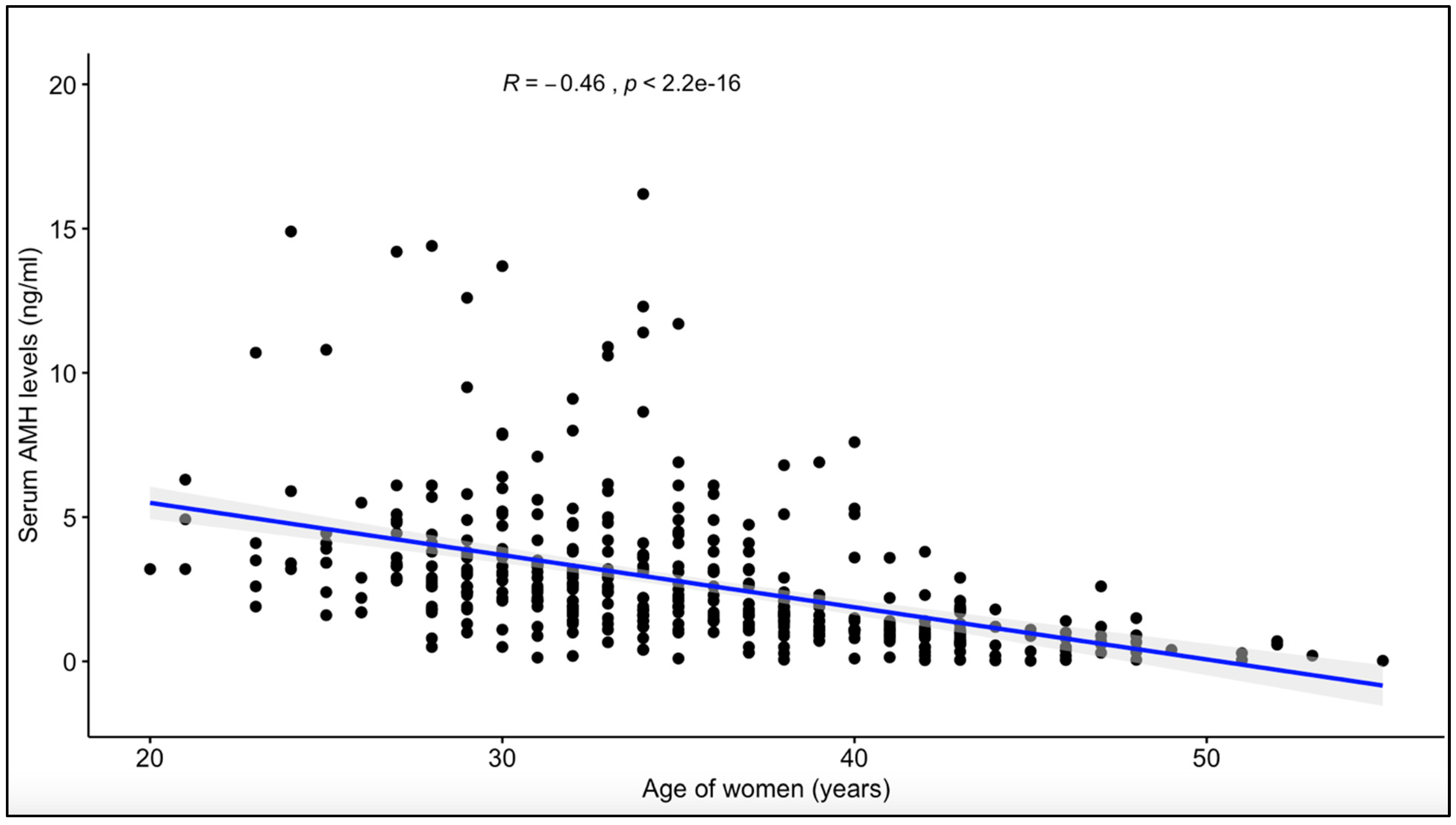
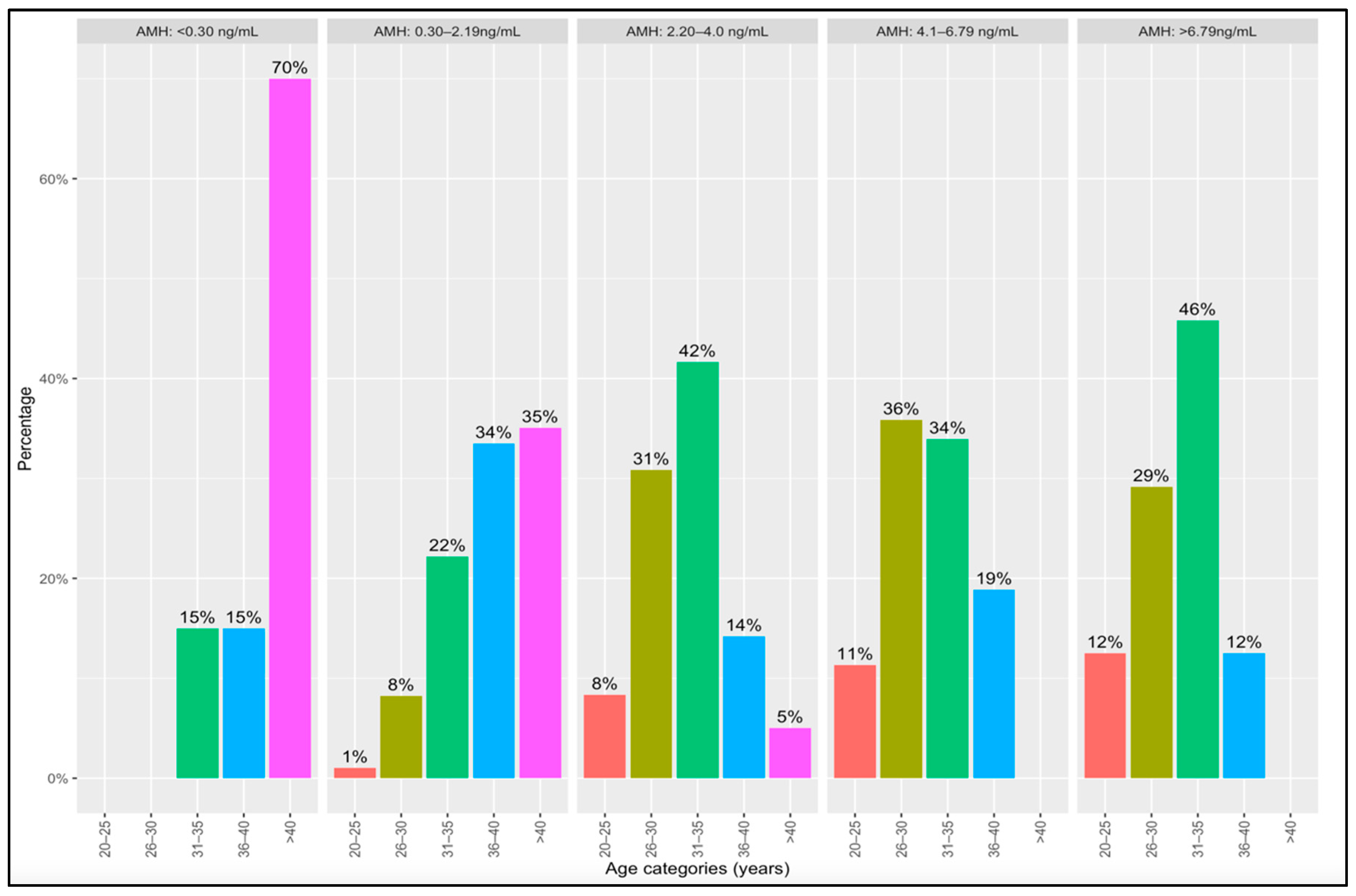
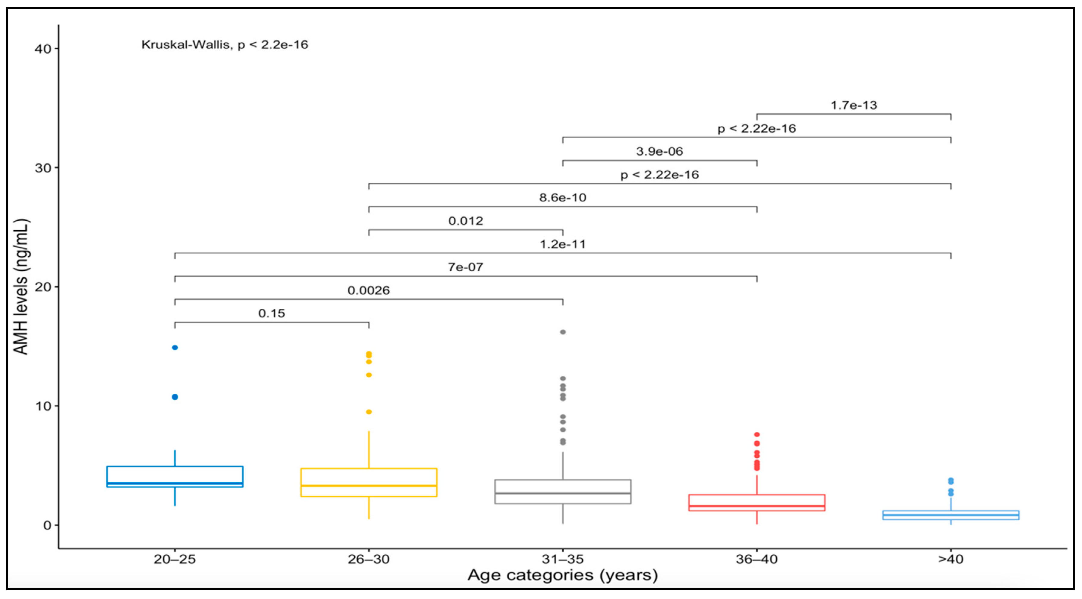
Data analysis was performed using the R statistical package (version 3.6.3, R Core Team, Vienna, Austria). Initial descriptive analysis was performed, and density plots of the AMH levels and women’s age were plotted. The correlation between AMH levels and age was determined using Pearson’s moment correlation coefficient. Non-normally distributed continuous variables were presented as median (interquartile range), and the normally distributed ones were presented as mean values ± standard deviation (SD) and compared across five ordered response groups. The AMH levels among the five groups were compared using the Kruskal–Wallis test. All p-values < 0.05 were considered statistically significant.

4. Results

Four hundred and twenty-six women were included in the study and had their baseline AMH determined. The study participants were categorized into five groups based on their ages. The mean (+SD) age was 35.25 ± 6.33 years (median 35 years (range: 20–55 years)). The mean AMH level was 2.80 ± 2.60 ng/mL. Women who were between 20–25 years had the highest average serum AMH concentration (4.85 ± 3.34 ng/mL), while those older than 40 age years had the lowest (0.94 ± 0.73 ng/mL) (Table 1). There were 28 (6.8%) women who were above 45 years [median age 47.5 years (IQR = 0.59)], and their median AMH level was 2.2 ng/mL (IQR = 0.55). Figure 1 shows the density plots for women’s age and AMH levels. The age distribution of the included women was not normally distributed (Figure 1), and this was confirmed by using the Shapiro–Wilk’s test (p < 0.004) indicating moderate positive skewness. Most of the women were in the age group between 30 and 40 years. The density plot of AMH levels showed gross deviation for normal distribution (significant positive skewness), and this was confirmed by the Shapiro–Wilk’s test (p < 2.2 × 10⁻¹⁶).
Table 2 summarizes the results of women’s AMH categories in the five age groups. Most of the women with very-low AMH levels (0.845 ng/mL) were older (>40 years of age) than those in the other groups. There were statistically significant differences in AMH levels across the five age groups (p < 2.2 × 10⁻¹⁶). There was a significant negative correlation between women’s age and their serum AMH levels (Figure 2).
Figure 3 presents AMH levels versus age categories of women treated for infertility. None of the women who were younger than 30 years had AMH levels lower than 0.30 ng/mL, while 70% of the women who had AMH levels of <0.30 ng/mL had advanced age (more than 40 years). Moreover, none of the women with advanced age had AMH levels greater than 4 ng/mL, with only 5% having AMH levels between 2.20 and 4.0 ng/mL. Furthermore, only 1% of women who had AMH levels between 0.30 to 2.19 ng/mL were between the ages of 20 to 25 years; all the rest (women between 20–25 years) had AMH levels of 2.20 ng/mL and above (Figure 3).
There was a significant negative correlation between women’s age and their serum AMH levels (correlation coefficient, R = −0.46; coefficient of determination = 21.3%).
Figure 4 shows the comparison of AMH levels among the various maternal age groups. There was no significant difference in median AMH levels (p-value: 0.15) between women in age groups 1 (20–25 years) and 2 (26–30 years). There was a significant difference in median AMH levels (p-value <0.000) between women in age groups 4 (36–40 years) and 5 (>40 years).

5. Discussion

In this study, we assessed the association between maternal age and AMH levels among women receiving IVF treatment. We determined that serum AMH levels varied significantly with women’s age. There was a significant negative correlation between serum AMH and age, which may be clinically useful in predicting ovarian response during IVF treatment. Our findings support the study by Meczekalski et al. [7], who determined that AMH seems the best endocrine biomarker for predicting age-related decline in ovarian reserve in healthy women. Clinical protocols combining women’s age, AMH levels, and other relevant ovarian reserve indicators are recommended in deciding the starting dose of gonadotropins for COS to improve outcomes in terms of both safety and cost effectiveness [6,17]. Previous studies have reported AMH levels for predicting reduced ovarian response [18,19,20]. For instance, Broer et al. reported that AMH had a sensitivity and specificity of 82% and 76%, respectively, in predicting excessive ovarian response to exogenous gonadotropins [20]. Thus, AMH is considered an important indicator of fertility potential in women of advanced maternal age [7]. In our study, the lowest and highest references or cutoffs were <0.3 and >6.79 ng/mL, respectively. However, ovarian response to COS was not studied in relation to AMH levels and women’s age, and this is a limitation of our study.
Our data also showed that 70% of women who were older than 40 years had AMH levels of <0.30 ng/mL and are likely to have suboptimal response (poor outcomes) to controlled ovarian stimulation and higher chances of cycle cancellations. Furthermore, only 1% of women between the ages of 20 to 25 years had AMH levels between 0.30 and 2.19 ng/mL. More recently, Zhang et al. determined that young women with relatively low levels of AMH undergoing IVF still yielded improved pregnancy outcomes compared to older counterparts [21]. Women with AMH levels ≥6.79 ng/mL and between the ages of 20 to 35 years may be considered “high responders” with an increased risk of ovarian hyperstimulation syndrome (OHSS)—a serious iatrogenic complication in IVF treatment following a standard COS protocol [20]. Similarly, women with PCOS are considered as high responders and constitute about 20% of patients undergoing COS [20]. Interestingly, in a recent study by Papler et al., there were no significant differences between obese and non-obese women concerning both biochemical and clinical pregnancy rates, following COS and IVF [22]. In this study, we excluded women diagnosed with PCOS to minimize the confounding effects on serum AMH levels. Several studies have demonstrated the clinical usefulness of AMH in refining the starting dose of gonadotropins for COS to maximize ovarian response and simultaneously minimize the risk of OHSS [18,23,24]. Unfortunately, a critical value for predicting the occurrence of OHSS could not be established in the present research because the outcome of ovarian stimulation was not included, and this is a limitation of this study. Similarly, obesity is an important factor in fertility treatment especially in PCOS.
The clinical relevance of AMH in predicting the likelihood of conception following assisted conception has been contentious in various studies [2]. In this study, we demonstrated significant negative correlation between women’s age and AMH, which serves as a robust tool for starting controlled ovarian stimulation and predicting ovarian response. Ebner et al. reported that low AMH levels (<1.66 ng/mL) are associated with oocyte quality [2]. However, subsequent studies have determined that AMH levels may be indicative of ovarian response to exogenous gonadotropins but not of oocyte quality [25,26,27]. Thus, low AMH levels may not be a strong indication for withholding fertility treatment. Meanwhile, Tal et al. concluded that AMH alone can potentially predict live birth after IVF and may be a clinically useful tool during counseling couples prior to fertility treatment, but its predictive accuracy is poor [27]. The findings we report in the present work, and previous studies by others, suggest that combined assessment and integration of AMH levels with maternal age before undergoing IVF treatment is clinically relevant in predicting women’s ovarian response and fertility outcome.
Our study has affirmed the inverse relationship between serum AMH levels and age among women undergoing IVF treatment. Further research to assess OHSS occurrence, the quality of oocytes retrieved, and the success rates of fertilization (embryos) and pregnancies (implantation) to facilitate the wholistic predictive value of combined AMH and women’s age is recommended.
The limitations of our study mainly relate to the non-inclusion of the clinical outcomes of controlled ovarian stimulation such as the fertilization and clinical pregnancy rates in association with AMH levels. Similarly, simultaneous assessment of the levels of FSH and AFC would have provided a more comprehensive understanding of the relevance of AMH in assisted reproductive technologies, and this omission is also considered a limitation of the study. In a recent study, Paola et al. demonstrated that the FSH starting dose for IUI calculated based on a validated nomogram [6] comprising AMH, AFC, and women’s age was significantly lower than the FSH dose prescribed [17]. In their study, approximately 15% of the stimulating cycles based on the nomogram had higher starting doses for FSH [17]. In our study, the association between AMH and women’s age was determined, but we did not include FSH and AFC. Another limitation is the potential influence of batch-to-batch inconsistencies in the determination of serum AMH levels as we did not freeze all the serum samples and the laboratory analyses were not conducted simultaneously at the end of the study. In addition, the specific etiologies of infertility of the recruited women were not included, and this may have influenced the findings. Despite the stated limitations, this study provides an important overview of the clinical relevance of AMH in relation to women’s age in the planning of ovarian stimulation protocols during ART, especially in low-resource settings.

6. Conclusions

This study presents age-specific AMH levels for women accessing IVF services in Ghana. It is inferential that women older than 40 years with AMH levels <0.3 ng/mL require adequate counseling on their need for oocyte donation before starting their first IVF treatment, given their predicted poor response to COS. There was a significant negative correlation (R = −0.46) between women’s age and serum AMH levels among women receiving assisted reproduction. It is suggested that combined evaluation of maternal age and AMH levels offers a benchmark in providing individualized planning of IVF treatment strategies for patients and in assisting practitioners to identify women at risk of low or high response to COS, thereby providing the opportunity to counsel them on their ovarian performance.

Author Contributions

D.M., K.A.-B., C.S.A. and F.K.A. conceived and designed the study; D.M. collected the data with supervision from C.S.A. and F.K.A.; K.A.-B. and D.M. conducted the data analysis; D.M. and K.A.-B. authored the article with significant contributions from C.S.A. and F.K.A. All authors approved the final manuscript. All authors have read and agreed to the published version of the manuscript.

Funding

This research received no external funding.

Institutional Review Board Statement

Ethical approval for this study (number: CHRPE/AP/276/17) was provided by the Committee on Human Research, Publication, and Ethics (CHRPE) of the School of Medicine and Dentistry, College of Health Sciences, Kwame Nkrumah University of Science and Technology (KNUST). We obtained written consent from all participants prior to the data collection.

Informed Consent Statement

Not applicable.

Data Availability Statement

The dataset for this study will be made available upon reasonable request from the corresponding author.

Acknowledgments

We are extremely grateful to all the staff of Airport Women’s Hospital for their support and co-operation during the recruitment and data collection. We are also grateful to all the women who consented to participate in this study.

Conflicts of Interest

The authors declare no conflict of interest.

References

  1. Seifer, D.B.; Golub, E.T.; Lambert-Messerlian, G.; Benning, L.; Anastos, K.; Watts, D.H.; Cohen, M.H.; Karim, R.; Young, M.A.; Minkoff, H.; et al. Variations in serum müllerian inhibiting substance between White, Black, and Hispanic women. Fertil. Steril. 2009, 92, 1674–1678. [Google Scholar] [CrossRef] [PubMed]
  2. Ebner, T.; Sommergruber, M.; Moser, M.; Shebl, O.; Schreier-Lechner, E.; Tews, G. Basal level of anti-Müllerian hormone is associated with oocyte quality in stimulated cycles. Hum. Reprod. 2006, 21, 2022–2026. [Google Scholar] [CrossRef] [PubMed]
  3. Broer, S.L.; Dólleman, M.; van Disseldorp, J.; Broeze, K.A.; Opmeer, B.C.; Bossuyt, P.M.M.; Eijkemans, M.J.C.; Mol, B.W.; Broekmans, F.J.M.; IPD-EXPORT Study Group. Prediction of an excessive response in in vitro fertilization from patient characteristics and ovarian reserve tests and comparison in subgroups: An individual patient data meta-analysis. Fertil. Steril. 2013, 100, 420–429. [Google Scholar] [CrossRef]
  4. Lee, M.M.; Donahoe, P.K.; Hasegawa, T.; Silverman, B.; Crist, G.B.; Best, S.; Hasegawa, Y.; Noto, R.; Schoenfeld, D.; MacLaughlin, D.T. Mullerian inhibiting substance in humans: Normal levels from infancy to adulthood. J. Clin. Endocrinol. Metab. 1996, 81, 571–576. [Google Scholar] [PubMed]
  5. Kanakatti Shankar, R.; Dowlut-McElroy, T.; Dauber, A.; Gomez-Lobo, V. Clinical utility of Anti-Mullerian Hormone in pediatrics. J. Clin. Endocrinol. Metab. 2022, 107, 309–323. [Google Scholar] [CrossRef]
  6. Yates, A.P.; Roberts, S.A.; Nardo, L.G. Anti-Müllerian hormone-tailored stimulation protocols improve outcomes whilst reducing adverse effects and costs of IVF. Hum. Reprod. 2012, 27. [Google Scholar] [CrossRef] [PubMed]
  7. Meczekalski, B.; Czyzyk, A.; Kunicki, M.; Podfigurna-Stopa, A.; Plociennik, L.; Jakiel, G.; Maciejewska-Jeske, M.; Lukaszuk, K. Fertility in women of late reproductive age: The role of serum anti-Müllerian hormone (AMH) levels in its assessment. J. Endocrinol. Investig. 2016, 39, 1259–1265. [Google Scholar] [CrossRef]
  8. Sills, E.S.; Alper, M.M.; Walsh, A.P. Ovarian reserve screening in infertility: Practical applications and theoretical directions for research. Eur. J. Obstet. Gynecol. Reprod. Biol. 2009, 146, 30–36. [Google Scholar] [CrossRef]
  9. Gleicher, N.; Weghofer, A.; Barad, D.H. Defining ovarian reserve to better understand ovarian aging. Reprod. Biol. Endocrinol. 2011, 9, 23. [Google Scholar] [CrossRef]
  10. Bukulmez, O.; Arici, A. Assessment of ovarian reserve. Curr. Opin. Obstet. Gynecol. 2004, 16, 231–237. [Google Scholar] [CrossRef]
  11. Hamdine, O.; Eijkemans, M.; Lentjes, E.; Torrance, H.; Macklon, N.; Fauser, B.; Broekmans, F. Ovarian response prediction in GnRH antagonist treatment for IVF using anti-Müllerian hormone. Hum. Reprod. 2015, 30. [Google Scholar] [CrossRef] [PubMed]
  12. Andersen, A.N.; Witjes, H.; Gordon, K.; Mannaerts, B. Predictive factors of ovarian response and clinical outcome after IVF/ICSI following a rFSH/GnRH antagonist protocol with or without oral contraceptive pre-treatment. Hum. Reprod. 2011, 26, 3413–3423. [Google Scholar] [CrossRef] [PubMed]
  13. Mawusi, D.; Yakass, M.B.; Abaidoo, C.S.; Addai, F.K. Use of Anti-Mullerian Hormone (AMH) for Testing of Ovarian Reserve: A Survey of Fifteen (15) Fertility Centres in Ghana. Adv. Reprod. Sci. 2021, 9, 9. [Google Scholar] [CrossRef]
  14. Arce, J.-C.; La Marca, A.; Klein, B.M.; Andersen, A.N.; Fleming, R. Antimüllerian hormone in gonadotropin releasing-hormone antagonist cycles: Prediction of ovarian response and cumulative treatment outcome in good-prognosis patients. Fertil. Steril. 2013, 99, 48. [Google Scholar] [CrossRef] [PubMed]
  15. Tuck, M.K.; Chan, D.W.; Chia, D.; Godwin, A.K.; Grizzle, W.E.; Krueger, K.E.; Rom, W.; Sanda, M.; Sorbara, L.; Stass, S.; et al. Standard operating procedures for serum and plasma collection: Early detection research network consensus statement standard operating procedure integration working group. J. Proteome Res. 2009, 8, 113–117. [Google Scholar] [CrossRef] [PubMed]
  16. Gassner, D.; Jung, R. First fully automated immunoassay for anti-Müllerian hormone. Clin. Chem. Lab. Med. (CCLM) 2014, 52, 1143–1152. [Google Scholar] [CrossRef]
  17. Di Paola, R.; Garzon, S.; Giuliani, S.; Laganà, A.S.; Noventa, M.; Parissone, F.; Zorzi, C.; Raffaelli, R.; Ghezzi, F.; Franchi, M.; et al. Are we choosing the correct FSH starting dose during controlled ovarian stimulation for intrauterine insemination cycles? Potential application of a nomogram based on woman’s age and markers of ovarian reserve. Arch. Gynecol. Obstet. 2018, 298. [Google Scholar] [CrossRef]
  18. La Marca, A.; Papaleo, E.; Grisendi, V.; Argento, C.; Giulini, S.; Volpe, A. Development of a nomogram based on markers of ovarian reserve for the individualisation of the follicle-stimulating hormone starting dose in in vitro fertilisation cycles. BJOG Int. J. Obstet. Gynaecol. 2012, 119, 1171–1179. [Google Scholar] [CrossRef]
  19. Nelson, S.M.; Yates, R.W.; Fleming, R. Serum anti-Müllerian hormone and FSH: Prediction of live birth and extremes of response in stimulated cycles—Implications for individualization of therapy. Hum. Reprod. 2007, 22, 204. [Google Scholar] [CrossRef]
  20. Broer, S.L.; Dólleman, M.; Opmeer, B.C.; Fauser, B.C.; Mol, B.W.; Broekmans, F.J.M. AMH and AFC as predictors of excessive response in controlled ovarian hyperstimulation: A meta-analysis. Hum. Reprod. Update 2011, 17, 34. [Google Scholar] [CrossRef] [Green Version]
  21. Zhang, B.; Meng, Y.; Jiang, X.; Liu, C.; Zhang, H.; Cui, L.; Chen, Z.-J. IVF outcomes of women with discrepancies between age and serum anti-Müllerian hormone levels. Reprod. Biol. Endocrinol. 2019, 17, 83. [Google Scholar] [CrossRef] [PubMed]
  22. Papler, T.B.; Bokal, E.V.; Zmrzljak, U.P.; Stimpfel, M.; Laganà, A.S.; Ghezzi, F.; Jančar, N. PGR and PTX3 gene expression in cumulus cells from obese and normal weighting women after administration of long-acting recombinant follicle-stimulating hormone for controlled ovarian stimulation. Arch. Gynecol. Obstet. 2019, 299, 863–871. [Google Scholar] [CrossRef]
  23. Nelson, S.M.; Yates, R.W.; Lyall, H.; Jamieson, M.; Traynor, I.; Gaudoin, M.; Mitchell, P.; Ambrose, P.; Fleming, R. Anti-Müllerian hormone-based approach to controlled ovarian stimulation for assisted conception. Hum. Reprod. 2009, 24, 867–875. [Google Scholar] [CrossRef] [PubMed]
  24. Rustamov, O.; Smith, A.; Roberts, S.A.; Yates, A.P.; Fitzgerald, C.; Krishnan, M.; Nardo, L.G.; Pemberton, P.W. The measurement of anti-Mullerian hormone: A critical appraisal. J. Clin. Endocrinol. Metab. 2014, 99, 723–732. [Google Scholar] [CrossRef] [PubMed]
  25. Smeenk, J.M.J.; Sweep, F.C.G.J.; Zielhuis, G.A.; Kremer, J.M.; Thomas, C.M.G.; Braat, D.D.M. Antimüllerian hormone predicts ovarian responsiveness, but not embryo quality or pregnancy, after in vitro fertilization or intracytoplasmic sperm injection. Fertil. Steril. 2007, 87, 223–226. [Google Scholar] [CrossRef]
  26. Fong, S.L.; Baart, E.B.; Martini, E.; Schipper, I.; Visser, J.A.; Themmen, A.P.N.; de Jong, F.H.; Fauser, B.J.C.M.; Laven, J.S.E. Anti-Müllerian hormone: A marker for oocyte quantity, oocyte quality and embryo quality? Reprod. Biomed. Online 2008, 16, 664–670. [Google Scholar] [CrossRef]
  27. Tal, R.; Tal, O.; Seifer, B.J.; Seifer, D.B. Antimüllerian hormone as predictor of implantation and clinical pregnancy after assisted conception: A systematic review and meta-analysis. Fertil. Steril. 2015, 103, 119–130. [Google Scholar] [CrossRef]
Figure 1. Density plot of women’s age and AMH levels.
Reprodmed 03 00020 g001
Figure 2. Correlation between women’s age and their serum AMH levels.
Reprodmed 03 00020 g002
Figure 3. Comparing AMH categories and maternal age categories.
Reprodmed 03 00020 g003
Figure 4. AMH levels versus age categories of women treated for infertility.
Reprodmed 03 00020 g004
Table 1. Serum AMH levels in five age categories of women undergoing IVF treatment.
Age GroupsN = 411; n (%)Median AMH (IQR)Mean AMH ± SD95% CI
20–2521 (5.1)3.5 (1.72)4.85 ± 3.343.33–6.37
26–3079 (19.2)3.3 (2.35)3.98 ± 2.833.34–4.61
31–35125 (30.4)2.7 (2.00)3.32 ± 2.632.86–2.26
36–4098 (23.8)1.6 (1.35)2.15 ± 1.541.85–2.46
>4088 (21.4)0.85 (0.74)0.94 ± 0.730.78 –1.09
N = total sample size; n = group sample size; IQR = interquartile range; SD = standard deviation; CI = confidence interval.
Table 2. Comparison of serum levels of AMH groups and maternal age categories.
AMH Categories
Age Group in Years(<0.30 ng/mL)n (%)(0.30–2.19 ng/mL) n (%)(2.20–4.0 ng/mL) n (%)(4.1–6.79 ng/mL) n (%)>6.79 ng/mL n (%)
1 (20–25)02 (1.0)10 (8.3)6 (11.3)3 (12.5)
2 (26–30)016 (8.3)37 (30.8)19 (35.8)7 (29.2)
3 (31–35)3 (15.0)43 (22.2)50 (41.7)18 (34.0)11 (45.8)
4 (36–40)3 (15.0)65 (33.5)17 (14.2)10 (18.9)3 (12.5)
5 (>40)14 (70.0)68 (35.1)6 (5.0)00
Total20 (100)194 (100)120 (100)53 (100)24 (100)
Publisher’s Note: MDPI stays neutral with regard to jurisdictional claims in published maps and institutional affiliations.

