
Effect of Higher Order Aberrations and Intraocular Scatter on Optical Quality Based on an Optical Eye Model
1
Tianjin Eye Hospital, Tianjin Eye Institute, Tianjin Key Laboratory of Ophthalmology and Visual Science, Nankai University Affiliated Eye Hospital, Nankai University Eye Institute, Clinical College of Ophthalmology of Tianjin Medical University, Tianjin 300020, China
2
Department of Opto-Electronic Engineering, Changzhou Institute of Technology, Changzhou 213022, China
*
Author to whom correspondence should be addressed.
Optics 2023, 4(2), 364-374; https://doi.org/10.3390/opt4020027
Submission received: 16 December 2022
/
Revised: 9 March 2023
/
Accepted: 23 May 2023
/
Published: 26 May 2023
(This article belongs to the Special Issue Advances in Vision Optics, Myopia Control and Refractive Surgery)
Abstract
:The impact of intraocular scatter and higher order aberrations (HOAs) on ocular optical quality was investigated. An optical eye model was constructed using the measured ocular aberrations, corneal surfaces, axial length, and scatter fraction, and the impact of HOAs and scatter on modulation transfer functions (MTFs) was studied based on the newly established optical eye model. For uniform intraocular scatter, the monochromatic and polychromatic MTF decreased as the HOAs or scatter fractions increased independently at each spatial frequency, which implied that both were essential for visual quality. In addition, the scatter effect on MTF was more significant for the eye with less HOA. The combined deterioration effect of these two factors on the MTF was less than their summation, suggesting a potential compensatory mechanism between HOAs and scatter.
1. Introduction
Wavefront aberration and intraocular scattering are two important factors that deteriorate the subjective visual performance of the human eye [1]. Low-order aberrations include defocus and astigmatism, which can be corrected using spectacles. Higher order aberrations (HOAs) need a complex adaptive optics system to correct [2,3,4], which is impossible in daily life. Therefore, it is important to assess the effects of the intraocular scatter and HOAs on the optical quality of the human eye.
Many efforts have been made to investigate the effects of intraocular scatter and HOAs on visual quality [5,6,7,8,9]. Fujikado et al. [5] reported that the loss of contrast sensitivity (CS) in the cataractous eye was predominantly owing to intraocular scatter and HOAs. Using multivariate regression analysis, Kamiya et al. [6] found that CS was significantly correlated with intraocular scatter, but not with HOAs. Bueno et al. [7] discovered that CS decreased with increasing scatter for all spatial frequencies. However, Lee et al. [8] reported that scattered light was not corrected using CS, suggesting that intraocular scatter plays a negligible role in visual quality. In 2017, Zhao et al. [9] modified an adaptive optics double-pass system to improve the measurement accuracy of intraocular scatter, which was subsequently used to explore the impact of HOAs and intraocular scatter on contrast sensitivity. The results indicate that the joint effect of HOAs and scatter was less than that of a simple summation, which suggested the existence of a compensatory mechanism in the human eye. Different studies have reported varying results.
Experimental studies have indicated various reasons for this inconsistency. These include variations in ocular HOAs [10], accuracy of measurement instruments [11], corneal moisture, ocular fatigue, and age [12]. In addition, differences in samples lead to different features [13], which complicate the extraction of inconsistent rules. Therefore, a theoretical approach is required to better understand the effects of HOAs and scatter of the human eye.
Mathematical models are effective tools for correlating the results of theoretical evaluations and clinical outcomes [14,15]. Eye models using optical ray tracing or other analytical and/or numerical methods have often been constructed using wavefront aberrations and other parameters. The retinal image can be calculated from the model, which is useful in Ophthalmology and Optometry. DeHoog and Doraiswamy [16,17] introduced the scatter fraction to a pseudophakic eye model to study the loss of optical quality from glistenings in intraocular lens. However, no chromatic aberrations or HOAs were observed in these eye models.
In this study, to assess the effect of intraocular scatters and HOAs on visual quality, an optical eye model with different HOAs and scatters was constructed using the optical ray-tracing software Zemax, and modulation transfer functions (MTFs) with different HOAs and scatterers were calculated to evaluate visual quality. An impact factor was also used to analyse their influence independently and jointly.
2. Methods
2.1. Construction of the Eye Model
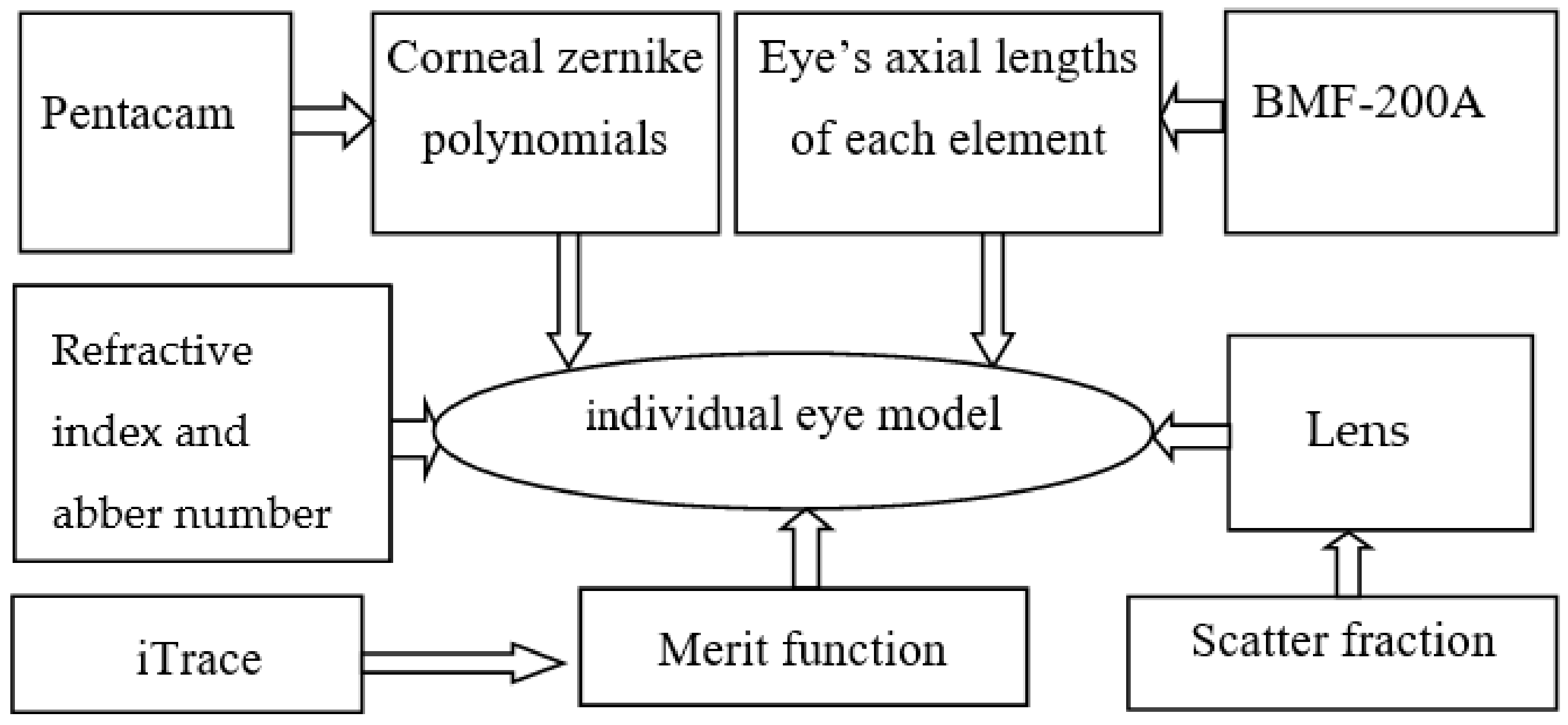
The optical eye model was constructed using Zemax (ZEMAX LLC., 2019, Bellevue, WA, USA) for optical quality evaluation, and detailed descriptions of the design procedures are available in previous studies [14,16,17]. In these models, the retinal image can be obtained through ray tracing, and a lens for correcting vision can be designed through optimisation. In this study, an optical eye model was modified by incorporating optical scattering and HOAs. It was started from an eye model and then built by replacing the measured ocular biometric parameters, including the front and back corneal radius of curvature, depth of cornea, anterior chamber, lens, and vitreous humour, after which the lens curvatures and elevation data were optimised corresponding to the measured ocular aberrations. These eye models have been used as effective tools in visual science to explain optical phenomena in vision and predict how changes in ocular biometry affect refraction and aberrations. In this study, we explored the effects of HOAs and scattering on vision. The construction for the new optical eye model is described below.
- (1)
- (2)
- The corneal surface was set as a Zernike Fringe sag surface, whose parameters can be directly measured in the clinic. The parameters include the curvature radius, depth, and elevation data of the anterior and posterior corneal surfaces represented by a set of 27 terms of Zernike polynomials.
- (3)
- The depths of the anterior chamber, crystalline lens, and vitreous body were replaced with the measured data of individual eyes [8] In Zemax, these data are written in a column of thickness in the Lens data.
- (4)
- The direct measurement of the elevation data of the crystalline lens is difficult. To ensure that the total aberrations of the eye model correspond to the measured aberrations of the actual eye, the operands ZERN in Zemax were added to the merit function, with coefficients defining the measured aberrations. The lens surfaces were set as Zernike Fringe Sag surfaces for optimisation to fit the aberrations of the actual eye. After optimisation, the Zernike coefficients of the lens surfaces were obtained.
- (5)
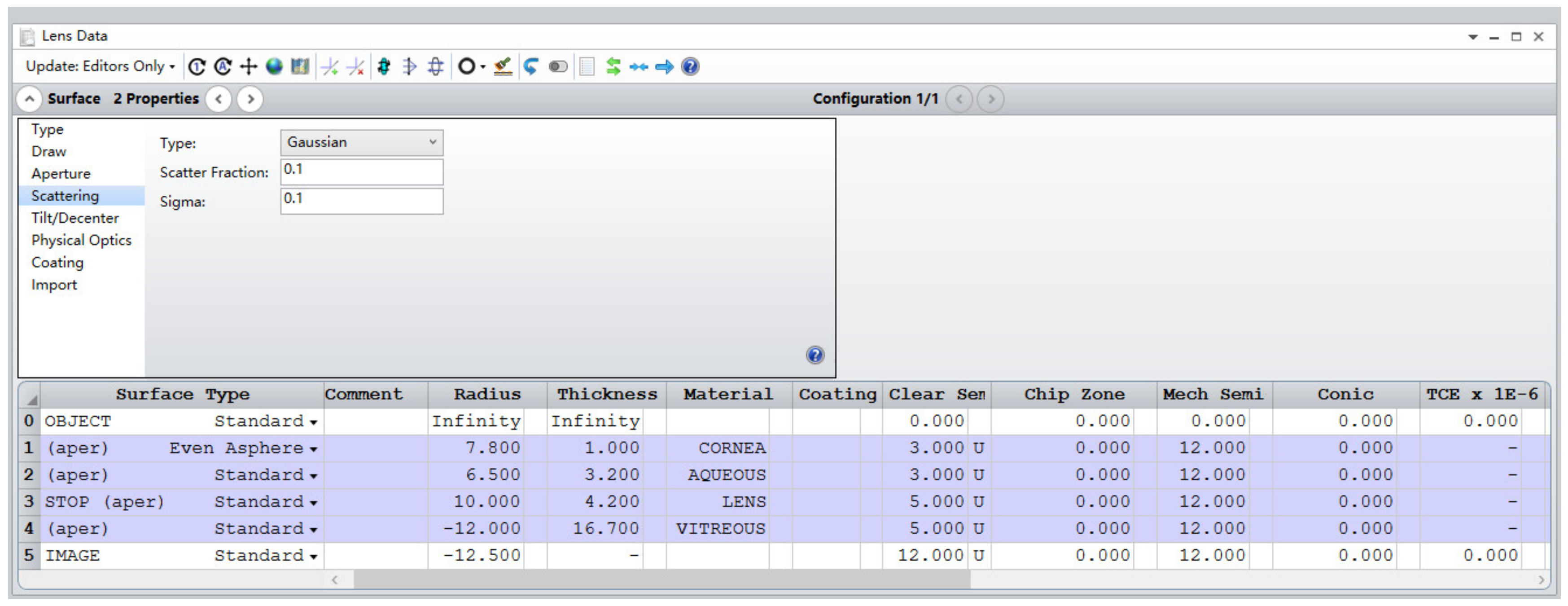
- The scattering types of the cornea and crystalline lens were set as Gaussian distributions with the parameters of the scatter fraction and a sigma [18].
2.2. Research Methods
In visual optics, the MTF has been proven clinically useful for assessing objective visual performance [1,2,14,16,17]. The area under the MTF (MTFA), which is the integration of the MTF from zero spatial frequency to a given spatial frequency (60 cpd in this study), is a single number that describes the visual quality, which was also used in this study. The calculation procedure is as follows:
- (1)
- The optical eye model in Zemax provides the MTFs in monochromatic and chromatic light at different HOAs and scatter levels.
- (2)
- MTFAs at different HOAs and scatter levels were calculated, and changes in monochromatic and chromatic MTFA were obtained.
- (3)
Here MTFA(HS) and MTFA(0) denote the MTFA values of the eye with specified scatter and HOAs and without scatter and HOAs, respectively. HS denotes the HOAs and scatter in the human eye. The impact factors were calculated for eyes with seven types of scatter and six types of HOAs. In this study, the investigation of the relationship between Factor(HS) and the sum of Factor(H) and Factor(S) was important.
3. Experiments
3.1. Subjects
The study protocol was conducted in accordance with the Declaration of Helsinki and was approved by the Ethics Committee of Tianjin Eye Hospital. Written informed consent was obtained from all the patients.
The averaged RMS error of HOAs, as obtained from previous studies, was approximately 0.4 ± 0.2 µm [1]. Therefore, six groups of eyes with RMS errors ranging from 0.1 to 0.6 µm and an interval of 0.1 µm were selected. In each group, 20 eyes, with the RMS deviation of (0.04 µm), were selected from the case library of Tianjin Eye Hospital as the subjects.
None of the subjects had undergone corneal refractive surgery or had ocular disease, and their ages ranged from 22–30 years. Furthermore, an aberration-free optical model eye was used for comparison, and its parameters are listed in Table 1.
3.2. Ocular Aberrations
An iTrace wavefront aberrometer (Tracy, CA, USA) was used to measure wavefront aberrations of the entire eye. The principle of the iTrace wavefront aberrometer is as follows [9]: 256 narrow near-infrared beams are projected onto the retina of a subject. As light propagates through an entrance pupil of 6 mm, the wavefront undergoes local phase shifts. The distribution of spots in the retina is captured by a CCD camera in the conjugate position of the retina through an exit pupil of 2 mm, where the wavefront can be neglected. Therefore, the wavefront aberrations of the 6 mm pupil were measured and expressed as Zernike polynomial expansions consisting of 27 terms.
3.3. Corneal Surface
A Pentacam (Oculus Co., Ltd., Wetzlar, Germany) was used to measure the corneal depth, curvature radius, and elevation data of the anterior and posterior corneal surfaces decomposed into 27 terms of Zernike polynomials.
The Pentacam obtained images of the anterior corneal surface using a rotating Scheimpflug camera. This rotating process obtained images in three dimensions and allowed precise measurement of the centre of the cornea. Accurate posterior elevation data are required for accurate pachymetry.
3.4. Axial Length of the Eye
A BMF-200 A/B Ultrasonic Diagnostic Instrument was used to measure the depth of each element of the eye, including the anterior chamber, crystalline lens, and vitreous body. The average data from five trials were considered as the final result.
3.5. Scatter Fraction
The scattering rate of the cornea and lens have been reported to be approximately 8% and 10% in statistics [19,20], respectively. Therefore, in this study, seven sets of scatter factors of the cornea and lens were introduced, and the scattering type was set as a Gaussian distribution, with the scatter fractions shown in Table 2. Sigma was set to 0.1, which did not significantly influence the MTF. In addition, a theoretical eye model without scatter was constructed for comparison.
4. Results
4.1. Eye Parameter
In this study, 120 eyes were divided into six groups. Owing to space limitations, we chose one eye in each group for presenting optical parameters.
Although the RMS error of the lower order aberration was approximately 90% of the RMS error of the total aberration, only the HOAs were considered in this study because defocus and astigmatism can be corrected with glasses. Table 3 lists the higher order aberrations of the six eyes, expressed as the 27 terms of Zernike polynomials. Owing to space limitations, only a few of them are presented.
Table 4 shows the corneal curvatures and corneal surfaces decomposed into 27 terms of Zernike polynomials for the subjects. Owing to space limitations, only a few are shown in this table.
Table 5 shows the axial length of each element in the eye, including the depths of the cornea, anterior chamber, crystalline lens, and vitreous body. The depth of the cornea was measured using the Pentacam, while other depths were measured using an Ultrasonic Diagnostic Instrument.
The photopic conditions in Zemax were selected as the incident light conditions. The lens parameters were obtained through optimisation. Table 6 lists the radius of curvature and Zernike polynomials, and only eight terms are shown owing to space limitations.
4.2. MTF of Different Scatter and HOAs
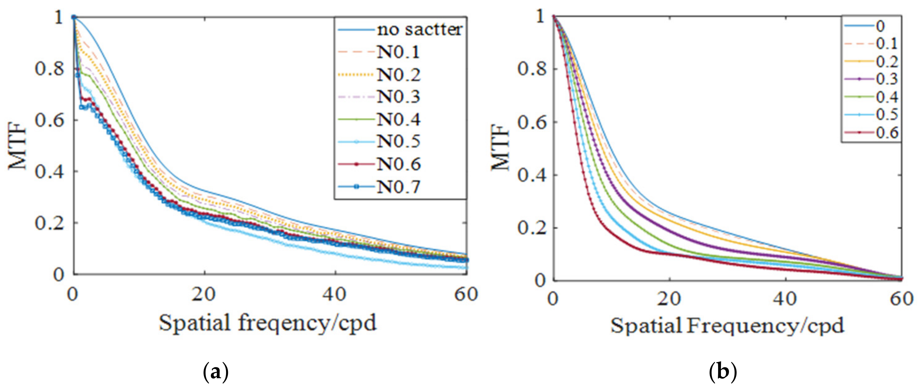
Figure 5a shows the MTFs of an aberration-free eye with different scatter fractions, as shown in Table 2. The MTF clearly decreases with increasing scatter, and lower spatial frequencies are associated with a more significant reduction in the MTF than higher spatial frequencies. Figure 5b shows the MTFs of a no-scatter eye with different HOA errors; the MTF decreases with increasing HOAs, suggesting that HOAs also affect the MTF negatively.
4.3. MTFA of Different HOAs and Scatter Factors
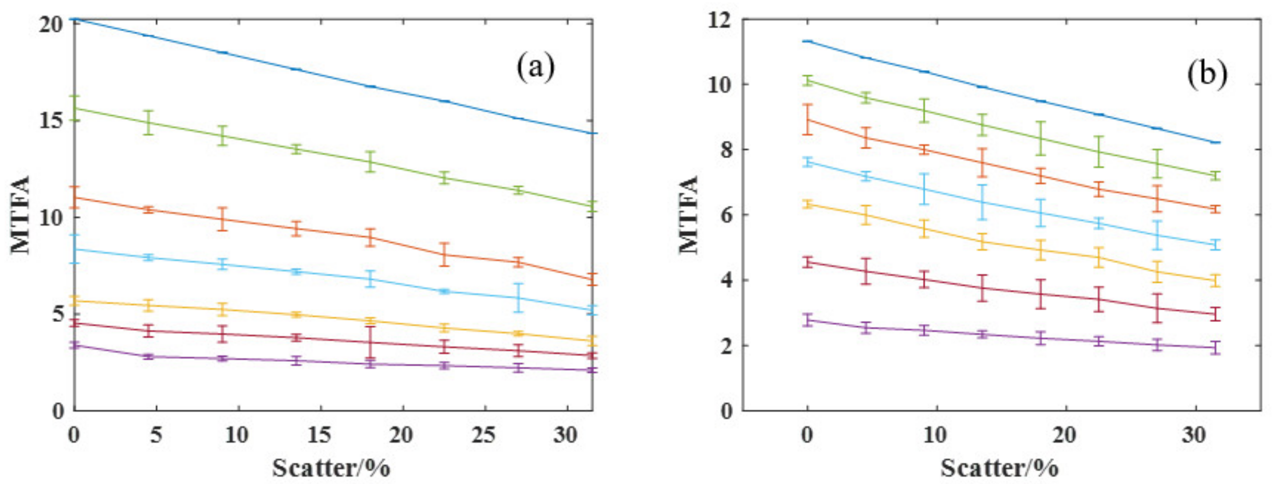
Figure 6a shows the monochromatic MTFA of the six groups of eyes with different scatterers (wavelength: 550 nm). All the MTFA clearly decrease with increasing scatter, and the rate of change decreases with increasing HOAs. Thus, the deteriorative effect of scattering on the visual performance decreases with increasing HOAs. Figure 6b shows the polychromatic MTFA of each group, and the same trend is observed. The chromatic aberration for everyone could be approximately 2 diopter across the visual spectrum, which results in the same effect as chromatic aberration on MTFA.
The MTFA shown in Figure 6a is referred to as a two-way analysis of variance (ANOVA) with HOAs and scatter fractions. The results indicated significant correlations between MTFA and HOAs (p < 0.001) and scatter (p < 0.001), suggesting a significant interaction between scatter and HOAs for monochromatic MTFA (p < 0.01). For the chromatic MTFA in Figure 6b, the results of ANOVA also indicated a significant interaction between the scatter and HOAs on visual quality (p = 0.017). Generally, p values of 0.05 or less were considered to be statistically significant, so that the optical quality of the human eye was influenced by HOAs, scatter and their interaction.
4.4. Compensation between HOAs and Scatter
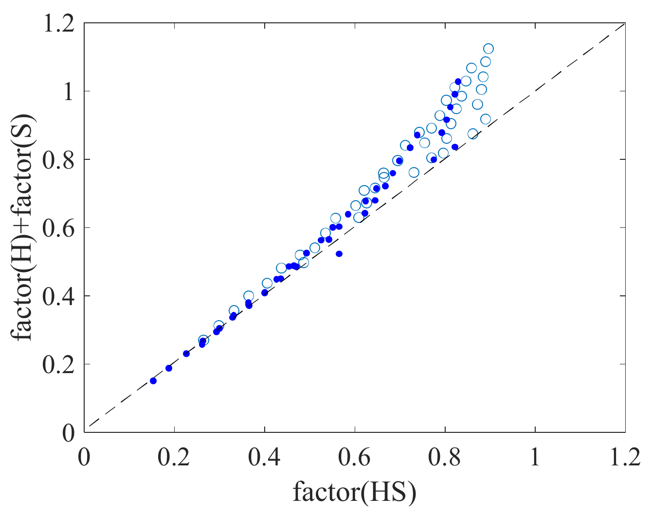
We further explored whether the combined effect of scatter and HOAs on MTF was the sum of the impact effects of scatter and HOAs. Figure 7 shows the impact factor (HS) as a function of the sum of the impact factor(H) and impact factor(S). The signals were clearly above the dashed line (y = x), indicating that there was no simple summation effect on the MTF for HOAs and intraocular scatter and suggesting a compensatory mechanism. As factor(HS) increases, the signals move upright, and the distances between the signals and dashed line increase rapidly, which indicates that larger compensation exists for larger HOAs and/or scatter on the MTF.
5. Discussions
Intraocular scatter is an important optical factor that affects visual quality, and has been the subject of many experimental studies. However, the results have been inconsistent for several reasons.
There are several instruments for measuring eye scatter in clinics, such as the Miller–Nadler Stray light meter, VistechMCT8000 meter, Mesotest, Nyktotest, OQAS, and C-Quant. Generally, the principles and procedures of these instruments are different, leading to inconsistent results.
Although the scatter fractions are the same, the scatter effects differ among subjects because the scattering particles differ in density, size, and distribution. This may explain why the preliminary data from the proposed study provided evidence that the effect of light scattering on visual performance varies among subjects [14].
The effect of scattering on visual quality may vary across subjects because of other influencing factors. In our study, the change rule could be easily obtained using statistics for the group with fewer HOAs. This may be difficult for eyes with larger wavefront aberrations, which may explain why different studies reported different conclusions.
In addition, in clinical practice, there are still some residual low-order aberrations in the eye, which make it difficult to establish reliable relationships between scattering and HOAs in visual performance.
Instead of the conventional experimental methods, an individual eye model was established here by non-contact measurements, numerical calculation, and optical modelling. High-order aberrations of individual eyes were obtained from the wavefront sensor. The software Zemax was chosen to construct the model with the Gullstrand–Le grand eye model as the initial configuration. Therefore, the individual eye model built in this study had the same wavefront aberration as the real eye.
In this study, the effect on visual performance was assessed using the established optical eye model, which overcomes the limitations of inconsistent and poor quantification of test instruments, differences among subjects, and enables us to determine the impact of HOAs and scatter on visual performance individually and jointly, as well as an in-depth analysis of their compensation. To the best of our knowledge, this is the first report on the quantitative estimation of the effects of HOAs and intraocular scatter based on an optical eye model.
This study has some limitations, and much future work is required. We only studied the effects of uniform forward scattering in the normal cornea and lens. For cataractous eyes, the forward scatter is not always uniform; therefore, further studies are required. The differences between the size, density, and distribution of the scattering particles have not yet been mentioned [15]. Additionally, the accuracy of the quantitative relationship was limited by the number of participants, scatter type, and distribution. However, this study suggests a new method for quantitatively studying the effects of scattering and HOAs on visual quality. Therefore, it may be an effective method for determining which factors play a more important role in the visual quality of an individual eye and then predicting the potential visual quality after refractive surgery.
6. Conclusions
An optical eye model was modified to study the optical quality of the eye with both scattering and HOAs using MTF. The results indicated that the effect of intraocular scatter on optical quality decreased with increasing HOAs, suggesting a compensatory mechanism between scatter and HOAs on visual quality.
The optical eye model was first adopted to analyse the influence of both scattering and HOAs on the optical quality of the eye. As a new method, it can be used to assess the effect of HOAs and scatter independently and subsequently assess the potential visual quality of the human eye. Therefore, it may be widely adopted for assessing ocular performance and clinical vision therapy.
Author Contributions
Conceptualisation, F.R. and Y.W.; methodology, F.R. and M.D.Z.; software, M.D.Z.; investigation, X.H.Z.; data curation, X.H.Z.; writing—original draft preparation, F.R.; writing—review and editing, F.R.; supervision, Y.W.; project administration, Y.W.; and funding acquisition, Y.W. All authors have read and agreed to the published version of the manuscript.
Funding
This research was funded by the National Natural Science Foundation of China grant number [82271118], the Tianjin Diversified Investment Fund for Applied Basic Research grant number [21JCZDJC01190], the Tianjin Health and Technology Project grant number [TJWJ2022XK036], and the Tianjin Key Medical Discipline (Specialty) Construction Project grant number [TJYXZDXK-016A].
Institutional Review Board Statement
The study was conducted in accordance with the Declaration of Helsinki, and approved by the Medical Ethics Committee of Tianjin Eye Hospital (protocol code 202032 and 2020.8).
Informed Consent Statement
Informed consent was obtained from all subjects involved in the study.
Data Availability Statement
Data underlying the results presented in this paper are not publicly available at this time but may be obtained from the corresponding author upon reasonable request.
Conflicts of Interest
The authors declare no conflict of interest.
Abbreviations
| HOA | higher order aberration |
| MTF | modulation transfer functions |
| MTFA | Area under MTF |
| CS | contrast sensitivity |
References
- Wang, Y.; Zhao, K.X. Wavefront Aberration and Clinical Vision Correction, 1st ed.; People’s Medical Press: Beijing, China, 2011; pp. 72–73. [Google Scholar]
- Liang, J.; Williams, D.R.; Miller, D.T. Supernormal vision and high resolution retinal imaging through adaptive optics. J. Opt. Soc. Am. A 1997, 14, 2884–2892. [Google Scholar] [CrossRef] [PubMed]
- Yang, Y.; Zhao, J.; Zhao, X.; Xiao, F.; Xie, J.; Liu, T.; Dai, Y. Objective visual performance evaluation with visual evoked potential measurements based on an adaptive optics system. Chin. Opt. Lett. 2018, 16, 86–90. [Google Scholar] [CrossRef]
- Morgan, J.W. Adaptive optics retinal imaging techniques and clinical applications. In Encyclopedia of Modern Optics, 2nd ed.; Guenther, R., Steel, D., Eds.; Elsevier: Oxford, UK, 2018; pp. 72–84. [Google Scholar]
- Fujikado, T.; Kuroda, T.; Maeda, N.; Ninomiya, S.; Goto, H.; Tano, Y.; Oshika, T.; Hirohara, Y.; Mihashi, T. Light scattering and optical aberrations as objective parameters to predict visual deterioration in eyes with cataracts. J. Cataract Refract. Surg. 2004, 30, 1198–1208. [Google Scholar] [CrossRef] [PubMed]
- Kamiya, K.; Shimizu, K.; Iijima, A.; Kobashi, H. Factors influencing contrast sensitivity function in myopic eyes. PLoS ONE 2014, 9, e113562. [Google Scholar] [CrossRef] [PubMed]
- Bueno, J.M.; Pérez, G.; Benito, A.; Artal, P. Impact of scatter on double-pass image quality and contrast sensitivity measured with a single instrument. Biomed. Opt. Express 2015, 6, 4841–4849. [Google Scholar] [CrossRef] [PubMed]
- Lee, H.; Lee, K.; Ahn, J.M.; Kim, E.K.; Sgrignoli, B. Double-pass system assessing the optical quality of pseudophakic eyes. Optom. Vis. Sci. 2014, 91, 437–443. [Google Scholar] [CrossRef] [PubMed]
- Zhao, J.; Xiao, F.; Zhao, H.; Dai, Y.; Zhang, Y. Effect of higher-order aberrations and intraocular scatter on contrast sensitivity measured with a single instrument. Biomed. Opt. Express 2017, 8, 2138–2147. [Google Scholar] [CrossRef] [PubMed]
- Quan, W.; Gao, B.Y.; Wang, C.W. Dynamic characteristic of the wavefront aberrations in human eye. Acta Photonic Sin. 2015, 44, 0117001. [Google Scholar] [CrossRef]
- Salmon, T.O.; van de Pol, C. Evaluation of a clinical aberrometer for lower-order accuracy and repeatability, higher-order repeatability, and instrument myopia. Optometry 2005, 76, 461–472. [Google Scholar] [CrossRef] [PubMed]
- Gaku, K.; Hiraoka, T.; Ueno, Y.; Mihashi, T.; Oshika, T. Influence of refractive status and age on corneal higher-order aberration. Vis. Res. 2021, 181, 32–37. [Google Scholar]
- Nancy, J.C.; Susana, M.; David, T. Ocular wavefront aberrations in the common marmoset Callithrix jacchus: Effects of age and refractive error. Vis. Res. 2010, 23, 2515–2529. [Google Scholar]
- Li, K.; Chen, X.; Bian, Y.; Xing, Y.; Li, X.; Liu, D.; Liu, Y. Design and optical analysis of a refractive aspheric intraocular lens with extended depth of focus. Optics 2023, 4, 146–155. [Google Scholar] [CrossRef]
- Osmers, J.; Kaiser, N.; Sorg, M.; Fischer, A. Adaptive finite element eye model for the compensation of biometric influences on acoustic tonometry. Comput. Methods Programs Biomed. 2021, 200, 105930. [Google Scholar] [CrossRef] [PubMed]
- Edward, D.; Doraiswamy, A. Evaluation of the impact of light scatter from glistenings in pseudophakic eyes. J. Cataract. Refract. Surg. 2014, 40, 95–103. [Google Scholar]
- Edward, D. Anand Doraiswamy. Evaluation of loss in optical quality of multifocal intraocular lenses with glistenings. J. Cataract. Refract. Surg. 2016, 42, 606–612. [Google Scholar]
- Lin, X.Y. Optical Design with Zemax, 2nd ed.; People’s Posts & Telecom Press: Beijing, China, 2019; p. 175. [Google Scholar]
- Iijima, A.; Shimizu, K.; Kobashi, H.; Saito, A.; Kamiya, K. Repeatability, Reproducibility, and Comparability of Subjective and Objective Measurements of Intraocular Forward Scattering in Healthy Subjects. Biomed. Res. Int. 2015, 2015, 925217. [Google Scholar] [CrossRef] [PubMed]
- Chen, X.; Zhao, M.; Lu, Y.; Wang, C.; Huang, Z. Comparison of visual quality and intraocular scattering between SMILE and Epi-LASIK by double-channel visual quality analysis system at a year after operation. Recent Adv. Ophthalmol. 2020, 40, 554–558. [Google Scholar]
Figure 1.
Flow chart for constructing the optical eye model.

Figure 2.
Scatter parameters in Zemax.

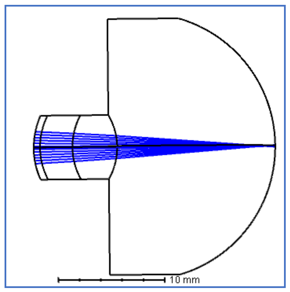
Figure 3.
Cross-sectional profile the of individual eye model.

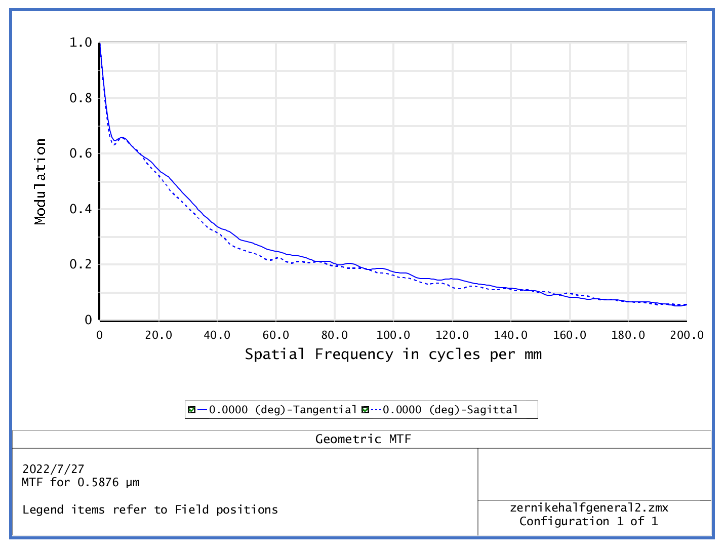
Figure 4.
MTF of the individual eye model.

Figure 5.
MTFs (a) with different scatter in Table 2 (b) with HOAs ranging from 0 to 0.6 µm.
Figure 5.
MTFs (a) with different scatter in Table 2 (b) with HOAs ranging from 0 to 0.6 µm.

Figure 6.
Impact of scatter on MTFA (area under modulation transfer function). (a) monochromatic light; (b) white light.
Figure 6.
Impact of scatter on MTFA (area under modulation transfer function). (a) monochromatic light; (b) white light.

Figure 7.
Combined impact of HOAs and scatter vs. the sum of the impacts of HOAs and scatter.


Table 1.
Aberration-free eye model.
| Surface Type | Radius /mm | Thickness /mm | Material | a2/ × 10−4 | a4/ × 10−4 | |
|---|---|---|---|---|---|---|
| Corneal front | Even asphere | 7.8 | 0.55 | Cornea | −8.01 | −1.49 |
| Corneal back | standard | 6.5 | 3.2 | Aqueous | - | - |
| Lens front | standard | 12 | 4.2 | Lens | - | - |
| Lens back | standard | −6 | 16.6 | Vitreous | - | - |
| Retina | standard | −12.5 | - | - | - | - |
Table 2.
Scatter fraction introduced into the model eye.
| Number | 0 | 1 | 2 | 3 | 4 | 5 | 6 | 7 | |
|---|---|---|---|---|---|---|---|---|---|
| Scatter/% | cornea | 0 | 0.02 | 0.04 | 0.06 | 0.08 | 0.10 | 0.12 | 0.14 |
| lens | 0 | 0.025 | 0.05 | 0.075 | 0.10 | 0.125 | 0.15 | 0.175 | |
Table 3.
Ocular aberrations/μm.
| Eye | Z7 | Z8 | Z9 | Z10 | Z11 | Z12 | Z13 | Z14 | Z15 | Z16 | Z17 |
|---|---|---|---|---|---|---|---|---|---|---|---|
| 1 | 0.052 | −0.0373 | 0.046 | 0.0022 | −0.0004 | −0.014 | −0.0047 | 0.0287 | 0.0074 | −0.0057 | 0.004 |
| 2 | 0.052 | −0.0347 | 0.0947 | 0.0097 | 0.0170 | 0.0912 | −0.0036 | −0.0363 | −0.0697 | 0.0473 | −0.002 |
| 3 | 0.028 | 0.1337 | 0.0721 | 0.0470 | −0.0164 | 0.0833 | 0.0252 | 0.0430 | −0.0095 | −0.0638 | 0.0236 |
| 4 | −0.036 | −0.1744 | −0.1148 | 0.0742 | 0.0455 | 0.1316 | 0.0812 | 0.0843 | 0.0962 | −0.0856 | 0.0906 |
| 5 | 0.194 | 0.1014 | −0.0785 | 0.0221 | −0.0198 | 0.1009 | 0.0402 | 0.0411 | −0.0444 | 0.0295 | −0.0625 |
| 6 | 0.343 | 0.0927 | −0.0785 | −0.0114 | 0.0128 | 0.1123 | −0.0739 | 0.0970 | −0.0191 | 0.0003 | −0.0199 |
Table 4.
Corneal parameters.
| Eye | Rc(mm) | Zernike Coefficient/μm | ||||||
|---|---|---|---|---|---|---|---|---|
| C7 | C8 | C9 | C10 | C11 | C12 | |||
| 1 | front | 7.90 | 0.0276 | −0.0196 | 0.0245 | 0.0011 | −0.0002 | −0.0074 |
| back | 6.55 | 0.0358 | −0.0238 | 0.0650 | 0.0067 | 0.0117 | 0.0626 | |
| 2 | front | 8.10 | 0.0343 | 0.1631 | 0.0880 | 0.0574 | −0.0201 | 0.1017 |
| back | 6.55 | −0.0235 | −0.1141 | −0.0751 | 0.0486 | 0.0298 | 0.0861 | |
| 3 | front | 8.39 | 0.1260 | 0.0658 | −0.0509 | 0.0144 | −0.0129 | 0.0655 |
| back | 6.59 | 0.4312 | 0.1165 | −0.0987 | −0.0143 | 0.0161 | 0.1411 | |
| 4 | front | 7.92 | 0.0276 | −0.0196 | 0.0245 | 0.0012 | −0.0002 | −0.0074 |
| back | 6.42 | 0.0519 | −0.0420 | −0.1507 | 0.0053 | 0.0540 | −0.0172 | |
| 5 | front | 7.81 | 0.3931 | 0.0948 | 0.0114 | 0.0604 | −0.0192 | 0.2680 |
| back | 6.67 | 0.1581 | 0.1927 | −0.1210 | −0.0738 | 0.0580 | 0.2142 | |
| 6 | front | 7.95 | −0.0804 | 0.2426 | 0.1508 | −0.0205 | 0.0144 | 0.2533 |
| back | 6.55 | −0.0557 | −0.0364 | 0.1420 | 0.0891 | −0.0262 | −0.0167 | |
Table 5.
Axial length of each component/mm.
| Number | Cornea | Anterior Chamber | Crystalline Lens | Vitreous Body |
|---|---|---|---|---|
| 1 | 0.56 | 3.36 | 3.82 | 17.33 |
| 2 | 0.57 | 3.07 | 3.96 | 18.16 |
| 3 | 0.53 | 3.24 | 3.46 | 18.66 |
| 4 | 0.56 | 3.21 | 3.43 | 18.63 |
| 5 | 0.61 | 3.20 | 4.19 | 18.41 |
| 6 | 0.58 | 3.32 | 3.94 | 17.72 |
Table 6.
Parameters of lens.
| Eye | Rc (mm) | Zernike Coefficients | ||||||||
|---|---|---|---|---|---|---|---|---|---|---|
| C7 | C8 | C9 | C10 | C11 | C12 | C13 | C14 | |||
| 1 | front | 10.14 | 0.2236 | 0.0628 | −0.0518 | 0.0215 | 0.0249 | 0.0078 | −0.0788 | 0.2236 |
| back | −6.52 | 0.1040 | −0.0238 | 0.0442 | −0.0254 | 0.0165 | 0.0164 | 0.1084 | 0.1040 | |
| 2 | front | 9.55 | 0.1635 | −0.0116 | −0.0176 | 0.0529 | 0.0496 | −0.0053 | −0.0115 | 0.1635 |
| back | −5.57 | 0.2442 | −0.0278 | −0.0066 | 0.0280 | 0.0270 | 0.0252 | −0.0874 | 0.2442 | |
| 3 | front | 9.37 | −0.0052 | −0.0291 | −0.0219 | 0.0650 | 0.0137 | −0.0242 | −0.0132 | −0.0052 |
| back | −5.85 | −0.0595 | −0.0862 | 0.0387 | −0.0325 | 0.0240 | 0.0237 | 0.0355 | −0.059 | |
| 4 | front | 10.2 | 0.0304 | −0.0086 | −0.0782 | 0.0989 | −0.0199 | 0.0318 | 0.0116 | 0.0304 |
| back | −6.0 | 0.1156 | −0.0611 | −0.0634 | 0.0141 | 0.0002 | −0.0150 | 0.0228 | 0.1156 | |
| 5 | front | 11.6 | 0.2236 | 0.0628 | −0.0518 | 0.0215 | 0.0249 | 0.0078 | −0.0788 | 0.2236 |
| back | −6.4 | 0.1040 | −0.0238 | 0.0442 | −0.0254 | 0.0165 | 0.0164 | 0.1084 | 0.1040 | |
| 6 | front | 10.4 | 0.1635 | −0.0116 | −0.0176 | 0.0529 | 0.0496 | −0.0053 | −0.0115 | 0.1635 |
| back | −5.88 | 0.2442 | −0.0278 | −0.0066 | 0.0280 | 0.0270 | 0.0252 | −0.0874 | 0.2442 | |
Disclaimer/Publisher’s Note: The statements, opinions and data contained in all publications are solely those of the individual author(s) and contributor(s) and not of MDPI and/or the editor(s). MDPI and/or the editor(s) disclaim responsibility for any injury to people or property resulting from any ideas, methods, instructions or products referred to in the content. |
© 2023 by the authors. Licensee MDPI, Basel, Switzerland. This article is an open access article distributed under the terms and conditions of the Creative Commons Attribution (CC BY) license (https://creativecommons.org/licenses/by/4.0/).
Share and Cite
MDPI and ACS Style
Rao, F.; Zhao, X.H.; Zhang, M.D.; Wang, Y. Effect of Higher Order Aberrations and Intraocular Scatter on Optical Quality Based on an Optical Eye Model. Optics 2023, 4, 364-374. https://doi.org/10.3390/opt4020027
AMA Style
Rao F, Zhao XH, Zhang MD, Wang Y. Effect of Higher Order Aberrations and Intraocular Scatter on Optical Quality Based on an Optical Eye Model. Optics. 2023; 4(2):364-374. https://doi.org/10.3390/opt4020027
Chicago/Turabian StyleRao, Feng, Xing Heng Zhao, Ming Dong Zhang, and Yan Wang. 2023. "Effect of Higher Order Aberrations and Intraocular Scatter on Optical Quality Based on an Optical Eye Model" Optics 4, no. 2: 364-374. https://doi.org/10.3390/opt4020027