
Disinfection of Transparent Screens by Side-Coupled UVA LED Radiation

1
Department of Medical Engineering and Mechatronics, Ulm University of Applied Sciences, Albert-Einstein-Allee 55, 89081 Ulm, Germany
2
Büchner Lichtsysteme GmbH, Uzstraße 2, 86465 Welden, Germany
*
Author to whom correspondence should be addressed.
Optics 2023, 4(2), 321-329; https://doi.org/10.3390/opt4020023
Submission received: 15 February 2023
/
Revised: 5 May 2023
/
Accepted: 10 May 2023
/
Published: 15 May 2023
(This article belongs to the Section Biomedical Optics)
Abstract
:(1) Background: Applications using touch screens are increasingly deployed in medical facilities, as well as in public areas. When touching the display with fingers, potentially pathogenic microorganisms such as methicillin-resistant Staphylococcus aureus (MRSA) can be transmitted. An automated process to decontaminate the device in between users would be highly useful. (2) Methods: Thin glass plates were superficially contaminated with the non-pathogenic Staphylococcus carnosus in a controlled manner. Subsequently, UVA radiation of 400 or 380 nm was laterally coupled into the glass plate, which acted as a light guide. Contact agar plates recorded the change in the staphylococci concentration over time. Additionally, the UVA radiation emitted by the glass plates was measured and the potential risk to humans assessed. (3) Results: Staphylococci concentration decreased as a result of UVA radiation for both wavelengths. At 400 nm, it took about 7.5 h and at 380 nm about 1 h until a reduction of 90% was reached. To meet higher disinfection requirements, disproportionately longer irradiation times were necessary. The potential UVA irradiation of humans in front of the glass pane was about 35 µW/cm2 or less and posed no risk to humans. (4) Conclusions: Side-coupled UVA radiation is in principle capable of safely automatically disinfecting microorganisms on touch screens. However, the required irradiation times are still in the hour range, so that a rapid disinfection within a minute or less is not yet possible with the presented setup. However, higher UVA intensities might reduce the current disinfection durations.
1. Introduction
Worldwide, the use of touch screens continues to increase [1,2]. Touch screen applications affect all areas of everyday and professional life. They are not only found on private notebooks, smartphones and tablets, but also, for example, on public vending machines for tickets for public transportation or other products. Medical devices employed in hospitals or doctors’ offices also increasingly feature touch screens [2].
When touching these screens, potentially pathogenic microorganisms and thus diseases can always be transmitted. All studies conducted to date, both inside and outside the healthcare sector, have found bacterial strains on touch screens, including the so-called ESKAPE pathogens (Enterococcus faecium, Staphylococcus aureus, Klebsiella pneumoniae, Acinetobacter baumannii, Pseudomonas aeruginosa, and Enterobacter spp.), which are of particular concern in medical environments [3]. Hitherto, none of the studies have reported even a single touch screen without microbial contamination. Staphylococci have always been detected [4].
To prevent the transmission of pathogens via displays, there would have to be fast automatic disinfection between two users. In theory, it is possible to reduce bacteria on surfaces within seconds by using strong UVC radiation sources (UVC: ultraviolet radiation between 100 and 280 nm) such as mercury vapor lamps, as has already been proven by Rudhart et al. (2022), Petersson et al. (2015), Moore et al. (2012) [5,6,7] and others. Unfortunately, UVC radiation could also harm humans in case of an accidental irradiation [8,9,10,11,12].
UVA radiation (UVA: ultraviolet radiation between 315 and 400 nm) poses a much smaller risk to humans compared to UVC [13], and all of the most often observed bacterial strains on touch screens can also be inactivated by UVA irradiation. These most frequently found bacterial strains and their necessary UVA log reduction doses can be found in Table 1. Unfortunately, the UVA log reduction doses are in the range of tens of J/cm2 and are therefore three to four orders of magnitude higher than typical UVC log reduction doses, which are in the range of several mJ/cm2 [14,15]. Therefore, the application of UVA radiation for disinfection requires significantly longer irradiation durations. Humans in front of or near the screen should be irradiated as little as possible and existing daily UVA exposure limits [13] should not be exceeded.
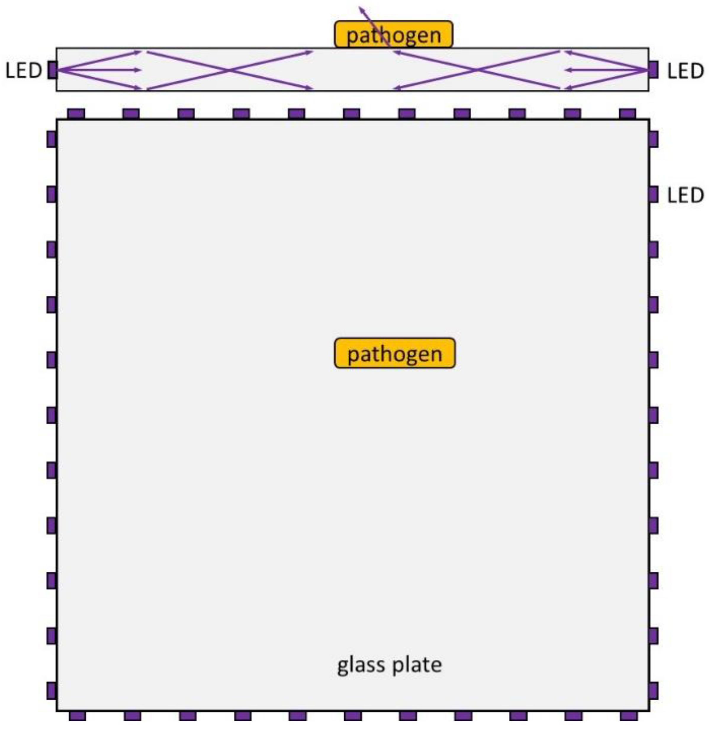
The aim of this study is to investigate the disinfection performance of UVA radiation without endangering/irradiating humans. The experiments were carried out with staphylococci on a transparent glass pane. UVA LED radiation of different wavelengths was coupled laterally into the glass plate, which acts as a light guide, as illustrated in Figure 1. The UVA radiation is assumed to remain within the glass plate due to total internal reflection. In principle, the radiation only escapes at the points where microorganisms or other contaminations are present. From a wave-optical point of view, this is not quite true. The so-called evanescent field protrudes slightly beyond the glass plate. Ideally, no human should be irradiated besides the short moment when the display is touched.
2. Materials and Methods
2.1. Irradiation Setup
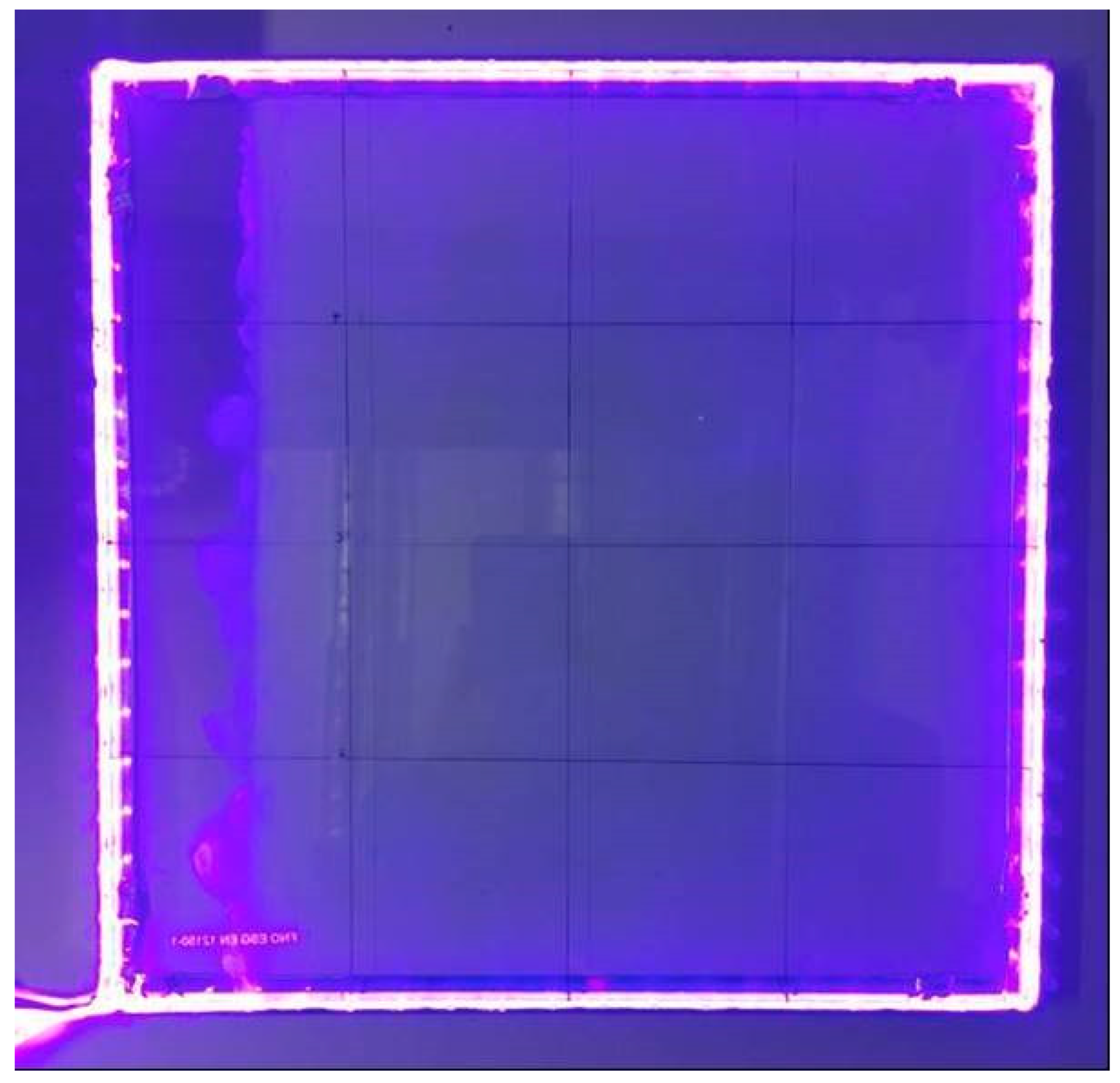
The irradiation setups consist of two glass plates; (1) white glass of BE Glass (Berlin, Germany) with the dimensions of 30 × 30 × 0.5 cm3, and (2) quartz glass of GVB (Herzogenrath, Germany) with the dimensions of 30 × 30 × 0.4 cm3. The spectrally resolved optical transmissions of both glasses is given in Figure 2. To the edges of the glass plates, two LED strips are pressed. The 71 LEDs of the first LED strip from Onfuro (Shenzen, China) exhibit a peak emission at about 400 nm with a UVA power of 9 mW at 3 mA per LED. The second LED strip of UnvarySam (unknown town, China) also contains 71 single LEDs with a peak emission at about 380 nm and an optical power of 9 mW at 10 mA each. The efficiency of coupling the LED radiation into the glass panes is unknown. The emission spectra of both LEDs can also be found in Figure 2, and a photograph of the combination of the white glass with the 71 × 400 nm LEDs in the dark is given in Figure 3. The temperature of the glass plates was measured with an infrared thermometer Ranger MX of Raytek (Berlin, Germany), and for measuring the UVA irradiation at a distance of 1–5 cm above the glass pane, an optical powermeter OPM150 from Artifex Engineering (Emden, Germany) was employed.
2.2. Microbial Procedures
As the available microbiology lab did not have the necessary biosafety level to work with pathogens such as Staphylococcus aureus, all experiments were performed with the non-pathogenic Staphylococcus carnosus (DSM 20501), which was provided by Deutsche Sammlung von Mikroorganismen und Zellkulturen (Brauschweig, Germany). S. carnosus was cultivated at 37 °C in M92 medium (trypticase soy yeast extract medium as described in [20]) up to an optical density of about 0.4. This corresponds to a concentration of about 6 × 10⁷ colony forming units (CFU)/mL and was diluted 1:3000 in phosphate-buffered saline (PBS) to a final concentration of 2 × 10⁴ CFU/mL.
For the purposeful contamination of the glass plates with staphylococci, the glass panes were first manually cleaned and disinfected with a 70% ethanol solution for about 10 min and thereafter exposed to the UVC radiation of a low-pressure mercury vapor lamp for at least half an hour. In order to obtain adequate bacterial concentrations and distribute them evenly over the glass plate surface, the following procedure was performed, which was previously described in [21]: An industrial paper towel (Glaeser, Ulm, Germany) was placed on the plate, and with a Pasteur pipette a volume of approximately 7 mL of bacterial test solution was manually and evenly distributed over the towel and the glass pane. After about 1.5 h, the towel was dry and was removed.
For the sampling, the glass plate was divided into 4 × 4 fields. The sampling was performed with caso contact agar plates of VWR (Darmstadt, Germany) at predefined time intervals of 0 h, 12 h, 24 h and 36 h for the 400 nm irradiation and for 0 h, 1 h, 2 h, 4 h and 6 h for the 380 nm irradiation. At 0 h, three samples were taken. For all other points of time, only two samples were taken: One at the outer row of sample fields, near the LEDs, and one at the four fields in the middle of the glass plate at the maximum distance from the LEDs. For each LED wavelength, five runs were performed and an additional 5 runs were performed without any LEDs as a reference. After sampling, the contact agar plates were cultivated at 37 °C in an incubator for around 20 h before colony counting for the unirradiated plates. The irradiated plates were cultivated for about 40 h. The reason for this procedure was that the unirradiated plates had up to a total of about 1000 colonies, which would have been difficult to count separately if the colonies had been too large. This was not a problem on the irradiated plates as the colony numbers were much lower. The intention of the longer cultivation time was to ensure that no staphylococci were missed, because they had suffered damage by the UVA irradiation, resulting in a prolonged growth but not in death. Additionally, plates were re-examined 1–2 days later for new colonies; however, none were observed.
As the experiment was not performed under the filtered air of a laminar flow box but on an open workbench in the lab, other microorganisms from the air could contaminate the glass plate and lead to colonies on the agar plates. However, all colonies that appeared different in color or morphology from familiar staphylococcus colonies were ignored and not counted. The risk of accidentally having counted contaminant colonies that looked very similar to S. carnosus colonies cannot be completely excluded and would have resulted in a seemingly slower bacterial reduction. However, we do not assume to have had a high concentration of bacteria in the air which form colonies that look like S. carnosus colonies, as we sometimes observed agar plates with zero colonies even after long irradiation.
3. Results
During irradiation, the temperature of the glass plates increased by about 0.5 °C to approximately 20 °C, which was assumed to have no large impact on bacterial survival. The UVA irradiation measured near the pane (distance 1–5 cm) varied between 20–35 µW/cm2 for different positions (corners, edges and center) above the glass plate that was visibly contaminated with bacteria. At a distance of 20 cm and 50 cm, the irradiation dropped to about 50% and 10%, respectively, of these maximum values.
The change in staphylococci concentration on the glass plates as a function of time can be found in Table 2 and Figure 4 for both wavelengths and without irradiation. Even without irradiation, the number of staphylococci decreases. The decrease appears to be approximately exponential. The plotted straight line with a slope of 0.0055/h in the semi-logarithmic plot corresponds to a log reduction duration of about 180 h.
When irradiated by the side-mounted LEDs, the staphylococci reduction is significantly faster. However, the course seems not to be exponential here, since the plotted linear trend line in this half-logarithmic representation describes the actual reductions poorly. The reduction is strong at the beginning and then slows down. If only the first measurement points for the shortest irradiation are considered, the log reduction time for irradiation of the 5 mm white glass plate with 400 nm is approximately 7.5 h, and for the staphylococci on the quartz glass plate approximately 1 h of 380 nm irradiation is required for a 90% decrease. For higher reduction demands, such as a 99% reduction, much higher irradiation durations are needed. For 400 nm, it would probably take around 20 h, and for the 380 nm LEDs, about 4–5 h.
4. Discussion
The staphylococcal reduction is much stronger with lateral coupling of the LED radiation than without radiation, and 380 nm irradiation reveals a three to four times stronger impact than 400 nm irradiation. This is perhaps an unexpectedly large difference, given that the total irradiated powers of both LEDs are the same and the two LED emission spectra even overlap. Presumably, the actual difference in antimicrobial efficacy between the two LED types might be smaller, but is amplified here by the different glass plates with their different thicknesses and absorption spectra.
In order to be able to compare the efficiency of this approach of lateral coupling of LED radiation with transillumination from above or below, the average energies required for a log reduction per area are considered. During the log reduction times for Staphylococcus carnosus of about 7.5 h (400 nm) and about 1 h (380 nm) determined here, each of the 71 LED strips emitted a total of 17,200 J (400 nm) and 2300 J (380 nm), respectively. Unfortunately, there are no published UVA disinfection results for S. carnosus or S. aureus. However, Santos et al. have published LD50 doses for irradiation of Staphylococcus saprophyticus in a NaCl solution [16], and these can be converted to a log reduction dose of 45.9 J/cm2. Irradiation was performed from above with a 365 nm UVA lamp. If Santos et al. had also applied this dose to an area of 30 × 30 cm2, the energy required for this would have been 41,310 J. Assuming that S. carnosus and S. saprophyticus have comparable log reduction doses, and the UVA-sensitivity of bacteria in saline solutions and on surfaces are similar, lateral coupling of LED radiation might be much more efficient than direct irradiation from above.
One can raise the question whether the assumption made above—S. carnosus and S. saprophyticus having comparable log reduction doses—is realistic, or if the non-pathogenic S. carnosus is possibly much more sensitive to UVA than, e.g., the pathogens S. saprophyticus or S. aureus. So far, there are no suitable studies on S. saprophyticus or S. aureus in the UVA range, but there are investigations for bacteria in solutions in adjacent spectral ranges at 222 nm (UVC), 254 nm (UVC), 405 nm (visible light) and 450 nm (visible light) [22,23]. At 222 nm, S. carnosus and S. aureus are equally photosensitive, at 254 nm, S. carnosus is more susceptible, and at 405 and 450 nm, S. aureus is more sensitive. So far, there has been no indication that pathogenic and nonpathogenic staphylococci exhibit large differences in the UVA photosensitivity.
In this study, only staphylococci were examined because staphylococci have been found most frequently on touch screens and because S. aureus, in the form of MRSA, is one of the best reported and most important pathogens. Table 1 shows that other relevant microorganisms require higher UVA log reduction doses in some cases, but these are not orders of magnitude higher, so it can be assumed that other relevant bacteria are also reduced with this kind of UVA irradiation.
The measured maximum UVA irradiation of below 35 µW/cm2 above the glass plate can be applied to calculate a theoretical UVA dose of 3 J/cm2 for a human that would stay for 24 h at a distance of 5 cm in front of it. However, even in this unrealistic scenario, the human irradiation is well below the allowed daily limit of 47 J/cm2 at 380 nm [24]. So this irradiation would be very safe and could continue even in the presence of humans.
We should mention that this irradiation was observed for a glass plate with bacterial contamination visible to the human eye. For a cleaned pane, it was much smaller and for a glass plate full of fingerprints, it was about 5× times higher (and revealed a slight fluorescence in a dark environment). Nevertheless, there is still no risk of exceeding daily exposure limits even under extreme circumstances. Additionally, there would be the option to turn off the UVA LEDs automatically if the display is touched or if someone is very near to the touch screen.
Unfortunately, the required disinfection durations of 7.5 and 1 h are still very long for a pathogen reduction of only 90%. They allow automatic disinfection over night or over the weekend, but not between different users who come every minute, even by applying more UVA LEDs and/or higher LED currents. Although UVA intensity might be increased by an order of magnitude, presumably resulting in an inverse decrease in disinfection time, this would still be insufficient for a disinfection within one minute.
It is also an open question as to whether such a glass panel, which exhibits little absorption in the UVA range and is at the same time a good light guide, can actually be integrated into touchscreens. Further development efforts and investigations are necessary here.
5. Conclusions
In UVA–transparent glass panels, the lateral coupling of UVA LED radiation is possible and can inactivate the staphylococci that are always detected on touch screens, which also include the well-known methicillin-resistant Staphylococcus aureus (MRSA). This is apparently done much more efficiently and safely than with direct UVA irradiation from below or above. However, disinfection in the minute range is not possible, at least not yet, and it remains to be seen whether the approach can be well integrated into touch screens.
Author Contributions
Conceptualization, B.S., A.-M.G. and M.H.; Methodology, B.S., A.-M.G., F.S., M.K. and M.H.; Validation, B.S., A.-M.G., F.S., M.K. and M.H.; Investigation, B.S., A.-M.G., F.S., M.K. and M.H.; Resources, M.K. and M.H.; Data Curation, B.S., A.-M.G., F.S., M.K. and M.H.; Writing—Original Draft Preparation, B.S., A.-M.G. and M.H.; Writing—Review and Editing, B.S., A.-M.G., F.S., M.K. and M.H.; Supervision, M.H.; Project Administration, M.H.; Funding Acquisition, M.K. and M.H. All authors have read and agreed to the published version of the manuscript.
Funding
This research was funded by the German Federal Ministry of Economics and Technology within the ZIM joint project “Clean Screen” (grant number KK5191602LU1).
Data Availability Statement
The data is given in the text.
Conflicts of Interest
M. Hessling has filed a German Patent application on surface disinfection in 2020 (DE102020116262A1). The other authors declare no conflict of interest.
References
- Data Bridge Market Research. Global Touch Screen Display Market, by Screen Types (Capacitive Touch Screens, Resistive Touch Screens, Surface Acoustic Wave Type Displays, Infrared Touch Screens, Others), Application (Personal Use, Professional Use)—Industry Trends and Forecast to 2029; Data Bridge Market Research: Maharashtra, India, 2022. [Google Scholar]
- Grand View Research. Commercial Display Market Size, Share & Trends Analysis Report By Product (Digital Signage, Display Monitor, Display TVs), by Technology, by Component, by Display Size, by Display Type, by Application, by Region, And Segment Forecasts, 2020–2025; San Francisco, CA, USA, 2019. [Google Scholar]
- Mulani, M.S.; Kamble, E.E.; Kumkar, S.N.; Tawre, M.S.; Pardesi, K.R. Emerging Strategies to Combat ESKAPE Pathogens in the Era of Antimicrobial Resistance: A Review. Front. Microbiol. 2019, 10, 539. [Google Scholar] [CrossRef] [PubMed]
- Hessling, M.; Haag, R.; Sicks, B. Review of microbial touchscreen contamination for the determination of reasonable ultraviolet disinfection doses. GMS Hyg. Infect. Control. 2021, 16, dgkh000401. [Google Scholar] [CrossRef]
- Petersson, L.P.; Albrecht, U.-V.; Sedlacek, L.; Gemein, S.; Gebel, J.; Vonberg, R.-P. Portable UV light as an alternative for decontamination. Am. J. Infect. Control. 2014, 42, 1334–1336. [Google Scholar] [CrossRef] [PubMed]
- Rudhart, S.A.; Günther, F.; Dapper, L.I.; Gehrt, F.; Stuck, B.A.; Hoch, S. Analysis of bacterial contamination and the effectiveness of UV light-based reprocessing of everyday medical devices. PLoS ONE 2022, 17, e0268863. [Google Scholar] [CrossRef] [PubMed]
- Moore, G.; Ali, S.; Cloutman-Green, E.A.; Bradley, C.R.; Wilkinson, M.A.C.; Hartley, J.C.; Fraise, A.P.; Wilson, A.P.R. Use of UV-C radiation to disinfect non-critical patient care items: A laboratory assessment of the Nanoclave Cabinet. BMC Infect. Dis. 2012, 12, 174. [Google Scholar] [CrossRef] [PubMed]
- Trevisan, A.; Piovesan, S.; Leonardi, A.; Bertocco, M.; Nicolosi, P.; Pelizzo, M.G.; Angelini, A. Unusual High Exposure to Ultraviolet-C Radiation. Photochem. Photobiol. 2006, 82, 1077–1079. [Google Scholar] [CrossRef] [PubMed]
- Zaffina, S.; Camisa, V.; Lembo, M.; Vinci, M.R.; Tucci, M.G.; Borra, M.; Napolitano, A.; Cannatà, V. Accidental Exposure to UV Radiation Produced by Germicidal Lamp: Case Report and Risk Assessment. Photochem. Photobiol. 2012, 88, 1001–1004. [Google Scholar] [CrossRef] [PubMed]
- Boudet, L.; Andujar, P.; Le Cam, M.; Pairon, J. Occupational sunburn caused by ultraviolet-C in a school restaurant. Contact Dermat. 2023. [Google Scholar] [CrossRef] [PubMed]
- International Commission on Illumination. UV-C Photocarcinogenesis Risks from Germicidal Lamps: Technical Report (CIE 187); CIE Central Bureau: Vienna, Austria, 2010. [Google Scholar]
- Rose, R.C. Erythema and conjunctivitis. Outbreak caused by inadvertent exposure to ultraviolet light. JAMA 1979, 242, 1155–1156. [Google Scholar] [CrossRef] [PubMed]
- International Commission on Non-Ionizing Radiation Protection. Guidelines on limits of exposure to ultraviolet radiation of wavelengths between 180 nm and 400 nm (incoherent optical radiation). Health Phys. 2004, 87, 171–186. [Google Scholar] [CrossRef] [PubMed]
- Masjoudi, M.; Mohseni, M.; Bolton, J.R. Sensitivity of Bacteria, Protozoa, Viruses, and Other Microorganisms to Ultraviolet Radiation. J. Res. Natl. Inst. Stand. Technol. 2021, 126, 1–77. [Google Scholar] [CrossRef]
- Kowalski, W. Ultraviolet Germicidal Irradiation Handbook; Springer: Berlin/Heidelberg, Germany, 2009. [Google Scholar]
- Santos, A.L.; Oliveira, V.; Baptista, I.; Henriques, I.; Gomes, N.C.M.; Almeida, A.; Correia, A.; Cunha, Â. Wavelength dependence of biological damage induced by UV radiation on bacteria. Arch. Microbiol. 2012, 195, 63–74. [Google Scholar] [CrossRef] [PubMed]
- Yagi, N.; Mori, M.; Hamamoto, A.; Nakano, M.; Akutagawa, M.; Tachibana, S.; Takahashi, A.; Ikehara, T.; Kinouchi, Y. Sterilization Using 365 nm UV-LED. In Proceedings of the 2007 29th Annual International Conference of the IEEE Engineering in Medicine and Biology Society, Lyon, France, 22–26 August 2007; pp. 5841–5844. [Google Scholar] [CrossRef]
- Lui, G.Y.; Roser, D.; Corkish, R.; Ashbolt, N.J.; Stuetz, R. Point-of-use water disinfection using ultraviolet and visible light-emitting diodes. Sci. Total. Environ. 2016, 553, 626–635. [Google Scholar] [CrossRef] [PubMed]
- Galezzo, M.-A.; Susa, M.R. Effect of single and combined exposures to UV-C and UV-A LEDs on the inactivation of Klebsiella pneumoniae and Escherichia coli in water disinfection. J. Water Sanit. Hyg. Dev. 2021, 11, 1071–1082. [Google Scholar] [CrossRef]
- Deutsche Sammlung von Mikroorganismen und Zellkulturen (DSMZ). M92: Trypticase Soy Yeast Extract Medium. 2012. Available online: https://www.dsmz.de/microorganisms/medium/pdf/DSMZ_Medium92.pdf (accessed on 30 April 2023).
- Hessling, M.; Spellerberg, B.; Hönes, K. Potential self-disinfection capacity of touch screen displays. J. Biophotonics 2019, 12, e201900118. [Google Scholar] [CrossRef] [PubMed]
- Gierke, A.-M.; Hessling, M. Investigation on Potential ESKAPE Surrogates for 222 and 254 nm Irradiation Experiments. Front. Microbiol. 2022, 13, 942708. [Google Scholar] [CrossRef] [PubMed]
- Hoenes, K.; Bauer, R.; Meurle, T.; Spellerberg, B.; Hessling, M. Inactivation Effect of Violet and Blue Light on ESKAPE Pathogens and Closely Related Non-pathogenic Bacterial Species—A Promising Tool Against Antibiotic-Sensitive and Antibiotic-Resistant Microorganisms. Front. Microbiol. 2021, 11, 612367. [Google Scholar] [CrossRef] [PubMed]
- Directive 2006/25/EC of the European Parliament and of the Council on the Minimum Health and Safety Requirements Regarding the Exposure of Workers to Risks Arising from Physical Agents (Artificial Optical Radiation); Official Journal of the European Union: Brussels, Belgium, 2006; pp. 38–59.
Figure 1.
Schematic representation of a microbially contaminated glass plate with UVA LEDs attached to the side. Due to total internal reflection, the glass plate acts as a light guide. At spots where contaminants are present, the radiation can escape and thus microorganisms can be irradiated. Top: cross-section of the glass plate and LEDs, bottom: top-view of the glass plate and LEDs.

Figure 2.
Transmission spectra of glasses and emission spectra of single LEDs. Both LED types emit 9 mW each.
Figure 2.
Transmission spectra of glasses and emission spectra of single LEDs. Both LED types emit 9 mW each.

Figure 3.
Photograph (in the dark) of irradiation setup with 400 nm LED strips around the glass plate. The visible dark lines on the glass plate define fields for sampling.
Figure 3.
Photograph (in the dark) of irradiation setup with 400 nm LED strips around the glass plate. The visible dark lines on the glass plate define fields for sampling.

Figure 4.
Change in the S. carnosus concentration on glass plates over time for the unirradiated plate and the plates irradiated by the laterally attached 400 and 380 nm LEDs. The given error bars illustrate the standard deviation of the single measurements. The dotted lines are linear fits in this half-logarithmic representation that would be exponential curves for a non-logarithmic Y axis.


Table 1.
Bacterial strains most often found on touchscreens—sorted by observation frequency—and their necessary log reduction doses. (Modified after [4]).
Table 1.
Bacterial strains most often found on touchscreens—sorted by observation frequency—and their necessary log reduction doses. (Modified after [4]).
| Bacteria Most Often Found on Touch Screens | UVA Log Reduction Doses [J/cm2] | |
|---|---|---|
| Staphylococcus spp. | 45.9 | [16] |
| Bacillus spp. | 50.7 | [16] |
| Micrococcus spp. | 98.8 | [16] |
| Pseudomonas spp. | 73.5 | [16] |
| Escherichia coli | 38.9 | [17] |
| Enterococcus spp. | 42 | [18] |
| Klebsiella spp. | 60 | [19] |
| Streptococcus spp. | unknown | |
| Corynebacterium spp. | unknown | |
| Acinetobacter spp. | 52.9 | [16] |
Table 2.
Change in the S. carnosus concentration on glass plates over time for the unirradiated plate and the plates irradiated by the laterally attached 400 and 380 nm LEDs. (The starting concentrations at 0 h were always set as reference without errors.).
Table 2.
Change in the S. carnosus concentration on glass plates over time for the unirradiated plate and the plates irradiated by the laterally attached 400 and 380 nm LEDs. (The starting concentrations at 0 h were always set as reference without errors.).
| Irradiation | Time [h] | Average Log Change | Standard Deviation of Single Measurements |
|---|---|---|---|
| no irradiation | 0 | 0 | |
| 12 | −0.117 | 0.110 | |
| 24 | −0.119 | 0.158 | |
| 36 | −0.190 | 0.137 | |
| 400 nm | 0 | 0 | |
| 12 | −1.611 | 0.170 | |
| 24 | −2.210 | 0.096 | |
| 36 | −2.578 | 0.316 | |
| 380 nm | 0 | 0 | |
| 1 | −1.128 | 0.545 | |
| 2 | −1.383 | 0.361 | |
| 4 | −1.867 | 0.363 | |
| 6 | −2.277 | 0.366 |
Disclaimer/Publisher’s Note: The statements, opinions and data contained in all publications are solely those of the individual author(s) and contributor(s) and not of MDPI and/or the editor(s). MDPI and/or the editor(s) disclaim responsibility for any injury to people or property resulting from any ideas, methods, instructions or products referred to in the content. |
© 2023 by the authors. Licensee MDPI, Basel, Switzerland. This article is an open access article distributed under the terms and conditions of the Creative Commons Attribution (CC BY) license (https://creativecommons.org/licenses/by/4.0/).
Share and Cite
MDPI and ACS Style
Sicks, B.; Gierke, A.-M.; Sommerfeld, F.; Klein, M.; Hessling, M. Disinfection of Transparent Screens by Side-Coupled UVA LED Radiation. Optics 2023, 4, 321-329. https://doi.org/10.3390/opt4020023
AMA Style
Sicks B, Gierke A-M, Sommerfeld F, Klein M, Hessling M. Disinfection of Transparent Screens by Side-Coupled UVA LED Radiation. Optics. 2023; 4(2):321-329. https://doi.org/10.3390/opt4020023
Chicago/Turabian StyleSicks, Ben, Anna-Maria Gierke, Florian Sommerfeld, Martin Klein, and Martin Hessling. 2023. "Disinfection of Transparent Screens by Side-Coupled UVA LED Radiation" Optics 4, no. 2: 321-329. https://doi.org/10.3390/opt4020023