
Design and Optical Analysis of a Refractive Aspheric Intraocular Lens with Extended Depth of Focus
by
 ,
,
Kunqi Li
1,†, 
Xiaoqin Chen
2,3,4,†,
Yayan Bian
1,
Yuwei Xing
1,
Xiaolan Li
1,
Dongyu Liu
1 and
Yongji Liu
1,4,* 1
Tianjin Key Laboratory of Micro-Scale Optical Information Science and Technology, Institute of Modern Optics, Nankai University, 38 Tongyan Road, Tianjin 300350, China
2
Tianjin Eye Hospital and Nankai University Eye Institute, Tianjin 300020, China
3
Tianjin Eye Hospital Optometric Center, Tianjin 300020, China
4
Nankai University Eye Institute, Nankai University Affiliated Eye Hospital, Nankai University, 38 Tongyan Road, Tianjin 300350, China
*
Author to whom correspondence should be addressed.
†
These authors contributed equally to this work.
Optics 2023, 4(1), 146-155; https://doi.org/10.3390/opt4010011
Submission received: 15 November 2022
/
Revised: 20 January 2023
/
Accepted: 28 January 2023
/
Published: 31 January 2023
(This article belongs to the Special Issue Advances in Vision Optics, Myopia Control and Refractive Surgery)
Abstract
:To obtain a continuous range of clear vision for pseudophakic eyes, a design of intraocular lens (IOL) with extended depth of focus (EDoF) was proposed. The IOL was optimized with a multi-configuration approach based on a pseudophakic eye model and the optical performances of the designed IOL were analyzed. The modulation transfer function (MTF) values remain above 0.2 at 50 lp/mm for object distance ranging from 0.35 m to infinity in both photopic vision and mesopic vision over a field of 4°. The optical performances remain stable when the pupil diameter changes from 2.25 mm to 5 mm. Besides, the presented theoretical analyses show the designed IOL has good optical performances for polychromatic light and corneal asphericity. The above shows that the IOL exhibits an excellent ability for pseudophakic eyes to see the object in a continuous range of distance.
1. Introduction
Cataract, the most common cause of blindness and visual impairment, is caused by crystalline lens opacity. Such crystalline lens is usually displaced by an implanted intraocular lens (IOL) to regain vision [1,2]. However, the pseudophakic eye would lose its ability of accommodation when the monofocal IOL replaces the crystalline lens, therefore failing to obtain clear vision over a range of object distances. To solve this problem, the design of multifocal IOL is proposed to obtain a clear vision for more object distances [3,4]. Bifocal IOL is the initial type of multifocal IOL, which is capable of providing functional distance vision and near vision, but leaving intermediate vision uncorrected [5]. Trifocal IOL thus is developed to provide clear vision for far, near, and intermediate distances [6], whereas it fails in achieving vision correction for a continuous range of object distance. Besides, the retinal images of different object distances overlapped on the retina, resulting in a reduced contrast of the retinal image due to the presence of the out-of-focus images [7,8]. One way to provide continuous clear vision over a large object distance is the accommodative IOLs. Special loops or materials are usually implemented to make it possible for the IOLs to move axially to provide clear vision over a continuous object distance. However, the accommodation range provided by accommodation IOLs is very limited [9,10,11,12]. The extended depth of focus (EDoF) IOL, though there is no international definition for this term, is the most commonly implemented way as an attempt to provide clear images over a continuous vision range from far to near. though its extended depth of focus performance also comes at the expense of image quality [13,14].
Various attempts have been made to design EDoF IOLs [15]. The light sword optical element, which is an axially asymmetric optical element, has been used as an IOL to extend the depth of focus [16,17], but with limited application in clinical practice. Fernández [18] proposed a multifocal IOL with through-focus performance for object distances ranging from 0.4 m to 5 m. The image quality of this IOL is barely affected by the pupil diameter variation. Jiang [19] proposed an aspheric diffractive IOL, which had a continuous depth of focus corresponding to the object distance ranging from 0.4 m to 8 m. One limitation of this design lies in the relatively small optical zone of 4.5 mm. One representative commercially available EDoF IOL is the TECNIS Symfony® IOL using diffractive technology by Johnson & Johnson Vision Care Inc. (Jacksonville, FL, USA). It is claimed to provide a 1.5 D through-focus range of vision [15,20,21], which could not meet most the of visual tasks from near (reading) to far (driving). One worth mentioning is that very little has been reported on how to design an EDoF and how to completely evaluate the performance of the EDoF IOLs during the design process.
In this paper, an IOL with an EDoF has been proposed along with a variety of evaluations of the performance of the IOL. The designed EDoF IOL demonstrates a large depth of defocus of 2.5 D with a reasonable optical performance.
2. Methods
2.1. The Pseudophakic Eye Model
The material of the IOL is polymethyl methacrylate (PMMA) with a refractive index of 1.494 and an Abbe number of 57.5. The initial central thickness of the lens was set to 0.68 mm. The design wavelength was 555 nm because the human eye is most sensitive to this wavelength. The IOL was optimized under a pupil diameter of 4.5 mm at a 4° field of view (FOV).
A pseudophakic eye model was constructed for the optimization of the IOL. The axial length of the Gullstrand-Le Grand eye model was optimized under a 3 mm pupil diameter to obtain a clear retinal image for an object at infinity, resulting in an axial length of 23.471 mm. The crystalline lens was then replaced by the IOL, which was 3.5 mm away from the posterior surface of the cornea. The structural parameters of the pseudophakic eye model are shown in Table 1. At this stage, the parameters of the IOL have not been determined. The Abbe number of the cornea, aqueous and vitreous are 55.8, 52.8, and 52.8, respectively [22].
2.2. The Optimization of the IOL
The pseudophakic eye model was constructed in the optical design software Zemax (ZEMAX LLC., 2009, Bellevue, WA, USA) to perform the optimization of the IOL as well as the analysis of the optical performance of the designed IOL. The anterior and posterior surfaces of the IOL were first optimized using the default merit function provided by Zemax to obtain the initial structure of the IOL. Both the anterior and posterior IOL surface types were represented by Equation (1)
where is the sag of the surface, is the radial distance from the optical axis, represents the vertex curvature of the surface, is the conic constant, and are the aspheric high-order polynomial coefficients from order 2 to order 10. was set as zero.
To obtain a large depth of focus under a pupil diameter of 2.5 mm, the refractive distribution of the IOL along the radial distance needs to be specially designed: the refractive power of the inner part of the IOL aperture (from center to 1.25 mm) covers a range from 0 D (corresponding to infinity) to 2.5 D (corresponding to 0.4 m). This was carried out by optimization using a customized Zemax programming language (ZPL) macro. The surface parameters including radii, the central thickness, were set as variables and optimized with the customized ZPL macro as the merit function. After this optimization, different radial positions with 1.25 mm of the IOL were assigned different refractive power. However, the optical quality given by the IOL fails to meet the requirements for real applications which means it needs further optimization.
A multi-configuration was established in Zemax for different object locations i.e., 5, 3, 1, 0.8, 0.6, 0.4 m with pupil diameters of 3 and 4.5 mm. Different weights were assigned to each configuration which were shown in Table 2. These variables set in the last step were optimized again to reach the least root-mean-square wavefront error by the default merit function. The optimization does not stop until all the modulation transfer function (MTF) value within a spatial frequency of 100 lp/mm is above zero. Then, the MTFA operand, which can set the MTF value at a specific spatial frequency, is added to the default merit function, to further improve the image quality of the IOL. Run the optimization until the MTF values are above 0.25 at 50 lp/mm for all object distances.
3. Results and Discussion
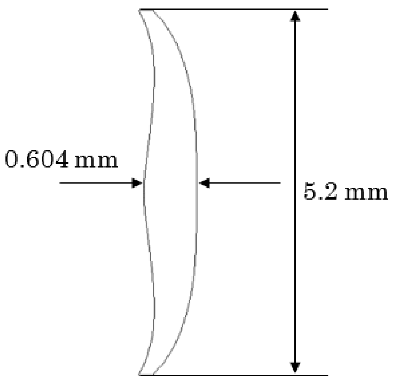
The designed IOL has an effective optical zone of 5.2 mm, a central thickness of 0.604 mm, and an edge thickness of 0.177 mm. The anterior and posterior surface parameters are shown in Table 3. The cross-section profile of the effective optical zone of the IOL is shown in Figure 1. This is obvious that both surfaces of the IOL are aspherical.
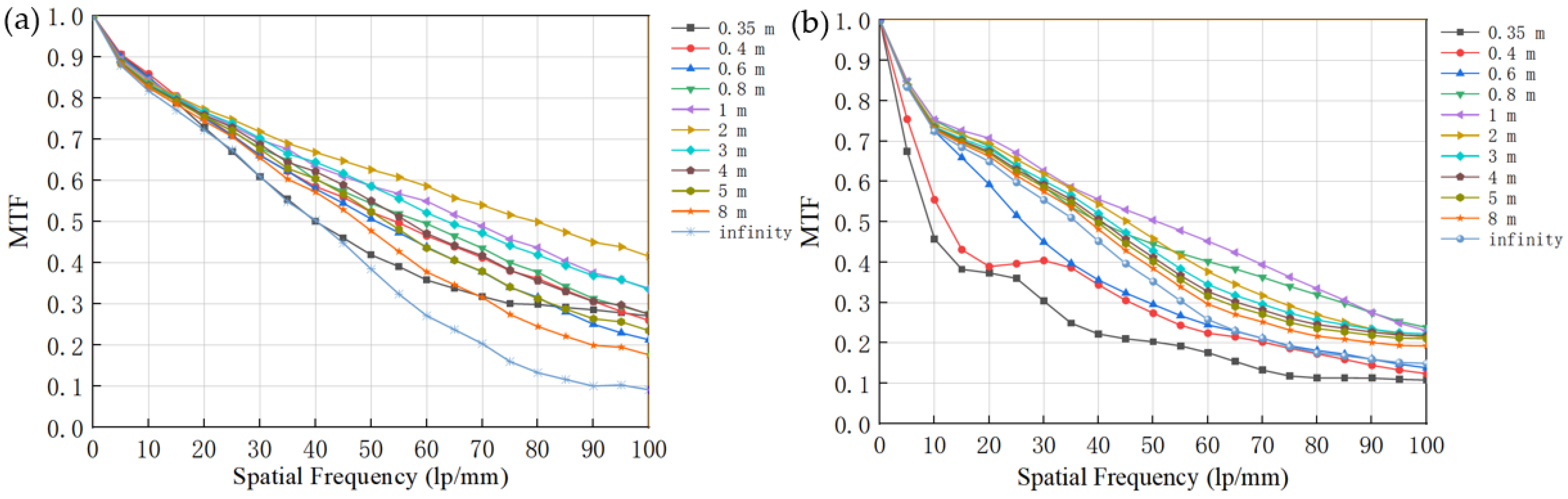
To evaluate the extended depth of focus feature, the MTFs of the pseudophakic eye model with the designed IOL in 0° field for 10 different object locations, which include six design object locations along with four non-design object locations selected randomly, ranging from 0.35 m to infinity, was illustrated in Figure 2. One important feature of a successful IOL design is that the optical quality should keep almost the same for different pupil diameters for human eyes usually work in different illuminations [23]. Here, the optical performance under pupil diameters of 3 and 4.5 mm, corresponds to photopic and mesopic vision respectively. During the day with good illumination, the pupil size of the elderly is usually 3 mm in diameter. Therefore, a pupil diameter of 3 mm is often used when analyzing the optical performance of the IOLs [24]. A pupil diameter of 4.5 mm for mesopic vision was chosen because the elders usually have the IOL implanted. In addition, the optical performance of IOL on an optical bench is usually performed under an aperture size no larger than 4.5 mm [25]. The MTF is presented in a unit of lp/mm. (1 cycle/degree = 0.297 lp/mm assuming a nodal point distance of 17 mm in image space). The MTF values are the average of tangential and sagittal directions.
Figure 2a shows the MTFs of the pseudophakic eye model under a pupil diameter of 3 mm (photopic vision). For all the designed object distances, which were 5, 4, 3, 2, 1, 0.8, 0.4, and 0.35 m, the MTFs at 100 lp/mm are above 0.25 while all the MTFs for all object locations are above 0.1 at 100 lp/mm, indicating good image quality in the photopic vision for all object locations. Meanwhile, the MTF values are higher than 0.5 at 50 lp/mm and remain above 0.21 at 100 lp/mm for object distances ranging from 0.6 m to 5 m, showing excellent optical performance for a large object range. It needs to be pointed out that the optical quality of the pseudophakic eye model for an object located at an intermediate distance from 0.6 m to 2 m is superior to these for objects located at far distances and a near distance of 0.35 m. Figure 2b shows MTFs of the pseudophakic eye model under a pupil diameter of 4.5 mm (mesopic vision). Generally speaking, the MTFs remain quite high for mesopic vision. The MTF values are higher than 0.2 at 50 lp/mm and remain above 0.1 at 100 lp/mm for all object locations. The MTF curves for the near objects located in 0.35 m, 0.4 m, and 0.6 m showed a relatively large drop in comparison with MTFs for other object distances. ISO11979-2-2014 standards state that the MTF at 100 lp/mm should be above 0.28 [26]. In the present design, not all MTF values meet this standard. It is mainly due to the large depth of focus. It is very hard to have a large depth of focus while maintaining high MTF values for all object distances.
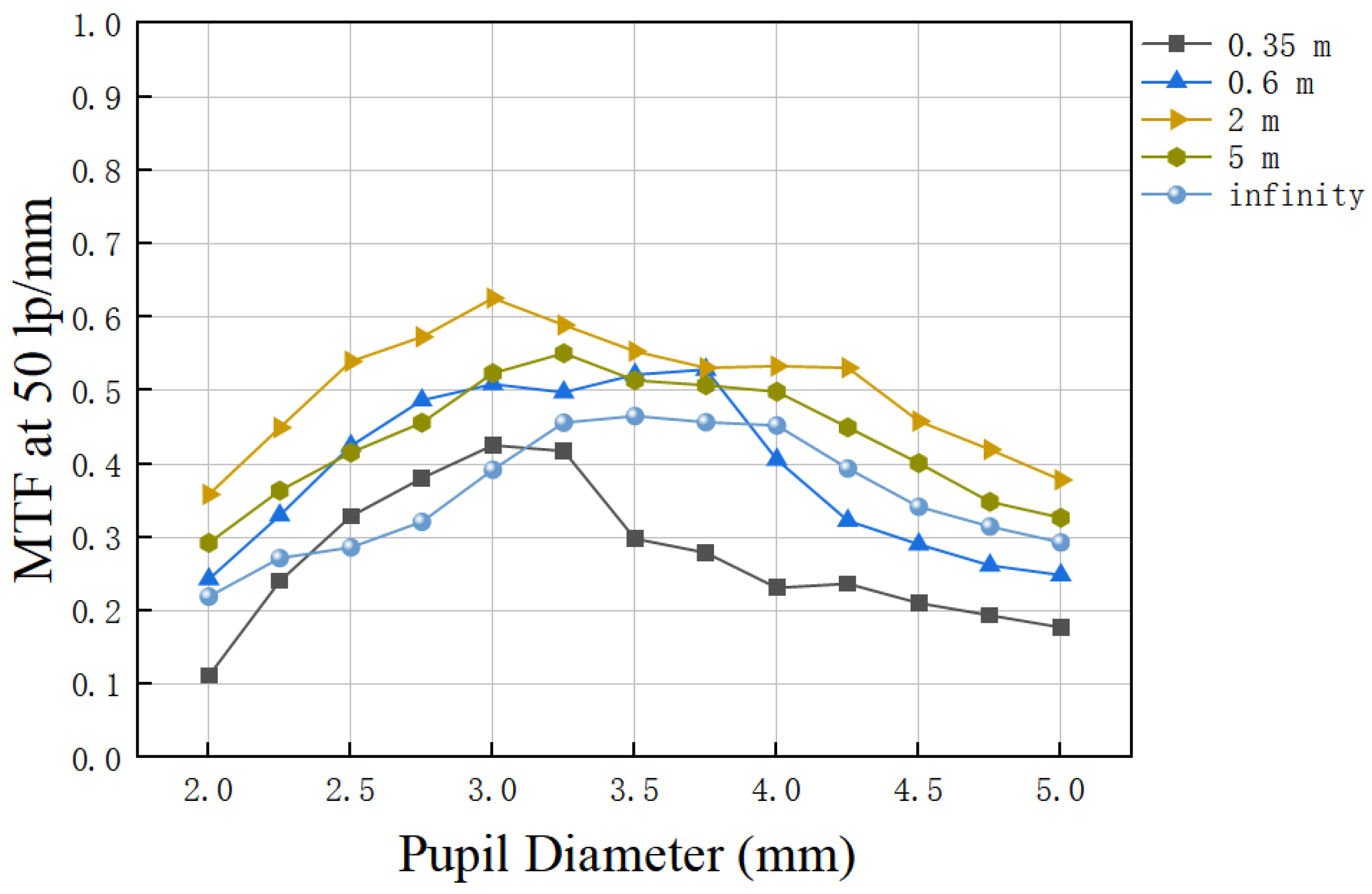
In addition, the MTFs at 50 lp/mm as a function of the pupil diameter for five randomly selected object distances were shown in Figure 3. All the MTFs under a pupil diameter around 3 mm show a value above 0.39. For pupil diameters less than 3 mm, the MTF declines rapidly because the diffraction plays a more important role in the image quality. When the pupil diameter increases from 3 mm, the MTF values drop slowly as more aberrations occurred. However, the MTF remains above 0.25 except for a very near object distance of 0.35 m. The analyses above indicate that the image quality of the eye model implanted with the designed IOL remains stable when the pupil diameter changes from 2.25 mm to 5 mm.
There are a large number of cone cells near the fovea of the retina, which allow us to distinguish details [27]. However, the cone density declines sharply with the increasing eccentricity of the fovea and drops by an order of magnitude at a distance of 1 mm from the foveal center (corresponding to 4° FOV) [28]. Therefore, the MTF curves of the pseudophakic eye model in a 4° FOV have been analyzed additionally at 10 object locations. The results are shown in Figure 4. The MTF values under photopic vision shown in Figure 4a are greater than 0.27 at 50 lp/mm and remain above 0.09 at 100 lp/mm for object distances ranging from 0.35 m to infinity. Figure 4b shows the MTFs of the eye model under a 4.5 mm pupil (mesopic vision). The MTF values are higher than 0.21 at 50 lp/mm and remain above 0.11 at 100 lp/mm for object distances ranging from 0.35 m to infinity. Only a slight decline in MTF at a 4° FOV is observed in comparison with that at 0° FOV. The results show that the designed IOL implanted in the eye model has quite good optical performance in both photopic vision and mesopic vision for 4° FOV.
Human eyes normally work in polychromatic light instead of monochromatic light. The optical performance of the pseudophakic eye model under polychromatic light was analyzed. The polychromatic light with wavelengths of 470 nm, 510 nm, 555 nm, 610 nm, and 650 nm, which represent the photopic visual spectrum, was selected to analyze the optical performance of the designed IOL. The weighting coefficients of the five wavelengths were set to 0.091, 0.503, 1, 0.503, and 0.107, respectively, based on the luminosity function curve of human eyes under the photopic condition. The MTF curves of the pseudophakic eye model at 0° FOV for 10 object locations under 3 mm and 4.5 mm pupil in polychromatic light are shown in Figure 5. It can be seen that the MTF curves show only a slight decline compared with these under monochromatic light in Figure 2. The MTF values are still greater than 0.1 at 100 lp/mm in both 3 mm and 4.5 mm pupil diameters for object distance ranging from 0.35 m to infinity in polychromatic light. The results show that the image quality of the designed IOL is not noticeably affected by chromatic aberration, which may be a concern for actual applications.
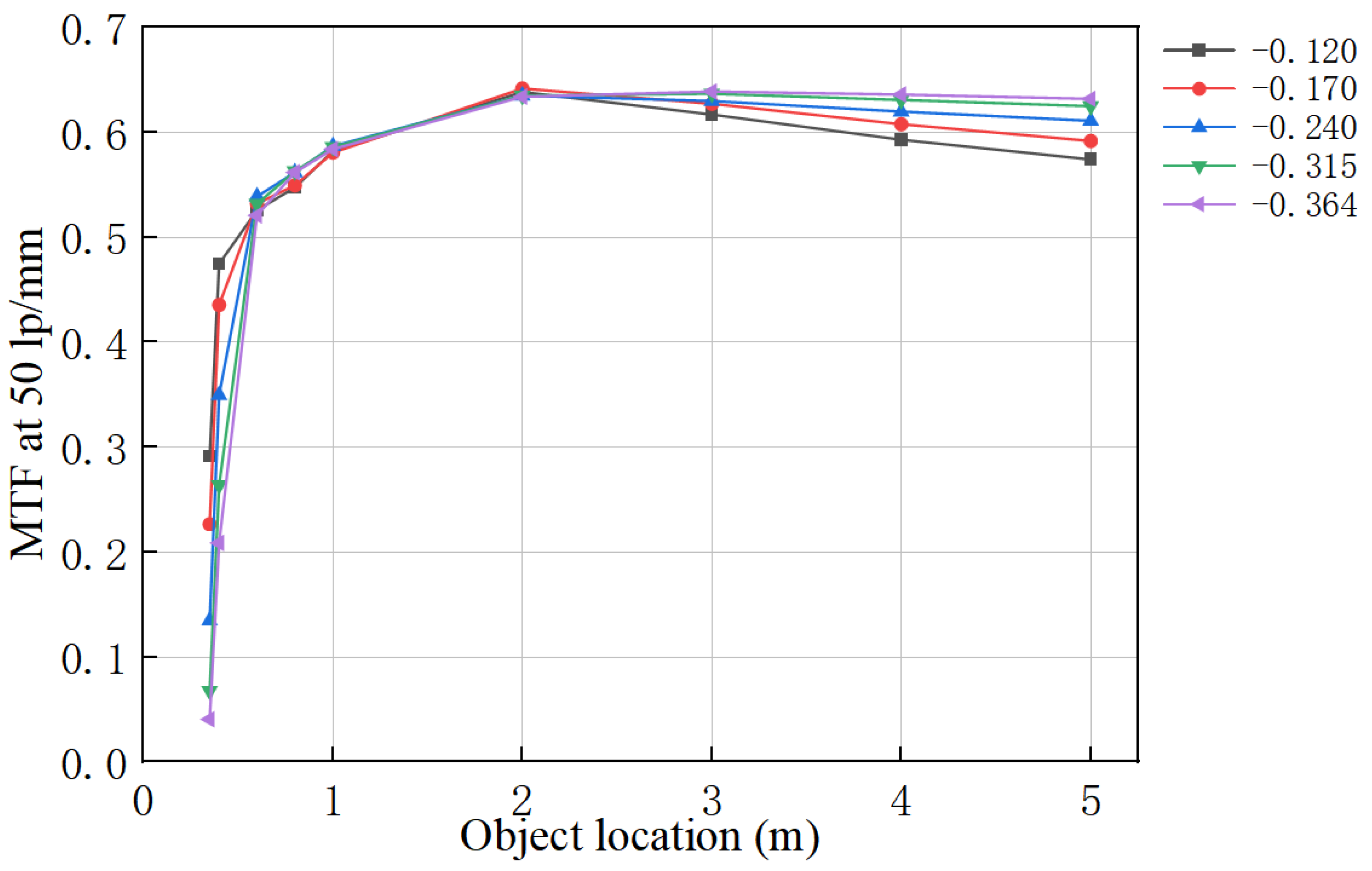
Corneal spherical aberration varies from individual to individual [29]. An ideal IOL should work well under different corneal spherical aberrations. Therefore, MTF at 50 lp/mm of the pseudophakic eye model with five different corneal spherical aberrations was analyzed at 0° FOV under 3 mm pupil diameter [18]. Here, we simulate different corneal spherical aberrations by setting different corneal aspheric coefficients, −0.120, −0.170, −0.240, −0.315, and −0.364 corneal aspherical coefficients correspond to corneal spherical aberrations of 0.42, 0.36, 0.27, 0.18, and 0.12 μm under a pupil diameter of 6 mm respectively, which can cover 90% of the population [30,31]. The results are shown in Figure 6. The MTFs at 50 lp/mm are affected slightly by the corneal spherical aberration only for near object distances of 0.35 m and 0.4 m. For large object distances, the corneal spherical aberration plays little role in the MTFs at 50 lp/mm.
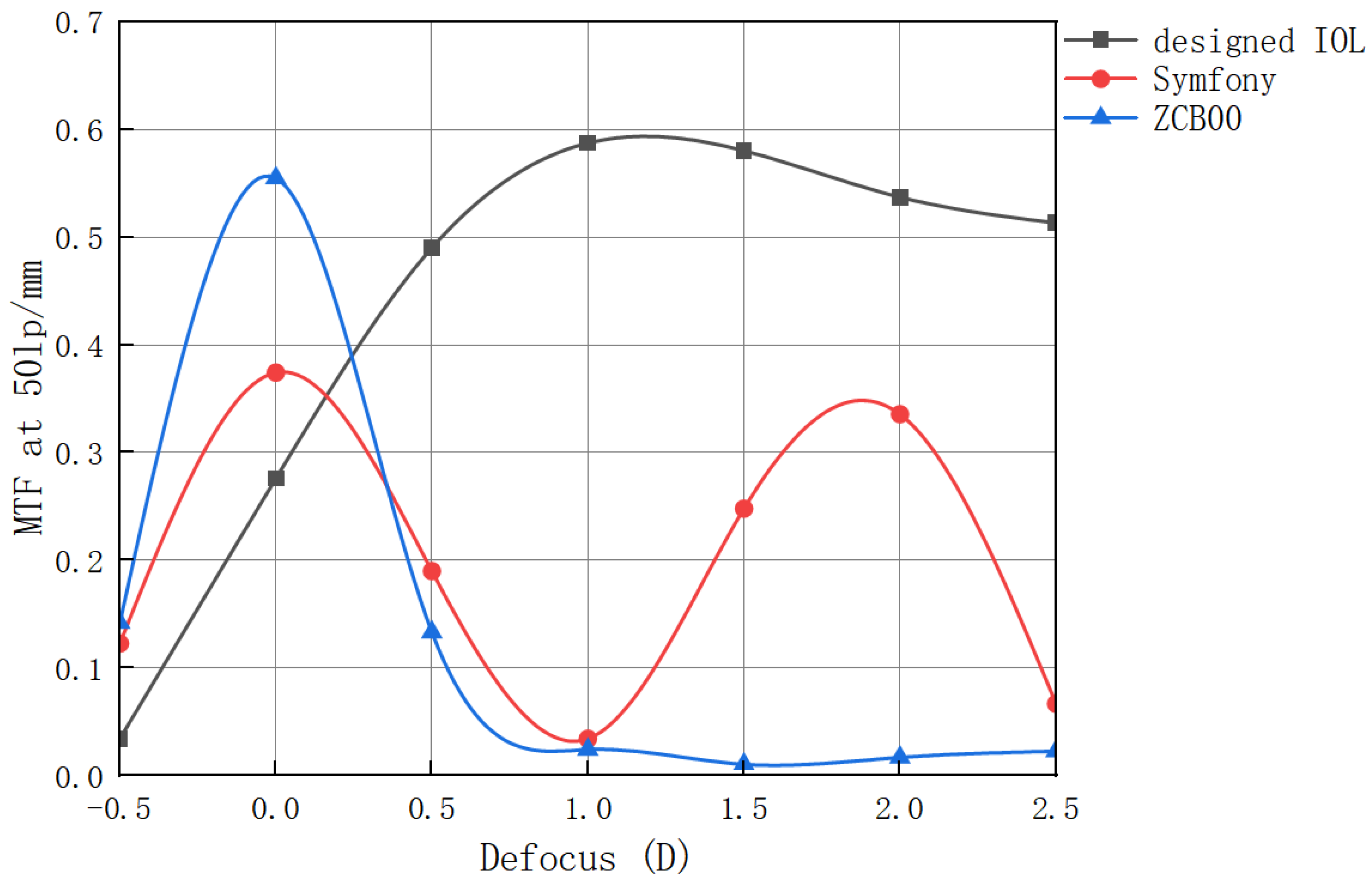
The designed IOL was compared to two commercial products. One is Tecnis ZCB00 (Abbott Laboratories, Chicago, IL, USA), which is a commercial monofocal IOL with an aspheric design, and the other is a commercial EDoF IOL TECNIS Symfony (Abbott Laboratories, Chicago, IL, USA). Since the through-focus data of commercial IOLs were evaluated under white light [32,33], the performance of the designed IOL was also simulated under white light according to the Abbe number provided in eye model. Figure 7 shows the through-focus MTF at 50 lp/mm in 0° FOV in white light for three different IOLs under 3 mm pupil diameter. It can be seen that the MTF of the designed IOL is lower than that of the two commercial products for defocus between about −0.5 D and +0.25 D, which corresponds to far vision, whereas the MTF of the designed IOL is much higher than that of the two commercial products at middle and near object distances. In addition, the designed IOL also demonstrates a larger depth of focus than the commercial ones.
When an IOL is implanted in the eye, it usually cannot be perfectly positioned because decentration and tilt are unavoidable in real practice [34]. We tested the optical performance of the IOL when there is decentration or tilt in the pseudophakic eye model. We found that the performance of IOL is sensitive to the tilt or the decentration of the IOL. This is probably due to the asphericity of the surfaces of the IOLs [35]. This is the limitation of this design. Considering the high optical performance of the IOL as shown in Figure 7, one solution to this limitation is to further design the IOL to improve its tolerance to decentration and tilt at the expense of some extent optical performance. This will be our next work. Another possible limitation is the effective optical zone diameter of 5.2 mm, which is less than 6 mm. This limitation can be solved by adding an optical zone with surface parameters of monofocal IOL.
4. Conclusions
In conclusion, the pseudophakic eye model implanted with the designed IOL achieves high-quality imaging for object distances ranging from 0.35 m to infinity over a field of 4°, providing a depth of focus of 2.5 D. The variation of pupil diameter, corneal asphericity, and working under polychromatic light, have limited effects on the performances of the designed IOL. These characteristics make the designed IOL has the potential for real application once it is more tolerant to decentration and tilt.
Author Contributions
Conceptualization, Y.B. and Y.L.; methodology, Y.B., X.C. and K.L.; software, Y.B., K.L. and Y.X.; data curation, Y.B., K.L. and Y.L.; validation, Y.B., X.C., K.L., X.L. and Y.L.; formal analysis, Y.B., X.C., K.L., Y.X., X.L., D.L. and Y.L.; investigation, Y.B. and X.C.; resources, Y.L.; writing—original draft preparation, Y.B.; writing—review and editing, Y.L. and X.C.; visualization, Y.B. and Y.L.; supervision, Y.L.; project administration, Y.L. and X.C.; funding acquisition, Y.L. and X.C. All authors have read and agreed to the published version of the manuscript.
Funding
This research was funded by the Tianjin Eye Hospital Optometric Center Science and Technology Fund: SGZX-2022-KY-00022.
Institutional Review Board Statement
Not applicable.
Informed Consent Statement
Not applicable.
Data Availability Statement
Data underlying the results presented in this paper are not publicly available at this time but may be obtained from the corresponding author upon reasonable request.
Conflicts of Interest
The authors declare no conflict of interest.
References
- Flaxman, S.R.; Bourne, R.R.A.; Resnikoff, S.; Ackland, P.; Braithwaite, T.; Cicinelli, M.V.; Das, A.; Jonas, J.B.; Keeffe, J.; Kempen, J.H.; et al. Vision Loss Expert Group of the Global Burden of Disease Study. Global causes of blindness and distance vision impairment 1990–2020: A systematic review and meta-analysis. Lancet Glob. Health 2017, 5, 1221–1234. [Google Scholar] [CrossRef] [PubMed] [Green Version]
- Yao, K.; Wu, R.; Xu, W.; Chen, P.; Yin, J. Combined phacoemulsification, foldable intraocular lens implantation and tra-beculectomy for cataract patients with glaucoma. [Zhonghua Yan Ke Za Zhi] Chin. J. Ophthalmol. 2000, 36, 330–333. [Google Scholar]
- Davison, J.A.; Simpson, M.J. History and development of the apodized diffractive intraocular lens. J. Cataract. Refract. Surg. 2006, 32, 849–858. [Google Scholar] [CrossRef] [PubMed]
- Packer, M.; Fine, I.H.; Hoffman, R.S. Refractive lens exchange with the array multifocal intraocular lens. J. Cataract. Refract. Surg. 2002, 28, 421–424. [Google Scholar] [CrossRef] [PubMed]
- Martínez de Carneros-Llorente, A.; Martínez de Carneros, A.; Martínez de Carneros-Llorente, P.; Jiménez-Alfaro, I. Comparison of visual quality and subjective outcomes among 3 trifocal intraocular lenses and 1 bifocal intraocular lens. J. Cataract. Refract. Surg. 2019, 45, 587–594. [Google Scholar] [CrossRef] [PubMed]
- Jonker, S.M.; Bauer, N.J.; Makhotkina, N.Y.; Berendschot, T.T.; van den Biggelaar, F.J.; Nuijts, R.M. Comparison of a trifocal intraocular lens with a +3.0 D bifocal IOL: Results of a prospective randomized clinical trial. J. Cataract. Refract. Surg. 2015, 41, 1631–1640. [Google Scholar] [CrossRef]
- de Vries, N.E.; Nuijts, R.M. Multifocal intraocular lenses in cataract surgery: Literature review of benefits and side effects. J. Cataract. Refract. Surg. 2013, 39, 268–278. [Google Scholar] [CrossRef]
- Breyer, D.R.H.; Kaymak, H.; Ax, T.; Kretz, F.T.A.; Auffarth, G.U.; Hagen, P.R. Multifocal Intraocular Lenses and Extended Depth of Focus Intraocular Lenses. Asia-Pac. J. Ophthalmol. 2017, 6, 339–349. [Google Scholar] [CrossRef]
- Liu, Y.; Wang, X.; Wang, Z. Intraocular Lens with Large Depth of Focus Based on the Residual Accommodation Power of the Human Eye. Chinese Patent 201510292026.2, 1 June 2015. the Application granted date: 18 May 2016. [Google Scholar]
- Cumming, J.S.; Slade, S.G.; Chayet, A. AT-45 Study Group. Clinical evaluation of the model AT-45 silicone accommodating intraocular lens: Results of feasibility and the initial phase of a Food and Drug Administration clinical trial. Ophthalmology 2001, 108, 2005–2009. [Google Scholar] [CrossRef]
- Langenbucher, A.; Huber, S.; Nguyen, N.X.; Seitz, B.; Gusek-Schneider, G.C.; Küchle, M. Measurement of accommodation after implantation of an accommodating posterior chamber intraocular lens. J. Cataract. Refract. Surg. 2003, 29, 677–685. [Google Scholar] [CrossRef]
- Pepose, J.S.; Burke, J.; Qazi, M.A. Benefits and barriers of accommodating intraocular lenses. Curr. Opin. Ophthalmol. 2017, 28, 3–8. [Google Scholar] [CrossRef]
- Yoo, Y.S.; Whang, W.J.; Byun, Y.S.; Piao, J.J.; Kim, D.Y.; Joo, C.K.; Yoon, G. Through-Focus Optical Bench Performance of Ex-tended Depth-of-Focus and Bifocal Intraocular Lenses Compared to a Monofocal Lens. J. Refract. Surg. 2018, 34, 236–243. [Google Scholar] [CrossRef] [PubMed] [Green Version]
- Gil, M.A.; Varón, C.; Cardona, G.; Buil, J.A. Visual acuity and defocus curves with six multifocal intraocular lenses. Int. Ophthalmol. 2020, 40, 393–401. [Google Scholar] [CrossRef] [PubMed]
- Kohnen, T.; Suryakumar, R. Extended depth-of-focus technology in intraocular lenses. J. Cataract. Refract. Surg. 2020, 46, 298–304. [Google Scholar] [CrossRef] [PubMed]
- Fan, R.Y.; Liu, Y.J. A New Element for Correcting Presbyopia- Intraocular Lens Based on Light Sword Element. Laser Optoelectron. Prog. 2015, 52, 051701-1-6. [Google Scholar] [CrossRef]
- Mira-Agudelo, A.; Torres-Sepúlveda, W.; Barrera, J.F.; Henao, R.; Blocki, N.; Petelczyc, K.; Kolodziejczyk, A. Compensation of Presbyopia With the Light Sword Lens. Investig. Opthalmology Vis. Sci. 2016, 57, 6870–6877. [Google Scholar] [CrossRef] [Green Version]
- Fernández, D.; Barbero, S.; Dorronsoro, C.; Marcos, S. Multifocal intraocular lens providing optimized through-focus perfor-mance. Opt. Lett. 2013, 38, 5303–5306. [Google Scholar] [CrossRef]
- Lai, J.; Liu, Y.; Wang, X.; Wang, Z. Multifocal Intraocular Lens to Correct Presbyopia; Optical Design & Testing VII: Beijing, China, 2016. [Google Scholar]
- Kohnen, T.; Böhm, M.; Hemkeppler, E.; Schönbrunn, S.; DeLorenzo, N.; Petermann, K.; Herzog, M. Visual performance of an extended depth of focus intraocular lens for treatment selection. Eye 2019, 33, 1556–1563. [Google Scholar] [CrossRef]
- Ganesh, S.; Brar, S.; Pawar, A.; Relekar, K.J. Visual and Refractive Outcomes following Bilateral Implantation of Extended Range of Vision Intraocular Lens with Micromonovision. J. Ophthalmol. 2018, 2018, 7321794. [Google Scholar] [CrossRef]
- Atchison, D.A.; Smith, G. Chromatic dispersions of the ocular media of human eyes. J. Opt. Soc. Am. A 2005, 22, 29–37. [Google Scholar] [CrossRef]
- Cardona, G.; López, S. Pupil diameter, working distance and illumination during habitual tasks. Implications for simulta-neous vision contact lenses for presbyopia. J. Optom. 2016, 9, 78–84. [Google Scholar] [CrossRef]
- Koch, D.D.; Samuelson, S.W.; Haft, E.A.; Merin, L.M. Pupillary size and responsiveness. Implications for selection of a bifocal intraocular lens. Ophthalmology 1991, 98, 1030–1035. [Google Scholar] [CrossRef]
- Lee, Y.; Łabuz, G.; Son, H.S.; Yildirim, T.M.; Khoramnia, R.; Auffarth, G.U. Assessment of the image quality of extended depth-of-focus intraocular lens models in polychromatic light. J. Cataract. Refract. Surg. 2020, 46, 108–115. [Google Scholar] [CrossRef]
- ISO 11979-2:2014; Ophthalmic Implants—Intraocular Lenses—Part 2: Optical Properties and Test Methods. ISO: Geneva, Switzerland, 2014. Available online: https://www.iso.org/standard/55682.html (accessed on 1 May 2022).
- Bringmann, A.; Syrbe, S.; Görner, K.; Kacza, J.; Francke, M.; Wiedemann, P.; Reichenbach, A. The primate fovea: Structure, function and development. Prog. Retin. Eye Res. 2018, 66, 49–84. [Google Scholar] [CrossRef]
- Charman, W.N. Correcting presbyopia: The problem of pupil size. Ophthalmic Physiol. Opt. 2017, 37, 1–6. [Google Scholar] [CrossRef] [Green Version]
- Kong, M.M.; Gao, Z.S.; Li, X.H.; Ding, S.H.; Qu, X.M.; Yu, M.Q. A generic eye model by reverse building based on Chinese population. Opt. Express 2009, 17, 13283–13297. [Google Scholar] [CrossRef]
- Du, W.; Lou, W.; Wu, Q. Personalized aspheric intraocular lens implantation based on corneal spherical aberration: A review. Int. J. Ophthalmol. 2019, 12, 1788–1792. [Google Scholar] [CrossRef]
- Beiko, G.H.; Haigis, W.; Steinmueller, A. Distribution of corneal spherical aberration in a comprehensive ophthalmology practice and whether keratometry can predict aberration values. J. Cataract. Refract. Surg. 2007, 33, 848–858. [Google Scholar] [CrossRef]
- Domínguez-Vicent, A.; Esteve-Taboada, J.J.; Del Águila-Carrasco, A.J.; Ferrer-Blasco, T.; Montés-Micó, R. In Vitro optical quality comparison between the Mini WELL Ready progressive multifocal and the TECNIS Symfony. Graefe’s Arch. Clin. Exp. Ophthalmol. 2016, 254, 1387–1397. [Google Scholar] [CrossRef]
- Abbott Medical Optics, Abbott Park, IL, USA, “TECNIS 1-Piece ZCB00 Brochure”. Available online: https://www.precisionlens.net/wp-content/uploads/PCB00-ZCB00-Brochure.pdf (accessed on 1 July 2022).
- Chen, X.Y.; Wang, Y.C.; Zhao, T.Y.; Wang, Z.Z.; Wang, W. Tilt and decentration with various intraocular lenses: A narrative review. World J. Clin. Cases 2022, 10, 3639–3646. [Google Scholar] [CrossRef]
- Borkenstein, A.F.; Borkenstein, E.M.; Luedtke, H.; Schmid, R. Impact of Decentration and Tilt on Spherical, Aberration Correcting, and Specific Aspherical Intraocular Lenses: An Optical Bench Analysis. Ophthalmic Res. 2022, 65, 425–436. [Google Scholar] [CrossRef] [PubMed]
Figure 1.
The cross-section profile of the effective optical zone of the optimized IOL.

Figure 2.
MTFs of the pseudophakic eye model for 0° field of view at 11 object locations. (a) Under 3 mm pupil diameter. (b) Under 4.5 mm pupil diameter.
Figure 2.
MTFs of the pseudophakic eye model for 0° field of view at 11 object locations. (a) Under 3 mm pupil diameter. (b) Under 4.5 mm pupil diameter.

Figure 3.
MTF at 50 lp/mm as a function of pupil diameter of the pseudophakic eye model for five object locations of 0.35, 0.6, 2, 5 m, and infinity.
Figure 3.
MTF at 50 lp/mm as a function of pupil diameter of the pseudophakic eye model for five object locations of 0.35, 0.6, 2, 5 m, and infinity.

Figure 4.
MTFs of the pseudophakic eye model in a 4° field for 11 object locations. (a) Under a pupil diameter of 3 mm. (b) Under a pupil diameter of 4.5 mm.
Figure 4.
MTFs of the pseudophakic eye model in a 4° field for 11 object locations. (a) Under a pupil diameter of 3 mm. (b) Under a pupil diameter of 4.5 mm.

Figure 5.
MTFs of the pseudophakic eye model at 0° field of view under polychromatic light for different object locations. (a) Under a pupil diameter of 3 mm. (b) Under a pupil diameter of 4.5 mm.
Figure 5.
MTFs of the pseudophakic eye model at 0° field of view under polychromatic light for different object locations. (a) Under a pupil diameter of 3 mm. (b) Under a pupil diameter of 4.5 mm.

Figure 6.
MTF at 50 lp/mm as a function of the object distance for pseudophakic eye models with different corneal aspherical coefficients of −0.120, −0.170, −0.240, −0.315 and −0.364. (3 mm pupil diameter and 0° field of view).
Figure 6.
MTF at 50 lp/mm as a function of the object distance for pseudophakic eye models with different corneal aspherical coefficients of −0.120, −0.170, −0.240, −0.315 and −0.364. (3 mm pupil diameter and 0° field of view).

Figure 7.
Through-focus MTF at 50 lp/mm at a 0° field in polychromatic light for the designed IOL and two commercial IOLs: Symfony and ZCB00 under 3 mm pupil.
Figure 7.
Through-focus MTF at 50 lp/mm at a 0° field in polychromatic light for the designed IOL and two commercial IOLs: Symfony and ZCB00 under 3 mm pupil.


Table 1.
Structural parameters of the pseudophakic eye model.
| Radius (mm) | Thickness (mm) | Refractive Index | |
|---|---|---|---|
| Anterior cornea | 7.8 | 0.5 | 1.376 |
| Posterior cornea | 6.6 | 3.5 | 1.336 |
| Pupil | Infinity | 0 | 1.336 |
| Anterior IOL | - | - | 1.494 |
| Posterior IOL | - | - | 1.336 |
| Retina | −12.5 | - | - |
Table 2.
Settings of the multi-configuration editor.
| Config 1 | Config 2 | Config 3 | Config 4 | Config 5 | Config 6 | Config 7 | Config 8 | Config 9 | Config 10 | Config 11 | Config 12 | |
|---|---|---|---|---|---|---|---|---|---|---|---|---|
| object distance (m) | 5 | 5 | 3 | 3 | 1 | 1 | 0.8 | 0.8 | 0.6 | 0.6 | 0.4 | 0.4 |
| aperture (mm) | 2.25 | 1.5 | 2.25 | 1.5 | 2.25 | 1.5 | 2.25 | 1.5 | 2.25 | 1.5 | 2.25 | 1.5 |
| weights | 0.1 | 0.2 | 0.01 | 0.02 | 0.04 | 0.08 | 0.01 | 0.02 | 0.01 | 0.02 | 0.18 | 0.36 |
Table 3.
The designed IOL surface parameters.
| Radius (mm) | a2 | a3 | a4 | a5 | ||
|---|---|---|---|---|---|---|
| anterior surface | 2.8433 | −66.5077 | −7.397 × 10⁻³ | 6.523 × 10⁻⁴ | −1.202 × 10⁻⁴ | 4.577 × 10⁻⁶ |
| posterior surface | −1.134 × 10−40 | −5.933 × 10⁵ | −0.0166 | 2.255 × 10⁻³ | −2.121 × 10⁻⁴ | 3.166 × 10⁻⁶ |
Disclaimer/Publisher’s Note: The statements, opinions and data contained in all publications are solely those of the individual author(s) and contributor(s) and not of MDPI and/or the editor(s). MDPI and/or the editor(s) disclaim responsibility for any injury to people or property resulting from any ideas, methods, instructions or products referred to in the content. |
© 2023 by the authors. Licensee MDPI, Basel, Switzerland. This article is an open access article distributed under the terms and conditions of the Creative Commons Attribution (CC BY) license (https://creativecommons.org/licenses/by/4.0/).
Share and Cite
MDPI and ACS Style
Li, K.; Chen, X.; Bian, Y.; Xing, Y.; Li, X.; Liu, D.; Liu, Y. Design and Optical Analysis of a Refractive Aspheric Intraocular Lens with Extended Depth of Focus. Optics 2023, 4, 146-155. https://doi.org/10.3390/opt4010011
AMA Style
Li K, Chen X, Bian Y, Xing Y, Li X, Liu D, Liu Y. Design and Optical Analysis of a Refractive Aspheric Intraocular Lens with Extended Depth of Focus. Optics. 2023; 4(1):146-155. https://doi.org/10.3390/opt4010011
Chicago/Turabian StyleLi, Kunqi, Xiaoqin Chen, Yayan Bian, Yuwei Xing, Xiaolan Li, Dongyu Liu, and Yongji Liu. 2023. "Design and Optical Analysis of a Refractive Aspheric Intraocular Lens with Extended Depth of Focus" Optics 4, no. 1: 146-155. https://doi.org/10.3390/opt4010011