Article

Marginal Bone Loss and Treatment Complications with Mandibular Overdentures Retained by Two Immediate or Conventionally Loaded Implants—A Randomized Clinical Trial

by
Beatriz Pardal-Peláez
*,
Abraham Dib
,
Yasmina Guadilla
,
Javier Flores-Fraile
,
Norberto Quispe-López
and
Javier Montero
Department of Surgery, Faculty of Medicine, University of Salamanca, 37008 Salamanca, Spain
*
Author to whom correspondence should be addressed.
Prosthesis 2023, 5(1), 295-309; https://doi.org/10.3390/prosthesis5010022
Submission received: 12 November 2022 / Revised: 2 February 2023 / Accepted: 7 March 2023 / Published: 13 March 2023
(This article belongs to the Collection Oral Implantology: Current Aspects and Future Perspectives)

Abstract

:
This study aimed to assess marginal bone loss and complication rates of mandibular overdentures retained on two implants with conventional and immediate loading protocols. Twenty edentulous patients were treated with mandibular two-implant-retained overdentures and new complete maxillary dentures. In one half of the sample, the implants were loaded immediately by VulkanLoc® abutments. In the counterpart group, these abutments were connected to the implants two months after implant placement (conventional protocol). Treatment outcomes were evaluated at 2, 6, and 12 months after implant placement. According to the pre- and post-insertion radiographs, there was a mean marginal bone loss of 0.25–0.59 mm (CI 95%) after 13.4 ± 2.1 months of follow-up. There were no significant differences between groups. The failure rate (percentage of implants failing per year) was slightly higher in the conventional loading group (14.0 ± 32.7%) than in the immediate loading group (8.3 ± 18.0%). The findings of the present study suggested that there were no differences in marginal bone loss observed at one year for immediately loaded implants (0.40–0.39 mm) versus conventionally loaded implants (0.44- 0.36 mm) placed for the retention of mandibular overdentures. There were no differences in primary and secondary stability of immediately loaded versus conventional implants; however, in the conventional loading group, stability increased significantly between implant placement compared at both 6 and 12 months post-placement.

1. Introduction

Complete edentulism is a frequent clinical condition with a high prevalence in elderly populations, as the oral pathology (caries and periodontal disease) is cumulative. Traditionally, patients with edentulous mandibles were rehabilitated with conventional removable complete dentures [1].
In the late 1970s, Brånemark et al. described dental implants together with the concept of osseointegration, a paradigm shift in oral rehabilitation [2]. In 2002, at the McGill consensus conference, and later in 2009 at the York consensus, the mandibular overdentures retained by two dental implants, whether splinted with bars or not (using balls, magnets, or Locator® attachments (Zest IP Holdings LLC, Delaware, DW, USA)), was established as one of the standards of care for edentulous mandibular patients, leaving behind the traditional rehabilitation with removable prostheses without dental implants [3,4,5]. Implant-retained overdentures are considered to restore function and improve the patient’s quality of life over conventional dentures [1,4,5].
The primary stability of the implants and the absence of micromovement have been considered two of the factors necessary to achieve the success rates described for dental implants. The success rates are 92% in the maxilla and 98% in the mandible at 10 years. For a long time, it was considered that if the implants were subjected to masticatory forces that caused micromovements (above 0.1 mm) during the healing period, soft tissue encapsulation (fibrointegration) of the implants could occur, resulting in implant failure. For this reason, and to minimize this risk, a no-load healing period of 3 to 4 months for the mandible and 6 to 8 months for the maxilla was established [1,2,6,7].
The period from tooth loss to rehabilitation with implants is perceived by patients as disabling and traumatic, and conventional mandibular removable prostheses present instability and lack of retention that compromise function, aesthetics and, ultimately, the patient’s quality of life. Shortening implant loading times in edentulous mandibles is beneficial; for this reason, there is a trend in implant dentistry to reduce treatment times to increase patient satisfaction [1,5,8,9].
In 2008, at the ITI consensus conference, the following definitions were established: conventional or delayed loading (loading of implants more than two months after implant placement); early loading (loading between 1 week and 2 months after implant placement); and immediate loading (loading within the first week after implant placement) [10].
The main objective of the present study was to assess the clinical performance of mandibular overdentures retained on two immediately loaded implants compared with implants with conventional loading used to treat completely edentulous patients. Clinical outcomes were assessed at 2, 6, and 12 months post-operatively.

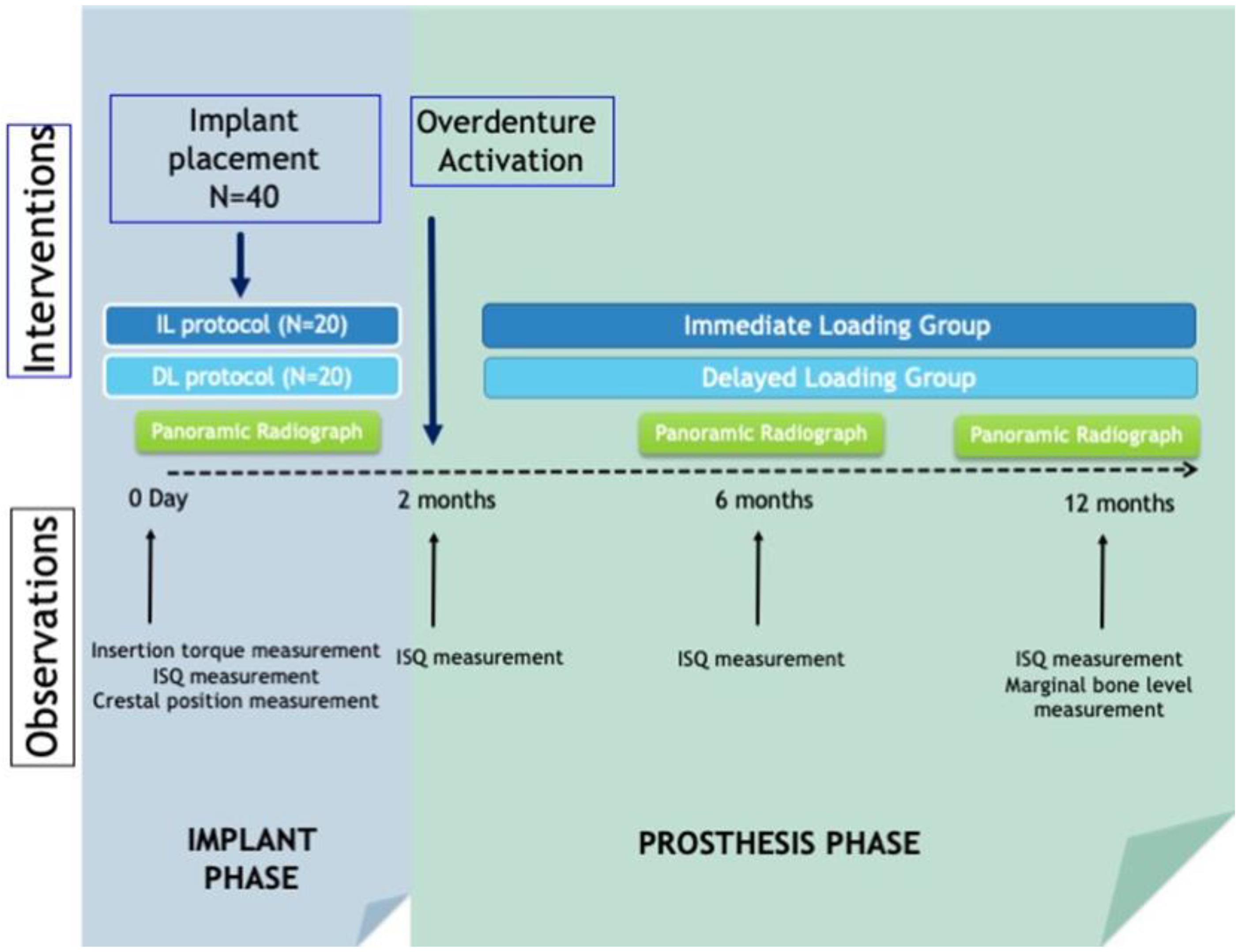
2. Materials and Methods

This research was conducted in full accordance with ethical principles, including the Declaration of Helsinki of the World Medical Association (https://www.wma.net/policies-post/wma-declaration-of-helsinki-ethical-principles-for-medical-research-involving-human-subjects/ (accessed on 7 January 2023)). The experimental protocol was approved by the Bioethics Committee of the university. All patients were informed about the aims, procedures, and duration of the study and were asked to provide written consent to participate and undergo the implant interventions and prosthetic rehabilitation according to the standard guidelines of the Spanish General Council of Dentists. Data were acquired and analyzed according to current legislation relating to privacy of personal data, clinical documentation, and biomedical research.
This triple-blind randomized clinical trial (RCT) aims to compare marginal bone loss of edentulous patients treated with mandibular overdentures retained by two implants that were subjected to either immediate loading or conventional loading (functional loading 2 months after implantation). The design of the study allows for two types of comparison: cross-comparison (comparing intra-subject results in different distinct observations) and parallel comparison (comparing the results of overdentures with or without immediate loading).
All participants were completely edentulous individuals who had been missing teeth for more than 10 years, regular users of conventional complete prostheses with sufficient remaining bone to receive the implants in the mandibular canine region (minimum height = 15 mm/minimum ridge width= 5 mm), and no evidence of systemic or psychic pathology that might contraindicate the implant treatment.

2.1. Surgical Protocol

All patients were operated under local anesthesia for the conventional placement (with flap rise) of two implants in the canine area. The minimum insertion torque was set at 40 Ncm. Primary stability was measured using a torque spanner (PCE-TM 80, Albacete, Spain) with a precision of ±1.5% and a measurement range from 0 to 147.1 Ncm in the clockwise and anti-clockwise directions and using Ostell® at baseline and also at 2, 6, and 12 months.
Bone quality was graded between 1 and 4 according to the Lekholm and Zarb [11] classification and the relationship of the implant to the bone crest was assessed directly and by digital panoramic radiography, with the prosthesis placed in maximum intercuspation to monitor the basal position of the bone crest.
In patients receiving immediate loading, Locator® type abutments, denoted VulkanLoc®, were placed where low retention Teflon (400 g: black) were placed, while in the conventionally loaded group, healing abutments of at least 3 mm in height were placed and conventional acrylic resin prosthesis with silicone reliners was placed over them. All patients underwent a new conventional full denture in the maxilla.
Two months after implant placement, patients in the immediate loading group had the black Teflon retention inserts replaced with more retentive inserts (680 g: blue), while those in the conventional loading group had the healing abutments replaced with VulkanLoc® abutments mounted directly on Teflon retention inserts with the same retentive properties (680 g: blue).
At 6 and 12 months, a panoramic radiograph was taken to evaluate the marginal bone loss based on the radiograph taken at the time of implant placement. For this purpose, an image analysis program (VixWinPro v1.5e, Gendex Dental Systems, Hatfield, PA, USA) was used to measure marginal bone loss based on the linear distance in mm between the following landmarks: implant shoulder (IS) and the most coronal bone-to-implant contact point (B). These quantitative parameters were measured in both the mesial and distal sides of the implants to obtain a mean estimate of marginal bone loss. Figure 1 shows the diagram of observations and interventions performed in the study.
This protocol was previously described in a paper published in 2021 by the same working group [5].

2.2. Evaluation of Complications

In addition to marginal bone loss, biological (dehiscence, mucositis, peri-implantitis, etc.), mechanical (loosening of screws, fractures, or damage to abutments, etc.) and minor prosthodontic complications (those that could be solved in an examining room); and major (those that had to be solved in the laboratory, such as relining or replacement of damaged attachments) were assessed per patient. These complication rates were evaluated in the short term and a 100-year complication rate calculation was performed.
The number of failed implants in each group and the failure rate of the implants were also recorded.
Furthermore, a linear regression analysis was performed to predict treatment outcomes in relation to age, sex, initial torque and ISQ of the implants placed, bone quality, gingival biotype, and the number of complications.

2.3. Data Analysis

Mean values and standard deviations (SD) were calculated for each quantitative outcome variable. Differences between immediately loaded and conventionally loaded implants were analyzed using the Student’s t-test, and the differences between observation time points within the same group were compared by pairwise Student’s t-tests or marginal homogeneity tests (for ordinal/multinomial variables) using baseline records as a reference. If the data distribution violated the principles of normality and homoscedasticity, non-parametric tests (Mann–Whitney) tests were used instead of parametric ones (Student’s t-tests) for intergroup comparisons and Wilcoxon tests rather than paired t-tests for within-group comparisons. Comparisons of nominal distributions between groups were carried out with Chi Square tests. Finally, backward stepwise linear regression models were calculated to predict marginal bone loss. The significance level was set at 0.05. All analyses were performed using SPSS 21.0 software (SPSS Inc., Chicago, IL, USA).

3. Results

3.1. Sample Description

The sample comprised 20 adult patients receiving 2 implants in the lower arch for the retention of overdentures on non-splinted implants, with a mean age of 66.3 ± 9.1 years. The sample size of this study was calculated based on the primary result with the greatest dispersion on the level of oral health-related quality of life (according to the Spanish version of the OHIP) [5].
Most implants were placed on type II quality (60.0%) covered by thin or medium soft tissue (88%) with a mean of 2.6 ± 2.2 mm of attached gingiva. The bone volume at the implant sites averaged 16.4 ± 5.3 mm in length and 6.9 ± 2.1 mm in width (measured 3 mm below the crest edge). No significant differences between groups were found.

3.2. Distribution and Characteristics of the Placed Implants

Table 1 shows the distribution of the 40 implants placed in the 20 selected patients, as well as their length and width measurements. The most frequently used implant size had a diameter of 3.75 mm (57.5%) or 4.2 mm (25%) and a length of 11.5 mm (55%) or 10 mm (35%).
Regarding the ISQ values, compared to the baseline values (70.4 ± 5.9), a significant increase was observed at the time of taking the one-year recordings at both canine sites (73.5 ± 4.8), as shown in Table 2. Within groups, the difference between pre-post ISQ values was only found to be significant for the conventional loading group. Similarly, a significant general improvement in peri-implant gingival health was observed, though this difference was only statistically significant for the immediate loading group. Despite the fact that the crestal position of the implants was equally distributed at baseline, one year after treatment the majority of implants were in the crestal position (65%), with this within-group change being statistically significant for the whole sample and for the conventional loading group.
Table 3 contains the marginal bone loss values one year after implant placement. According to the pre-post X-ray, it was observed that a mean of 0.25–0.59 mm (CI 95%) of marginal bone was lost after 13.4 ± 2.1 months of follow-up. There were no significant differences between groups.
Table 4 shows that there were no statistically significant differences between loading groups regarding the prevalence and annual rate of both biological and mechanical complications. Overall, an event rate of 3.4–7.3% of complications per 100 years was observed, mostly biological (mucositis, swelling, periimplantitis, etc.) rather than mechanical (abutment loosening/damage). In four patients, it was necessary to replace an implant due to mobility/infection (20%) although the distribution was equal in both groups. The failure rate (percentage of implants failing per year) was slightly higher in the conventional loading group (14.0 ± 32.7%) than in the immediate loading group (8.3 ± 18.0%). However, in general, the annual implant failure rate per 100 patients/years ranged between 0.1 and 2.3 failures.
The regression models summarized in Table 5 show that some clinical and patient-based outcomes are somewhat interrelated. Bone quality (D1 to D4 according to Leckholm and Zarb 11) is the main predictor for implant failure and marginal bone loss with high predictability. The odds ratio for implant failure increases from 1.3 to 25.8 as bone density decreases; however, the higher the bone density, the greater the marginal bone loss (OR: 0.15–1.02 mm per density change). The number of prosthetic complications is proportional to the mean insertion torque (OR: 0.01–0.24), whereas the number of biological complications depends on the number of minor prosthetic complications, mostly denture-based touch-ups.

4. Discussion

In this work, 20 patients were selected and a total of 40 implants were placed in the maxilla for the positioning of mandibular overdentures on Locator® abutments. Half of the sample underwent immediate loading and the other half underwent delayed or conventional loading.

4.1. Bone Loss Assessment

In general, it is considered that the assessment of marginal bone loss around implants should be carried out by means of correctly calibrated and parallel periapical radiographs [12,13,14].
However, in totally edentulous patients, it is sometimes complicated to obtain this type of radiograph, especially because the positioning of the plate in areas with significant mandibular bone resorption and where the insertion of the muscles of the floor of the mouth is high is uncomfortable and even painful, despite the use of devices for holding the plates and parallelizers [15,16]. In some of the published clinical trials comparing immediate loading versus conventional loading in mandibular overdentures, these measurements are performed on periapical radiographs and with the use of image processing software [12,13,14]. In this study, bone loss was assessed using properly calibrated panoramic radiographs, measuring the implants on the radiograph and comparing this measurement with the actual measurements of the placed implants as in other studies reported in the literature [15,16,17]. In these works, the authors are aware that this can lead to erroneous measurements as there is distortion and loss of detail in the central area of the radiograph, [15,16] where implants for mandibular complete dentures will usually not be present. In recent times, the availability and accessibility of CBCT also the allows assessment of marginal bone loss through its use, which improves the accuracy of the data as it allows the assessment of bone loss not only mesially and distally but also vestibularly and lingually, although it increases the radiation dose to the patient [18].
To determine the success of implants, many authors use Albrektsson and Zarb’s 1986 success criteria as a reference [14,15,16,18]. These consist of the following points to determine that an implant is successful: the absence of mobility; the absence of peri-implant radiolucency on radiography; vertical bone loss less than 2 mm during the first year and less than 0.2 mm annually after the first year of observation; and the absence of signs and symptoms such as pain, infection, neuropathies, paraesthesia, or insertion into the lower dental nerve canal.
Nowadays, it is considered that these criteria, without the evaluation of the implant-prosthetic complex, are not sufficient to evaluate the clinical efficiency of the implant, as there are other parameters such as the patient’s subjective criteria and aesthetics that must be taken into account to determine whether an implant is successful or not [19]. Since Albrektsson and Zarb established in 1986 a success rate of 85% at 5 years and 80% at 10 years taking into account their criteria [20], other evaluation scales have appeared such as the one established by Gallucci et al. in 2009, which, taking into account their criteria that also evaluated the peri-implant tissues, the prosthesis, and subjective parameters that allowed the patient to evaluate their treatment as good or excellent through a survey, established a success rate of 86.7% and a survival rate of 95.5% at 5 years [21].
Despite the fact that most studies evaluate marginal bone loss [13,14,15,18,22], as a fundamental element in determining the success of implants, it is true that there are marked differences in the duration of the observation times of each study published on this subject and, therefore, differences in the time of evaluation of implant bone loss, ranging from 12 to 84 months in the studies published on this subject [13,14,15,18,22]. In the present study, marginal bone loss was assessed 12 months after implant placement. It seems logical to think that the marginal bone loss that can be related to the type of load received by the implant is that measured within the first year of implant function.
A meta-analysis published in 2020 by the same working group [23] assessed marginal bone loss one year after placement of mandibular implants for the retention of mandibular prostheses (fixed or removable) including seven randomized clinical trials [12,13,14,15,16,18,22]. Of these, only five were estimated for the meta-analysis of bone loss assessment [12,13,15,18,22], and the weighted mean value of marginal bone loss was 0.12 mm [95% CI −0.03, 0.28], with a tendency for marginal bone loss to be lower in implants that were loaded deferred. In this paper [23], all studies included were clinical trials on implants for overdentures using either bar-splinted [15,16] or non-splinted implants with ball splints or Locator [13,14,18,22] and one fixed prosthetic work on four implants [12].
In the present work, there was no difference between the two groups in terms of marginal bone loss at 1 year after implant placement: 0.40 mm [95% CI 0.12, 0.68] in the immediate loading group and 0.44 mm [95% CI 0.19, 0.69] in the conventional loading group.
If we compare the marginal bone loss at one year in this study with other studies published on the same subject, the results are similar to those of the work of Romeo et al. in 2002 [15] who evaluated the marginal bone loss of 80 ITI implants for mandibular overdentures (4 implants per patient), although in this case, the implants were splinted with a Dolder bar. In this study, there was no difference in marginal bone loss between implants loaded immediately (−0.21 ± 0.12 mm) versus delayed (−0.19 ± 0.24 mm) [15]. In the work published by Schuster et al. in 2020 [17] in which 2 narrow implants were placed in 20 patients, again there was no difference in marginal bone loss between the immediate (−0.05 with a range of −0.85 to 0.75 mm) and delayed (−0.05 with a range of −1.08 to 1.06 mm) loading groups.
On the other hand, in the work by Schincaglia et al. published in 2016 [13] in which 2 implants per patient were placed for the retention of lower overdentures with Locator as in the present study (a total of 64 implants), it was observed that the marginal bone loss at 1 year was lower in immediately loaded implants (0.25 ± 0.5 mm) compared to conventionally loaded implants (0.54 ± 0.5 mm).
The works by Elsyad et al. published in 2014 and 2012 [18,22], which studied the placement of 2 mandibular implants per patient for the retention of lower overdentures using ball or Locator (a total of 72 implants each study), concluded that bone loss was significantly lower in those loaded with delayed loading (0.515 ± 0.38 mm and 0.87 ± 0.13 mm) versus those loaded immediately (1.002 ± 0.53 mm and 1.05 ± 0.18 mm).

4.2. Implant Stability

Another important element is the measurement of primary and secondary implant stability. Primary implant stability is directly dependent on bone quality and quantity, implant geometry, and placement technique, and secondary stability is related to the formation of secondary bone in contact with the implant [24].
Implant stability can be measured in several ways. A simple way to measure stability is the insertion torque. Implant placement motors are usually equipped with a torque meter, and most modern implant placement motors indicate the maximum torque of the implant at the time of placement. Most published works measure the insertion torque at the time of placement and use it as a determining parameter when deciding whether to carry out immediate loading on the implant. An insertion torque of 35 Ncm is considered sufficient to be able to safely perform immediate loading. In this context, we find that most studies consider a torque of 30 Ncm to 35 Ncm to be suitable for immediate loading [25,26,27].
In this work, immediate loading was performed on half of the sample and delayed or conventional loading on the other half. The mean torque values obtained in this clinical trial in the immediate loading group and the delayed loading group were 65.1 (±12.1) Ncm and 61.7 (±14.4) Ncm, respectively, with no differences between the insertion torque achieved in the two groups.
It is noteworthy that only a minority of published works assess implant stability using RFA (resonance frequency analysis) or percussive tip (Periotest®) techniques. It is considered that the measurement of stability is much more accurate if RFA is used, either by Ostell™ or Penguin RFA (Klockner, Barcelona, Spain), as the values obtained do not depend on the position of the implant, and generally several records are taken and the one with the lowest value is selected [28]
The ISQ is measured in values from 1 to 100, with 1 being minimum stability and 100 being maximum stability. ISQ values below 60 are considered to constitute a risk due to low implant stability [29]. For values between 60 and 65, immediate loading can be considered for splinted full-arch restorations, or it would be even better to perform two-stage surgery with conventional loading. Between 65 and 70, early or conventional loading can be considered and for values above 70, immediate loading can be considered in individual cases [29].
In the present study, ISQ was measured at the time of placement and at 2, 6, and 12 months after placement. The mean ISQ values obtained at the time of implant placement were 70.8 (±6.8) in the immediate loading group and 70.0 (±5.3) in the conventional loading group, with no significant differences between groups.
This is in line with the ISQ values obtained at the time of implant placement in the works of Elsyad et al. [22] and Acham et al. [30], with values of 68.85 (±1.23) and 74.5 (±4.4) for conventional loading and 71.95 (±1.98) and 76.2 (±4.2) for immediate loading implants, respectively. The differences between groups were significant in terms of initial implant stability on the day of surgery, with the ISQ value being higher in the immediately loaded group in both studies. This is important, as implants with greater primary stability were loaded immediately, while those with a lower ISQ were loaded conventionally, which may have influenced the result of the study. In this sense, in the work of Acham et al. [30], when the implants did not reach the 30 Ncm torque at the time of placement, if they belonged to the immediate loading group, they were transferred to the delayed loading group, which invalidates the comparison between groups in this parameter, as these cases in which the minimum torque conditions were not met were not removed from the study.
In the work of Schuster et al. [17], the primary stability of the two implants was also assessed at the time of placement and at 3, 6, and 12 months post-placement. The only time at which there were differences between the two groups was at the assessment 3 months after implant placement, with an ISQ of 57.0 (range 43.0–62.25) in the delayed loading group and 50.63 (36.5; 57.0) in the immediate loading group; significantly, the stability of the implants that had not yet been loaded was 8% higher than those that had been loaded immediately. At 6 and 12 months, there were no differences between groups, with ISQ values of 54.25 (range between 42.0 and 62.0) for the conventional loading group and 50.5 (range between 45.0 and 58.0) for the immediate loading group at 6 months and 53.88 (range between 44.0 and 66.75) for conventional loading and 56.5 (range between 48.0 and 60.75) for immediate loading at 12 months.
On the other hand, the ISQ values increase in the first year and decrease after 24 and 36 months, and these values become more equal after 1 year, reducing the differences between groups [22,30].
In the present study, the ISQ values at 2 months were 69.8 (±7.2) for the immediate loading group and 70.0 (±4.3) for the delayed loading group, which was significantly different from the values obtained at placement, which were 65.2 (±12.1) for the immediate loading and 61.7 (±14.4) for the conventional loading. At 6 months, the values were 73.3 (±5.2) in the immediate loading group and 72.3 (±2.8) in the conventional loading group, with no significant differences within each group with respect to the values at 2 months. At 12 months, the values were 72.8 (±5.7) and 74.2 (±3.9), respectively, with a significant difference in the values for the conventional loading group compared to those obtained at 6 months (Table 2). This means that in the conventional loading group, the ISQ values increased significantly from the time of implant placement to one year, which is not the case in the immediate loading group.
Some studies measure stability using the Periotest®. This consists of a percussive tip that strikes the implant 16 times for 4 s. The less stable the implant, the longer the contact time with the tip and the higher the value obtained. The Periotest® values range from −8 to +50, so that the higher the value, the lower the stability of the implant. The asymmetry in this scale is because the Periotest® is a device that was initially designed to evaluate dental mobility, with the lowest values (−8 to +9) corresponding to a degree 0 mobility of the tooth and the highest values (+30 to +50) corresponding to a degree III dental mobility [31]. In two published studies, Periotest® values of −4.0 (±2.1) and −3.39 (±0.04) were obtained for conventional loading and −5.5 (±1.5) and −4.2 (±0.31) for immediate loading. In both studies, there were differences in stability between groups in the first 3 months, with lower values in the immediate loading group; however, from that point on, there were no differences in stability between groups [18,30].
In the rest of the studies published on this subject, primary implant stability is measured by insertion torque at the time of placement, and no post-placement stability measurements are made [13,14,15]. On the other hand, there is no uniformity in relation to the system for measuring stability and the torque necessary to assess whether to perform immediate or early loading.

4.3. Implant Failure

In the present study, four patients had to have an implant replaced due to mobility and infection, two implants in the immediate loading group (10%, 2/20), and another two in the delayed loading group (10%, 2/20).
In the meta-analysis published by this same working group in 2020, which included nine studies comparing immediate versus delayed loading in complete mandibular restorations (seven studies on removable prostheses and two on fixed prostheses), it was concluded that conventional loading has fewer implant failures during the first year than immediate loading (odds ratio fixed effects 2.63 [95% CI: 1.22, 5.68]) [23].
Regarding the results of the individual studies that were part of this meta-analysis [23], three papers were included that studied lower overdentures placed on Locator®, one of them on four implants [30] and the other two on two implants [13,22] and two on ball retainers on one and two mandibular implants [18,32]. In three of these studies, there were two early implant failures (before the first year) in the immediate loading group (6.66%, 2/30 [18], 6.25%, 2/32 [22], and 6.25%, 2/32 [13]) and none in the conventional loading group [13,18,22]. In one paper, there were no early failures in either group [30] and in another paper, there were nine early failures in the immediate loading group (1.54%, 1/65) [32]. In these works, only implant failures that occurred during the first year after implant placement were considered, considering that this is the time in which the failure could be related to the type of loading the implants received.

4.4. Complications

Regarding complications, there were no differences between groups in relation to the prevalence and annual rate of biological (mucositis, inflammation, peri-implantitis, etc.) and mechanical (loosening or damage to screws) complications (rate of 3.4–7.3% of complications per 100 years). In other published studies, there were also no differences between groups in terms of biological and mechanical complications [13,17,30]. However, one of the studies indicates that the median number of appointments attended by patients who received immediate loading was significantly lower than in those who received conventional loading (16.5 vs. 22.5, p = 04, Mann–Whitney U test), as well as the need to reline the prosthesis due to pressure marks on the mucosa, which was 13 times in the immediate loading group compared to 42 times in the deferred loading group [30].
Another study indicates that the number of complications depends on the type of loading received, and that complications in the conventional loading group are mainly due to less adaptation of the soft tissues to the prosthetic components, which is associated with the possible fall of the healing abutments and the need for gingival reopening for repositioning [17].
Most of the published works do not perform an analysis of complications in relation to implant placement or the type of loading received.
In the present study, linear regression models showed that bone quality is the main predictor of implant failure and marginal bone loss with high predictability (Table 5). The risk of implant failure increases from 1.3 to 25.8 as bone density decreases.
On the other hand, the higher the bone density, the greater the marginal bone loss (OR: 0.15–1.02 mm). In a recently published meta-analysis by Atieh et al. [33], which included five studies comparing marginal bone loss with implant insertion torque, with regular torque being less than 50 Ncm and high torque greater than 50 Ncm, it was concluded that changes in peri-implant marginal bone level favored the regular insertion torque group but with no statistically significant differences between groups. In this case, it could be determined that bone loss around the implant may be greater in those implants placed with a torque higher than 50 Ncm and in those implants placed in the mandible, possibly due to the compression produced by the high insertion torque in a dense and poorly vascularized cortical bone. Another study analyzing marginal bone loss on periapical and panoramic radiographs concluded that bone loss around implants appeared to be associated with cortical bone characteristics [34].
In addition, other published studies have found no differences in marginal bone loss between implants placed in different types of bone [35] or have found significantly greater marginal bone loss in implants placed in type 4 bone [36].
Despite the importance of bone quality in implant survival and marginal bone loss, most published studies do not assess bone density, or, if they do, they are based exclusively on the tactile perception of the surgeon placing the implant at the time of placement or on the torque acquired by the implant at the time of placement, [12,13,15] without performing a radiological assessment of bone density using radiographs or CBCT.
In a study published in 2019 in which four mandibular implants were placed to support a fixed lower prosthesis [26], 83% of patients had type 2 or 3 bone and, out of the implants that failed in this work (5/160), four were placed in type 1 bone.
In the present study, most implants were placed in bone quality type 2 according to Leckholm and Zarb (60.0%) [5].
According to a systematic review with meta-analysis [37], the relative risk of implant failure is higher in implants inserted in bone quality type 1 than in those placed in bone qualities 2 and 3. Implants placed in bone quality 3 have a higher failure rate than those inserted in bone quality 2. On the other hand, implants placed in bone quality 4 have a higher failure rate than those placed in the other three types of bone quality; this is due to the fact that in lower quality bone it is more complicated to achieve adequate primary stability, which is related to a higher failure rate [37]. For this reason, it is considered that immediate or early loading should be ruled out in bone quality type 4 according to the classification of Lekholm and Zarb [11] and its equivalents D4 and D5 according to the classification of Misch [38] and that it can be performed in bone types 1 and 2. In the mandibular anterior sector, the quality of the bone is usually good, which favors adequate primary stability of the implants, favoring immediate or early loading [24,39].

4.5. Limitations and Continuity

It would be interesting to carry out more studies on the effect that other factors may have on marginal bone loss, such as the state of the peri-implant gingiva. It would also be useful to increase the sample size. The purpose of this research group is to continue carrying out studies in this line of research.

5. Conclusions

According to the results of this study:
  • There were no differences in marginal bone loss observed at one year in immediately loaded (0.40 ± 0.39 mm) versus conventionally loaded (0.44 ± 0.36 mm) implants placed for the retention of mandibular overdentures.
  • There were no differences in primary and secondary stability of immediately loaded versus conventional implants; however, in the conventional loading group, stability increased significantly between the time of implant placement at both 6 and 12 months post-placement.
  • The main predictive factor for implant failure as well as marginal bone loss is bone quality (β = 13.61; 95% CI: 1.3–25.8).

Author Contributions

Conceptualization, J.M.; methodology, J.M., Y.G., A.D., J.F.-F. and N.Q.-L.; validation, J.M.; formal analysis, J.M.; investigation, J.M., Y.G., A.D., J.F.-F., N.Q.-L. and B.P.-P.; resources, J.M.; data curation, Y.G., A.D., N.Q.-L., J.F.-F. and B.P.-P.; writing—original draft preparation, B.P.-P.; writing—review and editing, J.M.; visualization, J.F.-F.; supervision, J.M.; project administration, J.M.; funding acquisition, J.M. All authors have read and agreed to the published version of the manuscript.

Funding

This research received funding from Titanimplant, Barcelona (Spain) http://www.titanimplant.net (accessed on 7 January 2023) to support the direct costs of the exploratory, surgical, and prosthetic interventions of the present study.

Institutional Review Board Statement

The study was conducted in accordance with the Declaration of Helsinki, and approved by the Institutional Review Board (or Ethics Committee) of UNIVERSITY OF SALAMANCA (RUSAL_201500006834 13 June 2013).

Informed Consent Statement

Informed consent was obtained from all subjects involved in the study.

Data Availability Statement

Not applicable.

Acknowledgments

The study was conducted within the Research Group “Avances en Salud Oral” (Advances in Oral Health) of the University of Salamanca led by the first author. https://avancessaludoral.usal.es (accessed on 7 January 2023).

Conflicts of Interest

The authors declare no conflict of interest.

References

  1. Schimmel, M.; Srinivasan, M.; Herrmann, F.; Müller, F. Loading Protocols for Implant-Supported Overdentures in the Edentulous Jaw: A Systematic Review and Meta-Analysis. Int. J. Oral Maxillofac. Implant. 2014, 29, 271–286. [Google Scholar] [CrossRef] [PubMed]
  2. Brånemark, P.I.; Hansson, B.O.; Adell, R.; Breine, U.; Lindström, J.; Hallén, O.; Ohman, A. Osseointegrated implants in the treatment of the edentulous jaw. Experience from a 10-year period. Scand. J. Plast. Reconstr. Surg. Suppl. 1977, 16, 1–132. [Google Scholar]
  3. Feine, J.S.; Carlsson, G.E.; Awad, M.A.; Chehade, A.; Duncan, W.J.; Gizani, S.; Head, T.; Heydecke, G.; Lund, J.P.; MacEntee, M.; et al. The McGill consensus statement on overdentures. Mandibular two-implant overdentures as first choice standard of care for edentulous patients. Gerodontology 2002, 19, 601–602. [Google Scholar]
  4. Thomason, J.M.; Feine, J.; Exley, C.; Moynihan, P.; Müller, F.; Naert, I.; Ellis, J.; Barclay, C.; Butterworth, C.; Scott, B.; et al. Mandibular two implant-supported overdentures as the first choice standard of care for edentulous patients—the York Consensus Statement. Br. Dent. J. 2009, 207, 185–186. [Google Scholar] [CrossRef] [PubMed]
  5. Montero, J.; Dib, A.; Guadilla, Y.; Flores, J.; Pardal-Peláez, B. Functional and patient-centered treatment outcomes with man- dibular overdentures retained by two immediate or conven- tionally loaded implants. A randomized clinical trial. J. Clin. Med. 2021, 10, 3477. [Google Scholar] [CrossRef]
  6. Su, M.; Shi, B.; Zhu, Y.; Guo, Y.; Zhang, Y.; Xia, H.; Zhao, L. Comparison of implant success rates with different loading protocols: A meta-analysis. Int. J. Oral Maxillofac. Implant. 2014, 29, 344–352. [Google Scholar] [CrossRef] [Green Version]
  7. Adell, R.; Eriksson, B.; Lekholm, U.; Brånemark, P.I.; Jemt, T. Long-term follow-up study of osseointegrated implants in the treatment of totally edentulous jaws. Int. J. Oral Maxillofac. Implant. 1990, 5, 347–359. [Google Scholar]
  8. Papaspyridakos, P.; Chen, C.-J.; Chuang, S.-K.; Weber, H.-P. Implant Loading Protocols for Edentulous Patients with Fixed Pros-theses: A Systematic Review and Meta-Analysis. Int. J. Oral Maxillofac. Implant. 2014, 29, 256–270. [Google Scholar] [CrossRef]
  9. Borges, T.D.F.; Mendes, A. Mandibular Overdentures with Immediate Loading: Satisfaction and Quality of Life. Int. J. Prosthodont. 2011, 24, 534–539. [Google Scholar]
  10. Weber, H.-P.; Morton, D.; Gallucci, G.O.; Roccuzzo, M.; Cordaro, L.; Grutter, L. Consensus statements and recommended clinical procedures regarding loading protocols. Int. J. Oral Maxillofac. Implant. 2009, 24, 180–183. [Google Scholar]
  11. Lekholm, U.; Zarb, G.A. Patient selection and preparation. In Tissue Integrated Prostheses: Osseointegration in Clinical Dentistry; Quintessence Publishing: Chicago, IL, USA, 1985; pp. 199–210. [Google Scholar]
  12. Jokstad, A.; Alkumru, H. Immediate function on the day of surgery compared with a delayed implant loading process in the mandible: A randomized clinical trial over 5 years. Clin. Oral Implant. Res. 2013, 25, 1325–1335. [Google Scholar] [CrossRef] [Green Version]
  13. Schincaglia, G.P.; Rubin, S.; Thacker, S.; Dhingra, A.; Trombelli, L.; Ioannidou, E. Marginal Bone Response Around Immediate- and Delayed-Loading Implants Supporting a Locator-Retained Mandibular Overdenture: A Randomized Controlled Study. Int. J. Oral Maxillofac. Implant. 2016, 31, 448–458. [Google Scholar] [CrossRef] [Green Version]
  14. Alfadda, S.A. A Randomized Controlled Clinical Trial of Edentulous Patients Treated with Immediately Loaded Implant-Supported Mandibular Fixed Prostheses. Clin. Implant. Dent. Relat. Res. 2013, 16, 806–816. [Google Scholar] [CrossRef]
  15. Romeo, E.; Chiapasco, M.; Lazza, A.; Casentini, P.; Ghisolfi, M.; Iorio, M.; Vogel, G. Implant-retained mandibular overdentures with ITI implants: A comparison of 2-year results between delayed and immediate loading. Clin. Oral Implant. Res. 2002, 13, 495–501. [Google Scholar] [CrossRef]
  16. Chiapasco, M.; Abati, S.; Romeo, E.; Vogel, G. Implant-retained mandibular overdentures with Brånemark System MKII implants: A prospective comparative study between delayed and immediate loading. Int. J. Oral Maxillofac. Implant. 2001, 16, 537–546. [Google Scholar]
  17. Schuster, A.J.; Marcello-Machado, R.M.; Bielemann, A.M.; da Rosa Possebon, A.P.; Chagas Júnior, O.L.; Faot, F. Immediate vs conventional loading of Facility-Equator system in mandibular overdenture wearers: 1-year RCT with clinical, biological, and functional evaluation. Clin. Implant. Dent. Relat. Res. 2020, 22, 270–280. [Google Scholar] [CrossRef]
  18. Elsyad, M.A.; Al-Mahdy, Y.F.; Fouad, M.M. Marginal bone loss adjacent to conventional and immediate loaded two implants supporting a ball-retained mandibular overdenture: A 3-year randomized clinical trial. Clin. Oral Implant. Res. 2011, 23, 496–503. [Google Scholar] [CrossRef]
  19. Papaspyridakos, P.; Chen, C.J.; Singh, M.; Weber, H.P.; Gallucci, G.O. Success criteria in implant dentistry: A systematic review. J. Dent. Res. 2012, 91, 242–248. [Google Scholar] [CrossRef]
  20. Albrektsson, T.; Zarb, G.; Worthington, P.; Eriksson, A.R. The long-term efficacy of currently used dental implants: A review and proposed criteria of success. Int. J. Oral Maxillofac. Implants 1986, 1, 11–25. [Google Scholar]
  21. Gallucci, G.O.; Doughtie, C.B.; Hwang, J.W.; Fiorellini, J.P.; Weber, H.-P. Five-year results of fixed implant-supported rehabilitations with distal cantilevers for the edentulous mandible. Clin. Oral Implant. Res. 2009, 20, 601–607. [Google Scholar] [CrossRef]
  22. Elsyad, M.A.; Elsaih, E.A.; Khairallah, A.S. Marginal bone resorption around immediate and delayed loaded implants supporting a locator-retained mandibular overdenture. A 1-year randomised controlled trial. J. Oral Rehabil. 2014, 41, 608–618. [Google Scholar] [CrossRef] [PubMed]
  23. Pardal-Peláez, B.; Flores-Fraile, J.; Pardal-Refoyo, J.L.; Montero, J. Implant loss and crestal bone loss in immediate versus delayed load in edentulous mandibles: A systematic review and meta-analysis. J. Prosthet. Dent. 2020, 125, 437–444. [Google Scholar] [CrossRef] [PubMed]
  24. Turkyilmaz, I.; Tumer, C. Early versus late loading of unsplinted TiUnite surface implants supporting mandibular overdentures: A 2-year report from a prospective study. J. Oral Rehabil. 2007, 34, 773–780. [Google Scholar] [CrossRef] [PubMed]
  25. Cannizzaro, G.; Felice, P.; Soardi, E.; Ferri, V.; Leone, M.; Lazzarini, M.; Audino, S.; Esposito, M. Immediate loading of 2(all-on-2) versus 4 (all-on-4) implants placed with a flapless technique supporting mandibular cross-arch fixed prostheses: 1-year results from a pilot ran-domised controlled trial. Eur. J. Oral Implantol. 2013, 6, 121–131. [Google Scholar] [PubMed]
  26. Alfadda, S.A.; Chvartszaid, D.; Tulbah, H.I.; Finer, Y. Immediate versus conventional loading of mandibular implant-supported fixed prostheses in edentulous patients: 10-year report of a randomised controlled trial. Int. J. Oral Implantol. 2019, 12, 431–446. [Google Scholar]
  27. Mundt, T.; Passia, N.; Att, W.; Heydecke, G.; Freitag-Wolf, S.; Luthardt, R.G.; Kappel, S.; Konstantinidis, I.K.; Stiesch, M.; Wolfart, S.; et al. Pain and discomfort following immediate and delayed loading by overdentures in the single mandibular implant study (SMIS). Clin. Oral Investig. 2016, 21, 635–642. [Google Scholar] [CrossRef]
  28. García-Moreno, S.; González-Serrano, J.; López-Pintor, R.; Pardal-Peláez, B.; Hernández, G.; Martínez-González, J. Implant stability using piezoelectric bone surgery compared with conventional drilling: A systematic review and meta-analysis. Int. J. Oral Maxillofac. Surg. 2018, 47, 1453–1464. [Google Scholar] [CrossRef]
  29. La Escala ISQ-Osstell—Implant Stability. Available online: https://www.osstell.com/es/clinical-guidelines/the-isq-scale/ (accessed on 7 January 2023).
  30. Acham, S.; Rugani, P.; Truschnegg, A.; Wildburger, A.; Wegscheider, W.A.; Jakse, N. Immediate loading of four interforaminal im-plants supporting a locator-retained mandibular overdenture in the elderly. Results of a 3-year randomized, controlled, pro-spective clinical study. Clin. Implant. Dent. Relat. Res. 2017, 19, 895–900. [Google Scholar] [CrossRef]
  31. Medizintechnik Gulden. Periotest, el Instrumento de Medición Dental para la Implantología y la Odontología más Exigentes. Available online: http://www.med-gulden.com/downloads/03_spain/01_Informaci%F3ndeproducto/Proceso_de_producci%F3n.pdf (accessed on 7 January 2023).
  32. Kern, M.; Att, W.; Fritzer, E.; Kappel, S.; Luthardt, R.; Mundt, T.; Reissmann, D.; Rädel, M.; Stiesch, M.; Wolfart, S.; et al. Survival and Complications of Single Dental Implants in the Edentulous Mandible Following Immediate or Delayed Loading: A Randomized Controlled Clinical Trial. J. Dent. Res. 2017, 97, 163–170. [Google Scholar] [CrossRef] [Green Version]
  33. Atieh, M.A.; Baqain, Z.H.; Tawse-Smith, A.; Ma, S.; Almoselli, M.; Lin, L.; Alsabeeha, N.H.M. The influence of insertion torque values on the failure and complication rates of dental implants: A systematic review and meta-analysis. Clin. Implant. Dent. Relat. Res. 2021, 23, 341–360. [Google Scholar] [CrossRef]
  34. Merheb, J.; Graham, J.; Coucke, W.; Roberts, M.; Quirynen, M.; Jacobs, R.; Devlin, H. Prediction of Implant Loss and Marginal Bone Loss by Analysis of Dental Panoramic Radiographs. Int. J. Oral Maxillofac. Implant. 2015, 30, 372–377. [Google Scholar] [CrossRef] [Green Version]
  35. Barewal, R.M.; Stanford, C.; Weesner, T.C. A randomized controlled clinical trial comparing the effects of three loading protocols on dental implant stability. Int. J. Oral Maxillofac. Implant. 2012, 27, 945–956. [Google Scholar]
  36. Tawil, G.; Mawla, M.; Gottlow, J. Clinical and radiographic evaluation of the 5-mm diameter regular-platform Brånemark fixture: 2- to 5-year follow-up. Clin. Implant. Dent. Relat. Res. 2002, 4, 16–26. [Google Scholar] [CrossRef]
  37. Chrcanovic, B.R.; Albrektsson, T.; Wennerberg, A. Bone Quality and Quantity and Dental Implant Failure: A Systematic Review and Meta-analysis. Int. J. Prosthodont. 2017, 30, 219–237. [Google Scholar] [CrossRef] [Green Version]
  38. Misch, C.E. Contemporary Implant Dentistry. Implant. Dent. 1999, 8, 90. [Google Scholar] [CrossRef]
  39. Tawse-Smith, A.; Payne, A.G.; Kumara, R.; Thomson, W.M. Early Loading of Unsplinted Implants Supporting Mandibular Overdentures Using a One-Stage Operative Procedure with Two Different Implant Systems: A 2-Year Report. Clin. Implant. Dent. Relat. Res. 2002, 4, 33–42. [Google Scholar] [CrossRef]
Figure 1. Diagram of the observations and interventions scheduled in the study.
Prosthesis 05 00022 g001
Table 1. Distribution of the 40 implants among the study sample (n = 20).
Implant SizeAll Patients (n = 20 Patients with 40 Implants)Immediate Loading Group (n = 10 Patients with 20 Implants)Conventional Loading Group (n = 10 Patients with 20 Implants)
DiameterN%N%N%
3.3 mm717.5315.0420.0
3.75 mm2357.51365.01050.0
4.2 mm1025.0420.0630.0
LengthN%N%N%
10 mm1435.0525.0945.0
11.5 mm2255.01365.0945.0
13 mm410.0210.0210.0
Table 2. Distribution and variation of longitudinal implant-related variables in the study sample (n = 20). Comparisons by paired and unpaired Student’s t-tests.
BaselineAll Patients (n = 20 Patients with 40 Implants)Immediate Loading Group (n = 10 Patients with 20 Implants)Conventional Loading Group (n = 10 Patients with 20 Implants)
Implant insertion torque at different sitesMeanSDMeanSDMeanSD
#3362.914.464.513.861.215.5
#4364.013.065.911.962.014.4
Average63.513.165.212.161.714.4
Implant stability by ISQMeanSDMeanSDMeanSD
#3369.1 a7.369.2 a6.769.0 a8.1
#4371.7 a8.672.3 a10.371.0 a6.9
Average70.4 a5.970.8 a6.870.0 a5.3
Averaged Crestal position (aggregating mesial and distal sides of both implants)N%N%N%
Subcrestal6 a30.02 a20.04 a40.0
Crestal7 a35.04 a40.0 a3 a30.0
Supracrestal7 a35.04 a40.03 a30.0
At 2 months of follow-up
Implant stability by ISQMeanSDMeanSDMeanSD
#3367.0 a10.366.9 a12.567.1 a8.3
#4372.8 a4.872.7 a6.272.9 a3.3
Average69.9 a5.869.8 a7.270.0 a4.3
Percentage of healthy gingival sites57.5 a46.765.0 a47.450.0 a47.1
At 6 months of follow-up
Implant stability by ISQMeanSDMeanSDMeanSD
#3371.9 a4.373.0 a5.270.9 a3.0
#4372.9 a4.572.8 a5.872.9 a3.1
Average72.8 b4.173.3 a5.272.3 b2.8
Percentage of healthy gingival sites55.0 a45.660.0 a46.050.0 a47.1
At 12 months of follow-up
Implant stability by ISQMeanSDMeanSDMeanSD
#3372.9 b6.071.4 a7.474.6 b3.9
#4373.7 b5.073.5 a5.273.8 a5.0
Average73.5 b4.872.8 a5.774.2 b3.9
Percentage of healthy gingival sites87.5 b22.2100.0 b0.075.0 a26.4
Averaged Crestal position (aggregating mesial and distal sides of both implants)N%N%N%
Subcrestal3 b15.02 a20.01 b10.0
Crestal4 b20.02 a20.02 b20.0
Supracrestal13 b65.06 a60.07 b70.0
a,b: Different lower case letters within columns mean significant pre-post differences (p < 0.05) after paired t-test (for ISQ and gingival health) or marginal homogeneity test (for crestal position), which have preoperative values as reference. Significant differences p < 0.5 between groups after Student’s t-tests.
Table 3. Marginal bone loss in the study sample (n = 20) comparison between conventional and immediate loading groups by Chi Square and Student’s t-tests after 13.4 ± 2.1 months of follow-up.
All Patients (n = 20 Patients with 40 Implants)Immediate Loading Group (n = 10 Patients with 20 Implants)Conventional Loading Group (n = 10 Patients with 20 Implants)
Baseline Implant shoulder position (mm)MeanSDMeanSDMeanSD
#33−0.080.630.000.61−0.170.67
#43−0.060.46−0.050.28−0.080.60
All implants−0.080.51−0.030.40−0.130.62
Final Implant shoulder position (mm)MeanSDMeanSDMeanSD
#330.450.850.470.870.440.88
#430.210.590.290.420.140.74
All implants0.340.680.380.600.300.68
Marginal Bone Loss (mm)MeanSDMeanSDMeanSD
#330.540.500.470.530.610.50
#430.280.300.340.330.220.26
All implants0.40.370.400.390.440.36
CI 95%CI 95%CI 95%
lower limitupper limitlower limitupper limitlower limitupper limit
Averaged Marginal Bone loss (mm)0.250.590.120.680.190.69
MeanSDMeanSDMeanSD
Months of follow-up13.42.113.22.113.52.1
Table 4. Prevalence and rates of biological, mechanical, and technical complications among the study sample (n = 20).
Short-Term ComplicationsAll Patients (n = 20)Immediate Loading Group (n = 10)Conventional Loading Group (n = 10)
PrevalenceN%N%N%
Biological A1050.0440.0660.0
Mechanical B525.0330.022.0
None525.0330.022.0
Number of complicationsMeanSDMeanSDMeanSD
Average complications1.00.91.11.10.80.6
Biological complications0.80.80.81.00.80.6
Mechanical complications0.40.50.50.50.30.5
Prevalence of complications
Type of complicationsN%N%N%
Uneventful treatment735.0440.0330.0
Prosthetic complications1365.0660.0770.0
Minor C840.0550.0330.0
Major D630.0440.0220.0
Biological complications1260.0550.0770.0
Implant Failure420.0220.0220.0
MeanSDMeanSDMeanSD
Failure Ratio(%) E11.225.98.318.014.032.7
Event Rate Per 100 Yrs FMeanCI-95%MeanCI-95%MeanCI-95%
Total5.43.4–7.35.62.3–9.05.12.4–7.9
Prosthetic1.90.8–3.02.20.5–4.01.5−0.2–3.2
Biological3.51.9–5.23.40.5–6.33.61.4–5.9
Implant failure1.1−0.1–2.30.8−0.5–2.11.4−0.9–3.7
A Biological events such as implant-related dehiscence, mucositis-peri-implantitis, flare-ups, etc. B Mechanical events such as screw/abutment loosening or implant/abutment fracture/damage. C Minor prosthetic complications: those that can be easy/effectively solved in the dental chair. D Major prosthetic complications: those that need to be solved by dental lab (relining) or by changing damaged abutments. E The failure ratio was calculated by dividing the number of failed implants by the number of placed implants and the years of follow-up multiplied by 100%. F The estimated event rate per 100 years was calculated by computing the number of events per year and patient, multiplying it by 5 (i.e., 100/20 patients) (e.g., 100 patients observed for 1 year each, with only one complication, would have an event rate of 1 per 100 years).
Table 5. Linear regression analyses to predict clinical treatment outcomes as a function of age, sex, cohort, initial torque, bone quality, gingival biotype, initial ISQ, and number of complications (n = 20).
Dependent
Predictors
βErrorTp-ValueLower CI 95%Upper
CI 95%
Implant Failure a
Bone Quality13.6112.72.40.031.325.8
Prosthetic Complications b
Averaged insertion torque0.130.052.40.030.010.24
Biological Complications c
Minor prosthetic complications1.110.42.40.040.12.0
Marginal Bone Loss d
Bone Quality−0.590.21−2.80.01−0.15−1.02
a F = 5.9; p < 0.05. Corrected R2 = 0.29; b F = 5.8; p < 0.05. Corrected R2 = 0.29; c F = 5.8; p < 0.05. Corrected R2 = 0.28; d F = 8.0; p < 0.05. Corrected R2 = 0.27.
Disclaimer/Publisher’s Note: The statements, opinions and data contained in all publications are solely those of the individual author(s) and contributor(s) and not of MDPI and/or the editor(s). MDPI and/or the editor(s) disclaim responsibility for any injury to people or property resulting from any ideas, methods, instructions or products referred to in the content.

