Review

The Success Rate of the Adhesive Partial Fixed Prosthesis after Five Years: A Systematic Review

by
Maria Catarina Santos
1,
Luis Azevedo
2,
Patrícia Fonseca
1,3,
Pedro Couto Viana
4,
Filipe Araújo
1,3,
Eduardo Villarinho
5,
Gustavo Vicentis Oliveira Fernandes
3,6,* and
André Correia
1,3,*
1
Universidade Católica Portuguesa, Faculty of Dental Medicine, 3504-505 Viseu, Portugal
2
Division of Fixed Prosthodontics and Biomaterials, University Clinics for Dental Medicine, University of Geneva, 1205 Geneva, Switzerland
3
Universidade Católica Portuguesa, Center for Interdisciplinary Research in Health, 3504-505 Viseu, Portugal
4
Private Practice, 4200-060 Porto, Portugal
5
Department of Prosthodontics, Pontifical Universidade Católica do Rio Grande do Sul, Porto Alegre 91501-970, Brazil
6
Periodontics and Oral Medicine Department, University of Michigan School of Dentistry, Ann Arbor, MI 48109, USA
*
Authors to whom correspondence should be addressed.
Prosthesis 2023, 5(1), 282-294; https://doi.org/10.3390/prosthesis5010021
Submission received: 30 January 2023 / Revised: 13 February 2023 / Accepted: 15 February 2023 / Published: 7 March 2023

Abstract

:
Objective: Evaluation of the success and/or survival rates of resin-bonded fixed partial dentures (RBFPDs) reported in the scientific literature with a minimum average observation time of five years. Materials and Methods: Search conducted in Pubmed, Web of Science, and Cochrane databases in free-text format and MESH terms, until May 2021. The random-effects model was used for the estimated survival rate, percentage per year of estimated failure, and existing complications for the meta-analysis. Study heterogeneity was assessed by the inconsistency test (I2) and study quality by the Downs and Black scale. Results: Eleven articles were included, with 687 participants and 783 RBFPDs, with a mean observation time of 8.2 years, with success rates mentioned in three articles and survival rates reported in nine articles. A total of 142 failures were reported for 783 prostheses, the most frequent being debonding. The estimated failure rate was between 0.53% and 5.10% per year. The studies were of sufficient quality. In the meta-analysis, the survival rates showed a significant result (p < 0.001), with moderate heterogeneity (I2 = 58.76%). Conclusions: Within the limitations of this research, mainly related to the heterogeneity of the studies and their quality, it seems possible to conclude that RBFPDs are a viable clinical option for the rehabilitation of patients with single edentulous spaces, mainly when using a single retainer and a zirconia-ceramic prosthesis.

1. Introduction

Resin-bonded fixed partial dentures (RBFPDs) are a fixed prosthetic rehabilitation solution cemented to the dental pieces (mainly to the tooth enamel) with no possibility of mobility [1]. These prostheses were introduced in the 1970s. Since then, they have undergone significant modifications in terms of design and materials, as well as in terms of dental preparation, to increase their clinical longevity [2].
In 1973, Rochette [3] was the first to describe the design of these prostheses with a bridge and two lateral wings (retainers) with perforations inside its metal structure to increase the retention of the cementation material. However, some limitations due to these perforations were found, such as weakening of the metal retainer, exposure to resin wear, and limited metal adhesion to the tooth structure [4]. Thus, these prostheses were idealized as a temporary solution, which would not imply any dental preparation [3]. In 1982 and 1983, Livaditis et al. [5] and Thompson et al. [6,7] introduced the “Maryland bridges”, and the design of the metal retainers did not include perforations. The RBFPD retainers are commonly placed in the lingual/palatal and proximal areas of the abutment teeth, suggesting only minimal tooth reduction as the definitive procedure to facilitate the insertion axis and make room for the cementation material [8].
With the evolution of technologies and materials in dental prostheses and to mimic dental structures, other materials started to be used in retainers, such as ceramics, replacing metallic alloys [9]. In this way, superior aesthetics can be achieved, especially in the anterior area of the upper jaw [10]. Therefore, RBFPDs were developed to rehabilitate edentulous areas minimally invasively, mainly in anterior areas and particularly in younger patients. However, they can also be used in elderly patients [11], and this clinical application has also been reported in the posterior region of the jaws, with several publications in the literature [7,8,12,13].
The quality of the abutments and surrounding periodontal tissues plays an important role in the success of the RBFPD. The general principle is that the rigid support provided by abutments should overcome any stress levels applied on the pontics. This fact can be explained as the prerequisite that enough healthy abutments should exist to compensate for the missing tooth/teeth. Based on Robert’s Law and Ante’s Law, biting forces and the periodontal membrane area must be considered when selecting the abutment tooth [14]. It is recommended that RBFPDs have two rigid ends on the abutments; in some special cases, when replacing only one tooth, a cantilever RBFPD can be used.
Different designs for RBFPDs have been proposed for enhancing retention and resistance [15,16,17]. The standard for preparations has wings and occlusal rests on the abutment teeth, while other patterns have provided proximal slices, grooves, and extensive coverage on occlusal surfaces. Otherwise, clinicians have no general agreement about the best design for a long-lasting service of RBFPDs [15]. Therefore, conservative designs for RBFPDs can result in an excellent stress concentration in the abutment and the maximum stresses concentrated around the connector region [16]. Nevertheless, excessive stress on the periodontal ligament leads to the resorption of supporting bone and weakening of the abutment teeth, leading to the restoration’s failure [17].
From a laboratory’s point of view, its execution with current techniques and materials is demanding given the conservative preparation of the teeth (or even no preparation at all), which implies minimal thicknesses of the material present in the prosthesis retainers and much attention to the occlusal scheme [8,13]. In the clinical context, these prostheses demand both space evaluation and extremely conservative dental preparation and the cementation procedure/adhesion to the dental structure [13]. From a mechanical point of view, these prostheses present a design that is not retentive, and from an aesthetic point of view, when the abutment teeth are very translucent or when there are ample edentulous spaces, the aesthetics may be compromised, since when a metal structure is used, the metal may be visible [2,18]. Therefore, dental practitioners often see RBFPDs as a temporary solution rather than a long-term one [19].
Thus, the goal of this systematic review was to evaluate the success rate of the adhesive partial fixed prosthesis after five years of loading to provide information for clinicians about this system.

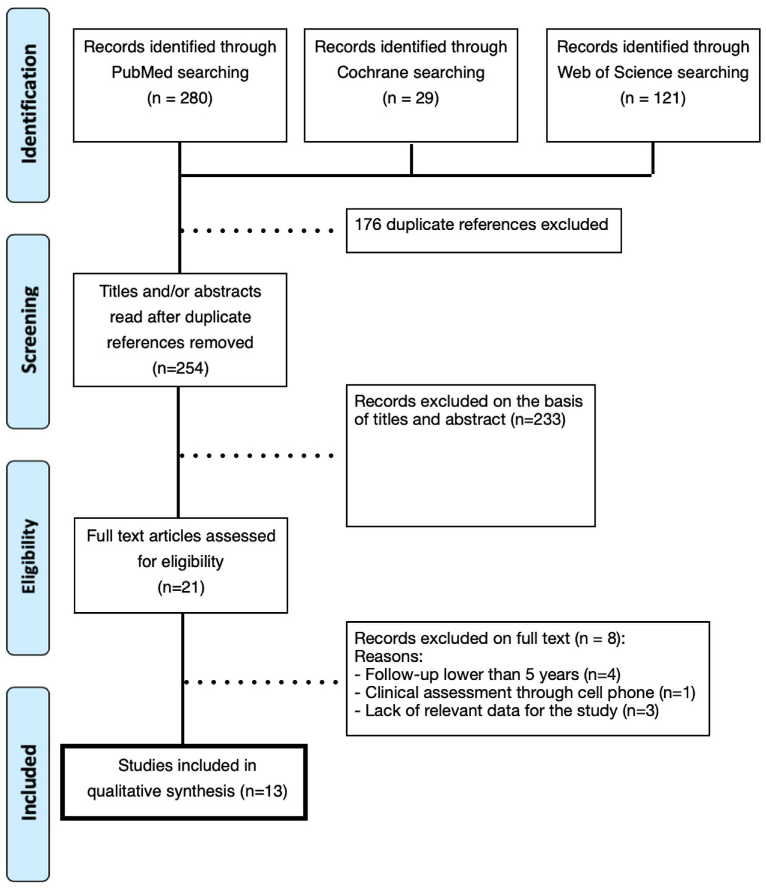
2. Materials and Methods

2.1. Search Strategy

This systematic review was based on the PRISMA standards [20] and defined the research question using the PICOT format [21] (Table S1)— “In a partially edentulous population (P) the realization of adhesive partial fixed prosthesis (I), compared to conventional partial fixed prosthesis (C), have superior clinical results (O) in a minimum follow-up of 5 years (T)?” This protocol was registered in PROSPERO (International Prospective Register of Systematic Reviews), organized by the Centre for Reviews and Dissemination (the University of York, National Institute for Health Research, United Kingdom) with the number CRD42021236749 and the title “Success rate of the adhesive partial fixed prosthesis after five years: systematic review and meta-analysis.”
The search was initiated on 12 November 2020, and conducted through 13 May 2021, using the following bibliographic databases: Medline (PubMed), Cochrane Library, and Web of Science. Two types of search strategies were used, with MESH terms (medical subject headings) “Denture, Partial, Fixed, Resin-Bonded” and with free text: Maryland bridge OR Resin-bonded fixed partial denture OR Resin-bonded fixed dental prosthesis, to which “boolean operators” were applied. In addition to the literature database search, a manual search was conducted. The filters applied in Medline were: language (English, Portuguese, Spanish, French), article type (clinical study, clinical trial, clinical trial protocol, clinical trial phase I, clinical trial phase II, clinical trial phase III, clinical trial phase IV, comparative study, controlled clinical trial, evaluation study, multicenter study, observational study, pragmatic clinical trial, randomized controlled trial, twin study, validation study), and journal category (dental journals). No filters were applied to the remaining databases.

2.2. Inclusion and Exclusion Criteria

The inclusion criteria involved human studies, controlled clinical trials, prospective and retrospective cohort studies, minimum follow-up of 5 years, articles indicating survival rate and/or success rate of prosthetic rehabilitation, studies indicating the characteristics of the infrastructure (e.g., materials and design), studies showing the location of the RBFPD, studies indicating the biological/mechanical complications, and studies with at least ten patients/ten RBFPDs.
The exclusion criteria were studies that included extensive dental preparations (such as inlays or partial/total crowns on at least one retainer), studies including acrylic or metal-acrylic prostheses, in vitro studies, pre-clinical studies, and systematic reviews.

2.3. Study Selection and Data Extraction

To reduce bias, two independent reviewers (M.C.S. and L.A.) selected data from each included article and recorded them in an Excel® sheet (version 15.17, Microsoft, Redmond, Washington, DC, USA). Any potential disagreement and/or discrepancy was resolved by consensus and in the presence of a third reviewer (A.C.).
The following variables were defined in this investigation: first author’s last name; year of publication; sample size; follow-up time; the number of dropouts; number and location of adhesive partial dentures (posterior/anterior and maxillary/mandibular); prosthetic framework material; type of preparation; cement used; biological complications; mechanical complications; and success/survival rates. Additional information was obtained by contacting the investigators when necessary.

2.4. Quality Assessment/Risk of Bias

Quality assessment of the included studies was performed by two independent reviewers (L.A. and M.C.S.) using the modified Downs and Black Quality Assessment scale [22,23], with the methodology designed to contemplate both randomized and non-randomized studies, having 27 scorable items. Divergences and doubts in the evaluations were resolved through a third reviewer (G.V.O.F.).

2.5. Statistical Analysis

The meta-analysis involved the analysis of effect sizes estimated from studies of different sizes, so the sampling variances for each effect size are not equal. Thus, each effect size is weighted by the inverse variance in the analyses. However, the random-effects model was used (5% being considered for significance level). The variability in effect sizes comes from sampling error and true differences in effects between studies. Thus, each effect was weighted by the inverse of the sum of the sampling variance and the between-study variance in the random-effects model for the estimated survival rate, percent per year of estimated failure, and existing complications. Heterogeneities across studies were quantified by the inconsistency test (I2), in which values above 75% were considered an indication of substantial heterogeneity.

3. Results

Through the search strategy, 430 articles were identified: 280 from Medline/PubMed, 29 from the Cochrane Library, and 121 from Web of Science. In the total number of articles (n = 430), 175 duplicate articles and one triplicate article were excluded. The remaining articles (n = 254) were systematically analyzed by two researchers (M.C.S. and L.A.), prioritizing only those that met the previously defined inclusion criteria. As such, 70 articles were selected by title and, subsequently, 21 articles by abstract. Then, the articles were read entirely, eleven articles were selected, and ten articles were excluded. The flowchart of this article’s selection is presented in Figure 1. The Cohen’s kappa obtained was k = 0.91. After the final 11 articles included in the study were read in full, they were quantitatively summarized to evaluate the success/survival rate, the average follow-up time, and the estimated failure rate (Table 1, Table 2 and Tables S2–S4).

3.1. Biological and Mechanical Complications

Biological and mechanical complications were analyzed in all studies to classify them as relative and absolute failures. A total of 142 failures were reported in the 11 studies, corresponding to 18.14%. In this context, regarding the relative failures, the most frequent one was re-cementation (n = 31, 21.83%), followed by chipping of the ceramic (n = 10, 7.04%), reversible trauma (n = 5, 3.52%), cementation error (n = 4, 2.82%), and finally, with only one failure, removal due to lack of aesthetics (n = 1, 0.70%). Regarding absolute failures, debonding (n = 53, 37.32%) is the most common, followed by fracture of the prosthesis framework (n = 12, 8.45%), loss due to trauma (n = 10, 7.04%) and caries (n = 10, 7.04%), periodontal problems (n = 4, 2.82%), and finally, fracture of the abutment tooth (n = 3, 2.11%).
Considering the total exposure time of the RBFPDs obtained by multiplying the total number of prostheses by the average follow-up, the estimated failure rate (%/year) was calculated by dividing the number of biological/mechanical complications by the total exposure time. Therefore, this rate ranges from 0.53%/year [12] to 5.10%/year [27].

3.2. Success/Survival Rate and Risk of Bias/Quality Assessment

This systematic review defined the success/survival rate as the absence of relative and absolute failures. Of the 11 studies evaluated, two showed a success rate [26,27], eight survival rates [12,24,27,28,29,30,31,33], and only one study had both [25]. The highest success rate was 93.1% in Kern et al.’s (2017) study [29]. In the Sasse and Kern (2014) study [32], the rate was 93%, and in Botelho et al. (2014) [25], it was 84.4%. Regarding the survival rate, the highest was 100% and was reported in the study of Sailer et al. [30], and the lowest was 62% in the study of Behr et al. [12]. A total score of more than 15 was obtained in all studies, and they were rated with sufficient quality (Table S2). The scores for each study are summarized in Table S5.

3.3. Statistical Analysis

The survival rate compared between the studies showed a significant result (p < 0.001). This significance was probably increased due to the results obtained in the study by Behr et al. (1998) [12], with the lowest rate obtained. Furthermore, heterogeneity among studies was moderate, with I2 = 58.76%. However, the data were analyzed using the funnel plot to check for bias in the data found, which was not observed (Figure S1).
For the estimated failure rate (% per year), a significant result can be observed for both the data obtained and the moderating effect of time analyzed, respectively, with p = 0.002 and p = 0.012. However, even with the statistical significance achieved, there was no heterogeneity in the studies, with I2 = 0.1%, and furthermore, no bias was observed after checking the result of the funnel plot (Figure S2), which presented a more similar distribution for this parameter.
The complications found were also evaluated statistically to verify any discrepancy between the included studies. However, even with a significant shift in the complications found in Garnett et al. (2006) [26], there was no statistically significant difference between the studies (p = 0.55), and the effect of time, as a moderating contrast, also had no significant influence on the complications observed (p = 0.39). However, it can be stated that there was high heterogeneity among the included studies, with a result of I2 = 93.36%, and there was one study which had bias, Garnett et al. (2006) [26], as shown in the funnel plot (Figure S3).

4. Discussion

This systematic review was performed to analyze the available clinical data on the long-term success and complications of FPDs to debate about the clinical/scientific evidence within the existing literature. Therefore, prospective and retrospective cohort studies were included to estimate the success and five-year survival rates of RBFPDs.
Based on the research carried out, the existing literature presents a limitation in the number of clinical studies of this type of RBFPD, with a heterogeneity of the relevant studies, which makes a comparative analysis between them difficult, both in terms of design (number of bridges and retainers) of the adhesive fixed partial dentures, in terms of surface preparation, and in terms of the success rate of the cement used. In addition, only three studies [34,35] mentioned the success rate and nine the survival rate, one of the most significant gaps found. The fact that there are no established and widely used success and/or survival criteria in each clinical trial adds to the comparison difficulty.
Comparing the maxilla and mandible applicability, RBFPDs were used more in the maxilla than in the mandible across all studies included in this systematic review [12,24,25,26,27,28,29,30,31,32,33]. The study of Botelho et al. [25] confirms the above, where 136 prostheses were placed in the maxilla and only 75 in the mandible. In Galiatsatos et al. [36], even if the difference is minimal, 30 prostheses were made in the maxilla and 24 in the mandible. In addition, RBFPDs are widely used in the anterior areas, as confirmed in the study of Kern et al. [29], where 115 prostheses were placed in the anterior area and zero in the posterior. This is also the case in five studies in this review [27,28,29,31,32].

4.1. Success/Survival or Retention Rates

Its applicability increases with high success/survival or retention rates when placed in the maxilla. In the study by Botelho et al. [25], the retention rate for maxillary molars was 85.7%, and for mandibular molars, 60%. In the same study, the highest retention, success, and survival rates were for mandibular canines with 100%, then maxillary incisors with a survival rate of 97.0%, and retention and success rates of 93.9% [25].
In Aggstaller et al.’s [24] study, the failure survival rate was calculated for the different locations after ten years. The maxilla and posterior region show a higher rate (84% and 82%, respectively) than the mandible and anterior parts (70% and 57%, respectively) [24]. Pjetursson et al., using multivariable Poisson regression, concluding that the failure rate of RBFPDs placed in the maxilla was lower than those placed in the mandible, at 1.07% and 2.36%, respectively, but this difference did not reach statistical significance (p = 0.370). Furthermore, the decay rate was lower for RBFPDs placed in anterior zones than those in posterior zones [2]. A similar result was found by El Mesbahi et al. [37], who reported no statistically significant difference for failures between maxilla and mandible (p > 0.05).
Comparing two studies 15 years apart, Behr et al. [12] and Sailer et al. [30], both with prostheses in the maxilla, mandible, and anterior and posterior zones, showed different survival rates. In Sailer et al.’s [30] study, that rate was 100%, whereas in Behr et al. [12], it was 62%. This fact may have a relationship with the follow-up periods of 6 and 11 years, respectively. Otherwise, even though there were differences among the survival rate of the studies, with a similar period of follow-up (around 6 years), a lower survival rate was observed compared to Sailer et al.’s study. This fact might be subject to interference from the professional abilities/qualities and experience of the clinicians. In the present review and in Alraheam et al.’s study [18], a trend towards a higher success rate was observed when using one retainer instead of two for a bridge replacement. However, according to Rosentritt et al. [38], the fabrication of the RBFPD with two retainers has been more widely used since, under the experimental conditions described in the study, it was concluded that a significantly higher force was required to fracture them compared to using only one retainer.
In this sense, Botelho et al. [39] compared two groups with the metal framework: the first composed of RBFPDs with one retainer and the second group with two. The estimated success rate was 100% for the first group, and for the second group, it was 75%, with an 80% debonding of the retainers in the second group [39]. Kern [28] also made the same comparison, i.e., one retainer and two retainers in anterior RBFPDs made of infiltrated alumina ceramic. In this way, he reported a success rate of 97.5% for only one retainer and 88.3% for two, the reason for the latter’s failure being the fracture of the structure [28].
In the study of Botelho et al. [25], retainers and bridges were used, presenting a survival rate of 90.0%, unlike Behr et al. [12], which used two retainers and only showed a 62% survival rate. Another similar example is the studies of Sasse and Kern [31], who made prostheses with a bridge and a retainer and obtained a survival rate of 93.3%. With a bridge and two retainers, Younes et al. [33] had a survival rate of 83%. Galiatsatos et al. [36] evaluated anterior ceramic (In-Ceram) RBFPDs with two conventional retainers for eight years. In this study, the success rate was 85.15%. The same was found in the systematic study of Alraheam et al. [18], showing that for one retainer, the success rate was 95.01%, and for two retainers, it was 88.96%, while the estimated five-year success rate for the metal denture framework was 88.18% and 84.41% for the non-metal framework. For zirconia, it was 92.07%, for In-Ceram™ alumina, it was 94.26%, and for FRC, it was 84.83%.
For zirconia-ceramic RBFPDs, based on the included studies, the survival rate increased by 0.1% over three years; in the study of Sasse and Kern [32], the rate was 93%, and in Kern et al. (2017) [29], it was 93.1%. In the study of Sasse and Kern [31] with a five-year follow-up, single-retainer zirconia-ceramic RBFPDs showed an excellent clinical outcome and survival of 93.3%. For RBFPDs made of fiber-reinforced composite, in an observation period between four months and 8.9 years, the reported success rate was between 64.7% and 100% [34]. On the other hand, Van Heumen et al. [40], at an average follow-up of five years, reported a success rate of 45%, much lower than that of Miettinen et al. [34] for FRC RBFPDs.
In the systematic review of Miettinen et al. [34], in a three-year follow-up, the success rates for RBFPDs were 82.8% for metal, 88.5% for FRC, and 72.5% for all-ceramic. In the systematic review of Thoma et al. (2017) [41], for a five-year follow-up, the survival rate of RBFPDs was 91.4%. In Pjetursson et al. [42], the meta-analysis of the included studies indicated an estimated five-year survival rate of conventional tooth-supported FPDs of 93.8%, and a five-year survival rate of exclusively implant-supported FPDs of 95.2%.
According to the systematic review of Miettinen et al. [34], the clinical performance of RBFPDs is comparable to conventional FPDs and implant-supported FPDs, which have a five-year survival rate of 94.4% and 93.8%, respectively [42]. Thus, compared with conventional fixed partial dentures, it seems possible to conclude that according to several studies included in this review [25,28,29,30,31,32], the clinical success rates of adhesive bridges are very similar, making this prosthetic option clinically feasible.

4.2. Failure rate and RBFPD Design

Aggstaller et al. [24] studied the estimated survival rate (Kaplan–Meier) after 10 years in a sample collected from 1985 to 2002, focusing on all kinds of failures (catastrophic failures (loss of the prosthesis) and relative failures (e.g., possibility of repair/re-bonding the RBFPD)). The estimated overall survival rate for all failures was 77%, and for catastrophic failures it was 88%. The differences were not statistically significant in the number of abutment teeth (two or three) and the number of bridges (one, two, or three).
Garnett et al. [26] presented a failure rate of 40.3% and a mean survival time of 59.3 months. Like Aggstaller et al. [24], this author did not find statistically significant differences between one or two retainers, although the mean survival time of the two retainers was 38.9 months, and for one retainer it ranged between 55.9 and 65.3 months. Younes et al. [33], also using two retainers, had six cases of debonding (14.3%), and Kern et al. [27] had no failures associated with debonding.
There were fewer failures in adhesive bridges, with only one retainer in most studies. This can be explained by the fact that teeth have a periodontal ligament that allows them some micromovement [43], i.e., since the bridges are bonded on only one retainer, any micromovement of the abutment tooth will not cause debonding. Eventually, it could change the contact point/surface with the natural tooth adjacent to the bridge. We did not find anything in the literature about the latter situation. On the other hand, by having the adhesive bridge bonded at both ends, i.e., a structure that acts as a ferrule between the abutment teeth, micromovement, which can occur during the entire functional activity of the stomatognathic system [43,44], can lead to high stresses/deformations/displacements in one of the retainers/abutment teeth and cause stripping of one of the retainers.
Galiatsatos et al. [36] installed anterior RBFPDs on 49 patients, and 48 were satisfied with the aesthetics. Only one patient reported a small aesthetic asymmetry problem since the area was to be rehabilitated extensively. In the same study, six debondings, eight total fractures, and nine partial fractures occurred in eight years [36]. In the study of Kern and Sasse [45], no debonding was reported in two all-ceramic RBFPDs in a ten-year follow-up.
Miettinen et al. [34] reported that the most frequent failure in metal–ceramic prostheses was decementation (92.6% of all failures). The other losses were fracture of the veneering ceramic, fracture of the metal framework, and caries [34]. The lack of aesthetics was mentioned in only one of the cases, as in Galiatsatos et al. [36] and Aggstaller et al. [24]. In RBFPDs made of fiber-reinforced composite, the failures were chipping of the coating composite and fracture of the fiber-reinforced structure [34].
In the study of Kern and Sasse [45], ceramic RBFPDs had a 26.1% failure rate when using two retainers in a ten-year follow-up and a 5.6% failure rate when using one retainer, but this was not statistically significant. In the systematic review of Alraheam et al. [18], frame debonding and retainer fracture were the most reported technical complications in the included studies, accounting for 82% and 15%, respectively. Caries and periodontal problems, as biological complications, presented rates of 1.7% and 0.6%, respectively [18]. Other failures reported were poor aesthetics, incorrect cementing procedure, and pulpal pathology [18].
For Thoma et al. [41], biological complications included caries in the abutment teeth and progression of periodontal disease, and technical complications included debonding and chipping of the veneering ceramic. Therefore, at a 5-year follow-up, debonding (loss of retention), as in the previous review, was also the most frequent complication, occurring in 15% of the included RBFPDs, followed by chipping of the veneering ceramic, at 4.1% [41].

4.3. Tooth/Teeth Preparation

Dental preparation generates controversy since some studies advocate the no-prep concept [12,26] and others the opposite; the latter do not carry out even minimal dental reductions [24,25,27,28,29,30,31,32,33,35]. Therefore, adequate enamel-preserving preparation can influence the retention of RBFPDs [46]. In the systematic review of Miettinen et al. [34], they examined studies of various degrees of preparation, i.e., either minimal enamel preparation of the abutment teeth, including guide planes, vertical grooves, and occlusal supports, or no preparation.
Galiatsatos et al. [36], for the fabrication of all-ceramic RBFPDs, reduced the preparation margins by approximately 0.5 mm on the lingual surface of the incisors, 1 mm above the gingival margin, and between 1 and 1.5 mm from the incisal edge. Compared to the study of Kern [27], where ceramic prostheses were also made, the connectors in the buccal–lingual and cervical–incisal directions were 2 mm and 3 mm thick, respectively. On the lingual surfaces where the wings are placed, a reduction between 0.5 mm and 0.7 mm was performed [27].
For metal–ceramic prostheses, Aggstaller et al. [24] considered occlusal reductions between 0.3 and 0.5 mm in the posterior teeth and anterior teeth, from 0.4 mm to 0.6 mm mesially and distally for the grooves, and 1 mm palatally for the pin. Younes et al. [33] reported that the occlusal thickness for metal varies between 0.5 and 0.6 mm.
In our opinion, there is one very relevant factor in this issue of tooth preparation that the authors rarely discuss: the intermaxillary relationship and occlusion. Being perfectly understandable that one wants to quantify the amount of reduction to fit the design and thickness of the prosthetic structure, we understand that the available prosthetic inter-arch space is a crucial factor in the “prep versus no-prep” decision. Another relevant factor is the prosthesis insertion axis related to the anatomy of the retainers because the amount of reduction will always depend on the space needed for prosthesis insertion vs. the minimum thickness of the prosthetic material.

4.4. Retention and Retainers

The surfaces of the retainers should be prepared to create roughness, usually blasted with 50 µm alumina oxide particles [25,27]. In Garnett et al.’s [26] study, the particles varied in size, between 50 and 250 µm.
Subsequently, resin cement is applied for the cementation of the retainers. In the present systematic review, Panavia™ resin cement was the most cited cement [25,26,27,28,32,33] but other cement-like substances, such as Microfill-pontic™, Variolink™, Tetric Flow™, Tetric Ceram™, Rely-X™, and HFO™, were also mentioned [12,24,29,30,31].
In the study of Galiatsatos et al. [36], the surfaces were blasted with alumina oxide particles (Korax 250, Bego) and coated with silane (Monobond S, Ivoclar Vivadent®). The rubber dam was placed, then a pumice stone was used to clean the tooth surface, and 37% phosphoric acid (Ultra-Etch, Ultradent®) was placed for 60 s [36]. Finally, Syntac Classic™ dentin adhesive (Ivoclar Vivadent®) was applied. The ceramic crowns were cemented with resin cement, Variolink II™ [34]. Similar to this study, Zitzmann et al. [47] also state that before the cement, phosphoric acid (35% to 37% for 30 to 60 s) should be applied to the tooth surface to achieve the desired mechanical retention.
Sasse and Kern [31] obtained excellent clinical results with resin cement modified with phosphate monomers and resin cement modified with a zirconia-ceramic primer. The results obtained in Rosentritt et al. [38] highlighted that the failure of RBFPDs depends on the success of tooth and surface preparation and that the survival rate of RBFPDs may increase with the development of a better bond between the cement, tooth, and zirconia.
In all the articles listed above, it was found that adhesive cementation (bonding) was always carried out with resin cement. The cementation protocol indeed varied with the evolution of the materials over the years, in terms of the treatment of the prosthesis’s internal surface and the abutment tooth itself, related to the indications of the cement manufacturers, both for the cement per se and for the abutment tooth.
In our opinion, it is essential to emphasize that this type of cementation is the only viable one for the clinical success of these types of rehabilitation. The adhesive cementation protocol is crucial and must be perfectly defined during the prosthesis placement appointment and always based on the latest scientific evidence on the internal surface treatment of the prosthetic structure, the treatment of the abutment tooth, and the properties of the cement.
In the prospective study of Sasse and Kern [31], single-retained zirconia RBFPDs fabricated by CAD/CAM technology are presented as a treatment alternative, offering good aesthetics, minimally invasive preparation, high biocompatibility, and a five-year follow-up rate of 93.3%. However, there is a lack of clinical studies comparing single-retained and double-retained RBFPDs fabricated by CAD/CAM technology [31]. As exceptions, in the study of Sailer [30], the frameworks were made of lithium disilicate (Empress E.max Press™, Ivolar Vivadent®), and in the study of Kern [27], alumina (In-ceram™, Vita Zahnfabrik® Germany) were used.
Again, the rehabilitation’s success depends on the prosthetic structure’s material because it is directly related to the cementation material used. This review seems to make it possible to conclude that ceramic frameworks have a higher clinical success rate than metal–ceramics, which we believe is directly related to the evolution/optimization of cementation materials and protocols, particularly in the last two decades.
However, it is again important to emphasize the utmost importance of the cementation protocol, the location in the anterior zone, and the framework’s design with only one retainer as crucial factors for clinical success. Overall, for the articles included in this systematic review, the studies prior to 2013 had a lower clinical success/survival rate than studies published after 2013, as observed also by Alraheam et al. [18]. This situation seems to be explained by the evolution of materials and cementation protocols.
The results of this review were comparable to a review published in 2017 by Thoma et al. [41] and in 2018 by Alraheam et al. [18]. Their findings, respectively, showed that the estimated 5-year survival rates for RBFPDs were 91.4% and 94.4%. For the same period, the overall survival rate found here was 83.81%.

4.5. Limitations and Future Perspectives

This review had as a first limitation the period of analysis for inclusion, which considered only RBFPDs assessed at least after 5 years; it excluded many articles that reported results after a shorter period. Moreover, the professional experience and ability within the different studies could not be compared. Another point is related to the cement and the volume of cement applied, which can vary according to the author/clinician. Furthermore, since there was a lack of available RCTs, a relatively lower level of evidence was used, mainly from prospective and retrospective clinical studies. Additionally, the long-term clinical studies on RBFDPs were still considered scarce. Therefore, when interpreting the results of this systematic review, note that the evidence level is not relatively strong, with moderate/high heterogeneity among the studies. Future studies must consider long-term and well-designed RCTs.

5. Conclusions

Within the limitations of this study, it was possible to verify that a higher success rate was observed when prostheses had only one retainer and for zirconia-ceramic prostheses, considering the descriptive analysis performed. For the cementation protocol, the anterior zone and the framework’s design with only one retainer were crucial factors for clinical success. Moreover, ceramic frameworks had a higher success rate than metal–ceramics, which we believe is directly related to the evolution/optimization of cementation materials and protocols, which may be suggestively observed in studies published after 2013, with higher success/survival rates. Within a 18,14% of failures, the most common absolute failure was debonding and the most common relative failure was re-cementation.

Supplementary Materials

The following supporting information can be downloaded at: https://www.mdpi.com/article/10.3390/prosthesis5010021/s1.

Author Contributions

Conceptualization, M.C.S. and A.C.; methodology, M.C.S., L.A. and A.C.; validation, M.C.S., L.A., P.F., G.V.O.F. and A.C.; formal analysis, M.C.S., L.A., G.V.O.F. and A.C.; investigation and data curation, M.C.S., L.A., P.F., G.V.O.F. and A.C.; writing—original draft preparation, M.C.S., L.A., P.F., P.C.V., F.A., E.V., G.V.O.F. and A.C.; writing—review and editing, M.C.S., G.V.O.F. and A.C.; visualization, M.C.S., G.V.O.F. and A.C.; supervision, A.C.; project administration, M.C.S. and A.C. All authors have read and agreed to the published version of the manuscript.

Funding

This research received no external funding.

Conflicts of Interest

The authors declare no conflict of interest with the study.

References

  1. Ferro, K.J.; Morgano, S.M.; Driscoll, C.F.; Freilich, M.A.; Guckes, A.D.; Knoernschild, K.L.; McGarry, T.J. The Glossary of Prosthodontic Terms: Ninth Edition. J. Prosthet. Dent. 2017, 117, e1-105. [Google Scholar]
  2. Pjetursson, B.E.; Tan, W.C.; Tan, K.; Brägger, U.; Zwahlen, M.; Lang, N.P. A systematic review of the survival and complication rates of resin-bonded bridges after an observation period of at least 5 years. Clin. Oral Implant. Res. 2008, 19, 131–141. [Google Scholar] [CrossRef] [PubMed]
  3. Rochette, A.L. Attachment of a splint to enamel of lower anterior teeth. J. Prosthet. Dent. 1973, 30, 418–423. [Google Scholar] [CrossRef] [PubMed]
  4. Williams, V.D.; Drennon, D.G.; Silverstone, L.M. The effect of retainer design on the retention of filled resin in acid-etched fixed partial dentures. J. Prosthet. Dent. 1982, 48, 417. [Google Scholar] [CrossRef] [PubMed]
  5. Livaditis, G.J.; Thompson, V.P. Etched castings: An improved retentive mechanism for resin-bonded retainers. J. Prosthet. Dent. 1982, 47, 52–58. [Google Scholar] [CrossRef] [PubMed]
  6. Thompson, V.P.; Del Castillo, E.; Livaditis, G.J. Resin-bonded retainers. Part I: Resin bond to electrolytically etched nonprecious alloys. J. Prosthet. Dent. 1983, 50, 771–779. [Google Scholar] [CrossRef]
  7. Thompson, V.P.; Livaditis, G.J. Etched casting acid etch composite bonded posterior bridges. Pediatr. Dent. 1982, 4, 38–43. [Google Scholar]
  8. Shimizu, H.; Kawaguchi, T.; Takahashi, Y. The current status of the design of resin-bonded fixed partial dentures, splints and overcastings. Jpn. Dent. Sci. Rev. 2014, 50, 23–28. [Google Scholar] [CrossRef] [Green Version]
  9. van Heumen, C.C.; Kreulen, C.M.; Creugers, N.H. Clinical studies of fiber-reinforced resin-bonded fixed partial dentures: A systematic review. Eur. J. Oral Sci. 2009, 117, 1–6. [Google Scholar] [CrossRef]
  10. Meijndert, L.; Raghoebar, G.M.; Meijer, H.J.A.; Vissink, A. Clinical and radiographic characteristics of single-tooth replacements preceded by local ridge augmentation: A prospective randomized clinical trial. Clin. Oral Implant. Res. 2008, 19, 1295–1303. [Google Scholar] [CrossRef]
  11. Creugers, N.H.; De Kanter, R.J.; Verzijden, C.W.; Van’t Hof, M.A. Risk factors and multiple failures in posterior resin-bonded bridges in a 5-year multi-practice clinical trial. J. Dent. 1998, 26, 397–402. [Google Scholar] [CrossRef] [PubMed]
  12. Behr, M.; Leibrock, A.; Stich, W.; Rammelsberg, P.; Rosentritt, M.; Handel, G. Adhesive-fixed partial dentures in anterior and posterior areas. Results of an on-going prospective study begun in 1985. Clin. Oral Investig. 1998, 2, 31–35. [Google Scholar] [CrossRef] [PubMed]
  13. Livaditis, G.J. Cast metal resin-bonded retainers for posterior teeth. J. Am. Dent. Assoc. 1980, 101, 926–929. [Google Scholar] [CrossRef]
  14. Rosenstiel, S.F.; Land, M.F.; Fujimoto, J. Section 1 Planning and Preparation. In Contemporary Fixed Prosthodontics; Mosby, Inc.: St. Louis, MO, USA, 2002; pp. 65–74. [Google Scholar]
  15. Lin, C.-L.; Hsu, K.-W.; Wu, C.-H. Multi-factorial Retainer Design Analysis of Posterior Resin-Bonded Fixed Partial Dentures: A Finite Element Study. J. Dent. 2005, 33, 711–720. [Google Scholar] [CrossRef] [PubMed]
  16. Tanoue, N. Longevity of Resin-Bonded Fixed Partial Dental Prostheses Made with Metal Alloys. Clin. Oral Investig. 2016, 20, 1329–1336. [Google Scholar] [CrossRef] [Green Version]
  17. Acharya, P.H.; Patel, V.V.; Duseja, S.S.; Chauhan, V.R. Comparative Evaluation of Peri-Implant Stress Distribution in Implant Protected Occlusion and Cuspally Loaded Occlusion on a 3 Unit Implant Supported Fixed Partial Denture: A 3D Finite Element Analysis Study. J. Adv. Prosthodont. 2021, 13, 79–88. [Google Scholar] [CrossRef]
  18. Alraheam, I.A.; Ngoc, C.N.; Wiesen, C.A.; Donovan, T.E. Five-year success rate of resin-bonded fixed partial dentures: A systematic review. J. Esthet. Restor. Dent. 2019, 31, 40–50. [Google Scholar] [CrossRef] [Green Version]
  19. Kravitz, N.D. The Maryland bridge retainer: A modification of a Maryland bridge. Am. J. Orthod. Dentofacial. Orthop. 2020, 157, 128–131. [Google Scholar] [CrossRef] [PubMed] [Green Version]
  20. Moher, D.; Liberati, A.; Tetzlaff, J.; Altman, D.G.; PRISMA Group. Preferred reporting items for systematic reviews and meta-analyses: The PRISMA statement. Ann. Intern. Med. 2009, 151, 264–269. [Google Scholar] [CrossRef] [Green Version]
  21. Donato, H.; Donato, M. Stages for undertaking a systematic review. Acta Med. Port. 2019, 32, 227–235. [Google Scholar] [CrossRef] [Green Version]
  22. Downs, S.H.; Black, N. The feasibility of creating a checklist for the assessment of the methodological quality both of randomised and non-randomised studies of health care interventions. J. Epidemiol. Community Health 1998, 52, 377–384. [Google Scholar] [CrossRef] [Green Version]
  23. Hooper, P.; Jutai, J.W.; Strong, G.; Russell-Minda, E. Age-related macular degeneration and low-vision rehabilitation: A systematic review. Can. J. Ophthalmol. 2008, 43, 180–187. [Google Scholar] [CrossRef] [PubMed]
  24. Aggstaller, H.; Beuer, F.; Edelhoff, D.; Rammelsberg, P.; Gernet, W. Long-term clinical performance of resin-bonded fixed partial dentures with retentive preparation geometry in anterior and posterior areas. J. Adhes. Dent. 2008, 10, 301–306. [Google Scholar] [PubMed]
  25. Botelho, M.G.; Ma, X.M.; Cheung, G.J.K.; Law, R.K.S.; Tai, M.T.C.; Lam, W.Y.H. Long-term clinical evaluation of 211 two-unit cantilevered resin-bonded fixed partial dentures. J. Dent. 2014, 42, 778–784. [Google Scholar] [CrossRef]
  26. Garnett, M.J.; Wassell, R.W.; Jepson, N.J.; Nohl, F.S. Survival of resin-bonded bridgework provided for post-orthodontic hypodontia patients with missing maxillary lateral incisors. Br. Dent. J. 2006, 201, 527–534. [Google Scholar] [CrossRef]
  27. Kern, M. Clinical long-term survival of two-retainer and single-retainer all-ceramic resin-bonded fixed partial dentures. Quintessence Int. 2005, 36, 141–147. [Google Scholar]
  28. Kern, M. Fifteen-year survival of anterior all-ceramic cantilever resin-bonded fixed dental prostheses. J. Dent. 2017, 56, 133–135. [Google Scholar] [CrossRef] [PubMed]
  29. Kern, M.; Passia, N.; Sasse, M.; Yazigi, C. Ten-year outcome of zirconia ceramic cantilever resin-bonded fixed dental prostheses and the influence of the reasons for missing incisors. J. Dent. 2017, 65, 51–55. [Google Scholar] [CrossRef] [PubMed]
  30. Sailer, I.; Bonani, T.; Brodbeck, U.; Hämmerle, C.H.F. Retrospective clinical study of single-retainer cantilever anterior and posterior glass-ceramic resin-bonded fixed dental prostheses at a mean follow-up of 6 years. Int. J. Prosthodont. 2013, 26, 443–450. [Google Scholar] [CrossRef] [Green Version]
  31. Sasse, M.; Kern, M. CAD/CAM single retainer zirconia-ceramic resin-bonded fixed dental prostheses: Clinical outcome after 5 years. Int. J. Comput. Dent. 2013, 16, 109–118. [Google Scholar]
  32. Sasse, M.; Kern, M. Survival of anterior cantilevered all-ceramic resin-bonded fixed dental prostheses made from zirconia ceramic. J. Dent. 2014, 42, 660–663. [Google Scholar] [CrossRef] [PubMed]
  33. Younes, F.; Raes, F.; Van Berghe, L.; De Bruyn, H. A retrospective cohort study of metal-cast resin-bonded fixed dental prostheses after at least 16 years. Eur. J. Oral Implantol. 2013, 6, 61–70. [Google Scholar]
  34. Miettinen, M.; Millar, B.J. A review of the success and failure characteristics of resin-bonded bridges. Br. Dent. J. 2013, 215, E3. [Google Scholar] [CrossRef] [Green Version]
  35. Sailer, I.; Hämmerle, C.H. Zirconia ceramic single-retainer resin-bonded fixed dental prostheses (RBFDPs) after 4 years of clinical service: A retrospective clinical and volumetric study. Int. J. Periodontics Restorative Dent. 2014, 34, 333–343. [Google Scholar] [CrossRef] [PubMed] [Green Version]
  36. Galiatsatos, A.A.; Bergou, D. Clinical evaluation of anterior all-ceramic resin-bonded fixed dental prostheses. Quintessence Int. 2014, 45, 9–14. [Google Scholar] [PubMed]
  37. El Mesbahi, N.; Hamza, M.; Bennani, A. Resin-bonded fixed partial dentures with metal framework a report of 49 cases followed for 2 years. Int. J. Info. Res. Rev. 2018, 5, 5782–5786. [Google Scholar]
  38. Rosentritt, M.; Ries, S.; Kolbeck, C.; Westphal, M.; Richter, E.-J.; Handel, G. Fracture characteristics of anterior resin-bonded zirconia-fixed partial dentures. Clin. Oral Investig. 2009, 13, 453–457. [Google Scholar] [CrossRef]
  39. Botelho, M.G.; Chan, A.W.; Leung, N.C.; Lam, W.Y.H. Long-term evaluation of cantilevered versus fixed-fixed resin-bonded fixed partial dentures for missing maxillary incisors. J. Dent. 2016, 45, 59–66. [Google Scholar] [CrossRef] [Green Version]
  40. van Heumen, C.C.; van Dijken, J.W.; Tanner, J.; Pikaar, R.; Lassila, L.V.; Creugers, N.H.; Vallittu, P.K.; Kreulen, C.M. Five-year survival of 3-unit fiber-reinforced composite fixed partial dentures in the anterior area. Dent. Mater. 2009, 25, 820–827. [Google Scholar] [CrossRef]
  41. Thoma, D.S.; Sailer, I.; Ioannidis, A.; Zwahlen, M.; Makarov, N.; Pjetursson, B.E. A systematic review of the survival and complication rates of resin-bonded fixed dental prostheses after a mean observation period of at least 5 years. Clin. Oral Implant. Res. 2017, 28, 1421–1432. [Google Scholar] [CrossRef]
  42. Pjetursson, B.E.; Brägger, U.; Lang, N.P.; Zwahlen, M. Comparison of survival and complication rates of tooth-supported fixed dental prostheses (FDPs) and implant-supported FDPs and single crowns (SCs). Clin. Oral Implant. Res. 2007, 18, 97–113. [Google Scholar] [CrossRef] [PubMed]
  43. Fanmgthh, C., Jr. Periodontia Clínica, 9th ed.; Guanabara Koogan: Rio de Janeiro, Brazil, 2004. [Google Scholar]
  44. Proffit, W.F. Tratamento Combinado Ortodôntico e Cirúrgico. In Ortodontia Contemporânea; Profitt, W.R., Fields, J.R., Eds.; Guanabara-Koogan: Rio de Janeiro, Brazil, 1995; pp. 555–590. [Google Scholar]
  45. Kern, M.; Sasse, M. Ten-year survival of anterior all-ceramic resin-bonded fixed dental prostheses. J. Adhes. Dent. 2011, 13, 407–410. [Google Scholar] [PubMed]
  46. Wei, Y.R.; Wang, X.D.; Zhang, Q.; Li, X.X.; Blatz, M.B.; Jian, Y.T.; Zhao, K. Clinical performance of anterior resin-bonded fixed dental prostheses with different framework designs: A systematic review and meta-analysis. J. Dent. 2016, 47, 1–7. [Google Scholar] [CrossRef]
  47. Zitzmann, N.U.; Özcan, M.; Scherrer, S.S.; Bühler, J.M.; Weiger, R.; Krastl, G. Resin-bonded restorations: A strategy for managing anterior tooth loss in adolescence. J. Prosthet. Dent. 2015, 113, 270–276. [Google Scholar] [CrossRef] [PubMed] [Green Version]
Figure 1. Article selection strategy, according to the PRISMA Flowchart (Preferred Reporting Items for Systematic Reviews and Meta-Analyses).
Prosthesis 05 00021 g001
Table 1. Data extraction table and descriptive analysis of the included articles.
Author/YearAdhesive Partial Fixed Prostheses
(n)
Bridges
(n)
Retainers
(n)
Preparation of Abutment TeethMaterial of the FrameworkCement
TotalMaxMandAntPost
Aggstaller et al., 2008 [24]84NRNRNRNR<3<3YesMetal-CeramicResin cement
(Microfill pontic™ or Variolink™)
Behr et al., 1998 [12]1203819401712Preparation with retention and without retentionMetal-CeramicResin cement
(Microfill-pontic™ and Variolink™)
Botelho et al., 2014 [25]2111367511110011YesMetal-CeramicResin cement
(Panavia EX™ or Panavia 21™)
Garnett et al., 2006 [26]73NRNRNRNR1<2Yes/NoMetal-CeramicResin cement
(Panavia EX™ or Panavia 21™)
Kern, 2005 [27]37241337012YesAlumina-CeramicResin cement
(Panavia TC™ (Kuraray®))
Kern, 2017 [28]2216622011YesZirconia-CeramicResin cement
(Panavia 21™)
Kern et al., 2017 [29]1088233115011YesZirconia-CeramicResin cement
(Panavia 21™ and
Multilink Automix™)
Sailer et al., 2013 [30]14869511YesLithium disilicateResin cement
(Tetric Flow™, Tetric Ceram™, Rely-X™, Panavia F™, HFO™, and Variolink™)
Sasse and Kern, 2013 [31]30191130011YesZirconia-CeramicResin cement
(Panavia 21™ and
Multilink Automix™)
Sasse and Kern, 2014 [32]42261642011YesZirconia-CeramicResin cement
(Panavia™)
Younes et al., 2013 [33]422913241812YesMetal-CeramicResin cement
(Panavia™ or Panavia EX™)
Max.: maxilla; Mand.: Mandible; Ant.: anterior; Post.: Posterior; NR: not reported.
Table 2. Table of data extraction and descriptive analysis of the included articles.
Author/YearFollow-Up Median
(Years)
Biological/Mechanical Complications
(n)
Estimated Failure Rate (%/Year)Total Exposure Time of RBFPDS
(Years)
Estimated Success Rate
≥5 Years
(T MAX)
Estimated Survival Rate
≥5 Years (T MAX)
Aggstaller et al., 2008 [24]6.31212/529.2 = 2.2784 × 6.3 = 529.2NR77%
Behr et al., 1998 [12]1177/1320 = 0.53120 × 11 = 1320NR62%
Botelho et al., 2014 [25]9.44646/1983.4 = 2.32211 × 9.4 = 1983.484.4%90%
Garnett et al., 2006 [26]113131/803 = 3.8673 × 11 = 803NR75%
Kern, 2005 [27]5.31010/196.1 = 5.1037 × 5.3 = 196.1NR83.1%
Kern, 2017 [28]15.744/345.4 = 1.1622 × 15.7 = 345.4NR90.9%
Kern et al., 2017 [29]7.71313/831.6 = 1.56108 × 7.7 = 831.693.1%NR
Sailer et al., 2013 [30]622/84 = 2.3814 × 6 = 84NR100%
Sasse and Kern, 2013 [31]5.444/162 = 2.4730 × 5.4 = 162NR93.3%
Sasse and Kern, 2014 [32]5.233/218.4 = 1.3742 × 5.2 = 218.493%NR
Younes et al., 2013 [33]71010/294 = 3.442 × 7 = 294NR83%
NR: not reported.
Disclaimer/Publisher’s Note: The statements, opinions and data contained in all publications are solely those of the individual author(s) and contributor(s) and not of MDPI and/or the editor(s). MDPI and/or the editor(s) disclaim responsibility for any injury to people or property resulting from any ideas, methods, instructions or products referred to in the content.

