Case Report

Computer-Guided Osteotomy with Simultaneous Implant Placement and Immediately Loaded Full-Arch Fixed Restoration: A Case Report

1
Department of Clinical Surgical, Diagnostic and Pediatric Sciences, University of Pavia, 27100 Pavia, Italy
2
Private Practice, 29011 Borgonovo Val Tidone, Italy
3
School of Dentistry, Department of General Surgery and Medical and Surgical Specialties, University of Catania, 95124 Catania, Italy
4
Private Practice, 15057 Tortona, Italy
5
School of Dentistry, Department of Biomedical, Surgical and Dental Sciences, University of Milan, 20142 Milan, Italy
*
Author to whom correspondence should be addressed.
Prosthesis 2023, 5(1), 221-233; https://doi.org/10.3390/prosthesis5010017
Submission received: 17 November 2022 / Revised: 18 January 2023 / Accepted: 5 February 2023 / Published: 9 February 2023
(This article belongs to the Collection Oral Implantology: Current Aspects and Future Perspectives)

Abstract

:
Aim: This case report aims to illustrate a clinical protocol that allows for the rehabilitation of patients requiring extensive osteotomy, simultaneous implant placement, and full-arch, screwed-in prosthetics in one session. This protocol allows for the improvement of the aesthetics and functionality of the fixed implant-supported prosthesis through the preoperative planning of all surgical procedures, including osteotomy, and of the prosthesis through the application of 3D-printing technology for the creation of surgical templates and prostheses. Methods: This case report concerns a 72-year-old patient, ASA1, who, following diagnosis, the establishment of a treatment plan, and the provision of informed consent, opted for an immediate, full-arch rehabilitation of the lower arch. The digital planning stage started with the correct positioning of the fixtures. The proper bone levels were found and used to guide the creation of the provisional screwed-in prothesis. Two templates with the same supports (landmarks/pins) were then 3D-printed: a positioning template, including a slit to assist the surgeon during the osteotomy, and a surgery template to assist the surgeon during the implants’ positioning. A screwed-in prosthesis encased in resin C&B MFH (NEXTDENT®, Soesterberg, The Netherlands) was delivered. Minimal occlusal adjustments were performed. Results: In a single clinical session, through careful planning and the pre-operative 3D printing of a prosthesis, a temporary implant-supported prosthetic rehabilitation was possible in a case that required an extended osteotomy. Clinically, the correspondence between the virtual design phase and the final realization was consistent. At a functional level, the provisional prosthesis required minimal occlusal adjustments and the DVO values obtained in the immediate post-operative period were found to be comparable to those of the virtual design. By planning the final position of the bone and the implants in advance, it was possible to deliver a full-arch prothesis with proper implant emergence, occlusal vertical dimensions, and occlusal relationship. Conclusion: This fully digital protocol allows the clinician to preview and plan the osteotomy and implant surgery as well as the delivery of the temporary, immediately loaded, complete, fixed prosthesis in patients who are candidates for post-extraction surgery with the need for severe osteotomy.

1. Introduction

In recent years, the possibility for the installation of an implant-supported fixed prosthesis has completely revolutionized the clinical approach to patients with partial and total edentulism [1]. It is now possible, in selected cases, to perform the extraction of the compromised dental elements, the placement of the implants, and the delivery of the immediate temporary prosthesis during the same surgery. This “Three in one” approach is sometimes challenged by intraoperative problems such as the need to perform major osteotomy [2]. This procedure can cause delays in the delivery of the temporary prosthesis. Therefore, it appears necessary to map and design the bone plain for the osteotomy in the preoperative phase and proceed with the creation of tools that allow the clinician to perform guided surgery that maximally reflects the digital programming process performed in the preliminary phase [3]. Performing all the steps of the “3 in 1” process in a guided way allows the clinician to arrive at the final delivery stage of the provisional prosthesis in a more predictable way. The previsualization of each phase and the computer-guided design of said phase is the key to the success of this full-arch, fully digital approach in a single session [4].
The acquirement of a reference bone plane through a precisely guided osteotomy [5] is essential to obtain a prosthesis—provisional or definitive—that is congruous in its interface with the soft tissues and guarantees the aesthetic and functional values established during the design phase without requiring further sessions. The method typically used for osteotomy is empirical, freehand, and sometimes guided by a measurement with a periodontal probe [6,7]. The precision of freehand osteotomy is low and variable depending on the experience of the operator; the risk of complications such as the injury of important anatomical structures adjacent to the site is also greater [8,9]. Measurements made with periodontal probes have been addressed in the literature by various studies [10]. However, even through the use of these techniques, the result is never suitably predictable, especially in reference to the angle of the plane and the intra- and inter-arch symmetry. It has been shown [11] that a reduction in intraoperative error is achieved if the more complicated phases are guided step by step. In recent years, programming software and 3D printers [12,13] have played pivotal roles in every area of medicine and dentistry. The forerunner of the guided technique was the placement of an implant using a 3D surgical guide after superimposing intraoral and CBCT scanning [14]. Studies [15,16,17] have confirmed that placing an implant with a surgical guide is more efficient and precise than the freehand technique. Furthermore, several studies [18,19] have investigated the difference in terms of angular and millimetric precision between the traditional guide stent performed in a dental laboratory and the digital one using 3D printers after combination with clinical information and a clinical report on DICOM data from CBCT [20]. 3D bioprinting is an important topic in several fields of medicine and its development has proven particularly successful in the fields of dentistry and tissue engineering. The progress made in the last decade is remarkable, but given its recent introduction, the technology still has many aspects that can be optimized and improved [21,22]. The digitization of dentistry, which has already been largely implemented with the advent of cone-beam computed tomography (CBCT) and intraoral scanning, has reached its peak through the combination of these data with 3D-printing design and manufacturing. Its applications in dentistry relate to various disciplines including prosthetics, oral and maxillofacial surgery, orthodontics, and oral implantology.
Still, there is little evidence in the literature regarding computer-guided osteotomy. The guidance of an osteotomy in a quantitatively and qualitatively precise manner is the first step to providing correct implant positioning and guiding the rehabilitation of the final prosthetic spaces [23,24,25]. Giant steps have been made in the field of orthopedic osteotomy using a digital guide [26,27], but not as many studies on the accuracy of digital osteotomy guidance have been conducted in the dental field.
Therefore, this case describes an approach to surgical and prosthetic complex rehabilitation and forms the basis for the research and development of a workflow that can predictably help clinicians. It allows for a computer-guided osteotomy, the placement of endo-osseous implants, and immediate loading through a preoperatively printed, fixed prosthesis. These procedures were performed in a single session through a fully digital workflow and with the assistance of 3D printers. Although the potential offered by the joint use of all these technologies is scientifically valid and proven, the task that must now be undertaken is to provide clinicians with scientifically validated protocols that allow them to implement entirely digital methods in daily practice [28].

2. Results

This clinical case was successful with respect to the adherence between the preoperative planning process and the outcome in terms of implant position and the aesthetics/function of the prothesis. Thanks to the digital design, surgical times were reduced, and the extension of the flap was limited, thereby reducing post-operative symptoms [29,30]. On the one hand, the guided osteotomy process preserved the hard tissues through a precise identification of the required extension; on the other hand, the time required to passivize and functionalize the provisional prothesis was reduced. The digital design of the prosthesis required few occlusal modifications, as it was quite adherent to the patient’s tissues, the occlusal relationship, and the DVO. The patient immediately showed satisfaction with the result and presented no significant postoperative symptoms or complications [31]. The incorporation of the osteotomy within guided surgery was fundamental both for the correct positioning of the implants and for the creation of a suitable temporary prosthesis that could be screwed in within the same session. Furthermore, there was a good degree of coherence between the digital design and the surgical execution in terms of apico-coronal discrepancy and the angulation and inclination of the implants placed with the surgical guide [32,33].

3. Discussion

The advent of 3D instrumentation has made it possible to design the osteotomy phase by taking advantage of a stereolithographic template that guides cutting tools more precisely [34]. The osteotomy is customized through CBCT based on the anatomical characteristics of the patient, thus improving the surgical approach, reducing the risk of fractures and injuries of noble structures, and accelerating completion times. Several studies in the literature [35] have evaluated and validated the accuracy of this procedure by noting a non-significant discrepancy between the planned surgery and the one performed, or between the pre- and post-procedural measurements obtained from CBCT images. The guided approach to osteotomy is also a field of investigation in maxillofacial surgery, orthopedics, and oncological surgery [36,37,38,39,40].
Scientific studies to date boast of the effectiveness and efficiency of the guided approach as long as the design is carefully curated with the use of the latest-generation equipment. The case report under analysis laid the foundations for the following observations, which may be analyzed in future studies:
(a)
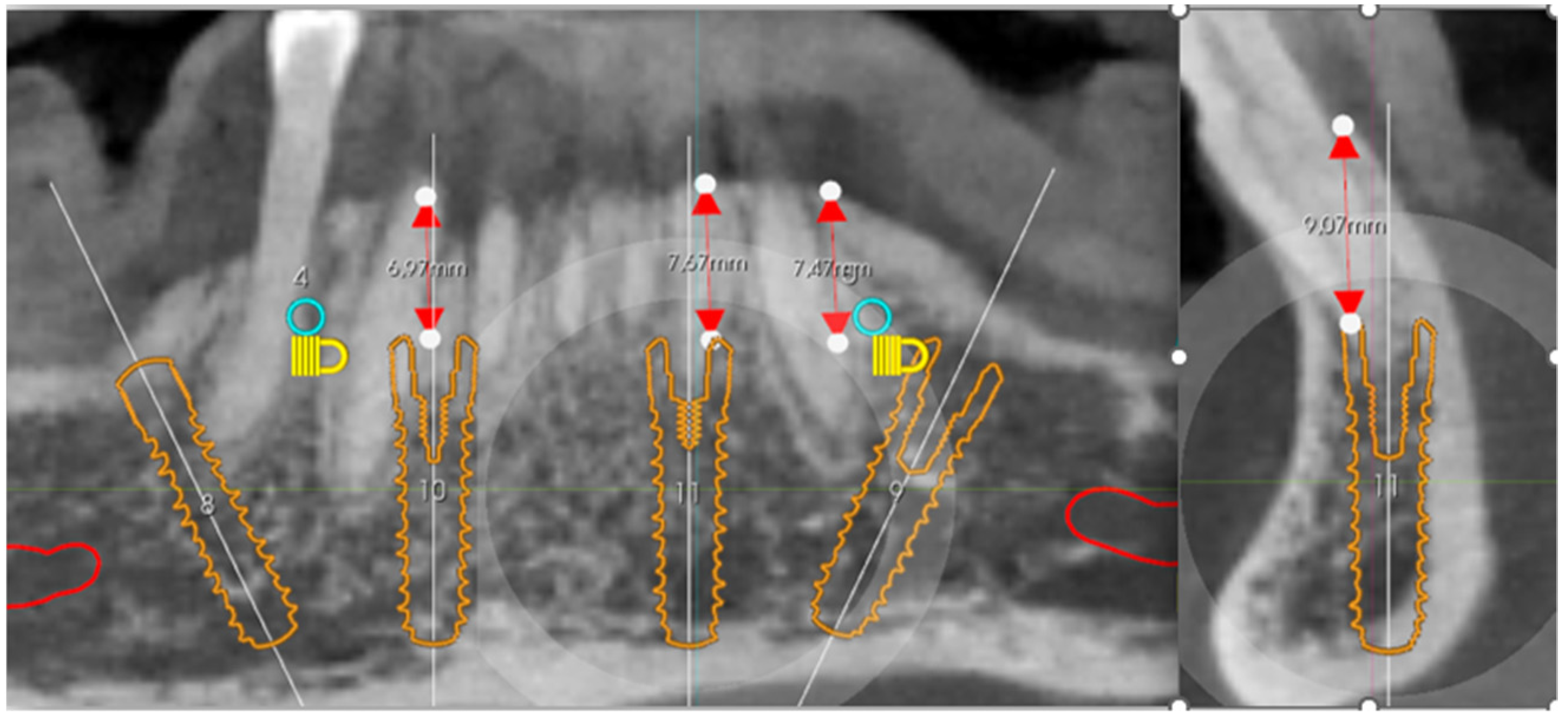
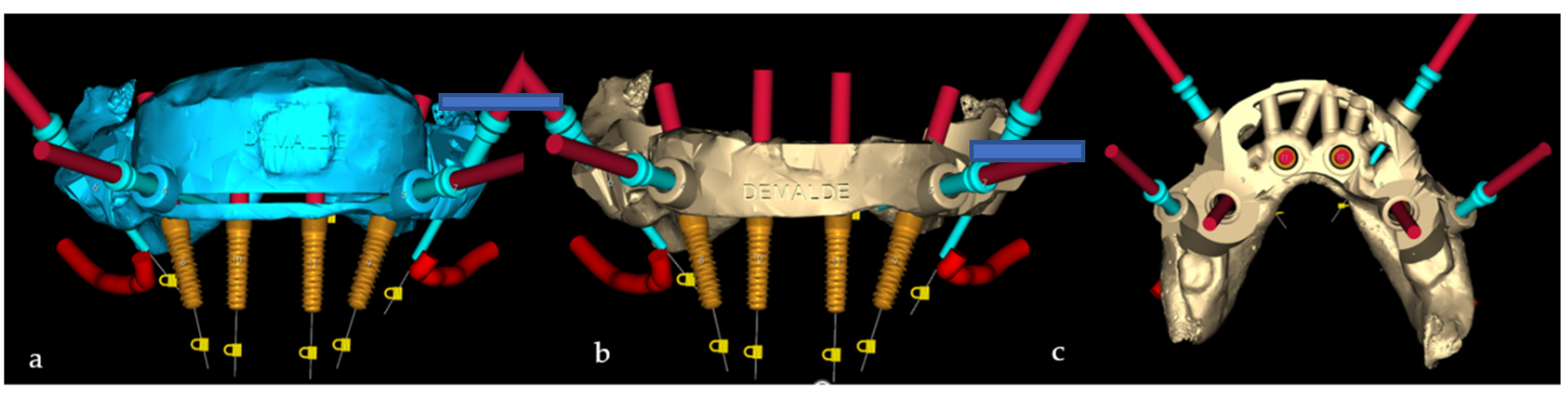
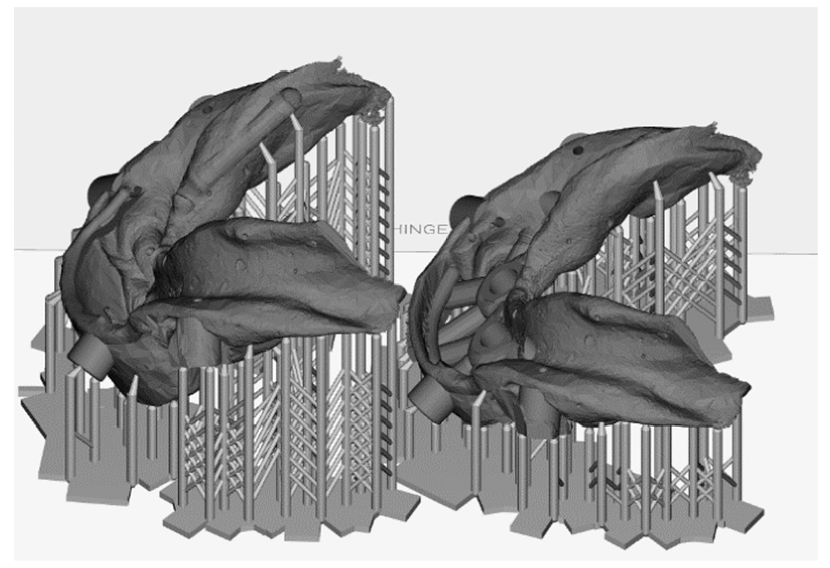
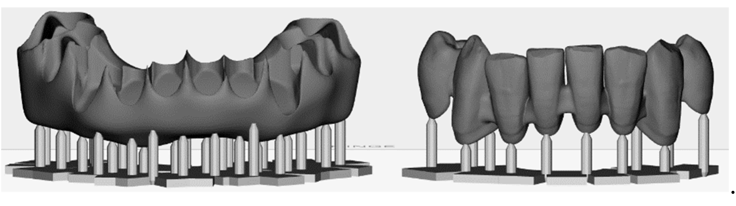
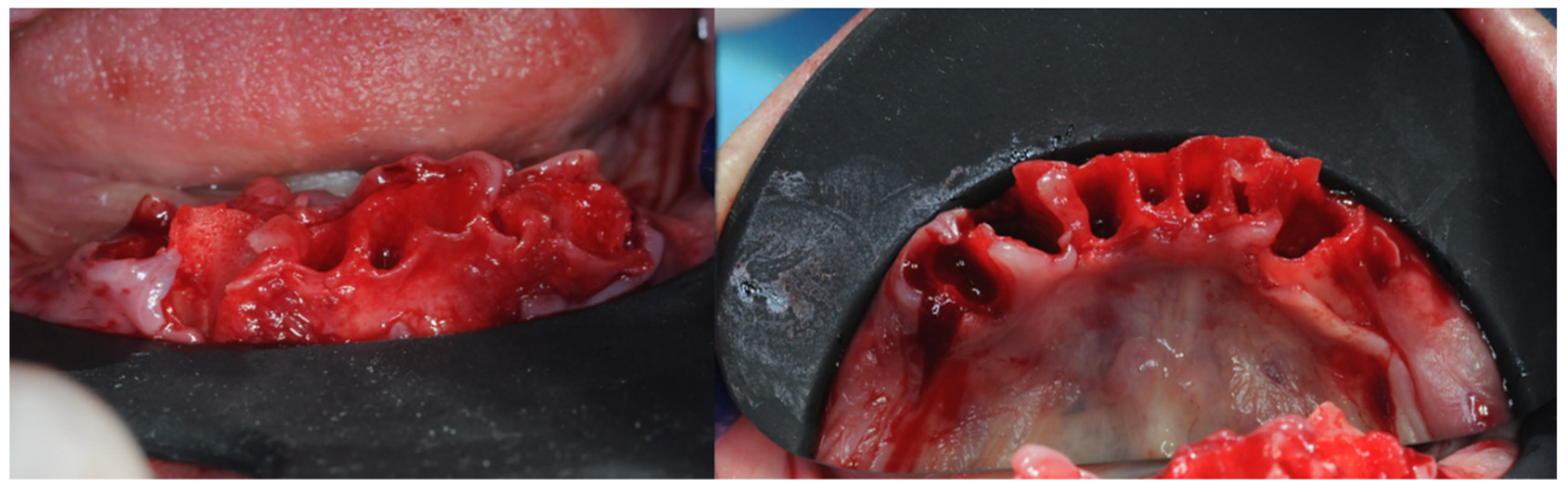
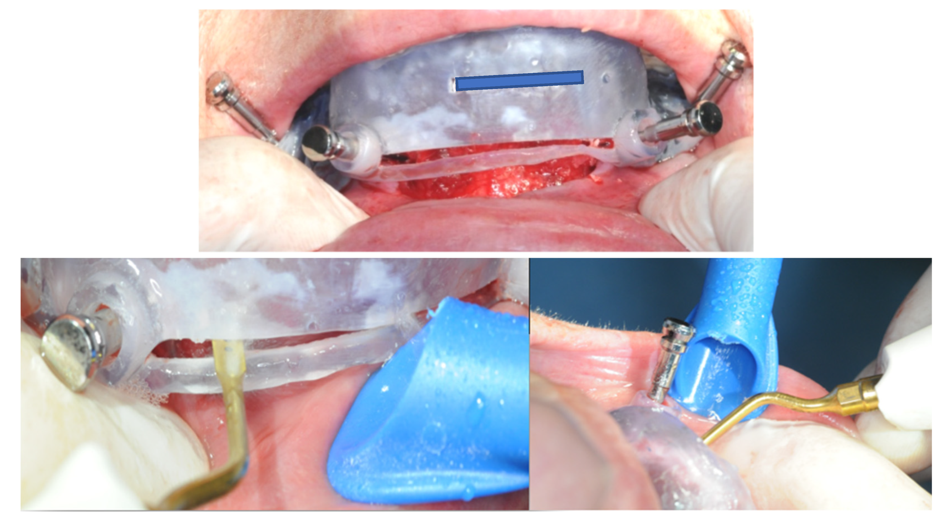
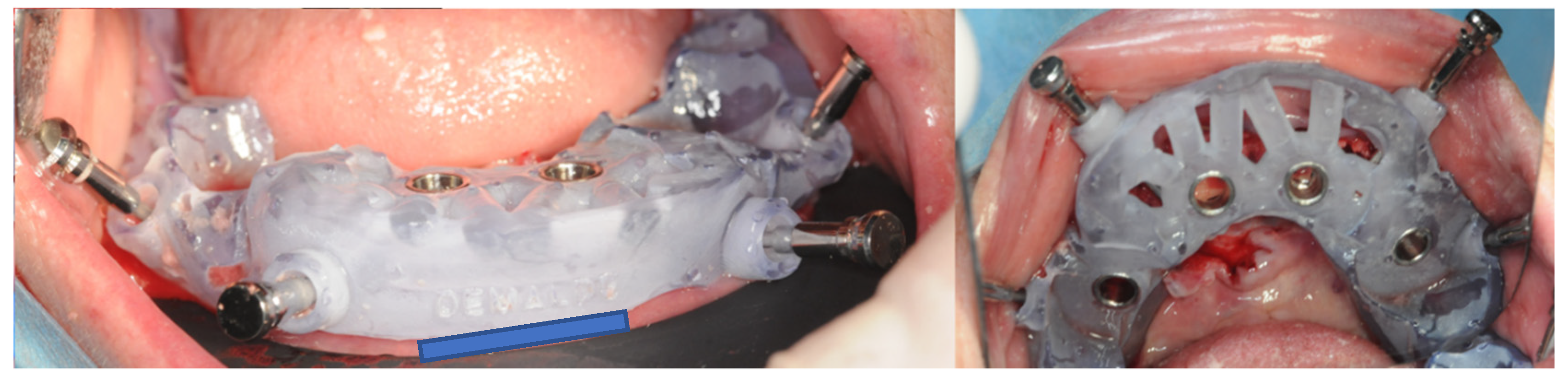
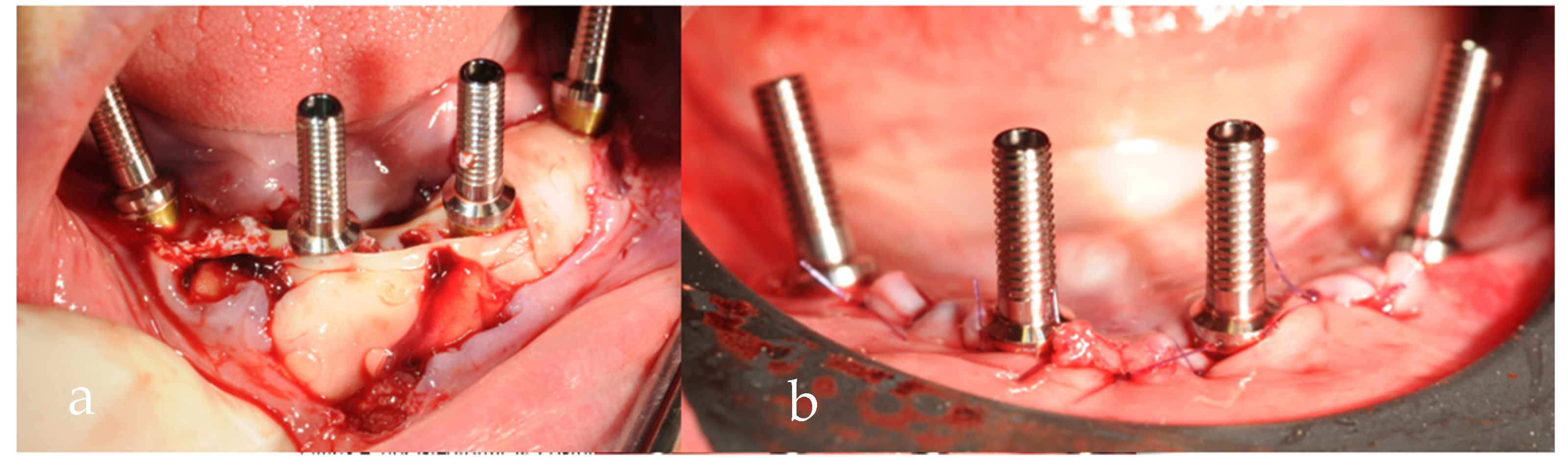
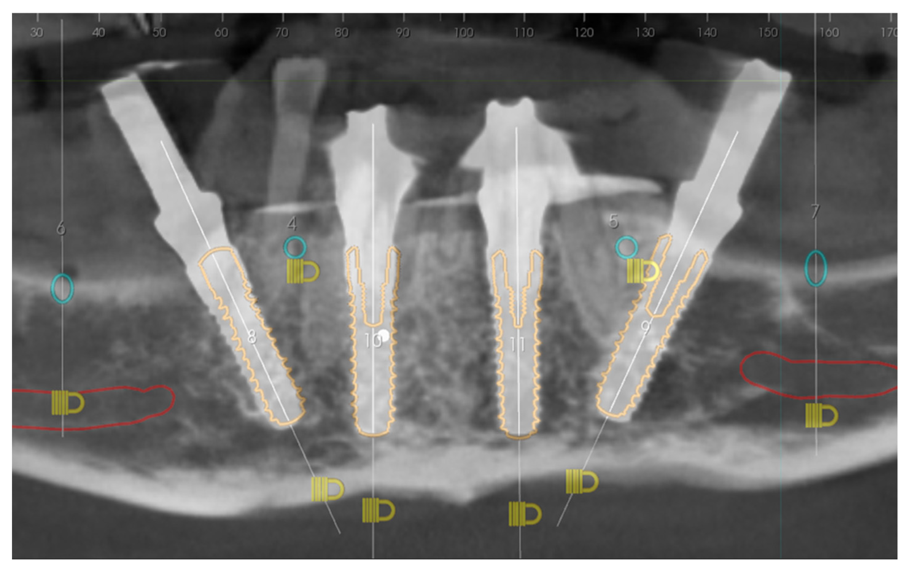
Support of the osteotomy and surgical guide (valence of the dental support). The design of the osteotomy positioning template provided support for all the dental elements present in the arch, from element 3.3 to element 4.4. The operative choice to keep the support elements for the osteotomy’s cutting template is supported by the literature. Currently, scientific studies such as that of Kholy et al. conducted in 2019 [41] have shown that the support provided by four stable elements or an entire arch is comparable in terms of implant positioning deviation with respect to the plan itself [33,42,43]. On the other hand, the positioning of implants starting from a template that has fewer dental elements as support is less predictable. The operative and design choice of the case was directed towards the realization of a first osteotomy guide template with dental support. However, in our experience, the amount of dental support is not a significant variable when the template is designed and stabilized by means of stabilization pins specially placed on the buccal side [44,45]. Any positioning discrepancies that could occur in full-arch rehabilitations due to the lack of dental elements or the presence of some of them with high mobility are bypassed by the presence of stable stabilization pins or landmarks. In the first instance, the creation of a predetermined bone plane provided greater predictability for the subsequent implant’s positioning, despite the fact that, due to the clinical needs of the case, the implant surgical guide was exclusively supported in a bone-mucosal fashion. Greater stability, positioning effectiveness, and design adherence were achieved by using the same positioning pins as the osteotomy guide as a reference point.
(b)
Arrangement of the slit for osteotomy and the role of piezosurgery. The slit prepared in the osteotomy guide was designed on the basis of the thickness of the inserts used with the piezosurgery approach in order to reduce the risk of the inclination of the drill in the surgical preparation of the osteotomy plane. The piezo-surgical approach to osteotomy has been extensively studied and, subsequently, validated in the literature on orthognathic surgery; it is more conservative with respect to the surrounding tissues, causing less bleeding and less severe damage to nerve structures [36]. In addition, regarding oral surgery, the piezosurgical technique was advanced above all for maxillary sinus lift and the extraction of impacted wisdom teeth, thus confirming its lower risk of postoperative complications compared to the traditional method using rotating burs [46,47,48].
(c)
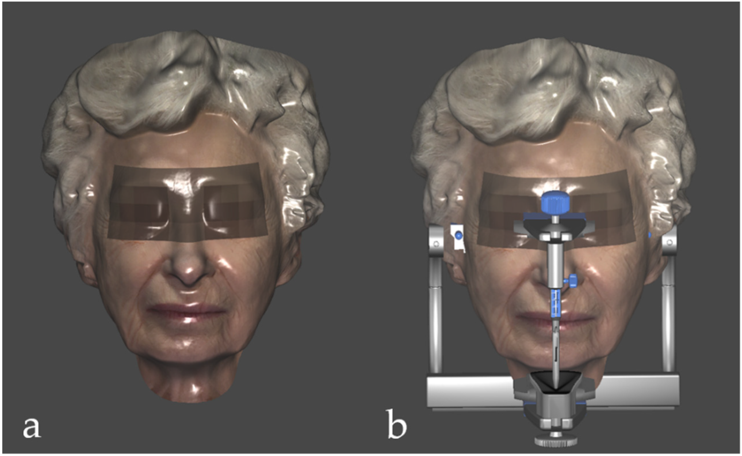
Facial scanning as an additional tool. Facial scanning represents a recent and innovative method with significant advantages in terms of the investigation and clinical planning of complex cases [49]. From an aesthetic perspective, this technique allows the clinician to program the digital smile design in a three-dimensional way, with a greater and more precise number of landmarks available [50,51]. From a functional perspective, the face scan combined with the scan of the arches, the CBCT, and the virtual articulator allow for the evaluation of the gnathological aspects of complex rehabilitation [52], which will be the subject of future research. Furthermore, it is offered as a means of making doctor–patient communication more effective [53].

4. Materials and Methods

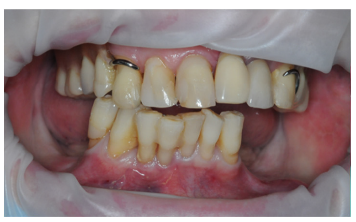
4.1. Selection of the Case

The patient (72 years of age) was in good health (ASA 1). Clinical examination revealed the presence of 6 extruded lower teeth, with mobility 3, pain on percussion, and relapsing periodontal abscesses. After discussing the case with the patient and providing all the explanations regarding the treatment alternatives, the patient opted for the treatment plan entailing the avulsion of the residual mandibular elements and an immediate, post-extractive, screwed, full-arch prosthesis of 12 elements, while the upper arch was subsequently finalized with a removable partial prosthesis. The patient was included in this digital research workflow as she met the following criteria: (1) age over 18 ya; (2) presented compensatory bone hyperplasia, surface irregularities, and the need for osteotomy; (3) had no serious systemic disease; (4) offered sufficient compliance for immediate loading implant surgery; (5) signed informed consent; (6) and agreed to the use of data for research purposes.

4.2. Data Acquisition

The necessary data acquired for the case study and 3D design were as follows: intraoral and facial photos and scans, CBCT, and analog transfer facebow on a compatible digital articulator. The instrument used to perform cone-beam CT was the CareStream® 81003D (Carestream Health Italia srl, Genova, Italy). The DICOM file obtained was exported to Implant3D® software (Medialab spa, Milano, Italy) (Figure 1). The acquisition of the intraoral scan of the dental arches was performed with the Primescan® scanner (Dentsply Sirona, Charlotte, NC, USA) and the STL data files were exported to Exocad® (Exocad gmbH, Darmstadt, Germany). Face scans were performed with Bellus3D® app (Bellus3D, Campbell, CA, USA) via iPhone12 (Figure 2).

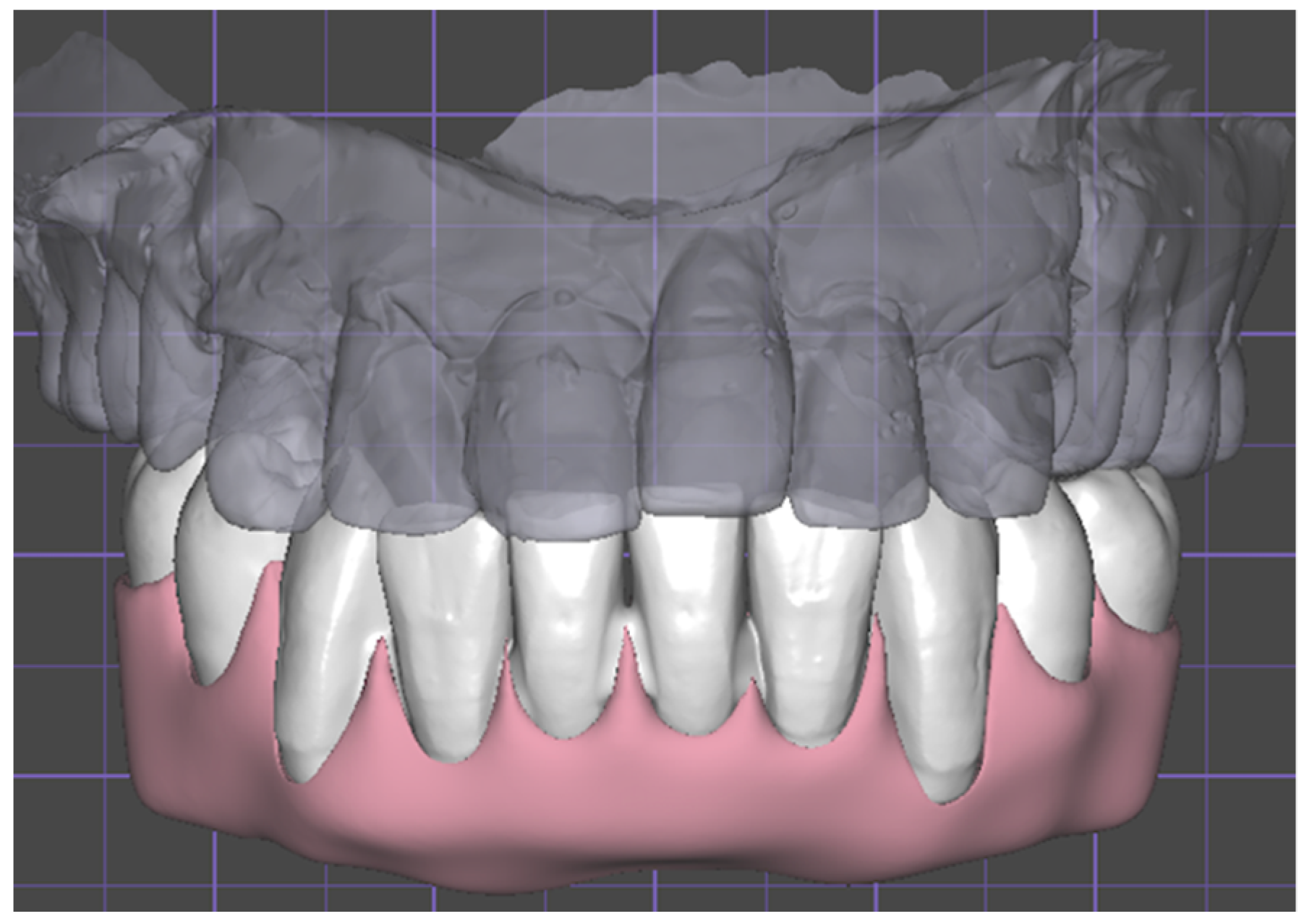
4.3. Diagnostic Wax-Up

Firstly, the residual dental elements in the lower arch were digitally extracted and the adequate DVO was found [54]. At this point, the digital smile design and the virtual articulator guided the positioning of the dental elements from position 3.5 to 4.5 of the lower provisional screwed-in prosthesis, and from positions 1.4 to 1.6 and 2.4 to 2.6 of the upper removable prothesis. (exocad®, Darmstadt, Germany). Once the desired positioning of the dental elements was obtained, the wax-up was exported to Implant3D® (Media Lab–S.p.A., Milan, Italy) in STL format to guide the planning of the implant’s installation. Only once the extent of the osteotomy had been established was the wax-up of the teeth integrated with that of the gum to complete the temporary screwed-in lower prosthesis (Figure 3).

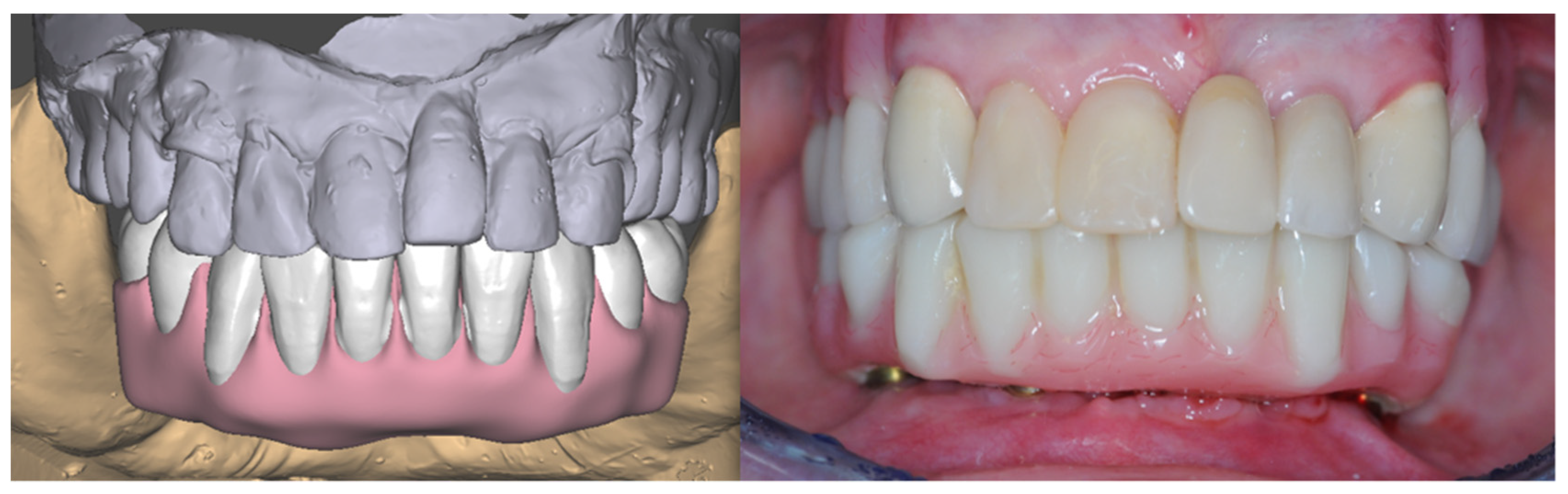
4.4. Design and 3D Printing of the Templates

Once the DICOM file had been imported and aligned with the STL files obtained from intraoral scans [55], the need to perform an osteotomy in order to correctly position the implants became evident (Figure 4).
A tooth-supported positioning template was created with two functions: to guide the positioning of the anchor pins for the implant template and to provide a reference to the surgeon during the execution of the osteotomy through the slit included in the lower part (Figure 5a). The creation of this template is essential, since once the extractions and the osteotomy have been performed, all the landmarks used during the design phase will be lost; therefore, only the anchor pins will guide the positioning of the implant template. At this point, the implant template is designed with the same anchor pins (Figure 5b,c). Once the STL files of both templates had been created, the data were sent to a 3D Printer (Moonray S, SprintRay Inc®, Los Angeles, CA, USA) (Figure 6). The templates were printed with KeyGuide resin (KeyPrint®, Singen, Germany). The printing process lasted for 90 min; afterwards, both templates were cleaned in an ultrasonic bath filled with isopropyl alcohol for 10 min, dried, and cured for 20 min with UV light at a temperature of 60 °C.

4.5. Prosthesis

Once the design of the surgical guides was completed and the extent of the osteotomy was established, it was possible to design the gingival part of the temporary prosthesis. The main focus was to give the metal-free prothesis adequate thickness to guarantee the necessary rigidity; secondarily, the contact surface between the prosthesis and the gingiva was crafted in order to avoid food stagnation and provide good cleanability (Figure 7). The prosthesis was then printed with C&B MFH resin (NextDent®) using a 3D printer (MoonRay S SprintRay®). The printing process lasted 70 min; afterwards, both templates were cleaned, dried, and cured following the same procedure as that of the surgical guides.

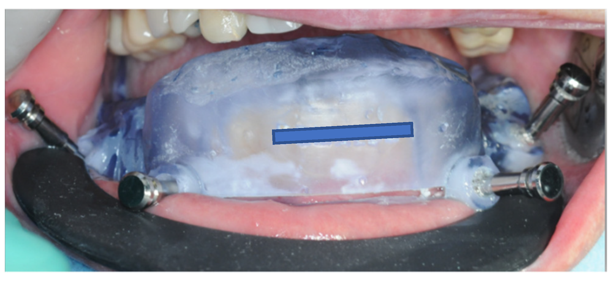
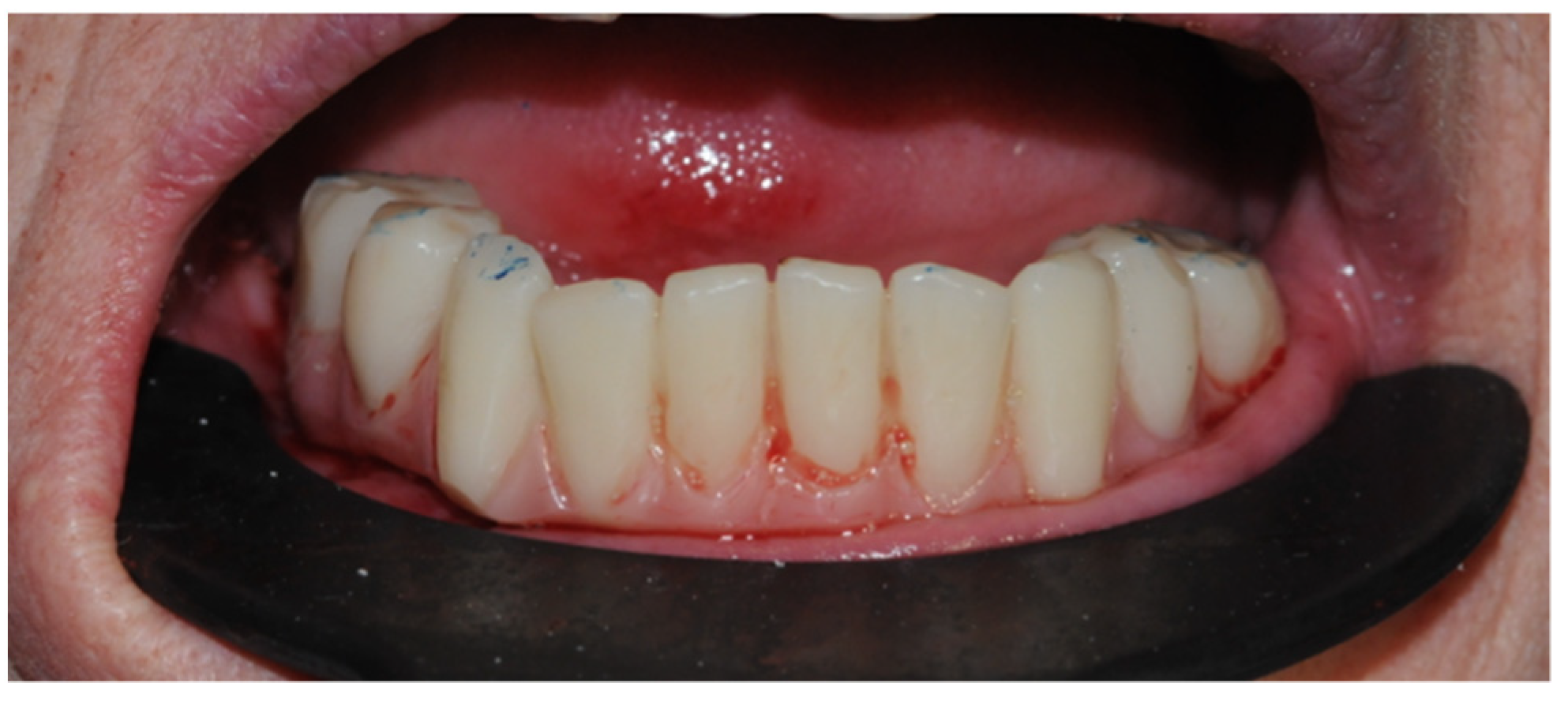
4.6. Surgical Procedure

The patient was prescribed Amoxicillin + Clavulanic Acid (875 mg + 125 mg) according to the following dosage regime: 2 g 1 h before surgery and then 1 g every 12 h for 6 days [56]. Before starting the surgical procedure (Figure 8), the patient’s oral cavity was rinsed with 0.30% chlorhexidine for one minute. The perioral surfaces were disinfected with gauze soaked in 0.30% chlorhexidine. After checking the fit of the positioning template and performing plexus anesthesia with mepivacaine and adrenaline (1:100,000), the anchor pins were inserted (Figure 9).
Once the template was removed, residual dental elements were extracted, the alveoli were curetted to remove any inflammatory tissue, and a full-thickness flap was elevated (Figure 10).
Once the first template had been re-applied, the osteotomy procedure begun. The Piezo insert (Piezosurgery Mectron®, Genova, Italy) was placed on the lower part of the slit and progressively cut away the bone. In this way, the osteotomy levels were compatible with those of the digital plan, and the implant positioning template did not present any interference with the underlying bone (Figure 11).
The anchor pins of the first template guided the positioning of the second template; thus, the implant alveoli creation process begun (Figure 12).
The guided surgical protocol used (Intra-Guide®, Intra-Lock, Salerno, Italy), which was employed for the incremental preparation of the implant site both in terms of diameter and length (Figure 13).
Once the implant site had been created, the implant was positioned on a mounter and screwed-in. All implants (4.0 × 13 mm Gold & Blue Intra-Lock®) were inserted with torques between 40 and 45 N/m. At this point, the bone surfaces were polished using osteotomic burs (Intra-Guide®, Intra-Lock, Salerno, Italy) mounted on a straight handpiece (Figure 14).
As planned, intermediate abutments with a height of 3 mm (Flat-One Wide Intra-Lock®) were screwed onto the implants with a torque of 35 N/m and the titanium provisional abutments were positioned to proceed with the passivation and cementation of the temporary prosthesis using CEM- LOCK (Intra-Lock®), a self-adhesive double-paste cement. To obtain better healing of hard and soft tissues and fewer post-operative symptoms, membranes of L-Prf (Intra-Spin Intra-Lock® Protocol) were placed before suturing the flap (Figure 15a,b).
The provisional prosthesis was screwed with a torque of 10 N/m, light occlusal adjustments were performed, and the patient was discharged following post-op CBCT and the administration of 1 g of paracetamol (Figure 16 and Figure 17).

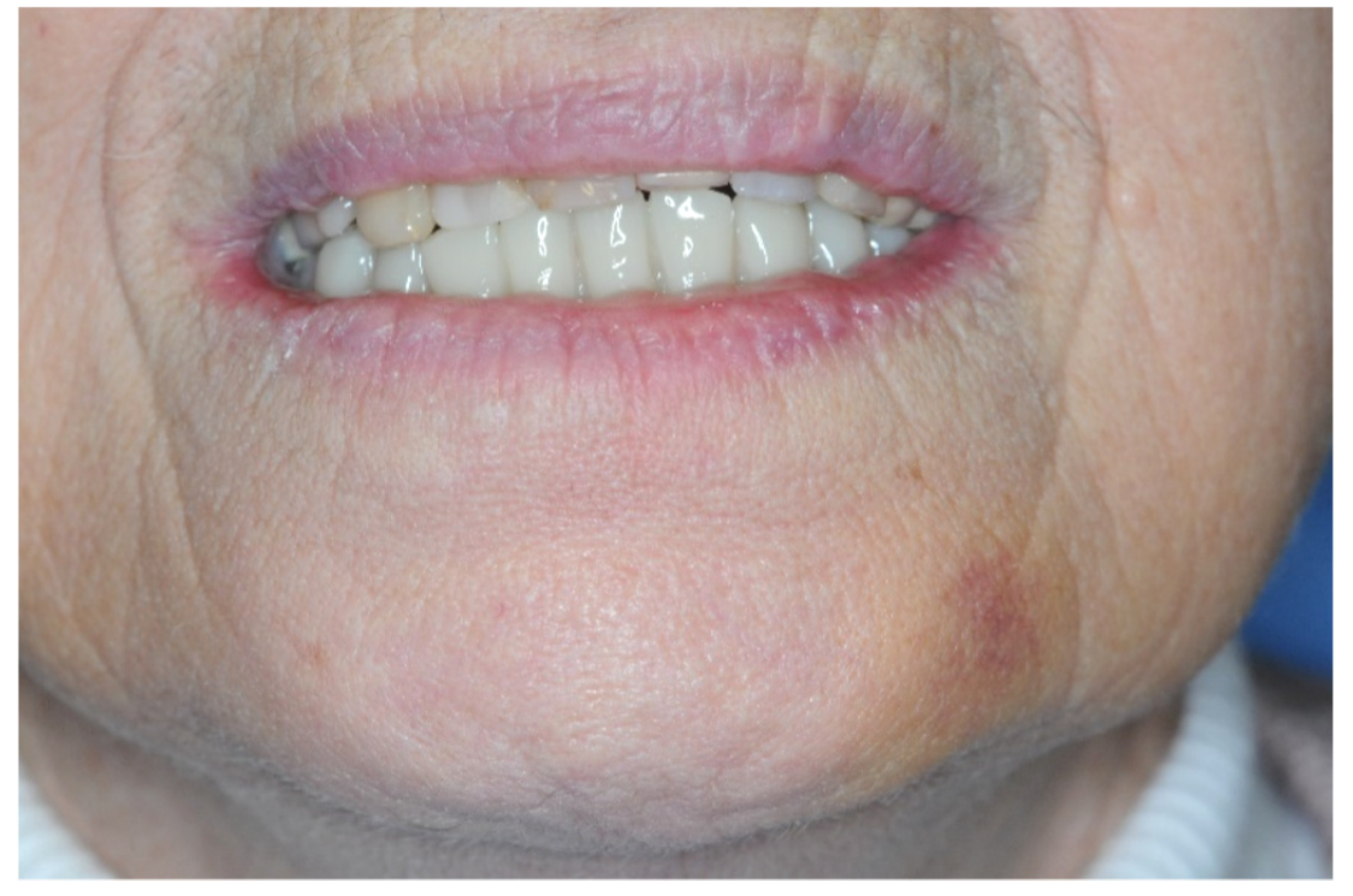
4.7. Evaluation of the Clinical Effect Immediately after the Operation

The patient was examined at 3 (Figure 18), 7, and 15 days after the surgery. The patient did not report any painful symptoms or usage of additional painkillers in the days following the operation. There was a slight bruising on the chin area, which disappeared before the 7-day checkup.
At 15 days, the residual sutures were removed. The implant’s placement was evaluated in the immediate post-operative period through the execution of a CBCT. The plan and the post-op X-rays were scrutinized to determine the consistency of the implant position. The software used for the comparison was Carestream Dental (Carestream®) (Figure 19 and Figure 20).

5. Conclusions

The result achieved in this case report suggests that the performance of an osteotomy should be as guided as the placement of the implant. A correct morphology of peri-implant bone tissue is fundamental; at the intraoperative level, it allows for a reduction in surgical time and accelerates the installation of the prosthesis, while in the long term, it guides the healing of soft tissues and ensures healthy peri-implant tissues.

Author Contributions

Conceptualization, C.T., M.C. and S.S.; methodology, G.I. and S.M.L.; validation, S.M.L.; resources, M.C.; data curation, C.T.; writing—original draft preparation, C.T., M.C., S.M.L., G.I., A.M. and S.S.; writing—review and editing, C.T., M.C., S.M.L. and R.R.y.B.; supervision, S.M.L. and R.R.y.B.; project administration, C.T. and A.M. All authors have read and agreed to the published version of the manuscript.

Funding

This research received no external funding.

Institutional Review Board Statement

Not applicable.

Informed Consent Statement

Informed consent was obtained from the subject involved in the study. Written informed consent has been obtained from the patient to publish this paper.

Data Availability Statement

Not applicable.

Conflicts of Interest

The authors declare no conflict of interest.

References

  1. Chan, M.H.; Nudell, Y.A. All-on-4 Concept Update. Dent. Clin. N. Am. 2020, 65, 211–227. [Google Scholar] [CrossRef] [PubMed]
  2. Beretta, M.; Poli, P.P.; Tansella, S.; Maiorana, C. Virtually guided alveolar ridge reduction combined with computer-aided implant placement for a bimaxillary implant-supported rehabilitation: A clinical report. J. Prosthet. Dent. 2018, 120, 168–172. [Google Scholar] [CrossRef] [PubMed]
  3. Chen, P.; Nikoyan, L. Guided Implant Surgery. Dent. Clin. N. Am. 2021, 65, 67–80. [Google Scholar] [CrossRef] [PubMed]
  4. Lu, J.Y.; Yu, J.Y.; Xie, C.Y.; Gao, J.; Yu, H.Y. Immediate precision of the digital osteotomy template in the digital stackable template: A clinical study. Hua Xi Kou Qiang Yi Xue Za Zhi 2021, 39, 732–738. [Google Scholar] [CrossRef]
  5. Sun, H.; Zhang, J.; Li, B.; Liu, Z.; Shen, S.; Wang, X. Accuracy of a new custom-made bone-supported osteotomy and repositioning guide system for reconstruction of the mandibular ramus using costochondral grafts: A preliminary study. Br. J. Oral Maxillofac. Surg. 2020, 58, 51–56. [Google Scholar] [CrossRef]
  6. Gangwani, K.D.; Shetty, L.; Kulkarni, D.; Seshagiri, R.; Chopra, R. Piezosurgery versus conventional method alveoloplasty. Ann. Maxillofac. Surg. 2018, 8, 181–187. [Google Scholar] [CrossRef]
  7. Zhu, M.; Liu, F.; Zhou, C.; Lin, L.; Zhang, Y.; Chai, G.; Xie, L.; Qi, F.; Li, Q. Does intraoperative navigation improve the accuracy of mandibular angle osteotomy: Comparison between augmented reality navigation, individualised templates and free-hand techniques. J. Plast. Reconstr. Aesthetic Surg. 2018, 71, 1188–1195. [Google Scholar] [CrossRef]
  8. Cushen, S.E.; Turkyilmaz, I. Impact of operator experience on the accuracy of implant placement with stereolithographic surgical templates: An in vitro study. J. Prosthet. Dent. 2013, 109, 248–254. [Google Scholar] [CrossRef]
  9. Rizzo, S.; Lupi, S.M.; Zampetti, P. Nerve damage resulting from oral surgery and medical legal implications. BioCensus 2009, 1, 86–94. [Google Scholar]
  10. Nejad, M.F.; Proussaefs, P.; Lozada, J. Combining guided alveolar ridge reduction and guided implant placement for all-on-4 surgery: A clinical report. J. Prosthet. Dent. 2016, 115, 662–667. [Google Scholar] [CrossRef]
  11. Li, J.; Chen, Z.; Dong, B.; Wang, H.; Joda, T.; Yu, H. Registering Maxillomandibular Relation to Create a Virtual Patient Integrated with a Virtual Articulator for Complex Implant Rehabilitation: A Clinical Report. J. Prosthodont. 2020, 29, 553–557. [Google Scholar] [CrossRef]
  12. Tian, Y.; Chen, C.; Xu, X.; Wang, J.; Hou, X.; Li, K.; Lu, X.; Shi, H.; Lee, E.-S.; Jiang, H.B. A Review of 3D Printing in Dentistry: Technologies, Affecting Factors, and Applications. Scanning 2021, 2021, 9950131. [Google Scholar] [CrossRef]
  13. Dawood, A.; Marti, B.M.; Sauret-Jackson, V. 3D printing in dentistry. Br. Dent. J. 2015, 219, 521–529. [Google Scholar] [CrossRef]
  14. Khorsandi, D.; Fahimipour, A.; Abasian, P.; Saber, S.S.; Seyedi, M.; Ghanavati, S.; Ahmad, A.; De Stephanis, A.A.; Taghavinezhaddilami, F.; Leonova, A.; et al. 3D and 4D printing in dentistry and maxillofacial surgery: Printing techniques, materials, and applications. Acta Biomater. 2021, 122, 26–49. [Google Scholar] [CrossRef]
  15. Ku, J.-K.; Lee, J.; Lee, H.-J.; Yun, P.-Y.; Kim, Y.-K. Accuracy of dental implant placement with computer-guided surgery: A retrospective cohort study. BMC Oral Heal. 2022, 22, 8. [Google Scholar] [CrossRef]
  16. Kim, M.-J.; Jeong, J.Y.; Ryu, J.; Jung, S.; Park, H.-J.; Oh, H.-K.; Kook, M.-S. Accuracy of digital surgical guides for dental implants. Maxillofac. Plast. Reconstr. Surg. 2022, 44, 35. [Google Scholar] [CrossRef]
  17. Flügge, T.; Kramer, J.; Nelson, K.; Nahles, S.; Kernen, F. Digital implantology—A review of virtual planning software for guided implant surgery. Part II: Prosthetic set-up and virtual implant planning. BMC Oral Health 2022, 22, 23. [Google Scholar] [CrossRef]
  18. Kernen, F.; Kramer, J.; Wanner, L.; Wismeijer, D.; Nelson, K.; Flügge, T. A review of virtual planning software for guided implant surgery-data import and visualization, drill guide design and manufacturing. BMC Oral Health 2020, 20, 251. [Google Scholar] [CrossRef]
  19. Kalaivani, G.; Balaji, V.R.; Manikandan, D.; Rohini, G. Expectation and reality of guided implant surgery protocol using computer-assisted static and dynamic navigation system at present scenario: Evidence-based literature review. J. Indian Soc. Periodontol. 2020, 24, 398–408. [Google Scholar] [CrossRef]
  20. Ma, B.; Park, T.; Chun, I.; Yun, K. The accuracy of a 3D printing surgical guide determined by CBCT and model analysis. J. Adv. Prosthodont. 2018, 10, 279–285. [Google Scholar] [CrossRef]
  21. Todaro, C.; Torchia, M.; Benazzo, M.; Baena, R.R.Y.; Canzi, P.; Lupi, S.M. Overview and Future Perspectives on Bone 3D Bioprinting. J. Pharm. Res. Int. 2022, 34, 32–48. [Google Scholar] [CrossRef]
  22. Suganna, M.; Kausher, H.; Ali, A.B.M.R.; Abed, M.M.; Albishi, W.S.; Al Hajji, F.A.; Sultan, N.A. Knowledge on Applications of 3D Design and Printing in Dentistry Among Dental Practitioners in Saudi Arabia: A Questionnaire-Based Survey. Cureus 2022, 14, e28379. [Google Scholar] [CrossRef] [PubMed]
  23. Testori, T.; Weinstein, T.; Scutellà, F.; Wang, H.-L.; Zucchelli, G. Implant placement in the esthetic area: Criteria for positioning single and multiple implants. Periodontology 2000 2018, 77, 176–196. [Google Scholar] [CrossRef] [PubMed]
  24. Pascual, D.; Vaysse, J. Chirurgie implantaire et prothèse guidées et assistées par ordinateur: Le flux numérique continu. Rev. Stomatol. Chir. Maxillo-Faciale Chir. Orale 2016, 117, 28–35. [Google Scholar] [CrossRef]
  25. Carpentieri, J.; Greenstein, G.; Cavallaro, J. Hierarchy of restorative space required for different types of dental implant prostheses. J. Am. Dent. Assoc. 2019, 150, 695–706. [Google Scholar] [CrossRef]
  26. Jones, G.G.; Jaere, M.; Clarke, S.; Cobb, J. 3D printing and high tibial osteotomy. EFORT Open Rev. 2018, 3, 254–259. [Google Scholar] [CrossRef]
  27. Shelton, T.J.; Monazzam, S.; Calafi, A.; Leshikar, H.B.; Haus, B.M. Preoperative 3D Modeling and Printing for Guiding Periacetabular Osteotomy. J. Pediatr. Orthop. 2021, 41, 149–158. [Google Scholar] [CrossRef]
  28. Hegedus, T.; Kreuter, P.; Kismarczi-Antalffy, A.A.; Demeter, T.; Banyai, D.; Vegh, A.; Geczi, Z.; Hermann, P.; Payer, M.; Zsembery, A.; et al. User Experience and Sustainability of 3D Printing in Dentistry. Int. J. Environ. Res. Public Health 2022, 19, 1921. [Google Scholar] [CrossRef]
  29. Joda, T.; Zarone, F.; Ferrari, M. The complete digital workflow in fixed prosthodontics: A systematic review. BMC Oral Health 2017, 17, 124. [Google Scholar] [CrossRef]
  30. Srinivasan, M.; Kamnoedboon, P.; McKenna, G.; Angst, L.; Schimmel, M.; Özcan, M.; Müller, F. CAD-CAM removable complete dentures: A systematic review and meta-analysis of trueness of fit, biocompatibility, mechanical properties, surface characteristics, color stability, time-cost analysis, clinical and patient-reported outcomes. J. Dent. 2021, 113, 103777. [Google Scholar] [CrossRef]
  31. HLerner, H.; Hauschild, U.; Sader, R.; Ghanaati, S. Complete-arch fixed reconstruction by means of guided surgery and immediate loading: A retrospective clinical study on 12 patients with 1 year of follow-up. BMC Oral Health 2020, 20, 15. [Google Scholar] [CrossRef] [Green Version]
  32. Bover-Ramos, F.; Viña-Almunia, J.; Cervera-Ballester, J.; Peñarrocha-Diago, M.; García-Mira, B. Accuracy of Implant Placement with Computer-Guided Surgery: A Systematic Review and Meta-Analysis Comparing Cadaver, Clinical, and In Vitro Studies. Int. J. Oral Maxillofac. Implant. 2018, 33, 101–115. [Google Scholar] [CrossRef]
  33. Smitkarn, P.; Subbalekha, K.; Mattheos, N.; Pimkhaokham, A. The accuracy of single-tooth implants placed using fully digital-guided surgery and freehand implant surgery. J. Clin. Periodontol. 2019, 46, 949–957. [Google Scholar] [CrossRef]
  34. De Stavola, L.; Fincato, A.; Bressan, E.; Gobbato, L. Results of Computer-Guided Bone Block Harvesting from the Mandible: A Case Series. Int. J. Periodontics Restor. Dent. 2017, 37, e111–e119. [Google Scholar] [CrossRef]
  35. Cristoforetti, A.; De Stavola, L.; Fincato, A.; Masè, M.; Ravelli, F.; Nollo, G.; Tessarolo, F. Assessing the accuracy of computer-planned osteotomy guided by stereolithographic template: A methodological framework applied to the mandibular bone harvesting. Comput. Biol. Med. 2019, 114, 103435. [Google Scholar] [CrossRef]
  36. Arnal-Burró, J.; Pérez-Mañanes, R.; Gallo-Del-Valle, E.; Igualada-Blazquez, C.; Cuervas-Mons, M.; Vaquero-Martín, J. Three dimensional-printed patient-specific cutting guides for femoral varization osteotomy: Do it yourself. Knee 2017, 24, 1359–1368. [Google Scholar] [CrossRef]
  37. Pérez-Mañanes, R.; Manaute, J.R.; Rodriguez, F.C.; Martín, J.V.; Burró, J.A. 3D Surgical Printing Cutting Guides for Open-Wedge High Tibial Osteotomy: Do It Yourself. J. Knee Surg. 2016, 29, 690–695. [Google Scholar] [CrossRef]
  38. Shi, J.; Lv, W.; Wang, Y.; Ma, B.; Cui, W.; Liu, Z.; Han, K. Three dimensional patient-specific printed cutting guides for closing-wedge distal femoral osteotomy. Int. Orthop. 2019, 43, 619–624. [Google Scholar] [CrossRef]
  39. Wang, F.; Zhu, J.; Peng, X.; Su, J. The application of 3D printed surgical guides in resection and reconstruction of malignant bone tumor. Oncol. Lett. 2017, 14, 4581–4584. [Google Scholar] [CrossRef]
  40. Park, J.W.; Kang, H.G.; Kim, J.H.; Kim, H.-S. The application of 3D-printing technology in pelvic bone tumor surgery. J. Orthop. Sci. 2021, 26, 276–283. [Google Scholar] [CrossRef]
  41. El Kholy, K.; Lazarin, R.; Janner, S.F.M.; Faerber, K.; Buser, R.; Buser, D. Influence of surgical guide support and implant site location on accuracy of static Computer-Assisted Implant Surgery. Clin. Oral Implant. Res. 2019, 30, 1067–1075. [Google Scholar] [CrossRef] [PubMed]
  42. Venezia, P.; Torsello, F.; Santomauro, V.; Dibello, V.; Cavalcanti, R. Full Digital Workflow for the Treatment of an Edentulous Patient with Guided Surgery, Immediate Loading and 3D-Printed Hybrid Prosthesis: The BARI Technique 2.0. A Case Report. Int. J. Environ. Res. Public Health 2019, 16, 5160. [Google Scholar] [CrossRef] [PubMed]
  43. Derksen, W.; Wismeijer, D.; Flügge, T.; Hassan, B.; Tahmaseb, A. The accuracy of computer-guided implant surgery with tooth-supported, digitally designed drill guides based on CBCT and intraoral scanning. A prospective cohort study. Clin. Oral Implant. Res. 2019, 30, 1005–1015. [Google Scholar] [CrossRef] [PubMed]
  44. Cunha, R.M.; Souza, F.; Hadad, H.; Poli, P.P.; Maiorana, C.; Carvalho, P.S.P. Accuracy evaluation of computer-guided implant surgery associated with prototyped surgical guides. J. Prosthet. Dent. 2021, 125, 266–272. [Google Scholar] [CrossRef] [PubMed]
  45. Tallarico, M.; Czajkowska, M.; Cicciù, M.; Giardina, F.; Minciarelli, A.; Zadrożny, Ł.; Park, C.-J.; Meloni, S.M. Accuracy of surgical templates with and without metallic sleeves in case of partial arch restorations: A systematic review. J. Dent. 2021, 115, 103852. [Google Scholar] [CrossRef]
  46. Al-Moraissi, E.; Elmansi, Y.; Al-Sharaee, Y.; Alrmali, A.; Alkhutari, A. Does the piezoelectric surgical technique produce fewer postoperative sequelae after lower third molar surgery than conventional rotary instruments? A systematic review and meta analysis. Int. J. Oral Maxillofac. Surg. 2016, 45, 383–391. [Google Scholar] [CrossRef]
  47. Cicciù, M.; Stacchi, C.; Fiorillo, L.; Cervino, G.; Troiano, G.; Vercellotti, T.; Herford, A.; Galindo-Moreno, P.; Di Lenarda, R. Piezoelectric bone surgery for impacted lower third molar extraction compared with conventional rotary instruments: A systematic review, meta-analysis, and trial sequential analysis. Int. J. Oral Maxillofac. Surg. 2021, 50, 121–131. [Google Scholar] [CrossRef]
  48. Gulnahar, Y.; Alpan, A. Comparison of postoperative morbidity between piezoelectric surgery and conventional rotary instruments in mandibular third molar surgery: A split-mouth clinical study. Med. Oral. Patol. Oral. Cir. Bucal. 2021, 26, e269–e275. [Google Scholar] [CrossRef]
  49. Hassan, B.; Gonzalez, B.G.; Tahmaseb, A.; Greven, M.; Wismeijer, D. A digital approach integrating facial scanning in a CAD-CAM workflow for complete-mouth implant-supported rehabilitation of patients with edentulism: A pilot clinical study. J. Prosthet. Dent. 2017, 117, 486–492. [Google Scholar] [CrossRef]
  50. Asutay, H.C.; Turkyilmaz, I.; Benli, M.; Martinez, J.L. Transforming smiles using an intraoral scanner and face scan application on smartphone. J. Dent. Sci. 2022, 17, 1413–1414. [Google Scholar] [CrossRef]
  51. Zhivago, P.; Turkyilmaz, I. A comprehensive digital approach to enhance smiles using an intraoral optical scanner and advanced 3-D sculpting software. J. Dent. Sci. 2021, 16, 784–785. [Google Scholar] [CrossRef]
  52. Amezua, X.; Iturrate, M.; Garikano, X.; Solaberrieta, E. Analysis of the influence of the facial scanning method on the transfer accuracy of a maxillary digital scan to a 3D face scan for a virtual facebow technique: An in vitro study. J. Prosthet. Dent. 2022, 128, 1024–1031. [Google Scholar] [CrossRef]
  53. Pérez-Giugovaz, M.G.; Mosier, M.; Revilla-León, M. An additively manufactured intraoral scan body for aiding complete-arch intraoral implant digital scans with guided integration of 3D virtual representation. J. Prosthet. Dent. 2022, 127, 38–43. [Google Scholar] [CrossRef]
  54. Nuytens, P.; D’Haese, R.; Vandeweghe, S. Reliability and Time Efficiency of Digital vs Analog Bite Registration Technique for the Manufacture of Full-Arch Fixed Implant Prostheses. J. Clin. Med. 2022, 11, 2882. [Google Scholar] [CrossRef]
  55. Moura, G.F.; Siqueira, R.; Meirelles, L.; Maska, B.; Wang, H.-L.; Mendonça, G. Denture scanning technique for computer-guided implant-supported restoration treatment of edentulous patients. J. Prosthet. Dent. 2021, 125, 726–731. [Google Scholar] [CrossRef]
  56. Rizzo, S.; Zampetti, P.; Baena, R.R.Y.; Svanosio, D.; Lupi, S.M. Retrospective analysis of 521 endosseous implants placed under antibiotic prophylaxis and review of literature. Minerva Dent. Oral Sci. 2010, 59, 75–88. [Google Scholar] [PubMed]
Figure 1. 3D reconstruction of the CBCT scan.
Prosthesis 05 00017 g001
Figure 2. (a) 3D face scan (b) 3D face scan with articulator.
Prosthesis 05 00017 g002
Figure 3. Digital projection of the provisional prosthesis.
Prosthesis 05 00017 g003
Figure 4. Implant positioning and osteotomy plan.
Prosthesis 05 00017 g004
Figure 5. (a) Osteotomic and positional guide; (b,c) implant Guide in sagittal and coronal view.
Prosthesis 05 00017 g005
Figure 6. 3D-printing preview of the guides.
Prosthesis 05 00017 g006
Figure 7. 3D-printing preview of the provisional prosthesis.
Prosthesis 05 00017 g007
Figure 8. Patient’s occlusion at T0.
Prosthesis 05 00017 g008
Figure 9. Insertion of the anchor pins in the positional guide.
Prosthesis 05 00017 g009
Figure 10. Details of the residual bone after tooth extraction.
Prosthesis 05 00017 g010
Figure 11. Details of the osteotomy executed with Piezosurgery.
Prosthesis 05 00017 g011
Figure 12. Details of the osteotomy executed with Piezosurgery.
Prosthesis 05 00017 g012
Figure 13. Intra-Guide Surgical Kit.
Prosthesis 05 00017 g013
Figure 14. Comparison of patient’s oral cavity pre- and post-osteotomy.
Prosthesis 05 00017 g014
Figure 15. (a) Detail of the L-Prf membranes and (b) suturing of the flap with 5/0 Polyglycolic Acid Sutures.
Prosthesis 05 00017 g015
Figure 16. Provisional prosthesis after surgery.
Prosthesis 05 00017 g016
Figure 17. Post-op CBCT.
Prosthesis 05 00017 g017
Figure 18. Details of the hematoma 3 days after surgery.
Prosthesis 05 00017 g018
Figure 19. Comparison between digital plan and post-op results.
Prosthesis 05 00017 g019
Figure 20. Comparison between digital plan and post-op CBCT.
Prosthesis 05 00017 g020
Disclaimer/Publisher’s Note: The statements, opinions and data contained in all publications are solely those of the individual author(s) and contributor(s) and not of MDPI and/or the editor(s). MDPI and/or the editor(s) disclaim responsibility for any injury to people or property resulting from any ideas, methods, instructions or products referred to in the content.

