Article

Zygomatic Implants Research: A Scientometric Analysis from 1990 to 2021

1
Department of Innovative Technologies in Medicine and Dentistry, University of Chieti-Pescara, 66100 Chieti, Italy
2
Faculty of Biosciences and Technology for Food, Agriculture and Environment, University of Teramo, 64100 Teramo, Italy
3
Institute of Biochemistry and Cell Biology (CNRIBBC/EMMA/Infrafrontier/IMPC), National Research Council, 00015 Rome, Italy
*
Author to whom correspondence should be addressed.
Prosthesis 2023, 5(1), 208-220; https://doi.org/10.3390/prosthesis5010016
Submission received: 20 December 2022 / Revised: 18 January 2023 / Accepted: 29 January 2023 / Published: 7 February 2023
(This article belongs to the Special Issue Bioactive Materials for Dental and Maxillofacial Repair)

Abstract

:
Zygomatic implants imply the use of the zygoma as the implant anchorage and have been proposed as a valuable alternative to the invasive classical procedures in cases of severe maxillary atrophy. Despite the numerous manuscripts published in this field, a quantitative analysis of the research products to infer the trends and the status identification of this specific issue was missing, as well as an objective map of this area. Thus, the present scientometric study analyzed all the research papers published within the interval 1990–2021 that included the keyword “zygomatic implants”. Research papers containing the keywords “zygomatic implants” were collected using Web of Science and analyzed with Cytoscape 3.7.2 and Sci software. A total of 654 studies were published between 1990 and 2020, reaching up to 11639 citations in total, with a mean of 17.8 citations per research study. Data show that the number of publications per year is rapidly increasing, as well as the sum of citations per year. While the USA was identified as the most productive country in this field, followed by Italy, Spain, and Brazil, the National Natural Science Foundation of China stands up as the major funding agency, followed by the National Institutes of Health (NIH) in the USA and the United States Department of Health and Human Services. The analysis of the keywords showed that “zygomatic fractures” represents the most common word within this field, with “complications” as the most recent keyword and “screws” as the keyword used for the longest time. The map of science representing the authors and their collaborations highlighted the existence of multiple small-size research groups that contribute to scientific production, forming highly clustered structures that do not collaborate between them. The present scientometric analysis demonstrates the rising interest in using the zygomatic implants technique as an alternative to the classical ones. The obtained data suggest that the scientific community involved in the study of such a field is highly fragmented, emphasizing the lack of communication among the scientists and research groups.

1. Introduction

Osseointegrated dental implants represent a routine procedure for partial or total edentulism in clinical practice [1,2,3,4]. In the presence of severe atrophies and insufficient bone volume, for instance, in the posterior maxilla, the insertion of standard dental implants often requires a regenerative procedure accompanied by important biological and operative costs [5,6]. Several techniques and surgical methods have been proposed to increase the residual bone volume prior to inserting the dental implants in this region, such as maxillary sinus augmentation [7,8,9], Le Fort I procedures [6], autografts [10,11] and bone substitutes [12,13]. A consistent alternative to bone grafting is represented by zygomatic implants, which possess a reduced morbidity due to the high stability of the cortical/trabecular ratio of this region during the course of human life and in presence of tooth loss [14,15]. In fact, the maxillary process of the zygomatic bone develops from the anterosuperior area and is prolonged to the anterior region producing the inferolateral margin of the eye orbit [15,16]. Biomechanically, the use of zygomatic implants for totally edentulous subjects should consider a mean vertical loading of 150 newtons (N) on the occlusal plane that could reach 300 N in the masseteric region [17]. Branemark et al. [18] described a method to carry out a zygomatic implant procedure, although multiple variants to the protocol have been proposed [19,20,21], mainly to preserve the integrity of the Schneiderian membrane. Moreover, the zygomatic fixture thread design and self-tapping apex could influence also the operativity, increasing the control of the dental implant insertion within this anatomical region [22]. In the literature, zygomatic implants reported a survival rate range between 91.2 and 100%, similar to the standard dental implants inserted in non-grafted sites [23]. The most recurrent complications are certainly represented by oedema and sinusitis, while rarer occurrences are represented by aspergillosis and the violation of the intracerebral and orbital spaces [23].
In the present study, all the scientific research products containing the keyword “zygomatic implants” and published between 1990 and 2021 were gathered in a scientometric study. Scientometry discipline has gained a key role in science as a quantitative method to analyze scientific production using mathematical and statistical methods, allowing the inferences of the trends within a field and the status identification of a specific issue. The combination of bibliographical collectors such as Web of Science, together with the available bioinformatic tools such as Cytoscape 3.7.2 and Sci software confers to the scientific community the possibility to create, investigate and visualize the maps of the Authors, Countries, Institutions, keywords, publications, and citations, as well as the Journals and funding Agencies [24,25,26]. While a scientometric analysis is used to map the scientific knowledge area objectively, a review manuscript is aimed to identify the research issues and their challenges based on scientometric results [27,28].
Due to the absence of a quantitative analysis of the research products within this field, in this study, the bibliometric parameters of the already published manuscripts are presented. As a result, the most productive Countries are evidenced, as well as the major funding agencies, the most common Journals where the scientific papers were published, and temporal analysis of the keywords. Last, a map of science with a topical visualization of the research collaborations is shown, inferring important information for this field of study that could be of great utility for policy making, optimization of the available funding, and research targeting for financial optimization, leading to an improved and enhanced communication among the research groups, the scientists and the government.

2. Materials and Methods

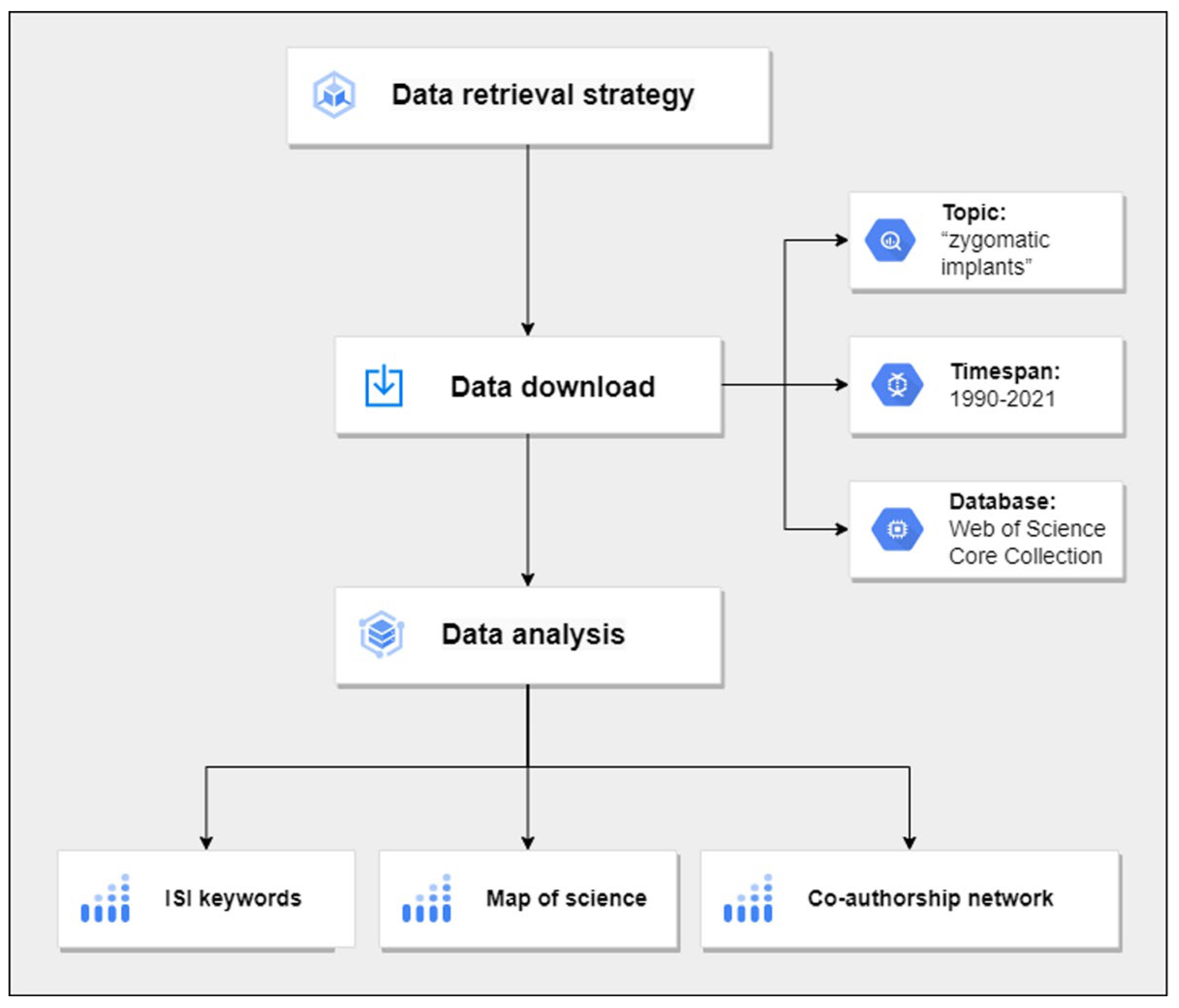
2.1. Data Collection

The present scientometric study was carried out to analyze the research products available within the field of zygomatic implants. All data were accessed from the Web of Science repository (https://webofscience.com/, accessed on 12 December 2021) up to December 2021 and are referred to the interval 1990–2021. The research products were then manually checked by three independent expert researchers prior to being included in the analysis. Figure 1 illustrates the experimental design of this study.
In the queries, “zygomatic implants” was used as a topic, obtaining a list of 654 studies with their attributes. All the following analyses have been carried out on this dataset:
  • Number of citable documents: including articles, reviews, and conference papers;
  • Number of cites per document: number of citations per document in specific years;
  • h-index: a topic/journal/author has an index h if among its Np papers, h has at least h citations each, and the other papers (Nph) have no more than h citations each.

2.2. Analysis of ISI Keywords
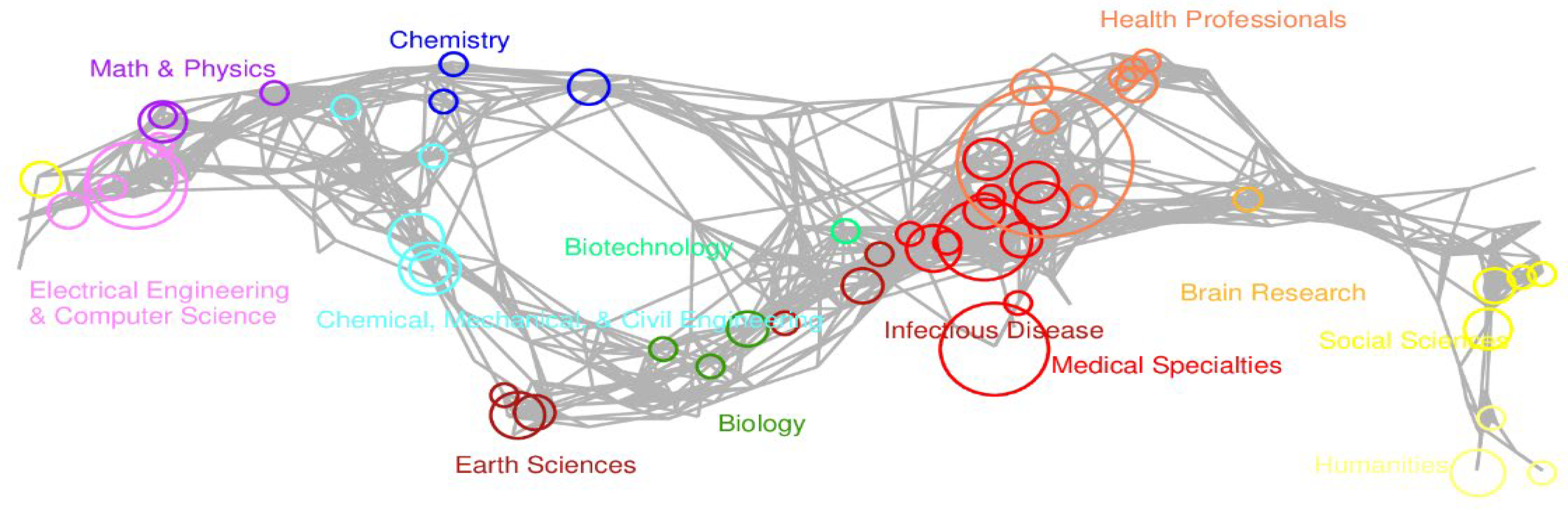
To analyze the keywords used in terms of frequency of use during the years, data were processed for temporal analysis using Sci2 Tool (Sci2 Team) [29]. This software generates a temporal visualization of bursts, detecting and analyzing the ISI keywords used within the research papers (Sci2 Team. (2009). Science of Science (Sci2) Tool. Indiana University and SciTech Strategies).

2.3. Map of Science

To explore the closeness (i.e., the distance on the map and thus the similarity in real life) between the scientific disciplines related to the study of zygomatic implants, a map of science using the Sci2 software was realized. A map of science is defined as the representation of a network built with a number N of subdisciplines (in this network, 554, represented as nodes) that are then grouped into N disciplines of science (13 in this case). Mapped subdisciplines are organized by their size and are related to the numbers (journals) and colors (disciplines).

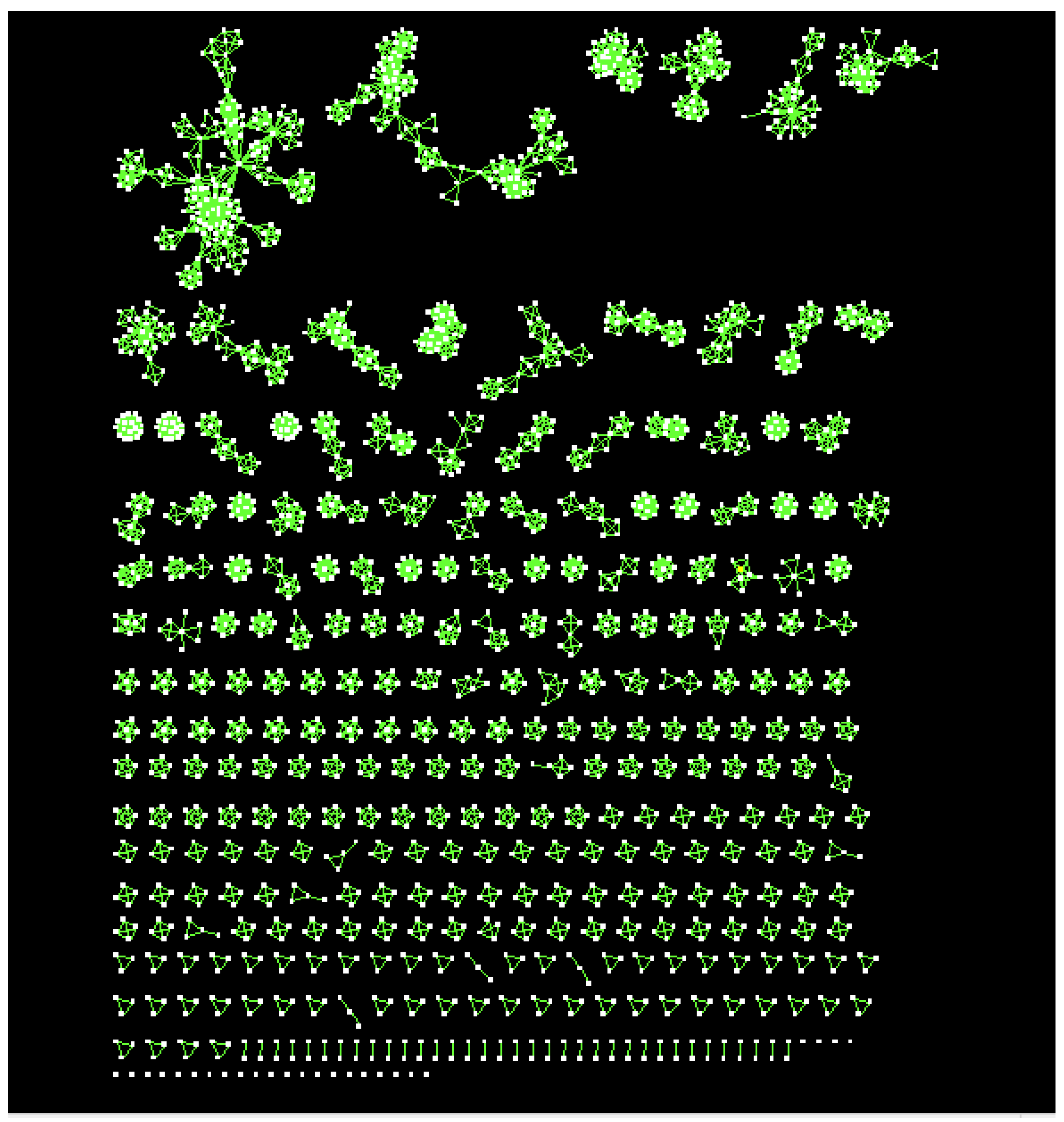
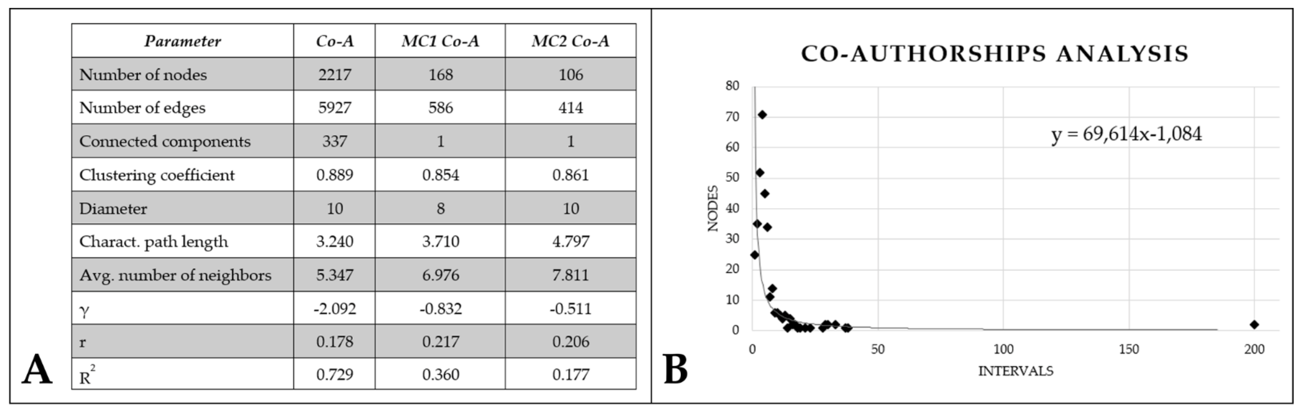
2.4. Co-Authorship Network

To study the dynamics of the co-authorships, an approach based on social networks was used, intended as an analysis of the social behavior of the Authors involved in the field and the collaborations among them [24]. To that, Authors were represented as nodes within the network and were linked by edges when Authors co-authored two or more publications. Cytoscape 3.7.2, an open-source software, was used for network creation, visualization, and analysis, considering the network as undirected [30]. To study the topology of the obtained networks, the main topological parameters listed in Table 1 were automatically computed. Then, the network’s topology (i.e., the statistical analysis of the network organization) was used to infer the social pattern of the Authors’ behavior.

3. Results

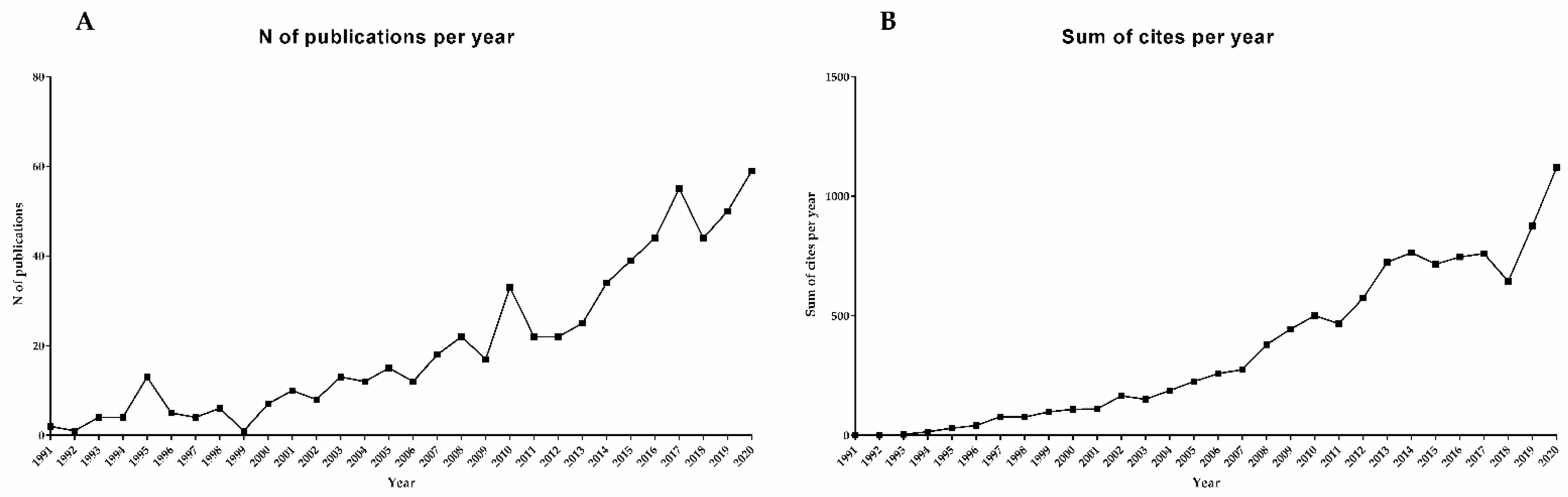
3.1. Zygomatic Implants Constitute a Constantly-Increasing Plot

In total, 654 research papers were found, characterized by the bibliometric parameters shown in Table 2. As illustrated in Figure 2, among the research products, 563 were original articles (including technical notes), 13 were book chapters, 62 corresponded to review manuscripts (including meta-analysis, commentaries, corrections, and systematic reviews), and 34 corresponded to proceedings or conference papers (among which 18 products were also original articles). The number of issues published per year is described in Figure 3A, demonstrating an exponential increase in interest regarding this issue. The time-trend of citations (sum of cites per year) reported in Figure 3B confirms the rise in the number of publications related to “zygomatic implants” by evidencing the exponential increase in the sum of cites per year. The distribution of cites per year, as shown in Figure 4, follows a power law with a negative exponent. The bibliometric data referred to papers published in 2020 and 2021 are not stable yet and thus cannot be included in the analysis.

3.2. USA, Italy, and Spain Are the Most Productive Countries

By investigating the number of issues published by each country, we could estimate the contribution of the different Countries in zygomatic implant research (Figure 5). As it is evident, most of the issues have been published in the USA (139), followed by Italy (71), Spain (65), and Brazil (58). Supplementary Material Table S1 contains the complete list, including all the Countries.

3.3. China and USA Localized the Major Funding Agencies

The main Agencies funding the research within this field were identified as well (Figure 6), with the National Natural Science Foundation of China (NSFC) as the major funding agency, followed by the National Institutes of Health (NIH) USA and the United States Department of Health Human Services. Supplementary Material Table S2 gathers the complete list of funding agencies.

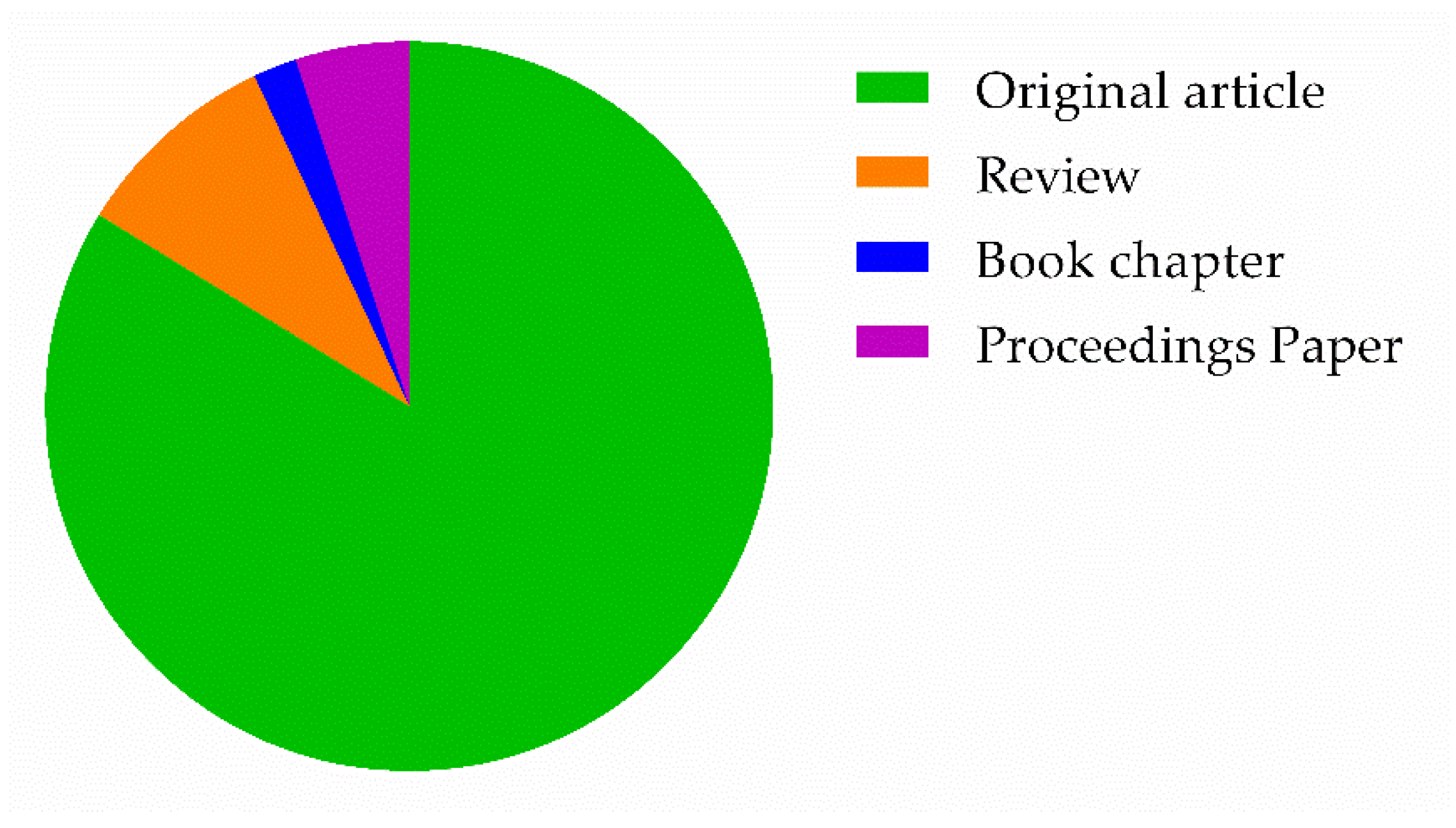
3.4. International Journal of Oral Maxillofacial Implants Published Most of the Research Articles

By focusing on the number of papers published by each Journal (Figure 7), results showed that the International Journal of oral maxillofacial implants published most of the research studies regarding this field (70 documents), followed by the Journal of oral and maxillofacial surgery (52) and the Journal of craniofacial surgery (36).

3.5. “Screws” Represent the Most Cited Keyword within the Published Studies

The ISI keywords cited in the papers were analyzed to identify the most important topics addressed, with a special focus on the time window in which they were approached. Table 3 shows the list of citation bursts identified. While the “weight” corresponds to the importance of the keyword, the “length” gives information about the number of years that the keyword has been considered a burst. In this analysis, “complications” stands as the most recent keyword, used mostly within the last three years but with a high score in terms of weight, with “zygomatic fractures” representing the most cited keyword (highest weight) within a timeframe (from 1994 to 2005) and “screw” the longest-lasting keyword (13 years, since 1994 to 2006) (Table 4).

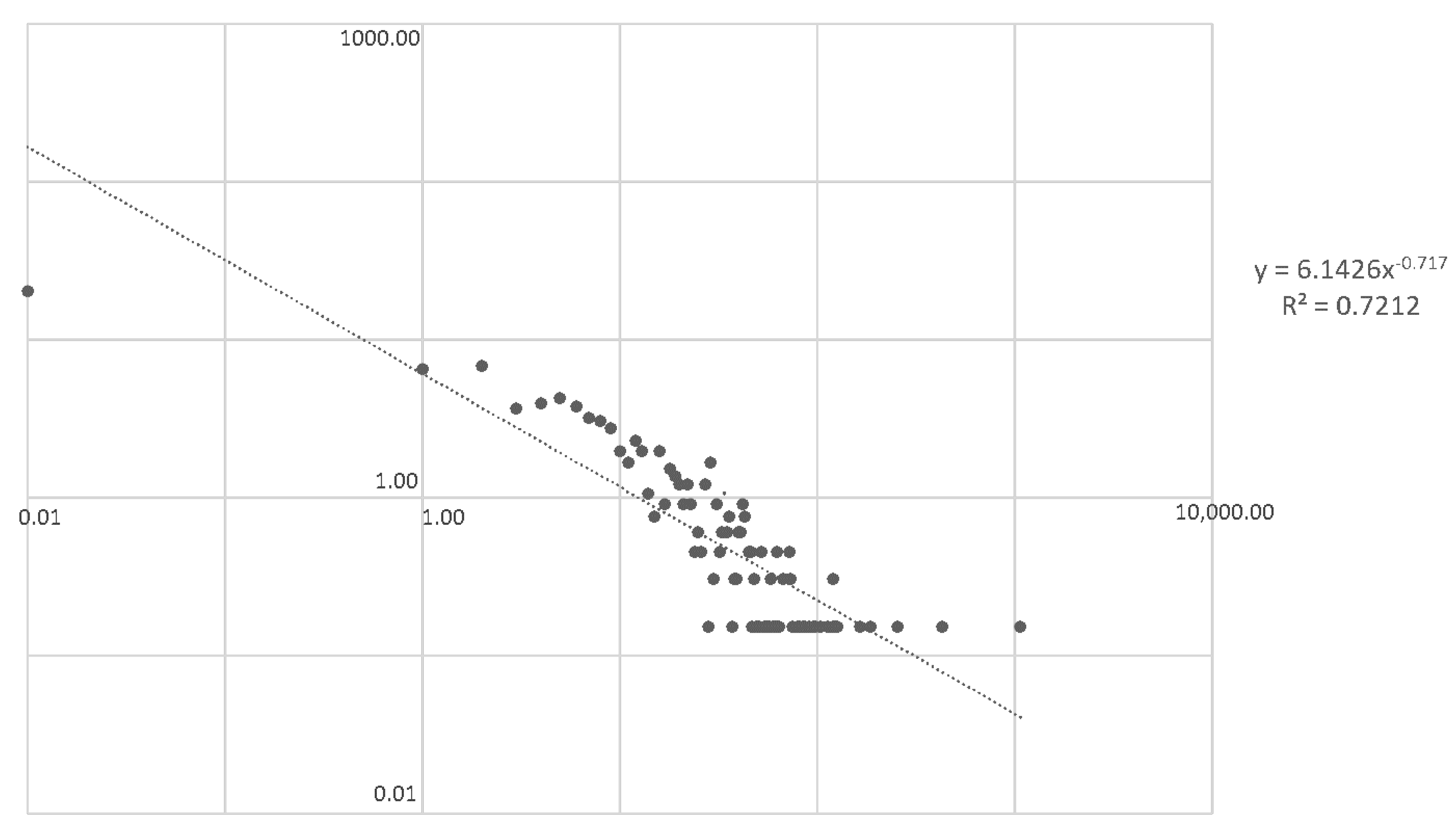
3.6. Lack of Communication among the Scientists Involved in the Field

To study the link among the different disciplines involved in zygomatic implant research, a map representing the co-citation of the documents was set up (Figure 7). To complete the analysis with the description of the authors and the co-authorship dynamics, it was set up and analyzed a co-authorship network (Figure 8). In the network, the Authors are represented as nodes and co-authorship as a link. The analysis of the network topology shows that the network is constituted by a high number of small-size (Main-Components-Co-Authorship Network) connected components (sub-networks), with the larger one reaching about 7.6% of the co-authorship network (Co-A). Thus, a unitary structure with big collaborations among the members is missing, while multiple small groups participate in this issue independently. As visually represented in Figure 8, all the components are characterized by the tendency to form highly clustered structures that do not communicate with each other. In Figure 9, the table shows the numeric data (Figure 9A), reaching a total of 337 components (i.e., co-authors) and 2217 nodes (i.e., connections among the authors), while the graph (Figure 9B) represents the components (subdivided into intervals, gathering > 98% of nodes) and the frequency of the nodes, with a negative exponent that emphasizes the low-degree of connection among the scientists in this area. Co-Authorship Networks 1 and 2 are the two larger subnetworks of Co-A (i.e., the two main groups from Figure 8).

4. Discussion

The “zygomatic implant” was first developed in 1998 by Branemark et al. to be applied in oral reconstruction and rehabilitation [18]. The technique proposed the use of the zygoma as the implant anchorage in cases of severe maxillary atrophy, representing in today’s society a viable alternative to the invasive classical procedures [31]. Indeed, while preventing the use of heavy bone graft techniques, it may decrease the treatment time and costs, with fewer complications and prosthodontic work, shorter rehabilitation times, and a decrease in patient morbidity [23,32]. In fact, maxillary sinus lifting complications such as infection, bone loss, implant displacement into the sinus, insufficient new bone regeneration, and the formation of oroantral fistulas may lead to the failure of sinus bone grafting [33].
Due to the importance of zygomatic implants within the dental field and the absence of quantitative analysis focused on the manuscripts published that allow the inference of important trends, the present in silico study analyzed the research studies published within the interval 1990–2021 that included the keyword “zygomatic implants”. In total, 654 papers were published during this time frame, gathering up to 11,639 citations in total (a mean of 17.8 citations per study). In this regard, as observed in Figure 3, the number of publications per year is rapidly increasing, thus the sum of cites per year. The low exponent value obtained when analyzing the distribution of cites per year is in keeping with the Bradford law [34], supporting the rising interest in the use of this technique as an alternative to the classical ones.
The present study realized a complete analysis of the scientific production gathering the geographical distribution of the studies, the major funding organizations, the most productive Countries, the most used words (i.e., the keywords), and their timeframe within these last decades and finally a topical visualization of the Authors-network and their collaborations. By analyzing the geographical distribution of the published papers, the USA was identified as the most productive Country (139), followed by two European Countries, Italy (71) and Spain (65), and a South American Country, Brazil (58). While the People’s Republic of China lay in 5th place in terms of scientific production (52), it possesses the major funding organization, i.e., the National Natural Science Foundation of China (NSFC), which is followed by the National Institutes of Health (NIH) USA and the United States Department of Health Human Services. In line with this information, it was observed that the majority of the studies were published in Journals based in the USA, with the International Journal of oral maxillofacial implants in the lead (70 research papers).
The analysis of the keywords in terms of importance and timeframe allowed the inference of important data. “Zygomatic fractures” stands as the most used word within this field, mostly during the first 15 years, while “complications” stands up as the newest keyword, principally employed from 2019. It might be probably due to the potential long-term complications derived from the use of this technique, which is possible to observe due to the increasing use of this method in clinical practice and the existence of long-standing patients with this type of implant. On the contrary, the decrease in the use of “zygomatic fractures” as a keyword may be due to the advances achieved in the field, which have allowed a safer and more efficient procedure. Of note, “guided placement” did not appear as one of the most used keywords, even if it represents one of the most controversial issues within the field, which could be possibly due to the fact that it represents a relatively new issue.
To study the connections between the disciplines and their Authors, two strategies were followed: the creation of a map including the co-citation of the documents and the creation of a network in which the Authors are represented and the collaborations (co-authorship) among them. This kind of analysis allows inferring the degree of collaboration between the different authors and research groups by quantifying the number of connections or collaborations (nodes). As evidenced by the results, this scientific issue is characterized by a high number of small-sized research groups that contribute to the scientific production, forming highly clustered structures and standing out the absence of collaboration between them. These data suggest that the scientific community involved in studying such an area is highly fragmented, emphasizing the lack of communication among the scientists involved in this field.
Scientometric analysis has gained enormous importance in several fields with multiple applications, from the business, entrepreneurship, and financial sectors [35,36] to health and medical applications [24,37]. However, this kind of analysis presents some limitations that should be considered. In the present scientometric study, the results achieved are correlated to the literature found on WOS collection within the period ranging from 1990 to 2021. Thus some studies could have been unintentionally omitted (mostly those after December 2021 or published within the timeframe but are not yet available), despite the efforts made by the scientific community working on this document library. Moreover, although the bibliographic search for this study included the research papers published until December 2021, the papers published in 2020 and 2021 are not stable yet and thus could not be included in this specific analysis. Further studies are needed to evaluate the trends among the Authors, specifically their collaborations and networks, promoting communication among them to improve the research quality and advances.

5. Conclusions

Zygomatic implantation is still not a mainstream procedure in the daily clinic, and indications need to be strictly controlled to avoid complications [38] or medical malpractice, especially considering the rising interest in this issue during the last 20 years, as exposed in this study. The current scientometric study evidences the need for collaboration among the experts in this field to share knowledge and contributes to the establishment of this practice as a routine, improving the life quality of the patients while avoiding important complications. Moreover, the information presented within this study could be of great utility for policy making, optimization of the available funding, and research targeting for financial optimization, leading to improved and enhanced communication among the research groups, the research scientists, and the government.

Supplementary Materials

The following supporting information can be downloaded at: https://www.mdpi.com/article/10.3390/prosthesis5010016/s1, Table S1: complete list of most productive Countries; Table S2: complete list of funding agencies.

Author Contributions

Conceptualization, A.S., L.V., A.T. and N.B.; methodology, M.R.-S. and N.B.; software, A.T. and N.B.; validation, A.S., A.T., C.B. and L.V.; formal analysis, A.T. and M.R.-S.; investigation, M.R.-S.; data curation, M.R.-S.; writing—original draft preparation, M.R.-S., A.T., F.L. and A.S.; writing—review and editing, M.R.-S., A.T., F.L., C.B. and A.S. All authors have read and agreed to the published version of the manuscript.

Funding

This research received no external funding.

Institutional Review Board Statement

Not applicable.

Informed Consent Statement

Not applicable.

Data Availability Statement

All experimental data to support the findings of this study are available by contacting the corresponding author upon request. The authors have annotated the entire data-building process and empirical techniques presented in the paper. The data underlying this article are not freely available by agreement with our partners to protect their confidentiality.

Conflicts of Interest

The authors declare no conflict of interest.

References

  1. Beschnidt, S.M.; Cacaci, C.; Dedeoglu, K.; Hildebrand, D.; Hulla, H.; Iglhaut, G.; Krennmair, G.; Schlee, M.; Sipos, P.; Stricker, A.; et al. Implant Success and Survival Rates in Daily Dental Practice: 5-Year Results of a Non-Interventional Study Using CAMLOG SCREW-LINE Implants with or without Platform-Switching Abutments. Int. J. Implant Dent. 2018, 4, 33. [Google Scholar] [CrossRef] [PubMed]
  2. Eskow, C.C.; Oates, T.W. Dental Implant Survival and Complication Rate over 2 Years for Individuals with Poorly Controlled Type 2 Diabetes Mellitus. Clin. Implant Dent. Relat. Res. 2017, 19, 423–431. [Google Scholar] [CrossRef] [PubMed]
  3. Albrektsson, T.; Berglundh, T.; Lindhe, J. Osseointegration: Historic Background and Current Concepts. Clin. Periodontol. Implant Dent. 2003, 4, 809–820. [Google Scholar]
  4. Inchingolo, F.; Tatullo, M.; Marrelli, M.; Inchingolo, A.M.; Inchingolo, A.D.; Dipalma, G.; Flace, P.; Girolamo, F.; Tarullo, A.; Laino, L.; et al. Regenerative Surgery Performed with Platelet-Rich Plasma Used in Sinus Lift Elevation before Dental Implant Surgery: An Useful Aid in Healing and Regeneration of Bone Tissue. Eur. Rev. Med. Pharmacol. Sci. 2012, 16, 1222–1226. [Google Scholar] [PubMed]
  5. Scarano, A.; Carinci, F.; Assenza, B.; Piattelli, M.; Murmura, G.; Piattelli, A. Vertical Ridge Augmentation of Atrophic Posterior Mandible Using an Inlay Technique with a Xenograft without Miniscrews and Miniplates: Case Series. Clin. Oral Implant. Res. 2011, 22, 1125–1130. [Google Scholar] [CrossRef]
  6. Hernández-Alfaro, F.; Ragucci, G.M.M.; Méndez-Manjón, I.; Giralt-Hernando, M.; Guijarro-Martínez, R.; Sicilia-Blanco, P.; Ventura-Martínez, N.; Valls-Ontañón, A. Rehabilitation of the Severely Atrophic Maxilla Using LeFort I Maxillary Advancement and Simultaneous Zygoma Implant Placement: Proof of Concept. Int. J. Oral. Implantol. 2019, 12, 359–372. [Google Scholar]
  7. Arosio, P.; Greco, G.B.; Zaniol, T.; Iezzi, G.; Perrotti, V.; Di Stefano, D.A. Sinus Augmentation and Concomitant Implant Placement in Low Bone-Density Sites. A Retrospective Study on an Undersized Drilling Protocol and Primary Stability. Clin. Implant Dent. Relat. Res. 2018, 20, 151–159. [Google Scholar] [CrossRef]
  8. Tatum, H. Maxillary and Sinus Implant Reconstructions. Dent. Clin. N. Am. 1986, 30, 207–229. [Google Scholar] [CrossRef]
  9. AlMugeiren, O.M.; AlGhamdi, R.M.; Bin Sebayel, N.H.; AlJaruf, H.A.; AlKhamis, D.A.; AlSuwayyid, L.S. Anterior Mandibular Alveolar Bone Measurements between Diabetic and Non-Diabetic Individuals Using Cone-Beam Computed Tomography. Eur. Rev. Med. Pharmacol. Sci. 2022, 26, 5476–5484. [Google Scholar] [CrossRef]
  10. Eser, C.; Gencel, E.; Gökdoğan, M.; Kesiktaş, E.; Yavuz, M. Comparison of Autologous and HeterologousBone Graft Stability Effects for Filling MaxillaryBone Gap After Le Fort I Osteotomy. Adv. Clin. Exp. Med. 2015, 24, 341–348. [Google Scholar] [CrossRef]
  11. Minetti, E.; Palermo, A.; Contessi, M.; Gambardella, U.; Schmitz, J.; Giacometti, E.; Celko, M.; Trisi, P. Autologous Tooth Graft for Maxillary Sinus Augmentation: A Multicenter Clinical Study. Int. J. Growth Factors Stem Cells Dent. 2019, 2, 45–51. [Google Scholar] [CrossRef]
  12. Chanavaz, M. Maxillary Sinus: Anatomy, Physiology, Surgery, and Bone Grafting Related to Implantology--Eleven Years of Surgical Experience (1979–1990). J. Oral Implantol. 1990, 16, 199–209. [Google Scholar] [PubMed]
  13. Scarano, A.; Degidi, M.; Iezzi, G.; Pecora, G.; Piattelli, M.; Orsini, G.; Caputi, S.; Perrotti, V.; Mangano, C.; Piattelli, A. Maxillary Sinus Augmentation With Different Biomaterials: A Comparative Histologic and Histomorphometric Study in Man. Implant Dent. 2006, 15, 197–207. [Google Scholar] [CrossRef] [PubMed]
  14. Uchida, Y.; Goto, M.; Katsuki, T.; Akiyoshi, T. Measurement of the Maxilla and Zygoma as an Aid in Installing Zygomatic Implants. J. Oral Maxillofac. Surg. 2001, 59, 1193–1198. [Google Scholar] [CrossRef] [PubMed]
  15. Nkenke, E.; Hahn, M.; Lell, M.; Wiltfang, J.; Schultze-Mosgau, S.; Stech, B.; Radespiel-Tröger, M.; Neukam, F.W. Anatomic Site Evaluation of the Zygomatic Bone for Dental Implant Placement: Zygomatic Bone for Dental Implants. Clin. Oral Implant. Res. 2003, 14, 72–79. [Google Scholar] [CrossRef] [PubMed]
  16. Karlan, M.S.; Cassisi, N.J. Fractures of the Zygoma: A Geometric, Biomechanical, and Surgical Analysis. Arch. Otolaryngol.—Head Neck Surg. 1979, 105, 320–327. [Google Scholar] [CrossRef]
  17. Gümrükçü, Z. Biomechanical Evaluation of Zygomatic Implant Use in Patients With Different Buccal Maxillary Defect Levels. Int. J. Oral Maxillofac. Implant. 2019, 34, e115–e122. [Google Scholar] [CrossRef] [PubMed]
  18. Brånemark, P.-I.; Gröndahl, K.; Ohrnell, L.-O.; Nilsson, P.; Petruson, B.; Svensson, B.; Engstrand, P.; Nannmark, U. Zygoma Fixture in the Management of Advanced Atrophy of the Maxilla: Technique and Long-Term Results. Scand. J. Plast. Reconstr. Surg. Hand Surg. 2004, 38, 70–85. [Google Scholar] [CrossRef]
  19. Blanc, O.; Shilo, D.; Weitman, E.; Capucha, T.; Rachmiel, A. Extramaxillary Zygomatic Implants: An Alternative Approach for the Reconstruction of the Atrophic Maxilla. Ann. Maxillofac. Surg. 2020, 10, 127–132. [Google Scholar] [CrossRef]
  20. Maló, P.; Nobre, M.d.A.; Lopes, I. A New Approach to Rehabilitate the Severely Atrophic Maxilla Using Extramaxillary Anchored Implants in Immediate Function: A Pilot Study. J. Prosthet. Dent. 2008, 100, 354–366. [Google Scholar] [CrossRef]
  21. Boyes-Varley, J.G.; Howes, D.G.; Lownie, J.F.; Blackbeard, G.A. Surgical Modifications to the Brånemark Zygomaticus Protocol in the Treatment of the Severely Resorbed Maxilla: A Clinical Report. Int. J. Oral Maxillofac. Implant. 2003, 18, 232–237. [Google Scholar]
  22. Scarano, A.; Conte, R.; Murmura, G.; Lorusso, F.; Harrath, A.H. Satisfaction Grade Assessment of Patients Treated with Zygomatic Implants with Self-Tapping Apex and Machined Body. J. Biol. Regul. Homeost. Agents 2019, 33, 1651–1656. [Google Scholar] [CrossRef] [PubMed]
  23. Chrcanovic, B.R.; Albrektsson, T.; Wennerberg, A. Survival and Complications of Zygomatic Implants: An Updated Systematic Review. J. Oral Maxillofac. Surg. 2016, 74, 1949–1964. [Google Scholar] [CrossRef] [PubMed]
  24. Bernabò, N.; Greco, L.; Mattioli, M.; Barboni, B. A Scientometric Analysis of Reproductive Medicine. Scientometrics 2016, 109, 103–120. [Google Scholar] [CrossRef]
  25. Ho, Y.S. The Top-Cited Research Works in the Science Citation Index Expanded. Scientometrics 2013, 94, 1297–1312. [Google Scholar] [CrossRef]
  26. Li, Z.; Ho, Y.S. Use of Citation per Publication as an Indicator to Evaluate Contingent Valuation Research. Scientometrics 2008, 75, 97–110. [Google Scholar] [CrossRef]
  27. Blümel, C.; Schniedermann, A. Studying Review Articles in Scientometrics and beyond: A Research Agenda. Scientometrics 2020, 124, 711–728. [Google Scholar] [CrossRef]
  28. Chen, C.; Song, M. Visualizing a Field of Research: A Methodology of Systematic Scientometric Reviews. PLoS ONE 2019, 14, e0223994. [Google Scholar] [CrossRef]
  29. Sci2 Tool: A Tool for Science of Science Research and Practice. Available online: https://sci2.cns.iu.edu/user/index.php (accessed on 10 November 2022).
  30. Shannon, P.; Markiel, A.; Ozier, O.; Baliga, N.S.; Wang, J.T.; Ramage, D.; Amin, N.; Schwikowski, B.; Ideker, T. Cytoscape: A Software Environment for Integrated Models of Biomolecular Interaction Networks. Genome Res. 2003, 13, 2498–2504. [Google Scholar] [CrossRef]
  31. Alqutaibi, A.Y.; Aboalrejal, A. Zygomatic Implants Are a Reliable Treatment Option for Patients with Atrophic Maxilla. J. Evid.-Based Dent. Pract. 2017, 17, 402–404. [Google Scholar] [CrossRef]
  32. Sharma, A.; Rahul, G.R. Zygomatic Implants/Fixture: A Systematic Review. J. Oral Implantol. 2013, 39, 215–224. [Google Scholar] [CrossRef] [PubMed]
  33. Scarano, A.; Cholakis, A.K.; Piattelli, A. Histologic Evaluation of Sinus Grafting Materials After Peri-Implantitis-Induced Failure: A Case Series. Int. J. Oral Maxillofac. Implant. 2017, 32, e69–e75. [Google Scholar] [CrossRef] [PubMed]
  34. Bradford, S.C. Documenmation; Public Affairs Press: Washington, DC, USA, 1950; Volume 19, p. 154. [Google Scholar]
  35. Hassan, M.K.; Rabbani, M.R.; Brodmann, J.; Bashar, A.; Grewal, H. Bibliometric and Scientometric Analysis on CSR Practices in the Banking Sector. Rev. Financ. Econ. 2022, 2, 1–20. [Google Scholar] [CrossRef]
  36. Škare, M.; Blanco-Gonzalez-Tejero, C.; Crecente, F.; del Val, M.T. Scientometric Analysis on Entrepreneurial Skills—Creativity, Communication, Leadership: How Strong Is the Association? Technol. Forecast. Soc. Chang. 2022, 182, 121851. [Google Scholar] [CrossRef]
  37. Liu, C.; Liu, Z.; Zhang, Z.; Li, Y.; Fang, R.; Li, F.; Zhang, J. A Scientometric Analysis and Visualization of Research on Parkinson’s Disease Associated With Pesticide Exposure. Front. Public Health 2020, 8, 91. [Google Scholar] [CrossRef]
  38. Mavriqi, L.; Lorusso, F.; Conte, R.; Rapone, B.; Scarano, A. Zygomatic Implant Penetration to the Central Portion of Orbit: A Case Report. BMC Ophthalmol. 2021, 21, 121. [Google Scholar] [CrossRef]
Figure 1. Experimental design of the study. The flowchart illustrates the step followed from data collection to data analysis.
Prosthesis 05 00016 g001
Figure 2. Graphical distribution of the research products. Among the 654 documents, 563 were original articles, 62 were review manuscripts, 13 were book chapters, and 34 were proceedings or conference papers.
Prosthesis 05 00016 g002
Figure 3. Number of publications and sum of cites per year. (A) Time-trend of the number of issues published per year (R2 = 0.739); (B) Time-trend showing the sum of cites per year (R2 = 0.975).
Prosthesis 05 00016 g003
Figure 4. Distribution of cites per year. The distribution of cites per year from 1991 to 2020 shows a power law with a negative exponent (R2 = 0.721), in keeping with the Bradford law, demonstrating the rising interest in this field of study.
Prosthesis 05 00016 g004
Figure 5. The USA, Italy, and Spain are the most productive Countries. The graphical representation shows the number of issues per Country for the 12 most productive Countries.
Prosthesis 05 00016 g005
Figure 6. Main funding agencies in zygomatic implant research.
Prosthesis 05 00016 g006
Figure 7. Map of science. Visual representation of 554 subdiscipline nodes connecting a total of 55 subdisciplines and 13 disciplines (from left to right: Electrical Engineering and Computer Sciences; Math and Physics; Chemical, Mechanical and Civil Engineering; Chemistry; Earth Sciences; Biotechnology; Biology; Infectious Disease; Medical Specialties; Health Professionals; Brain Research; Humanities; Social Sciences). Each discipline has a distinct color and is labeled. Overlaid are circles, each representing all records per unique subdiscipline. The circle area is proportional to the number of fractionally assigned records. Minimum and maximum data values correspond to 0 and 26, respectively. Mapped subdisciplines are shown by size related to the number of matching journals and colors for the discipline. Created with Sci2 Tool (https://sci2.cns.iu.edu; accessed on 17 January 2023).
Prosthesis 05 00016 g007
Figure 8. Graphical visualization of the Co-A. Visual representation of the co-authorships, in which the Authors are represented as nodes and the co-authorships as links. This schematic image shows how the scientific community is highly fragmented, with few groups collaborating and multiple small, isolated groups.
Prosthesis 05 00016 g008
Figure 9. Topological analysis of the co-authorship network. Table (A) shows the numerical data extracted from the analysis, reaching a total of 2217 connections between the 337 authors. Co-A = Co-Authorship Network, MC1 Co-A and MC2 Co-A = Main-Component- Co-Authorship Network 1 and 2 are the two larger subnetworks of Co-A (i.e., the two main groups from Figure 8). Graph (B) represents the intervals in which the components (authors) were subdivided and the frequency of the nodes (connections) among them.
Prosthesis 05 00016 g009
Table 1. Main topological parameters assessed. List of the main topological parameters examined within the network and their definitions.
ParameterDefinition
Connected componentNumber of networks in which every two vertices, a vertex is connected to the others by links, with no further connections within the network.
Number of nodes (N)Number of authors included in the network.
Number of edgesNumber of interactions between the nodes present in the network.
Clustering coefficientRepresents the tendency of the nodes to form clusters. Calculated as CI = 2nI/(kI − 1), being nI the number of links that connect the neighbors kI of the node I. Since 0 ≤ CI ≤ 1, the closer to 1, the higher the tendency to form clusters.
Diameter of the networkLongest path among the shortest paths calculated within the network.
Path length (characteristic)Expected distance existing between two linked nodes.
Number of neighbors (average)Mean number of connections for each node.
Node degree (k)Number of interactions for each node.
Node degree distribution (P(k))Probability of a node to possess k links.
γNode degree exponent within the equation.
R2Coefficient of the node degree versus the number of nodes applied to logarithmized data.
Table 2. Bibliometric parameters refer to the studied dataset.
ParameterValue
H-index48
Average citations per item17.8
Sum of the times cited11,639
Citing articles7041
Table 3. Main Journals publishing zygomatic implants documents. The table shows the number of documents per Journal among the 20 Journals with more documents published in this issue.
Journal TitleNumber of Documents
INTERNATIONAL JOURNAL OF ORAL MAXILLOFACIAL IMPLANTS70
JOURNAL OF ORAL AND MAXILLOFACIAL SURGERY52
JOURNAL OF CRANIOFACIAL SURGERY36
INTERNATIONAL JOURNAL OF ORAL AND MAXILLOFACIAL SURGERY27
CLINICAL IMPLANT DENTISTRY AND RELATED RESEARCH22
EUROPEAN JOURNAL OF ORAL IMPLANTOLOGY19
PLASTIC AND RECONSTRUCTIVE SURGERY19
JOURNAL OF ORAL IMPLANTOLOGY16
AMERICAN JOURNAL OF ORTHODONTICS AND DENTOFACIAL ORTHOPEDICS15
JOURNAL OF CRANIO MAXILLOFACIAL SURGERY14
JOURNAL OF PROSTHETIC DENTISTRY13
ANGLE ORTHODONTIST11
JOURNAL OF PROSTHODONTICS IMPLANT ESTHETIC AND RECONSTRUCTIVE DENTISTRY11
ANNALS OF MEDICINE AND SURGERY8
INTERNATIONAL JOURNAL OF IMPLANT DENTISTRY8
MEDICINA ORAL PATOLOGIA ORAL Y CIRUGIA BUCAL8
ORAL AND MAXILLOFACIAL SURGERY HEIDELBERG8
CASE REPORTS IN DENTISTRY7
CLINICAL ORAL IMPLANTS RESEARCH7
BRITISH JOURNAL OF ORAL MAXILLOFACIAL SURGERY6
Table 4. List of citation bursts among the ISI keywords. “Complications” stands as the most recent keyword, with “screws” representing the most cited keyword and followed by “zygomatic fractures”, “follow-up”, and “edentulous maxilla”.
WordLevelWeightLengthStartEnd
COMPLICATIONS18.68540576132019-
EDENTULOUS MAXILLA17.719396763520122016
FOLLOW-UP17.986919934520102014
ZYGOMATIC FRACTURES112.663504371219942005
SCREWS110.204888371319942006
Disclaimer/Publisher’s Note: The statements, opinions and data contained in all publications are solely those of the individual author(s) and contributor(s) and not of MDPI and/or the editor(s). MDPI and/or the editor(s) disclaim responsibility for any injury to people or property resulting from any ideas, methods, instructions or products referred to in the content.

