Article

Same-Day Digital Dentistry Restorative Workflow for Single Immediate Provisionalization of Narrow-Diameter Implants: An Exploratory Prospective Study

by
Janina Golob Deeb
1,
Nitya G. Reddy
1,
Liam J. Hopfensperger
2,
April L. Harris
2 and
Sompop Bencharit
3,*
1
Department of Periodontics, School of Dentistry, Virginia Commonwealth University, Richmond, VA 23298, USA
2
Department of Oral and Craniofacial Molecular Biology, Philips Institute for Oral Health Research, School of Dentistry, Virginia Commonwealth University, Richmond, VA 23298, USA
3
Department of Oral Rehabilitation, Office of Oral Health Innovation, College of Dental Medicine, Medical University of South Carolina, Charleston, SC 29425, USA
*
Author to whom correspondence should be addressed.
Prosthesis 2023, 5(1), 197-207; https://doi.org/10.3390/prosthesis5010015
Submission received: 17 December 2022 / Revised: 19 January 2023 / Accepted: 28 January 2023 / Published: 6 February 2023
(This article belongs to the Collection Oral Implantology: Current Aspects and Future Perspectives)

Abstract

:
This study evaluated the two-year clinical outcomes of 3.1 mm diameter dental implants, immediately provisionalized and later restored using same-day dentistry, in 10 patients receiving 11 narrow-diameter (3.1 mm) single implants. Each implant was placed and immediately restored with a provisional crown after placement. At least 2 months after placement, the implant was restored with a prefabricated titanium abutment and an all-ceramic crown using a same-day dentistry protocol. Clinical outcomes, including apical bone loss, probing depths, gingival index, and surgical and prosthetic complications, were documented. There was no implant failure over the course of two years. No surgical complications were reported. Two cases lost provisional crowns. One crown needed to be remade due to esthetic concern. The cumulative two-year survival rate of the implants was 100%. Implant bone loss after two years of functional loading was −0.56 ± 0.54 mm and −0.32 ± 0.68 mm for mesial and distal crestal bone, respectively. Two prosthetic complications included recementation of a crown and remaking of a crown. This exploratory study suggests that immediate provisionalization and a same-day restorative dentistry digital workflow protocol for narrow-diameter implants appear to be predictable clinical procedures with no reported surgical complications and minimal prosthetic complications.

1. Introduction

Narrow-diameter dental implants (NDIs) have been advocated for use when the edentulous residual ridge is too small for conventional-diameter dental implants, to avoid augmentation and other invasive surgical procedures [1]. Various diameter sizes for NDIs are available commercially. NDIs can be classified into three categories based on diameter size, including Category 1, <3.0 mm (“mini-implants”); Category 2, 3.0–3.25 mm; and Category 3, 3.30–3.50 mm [1,2]. A meta-analysis suggested that Category 1 implants had a lower survival rate, while Categories 2 and 3 featured similar implant survival rates compared to conventional-diameter implants [1]. NDIs also have demonstrated similar clinical outcomes in terms of implant survival, prosthetic success, and marginal bone loss [3,4]. The first-year marginal bone loss for NDIs was reported to be approximately 0.78 ± 0.64 mm, which is often less than or similar to conventional-diameter implants [5]. A five-year retrospective study of NDIs also concluded that long-term survival rates and marginal bone loss for NDIs is similar to conventional-diameter implants [6]. NDIs are therefore considered a predictable treatment option and offer an alternative to costly, time-consuming, and sometimes unpredictable additional surgical procedures [3,7]. NDIs may therefore be advantageous over conventional dental implants when a small residual ridge is present in the esthetic zone [8,9,10,11,12].
While NDIs are now widely used in the esthetic zone, where immediate provisionalization and loading are commonly prescribed, there is little information on the immediate loading of NDIs as a single implant restoration. A meta-analysis of overdenture studies suggests that NDIs can be immediately loaded to retain mandibular overdentures [13]. The survival rate of NDIs (Categories 2 and 3; 3.0 to 3.5 mm) was reported to be as high as >99% for a fixed single implant restoration with immediate provisionalization [14]. A one-stage surgical protocol for NDI placement for the restoration of a congenitally-missing maxillary lateral incisor appeared to be as successful and predictable as a two-stage protocol [15]. A one-year prospective study of immediate provisionalization of 3.0 mm NDIs reported 100% implant success rate, with minimal marginal bone loss of −0.35 ± 0.35 mm [16].
Recent advances in digital dentistry allow same-day CAD/CAM dentistry to become a routine practice [17,18]. For NDIs, in general, the digital and prosthetic options are often limited due to the size of the implants. There is little information on the fabrication of NDI implant crowns using a same-day digital dentistry protocol. This prospective exploratory study, therefore, aimed to evaluate clinical outcomes when single NDIs were immediately provisionalized and later restored using a same-day dentistry restorative workflow.

2. Materials and Methods

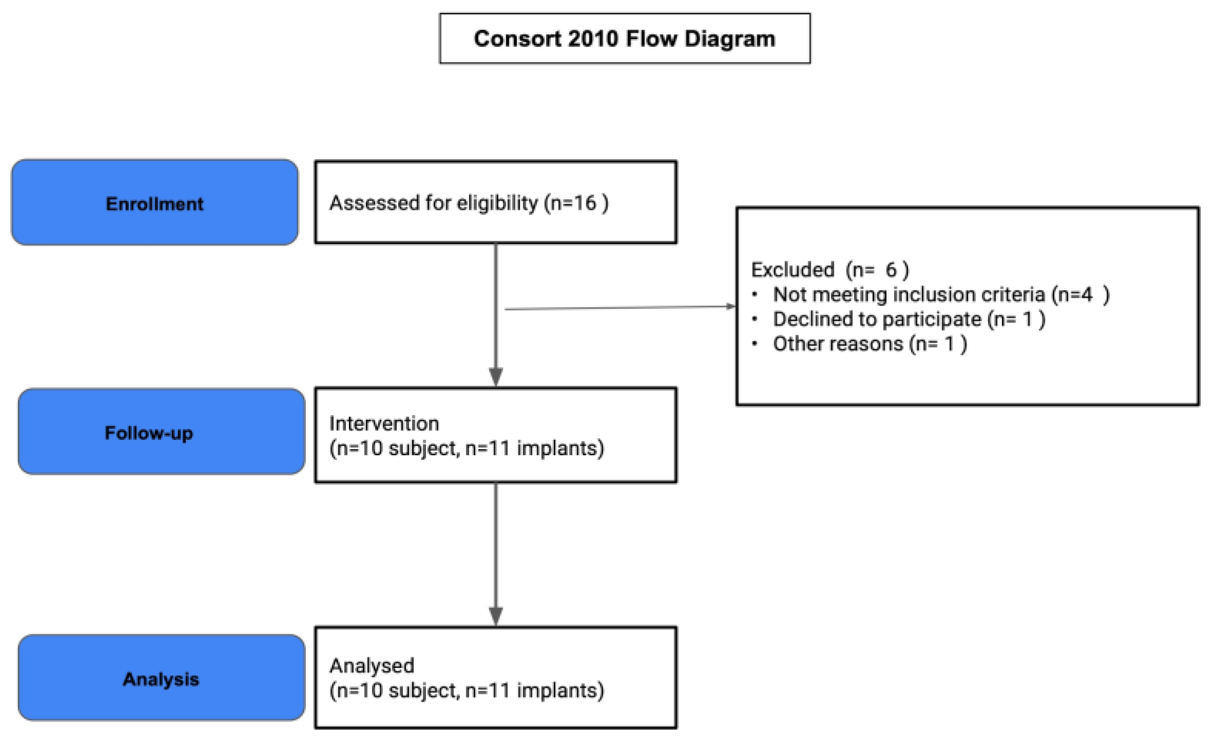
2.1. Study Population

The study protocol for this prospective study was approved by the Western Institutional Review Board (WIRB #20180697) and was registered at clinicaltrials.gov (registration no. NCT03917927). This study was part of a multicenter clinical trial that took place at three centers in the US and two centers abroad. While all centers shared common protocol, the cohort presented here was unique in terms of a digital dentistry component that may or may not be available in other centers. All clinical works presented here were performed at a single study site. Written consent, with a witness, was obtained from all participants. The study subjects enrolled in the study were patients who required single implant restoration in the esthetic zone, in the maxillary and mandibular anterior and premolar areas. The following inclusion criteria and exclusion criteria were used to screen potential candidates for the study (Table 1). The implants were planned to be placed in a healed edentulous site or immediately after single tooth extraction. Bone augmentation was not allowed in the study, and patients deemed to require bone augmentation would be excluded. All subjects signed the informed consent form and approved the treatment plan.

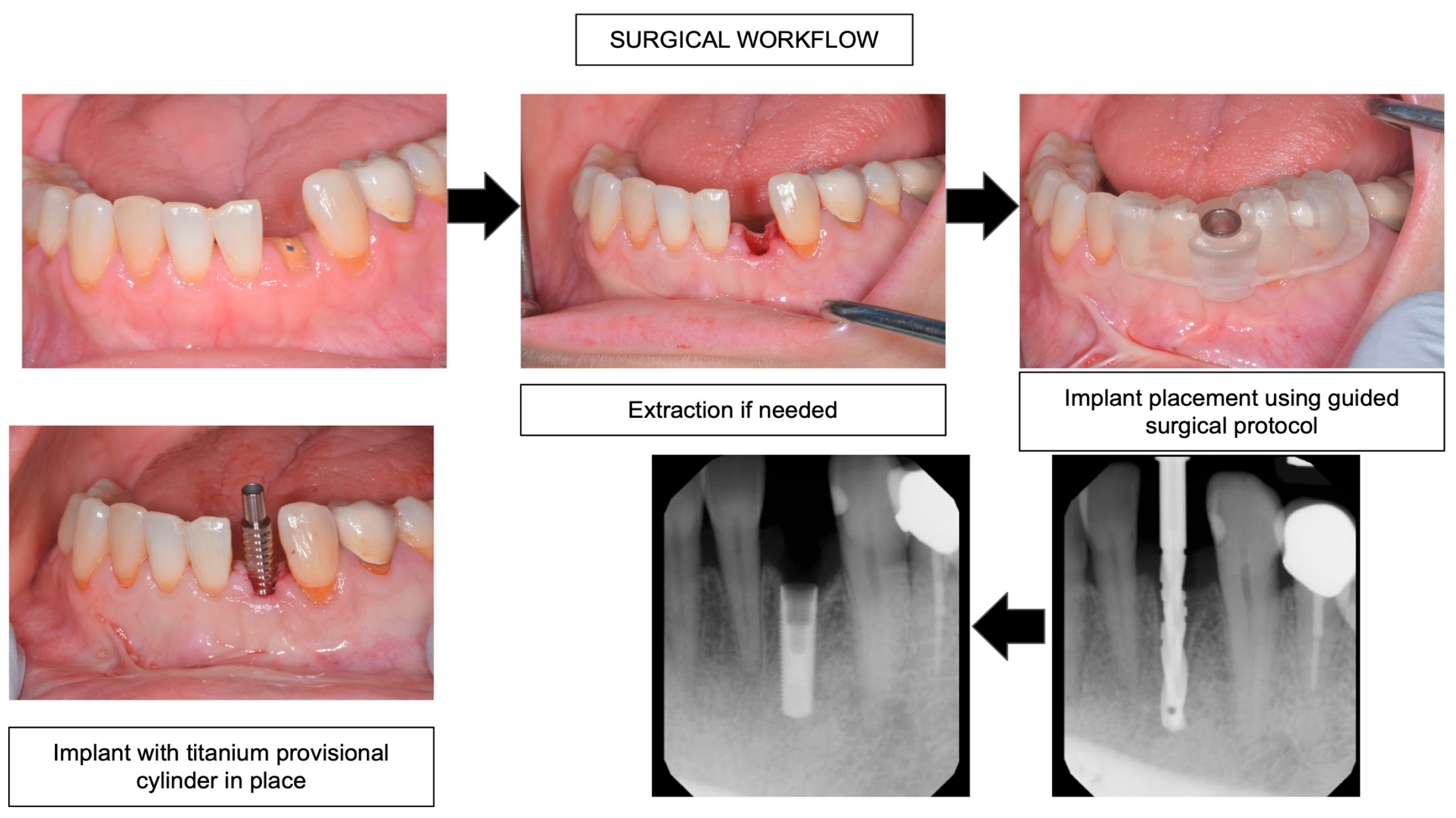
2.2. Clinical Protocol

Potential subjects were screened and clinical evaluations, including baseline oral health indices such as gingival and plaque indices, were documented (Figure 1). The edentulous site or unrestorable remaining tooth, as well as adjacent teeth, were clinically and radiographically evaluated. Preoperative intraoral scans were used to create a diagnostic wax-up. Preoperative cone-beam computed tomography (CBCT) was used to determine the available residual bone dimension and quality appropriate for a 3.1 mm diameter NDI implant (3.1 mmD Eztetic Implant, ZimVie, Palm Beach Garden, FL, USA). A preoperative antibiotic, 2 g Amoxicillin, was given approximately one hour prior to surgery. The patient was asked to rinse with 0.12% chlorhexidine gluconate for 30 s. The surgical procedures were performed under local anesthesia. Full thickness flaps were performed only in cases when the residual ridge was narrow and perforation might be expected. Otherwise, a flapless guided surgical protocol was employed. Figure 2 demonstrates a surgical workflow case with an extraction. The implant was placed using the manufacturer’s recommended protocol, using 2.1 mm/1.6 mm, 2.3 mm, and 2.8 mm diameter size osteotomy drills to the planned appropriate depth. Osteotomy procedures were performed using copious irrigation at a speed of 900 RPM. A bone tap was used when the presence of dense bone was determined. The implant was placed in the crestal position using 15–30 RPM speed. Primary stability with >35 Ncm insertion torque was chosen for the implants to be immediately provisionalized at the surgical appointment. Final seating torque was recorded. Resonance frequency analysis (RFA) was performed using the Osstell Smartpeg (Osstell, Göteborg, Sweden). The implant stability quotient (ISQ) value for each implant was then recorded, at the time of both implant placement and prosthetic insertion.
After the implant was placed, a provisional titanium prefabricated abutment (ZimVie, Palm Beach Gardens, FL, USA) and bis-acryl resin (Integrity, Charlotte, NC, USA) were used to fabricate a screw- or cement-retained provisional implant crown (Figure 3). The provisional crown was fabricated using a polyvinyl siloxane matrix fabricated from the preoperative diagnostic wax up, produced using a digital diagnostic wax up (3Shape Lab Studio) and stereolithographic 3D printing technology (Gray Resin, Formlabs and From 3B, Formlabs). The occlusal contacts of the crown were relieved. There was no occlusal contact in the maximum intercuspal position or in the lateral excursions. All implants were provisionalized within the same surgical visit.

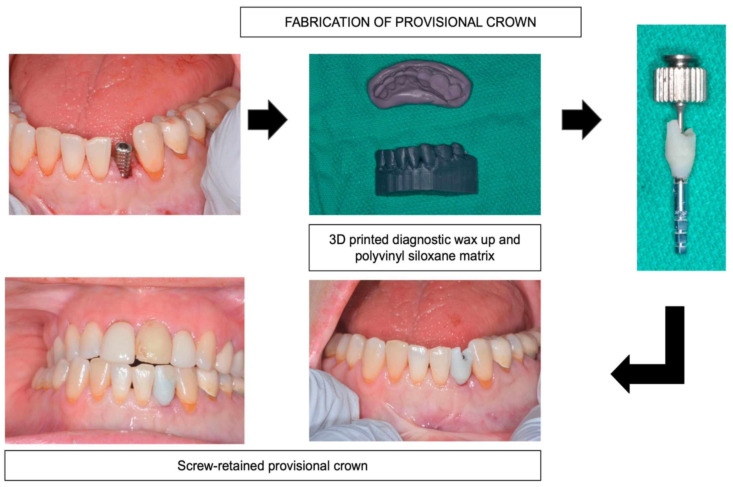
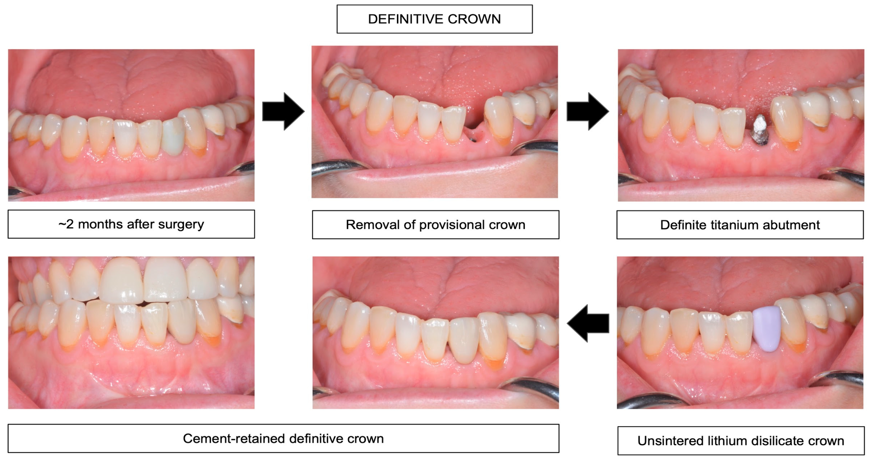
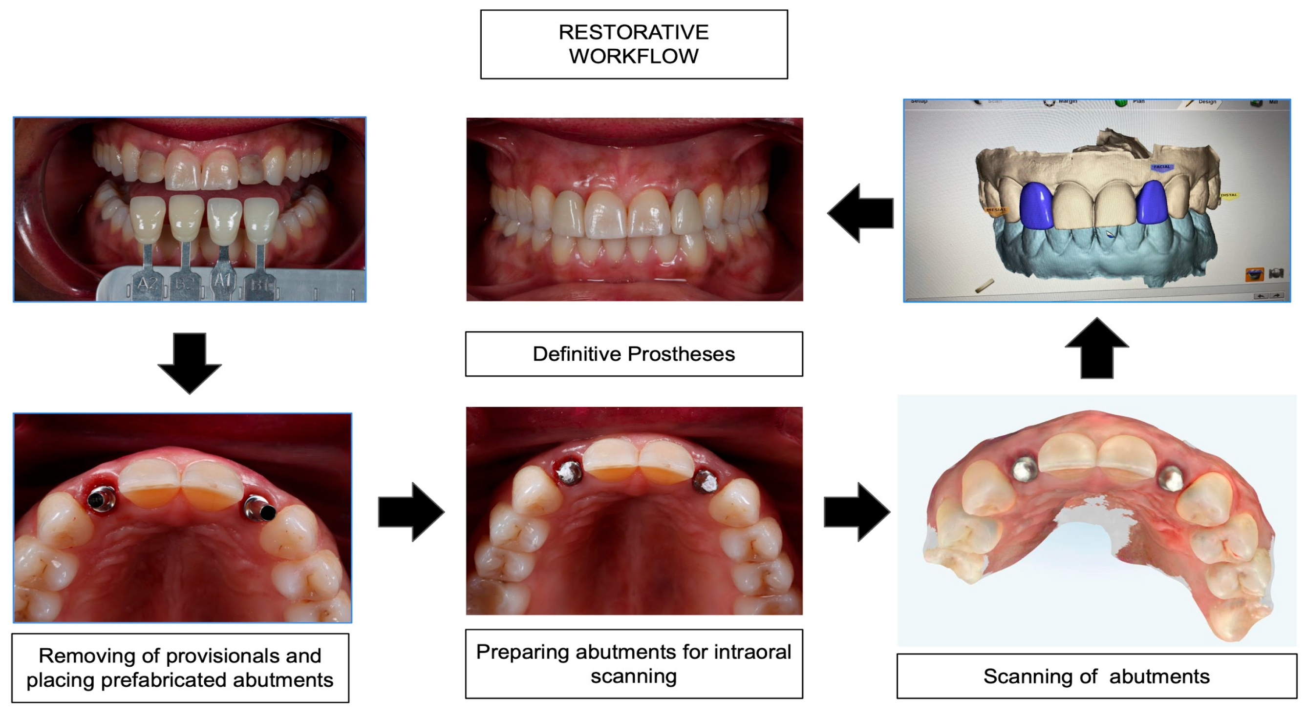
2.3. Same-Day Digital Dentistry Fabrication of Definitive Prosthesis

About eight weeks after implant placement, the prefabricated titanium implant abutment (Hex-lock Contour Abutment, ZimVie, Palm Beach Garden, FL, USA) was placed and tightened to 30 Ncm, according to the manufacturer’s recommendation. The abutment was then prepared intraorally with a high-speed handpiece and diamond burs, with copious cooling with air and water. Intraoral scanning was performed using either the Planmeca Emerald intraoral scanner (Planmeca, Helsinki, Finland) or the iTero Element II (Align Technology, San Jose, CA, USA). The interim prosthesis was then used as a guide to design the definitive implant crown using Planmeca PlanCAD software (Planmeca, Helsinki, Finland). The crown was then milled using CAD lithium disilicate material (IPS e.max CAD, Ivoclar, Liechtenstein). After milling, the unsintered crown was inserted to determine the proximal fit and customize the crown surface to mimic the adjacent and contralateral tooth surface morphology, texture, and characterization. The crown was then stained with feldspathic ceramics and sintered. The implant screw access hole was then filled with a Teflon tape barrier. The crown was then conditioned (Monobond Etch & Prime, Ivoclar, Liechtenstein), rinsed, air-dried, and luted with resin cement (Variolink Esthetics DC, Ivoclar, Liechtenstein). Figure 4 and Figure 5 demonstrate definitive crown fabrication and clinical restorative workflow.

2.4. Clinical Evaluation of Peri-Implant Soft Tissue Health

The peri-implant soft tissue esthetics, implant mobility, bleeding on probing, implant sulcus depth, gingival inflammation, and gingival and plaque indices were assessed at the time of implant placement with the provisional prosthesis, at the time of definitive prosthesis placement, and at 6-, 12-, and 24-month recalls. The Modified Gingival Index (MGI) was defined as: 0 for normal gingiva, absence of inflammation; 1 for mild inflammation, slight change in color, little change in texture of any portion of, but not entire, marginal or papillary gingival unit; 2 for mild inflammation, criteria as above but involving the entire marginal or papillary gingival unit; 3 for moderate inflammation, redness, edema, glazing, and/or hypertrophy of the marginal or papillary gingival unit; and 4 for severe inflammation, marked redness, edema, and/or hypertrophy of the marginal or papillary gingival unit, spontaneous bleeding, congestion, or ulceration. The Plaque Index (PI) was defined as: 1 for no plaque in the gingival area; 2 for a thin film of plaque at the gingival margin and area adjacent to the restoration, visible only when scraped with an explorer; 3 for a moderate amount of plaque along the gingival margin, the area adjacent to the restoration, and within the gingival sulcus, interproximal space free of plaque, plaque visible with the naked eye; and 4 for heavy accumulation of plaque at the gingival margin, the adjacent area of the restoration, and within the gingival sulcus, interproximal space filled with plaque.

2.5. Radiographic Evaluation

Standardized periapical radiographs were taken at the implant placement visit, at the visit of digital scanning and prosthesis fabrication, and then at one-year and two-year follow-up visits. The radiographs were used to compare the mesial and distal implant marginal bone changes. Periapical radiographs for each patient were used to determine the crestal bone levels, comparisons, and analysis between visits. Accuracy of crestal bone analysis was assured based on standardized radiographs. Attempts were made to ensure that the periapical radiographs were taken from the same position at each designated interval to reduce the errors in measuring crestal bone levels. Repeatable positioning of periapical radiographs was made possible by modifying a plastic Rinn bite block with impression material to register the incisal/occlusal position of the implant after placement. At the designated intervals, radiographs of study implants were obtained and the crestal bone level on the mesial and distal coronal aspects of each implant were measured. The distance between the crestal bone level and the point where the bone attaches to the implant was measured. To correct for any foreshortening that may have occurred due to minor angulation errors, the measured value was expressed as an absolute value in millimeters and as a normalized value. The normalized value was determined by multiplying the absolute value by the difference between the observed length of the implant on the radiograph and the actual length of the implant. An impartial evaluator, using standard operating procedures to ensure accuracy and precision, evaluated all radiographs. The measured dimensions were compared against known dimensions (length/diameter) and the percent difference were calculated and used to “normalize” the observed dimensions. These normalized values from the radiograph obtained at the immediate post-placement surgery date were considered the baseline against which all subsequent values were compared to assess crestal bone level changes. Periapical radiographs were also examined for evidence of other possible peri-implant pathologies.

3. Results

Ten patients who enrolled in the study received a total of eleven NDIs. Table 2 summarizes the patients’ demographics and implant data. The average age of the patients at the time of implant placement was 47.5 ± 18.0 years. No implant failure was reported. Thus, the implant survival rate was 100%. It took an average of 127.4 ± 53.0 days from the implant surgery to the day of definitive crown fabrication and insertion. The most common implant length used was 11.5 mm, which accounted for over half of the implants used (six out of eleven). Two implants were placed immediately after extractions. The ISQ values were 73.50 ± 12.01 at the time of the placement, and 76.59 ± 11.87 at the time of prosthetic insertion. The mesial and distal crestal bone loss values were −0.56 ± 0.54 mm and −0.32 ± 0.68 mm. Negative values signified bone resorption. The probing depth changes are shown in Table 3. Implant mobility was not found at any time point. The gingival inflammatory (GI) scores, bleeding on probing (BOP) scores, modified gingival index (MGI) scores, and plaque index (PI) scores are shown in Table 4.

4. Discussion

The main results of this exploratory study present the success of immediate provisionalization of single NDIs together with a same-day dentistry prosthetic fabrication protocol. This treatment protocol of immediate provisionalization of NDIs demonstrated a high implant success rate of 100%. This option allows clinicians to use titanium prefabricated abutments instead of Ti-based abutments, which are often not available for NDIs [16,19].
Compared to other studies, this protocol demonstrated comparable results with minimal mesial-distal peri-implant bone loss of ~0.3 to 0.6 mm after two years of implant placement. These bone remodeling value are similar to the ~0.35 mm bone resorption average from a study reporting on immediate provisionalization of NDIs after one year following implant placement [16], as well as from a study with delayed provisionalized protocol [20]. Immediate or delayed provisionalization appeared to have no effect on bone loss [16,20]. Long-term bone loss of <0.2 mm per year after the first year has been reported for NDIs [21]. While the peri-implant bone and gingival health, along with implant success, results of the current study were almost identical with previously reported outcomes by Oyama et al. [16], the prosthetic complications were different. Only two crowns in this study required recementation or remake, accounting for 18% of minor prosthetic complications with no complications observed during the provisional stage. On the other hand, in Oyama et al. [16], more prosthetic complications at the provisional crown stage were reported, including seven fractured provisionals, two de-bonded provisionals, and three loosened provisional abutment screws, all within the first three months following implant surgery, accounting for 70% of prosthetic complications. This may relate to the different implant design, provisional abutment, and implant connection, as well as the difference between using titanium prefabricated provisional abutments with bisacryl resin in this study and the use of titanium-based polyetheretherketone (PEEK) abutments with flowable composite resin in Oyama et al. [16].
There are multiple factors that affect the preservation of crestal peri-implant bone. Immediate provisionalization of conventional dental implants and NDIs has been shown to preserve crestal peri-implant bone and prevent the first year of peri-implant bone resorption [20,22]. Other factors, however, such as the implant fixture platform switching design [23], the microthread cervical design [24], the stability of the implant-abutment friction-fit design, and reduction of micromovement/microgaps [24,25], may also have a favorable effect on the reduction of the crestal peri-implant bone resorption. Note here that, in this study protocol, flap surgery was employed in most cases, since the alveolar ridges were narrow. Flapless surgery was employed for immediate placement implants and large alveolar ridge width. It is possible that the bone resorption may have been reduced further if flapless surgery were used exclusively [26,27,28,29].
Advances in digital technology, including intraoral scanning, CAD/CAM prosthetic design, and milling of the definitive implant restoration, allow for same-day implant crown fabrication [17,18,30,31]. The majority of same-day implant crown fabrications rely, however, on the availability of titanium-based (Ti-based) prefabricated abutments, together with a ceramic block with pre-milled implant screw access [17,18]. This option is often missing for NDIs. Therefore, in this study, a prefabricated titanium implant abutment was used with a monolithic lithium disilicate crown fabricated using CAD/CAM technology. One of the issues identified during this pilot clinical trial was the grayness of the titanium abutment showing through at the facial cervical gingiva, and in one case the crown needed to be replaced after additional preparation at the facial margin to drop the crown margin slightly deeper sub-gingivally. Titanium abutment preparation is therefore an important issue to ensure optimization of the scanning ability, as well as the milling capacity, of the restoration [17,32]. The same-day dentistry protocol for NDIs appears otherwise to be very successful, and, in most cases, the patients were pleased with the treatment outcome, esthetically, and with the speed of the treatment. The monolithic lithium disilicate crown was prescribed in this study to allow the same-day dentistry protocol without compromising the esthetics of the restorations, since zirconia restoration would have taken longer and likely would not be done in the same day [18].
It is also important to note here that, after enrolling and performing implant placement for the first seven patients, our clinic facility had to close for three months due to the COVID-19 pandemic. Some of our short-term care, such as surgical recalls and restorative work, had to be delayed. Some of the patients also refused to come back for the clinical follow-up until after the peak of the pandemic. While the COVID-19 pandemic undoubtedly had some adverse effects on clinical protocol, same-day crown fabrication reduces treatment visits, which eases patients’ anxiety about seeing a dentist during the pandemic. Several previous studies demonstrated that patients prefer digital impressions over conventional elastomeric impressions for a single implant [33,34,35,36,37]. Digital workflow for a single implant restoration has been shown to be more effective and less expensive, with shortened treatment time and a reduced number of clinical visits [37,38,39,40].
It is important to discuss some limitations of this pilot study and possible future clinical studies. First, the sample size of this study was limited. The sample size of ten was planned for this study as a pilot exploratory clinical trial to provide baseline data for future, larger clinical studies. In the future, there is potential for a clinical trial for immediate fabrication and insertion of the definitive crown together with guided implant surgery and CAD/CAM same-day dentistry. Second, only titanium prefabricated abutments were used here. Other types of abutments, such as zirconia-fused titanium base, may result in a different, or even a better, esthetic outcome. Third, in this study, while digital implant planning was employed, guided implant surgery was not applied. In the future, guided implant surgery can be applied, possibly together with a definitive restoration, fabricated ahead of surgery. More importantly, digital smile design can be an important part of routine treatment planning [41]. A future study which incorporates fully digital smile design and guided implant surgery will be important to provide insight into contemporary practices. Finally, the periapical radiograph is limited to 2D data, and the superimposition of facial and lingual bones can potentially alter the results.

5. Conclusions

NDIs are a viable alternative treatment for anterior and premolar implant sites without additional grafting or generative surgery. This exploratory study demonstrated that not only can NDIs be immediately provisionalized after implant placement in esthetic zone, but also that titanium preparable prefabricate abutments can be used together with same-day dentistry protocol with 100% implant survival rate over 24 months and minimal peri-implant bone loss or complications.

Author Contributions

Conceptualization, S.B.; methodology, S.B.; software, S.B.; validation, J.G.D. and S.B.; formal analysis, S.B.; investigation, S.B., J.G.D., A.L.H. and N.G.R.; resources, S.B.; data curation, A.L.H., S.B. and J.G.D.; writing—original draft preparation, S.B.; writing—review and editing, S.B. and L.J.H.; visualization, S.B. and J.G.D.; supervision, S.B. and J.G.D.; project administration, S.B. and A.L.H.; funding acquisition, S.B. All authors have read and agreed to the published version of the manuscript.

Funding

This research was funded by ZimVie, grant number FP00005953.

Institutional Review Board Statement

The study was conducted in accordance with the Declaration of Helsinki, approved by the Western Institutional Review Board (WIRB #20180697 approved on 16-MAY-2018), and was registered at clinicaltrials.gov (registration no. NCT03917927).

Informed Consent Statement

Informed consent was obtained from all subjects involved in the study.

Data Availability Statement

All relevant data are presented in the manuscript.

Acknowledgments

The authors thank the faculty and staff of the Virginia Commonwealth Center of Digital Dentistry and the Graduate Periodontology Clinic for their clinical support.

Conflicts of Interest

Author S.B. is a paid consultant and speaker for ZimVie, the sponsor of this research. However, the sponsor had no role in the interpretation of data, in the writing of the manuscript, or in the decision to publish the results.

References

  1. Schiegnitz, E.; Al-Nawas, B. Narrow-Diameter Implants: A Systematic Review and Meta-Analysis. Clin. Oral Implants Res. 2018, 29 (Suppl. 16), 21–40. [Google Scholar] [CrossRef] [PubMed]
  2. Klein, M.O.; Schiegnitz, E.; Al-Nawas, B. Systematic Review on Success of Narrow-Diameter Dental Implants. Int. J. Oral Maxillofac. Implants 2014, 29, 43–54. [Google Scholar] [CrossRef]
  3. Zhang, X.M.; Liu, B.L.; Qian, S.J.; Shi, J.Y.; Zhang, X.; Lai, H.C. Clinical evaluation of narrow-diameter implants versus standard-diameter implants with lateral bone augmentation in posterior jaws: Three-year results of a randomized controlled trial. Clin. Oral Implants Res. 2022, 33, 1245–1253. [Google Scholar] [CrossRef] [PubMed]
  4. Alrabiah, M. Comparison of Survival Rate and Crestal Bone Loss of Narrow Diameter Dental Implants versus Regular Dental Implants: A Systematic Review and Meta-Analysis. J. Investig. Clin. Dent. 2019, 10, e12367. [Google Scholar] [CrossRef] [PubMed]
  5. Worni, A.; Hicklin, S.P.; Mericske-Stern, R.; Enkling, N. Performance and Marginal Bone Level Alteration around Immediately Loaded Narrow-Diameter Implants. A Prospective Clinical Study: Results after 1 Year. Quintessence Int. 2018, 49, 267–276. [Google Scholar]
  6. Geckili, O.; Mumcu, E.; Bilhan, H. Radiographic Evaluation of Narrow-Diameter Implants after 5 Years of Clinical Function: A Retrospective Study. J. Oral Implantol. 2013, 39, 273–279. [Google Scholar] [CrossRef]
  7. Sierra-Sánchez, J.-L.; Martínez-González, A.; García-Sala Bonmatí, F.; Mañes-Ferrer, J.-F.; Brotons-Oliver, A. Narrow-Diameter Implants: Are They a Predictable Treatment Option? A Literature Review. Med. Oral Patol. Oral Cir. Bucal 2014, 19, e74–e81. [Google Scholar] [CrossRef]
  8. Cruz, R.S.; Lemos, C.A.A.; de Batista, V.E.S.; Yogui, F.C.; Oliveira, H.F.F.; Verri, F.R. Narrow-Diameter Implants versus Regular-Diameter Implants for Rehabilitation of the Anterior Region: A Systematic Review and Meta-Analysis. Int. J. Oral Maxillofac. Surg. 2021, 50, 674–682. [Google Scholar] [CrossRef]
  9. Parize, H.; Bohner, L.; Gama, L.; Porporatti, A.; Mezzomo, L.; Martin, W.; Gonçalves, T. Narrow-Diameter Implants in the Anterior Region: A Meta-Analysis. Int. J. Oral Maxillofac. Implant. 2019, 34, 1347–1358. [Google Scholar] [CrossRef]
  10. Davarpanah, M.; Martinez, H.; Tecucianu, J.F.; Celletti, R.; Lazzara, R. Small-diameter implants: Indications and contraindications. J. Esthet. Restor. Dent. 2000, 12, 186–194. [Google Scholar] [CrossRef]
  11. Ghazal, S.; Huynh-Ba, G.; Aghaloo, T.; Dibart, S.; Froum, S.; O’Neal, R.; Cochran, D. Randomized Controlled Multicenter Clinical Study Evaluating Crestal Bone Level Change of Narrow-Diameter Versus Standard-Diameter Ti-Zr Implants for Single Tooth Replacement in Anterior and Premolar Region. Int. J. Oral Maxillofac. Implant. 2019, 34, 708–718. [Google Scholar] [CrossRef] [PubMed]
  12. Roccuzzo, A.; Imber, J.; Lempert, J.; Hosseini, M.; Jensen, S.S. Narrow Diameter Implants to Replace Congenital Missing Maxillary Lateral Incisors: A 1-year Prospective, Controlled, Clinical Study. Clin. Oral Implant. Res. 2022, 33, 844–857. [Google Scholar] [CrossRef] [PubMed]
  13. Marcello-Machado, R.M.; Faot, F.; Schuster, A.J.; Nascimento, G.G.; Del Bel Cury, A.A. Mini-Implants and Narrow Diameter Implants as Mandibular Overdenture Retainers: A Systematic Review and Meta-Analysis of Clinical and Radiographic Outcomes. J. Oral Rehabil. 2018, 45, 161–183. [Google Scholar] [CrossRef] [PubMed]
  14. Degidi, M.; Piattelli, A.; Carinci, F. Clinical Outcome of Narrow Diameter Implants: A Retrospective Study of 510 Implants. J. Periodontol. 2008, 79, 49–54. [Google Scholar] [CrossRef]
  15. Degidi, M.; Nardi, D.; Piattelli, A. Immediate versus One-Stage Restoration of Small-Diameter Implants for a Single Missing Maxillary Lateral Incisor: A 3-Year Randomized Clinical Trial. J. Periodontol. 2009, 80, 1393–1398. [Google Scholar] [CrossRef]
  16. Oyama, K.; Kan, J.Y.K.; Rungcharassaeng, K.; Lozada, J. Immediate Provisionalization of 3.0-Mm-Diameter Implants Replacing Single Missing Maxillary and Mandibular Incisors: 1-Year Prospective Study. Int. J. Oral Maxillofac. Implants 2012, 27, 173–180. [Google Scholar]
  17. Bencharit, S.; Clark, W.A.; Stoner, L.O.; Chiang, G.; Sulaiman, T.A. Recent Advancements in CAD/CAM Same-Day Dentistry in Practice and Education. Preprints 2021. 2021110490. [Google Scholar]
  18. Blatz, M.B.; Conejo, J. The Current State of Chairside Digital Dentistry and Materials. Dent. Clin. N. Am. 2019, 63, 175–197. [Google Scholar] [CrossRef]
  19. Pelekanos, S. The Single Implant-Crown Complex in the Aesthetic Zone: Abutment Selection and the Treatment Sequencing. In Implant Aesthetics; Karateew, E., Ed.; Springer: Cham, Switzerland, 2017; pp. 301–336. [Google Scholar]
  20. Lambert, F.E.; Lecloux, G.; Grenade, C.; Bouhy, A.; Lamy, M.; Rompen, E.H. Less Invasive Surgical Procedures Using Narrow-Diameter Implants: A Prospective Study in 20 Consecutive Patients. J. Oral Implantol. 2015, 41, 693–699. [Google Scholar] [CrossRef]
  21. Froum, S.; Shi, Y.; Fisselier, F.; Cho, S.-C. Long-Term Retrospective Evaluation of Success of Narrow-Diameter Implants in Esthetic Areas: A Consecutive Case Series with 3 to 14 Years Follow-Up. Int. J. Periodontics Restor. Dent. 2017, 37, 629–637. [Google Scholar] [CrossRef]
  22. Block, M.S.; Mercante, D.E.; Lirette, D.; Mohamed, W.; Ryser, M.; Castellon, P. Prospective Evaluation of Immediate and Delayed Provisional Single Tooth Restorations. J. Oral Maxillofac. Surg. 2009, 67, 89–107. [Google Scholar] [CrossRef]
  23. Hagiwara, Y. Does Platform Switching Really Prevent Crestal Bone Loss around Implants? Jpn. Dent. Sci. Rev. 2010, 46, 122–131. [Google Scholar] [CrossRef]
  24. De Kok, I.J.; Chang, S.S.; Moriarty, J.D.; Cooper, L.F. A Retrospective Analysis of Peri-Implant Tissue Responses at Immediate Load/provisionalized Microthreaded Implants. Int. J. Oral Maxillofac. Implants 2006, 21, 405–412. [Google Scholar]
  25. Liu, Y.; Wang, J. Influences of Microgap and Micromotion of Implant–abutment Interface on Marginal Bone Loss around Implant Neck. Arch. Oral Biol. 2017, 83, 153–160. [Google Scholar] [CrossRef] [PubMed]
  26. Pisoni, L.; Ordesi, P.; Siervo, P.; Bianchi, A.E.; Persia, M.; Siervo, S. Flapless Versus Traditional Dental Implant Surgery: Long-Term Evaluation of Crestal Bone Resorption. J. Oral Maxillofac. Surg. 2016, 74, 1354–1359. [Google Scholar] [CrossRef]
  27. Mishra, P.; Tandan, A.; Shivakumar, G.C.; Choudhary, K.; Shivakumar, S.; Sharma, N.; Saxena, A. Evaluation of Crestal Bone Loss in Flap vs Flapless Implant Placement Technique: An In Vivo Comparative Study. J. Contemp. Dent. Pract. 2022, 23, 56–60. [Google Scholar]
  28. Vohra, F.; Al-Kheraif, A.A.; Almas, K.; Javed, F. Comparison of crestal bone loss around dental implants placed in healed sites using flapped and flapless techniques: A systematic review. J. Periodontol. 2015, 86, 185–191. [Google Scholar] [CrossRef]
  29. Al-Juboori, M.J.; Bin Abdulrahaman, S.; Jassan, A. Comparison of Flapless and Conventional Flap and the Effect on Crestal Bone Resorption during a 12-Week Healing Period. Dent. Implantol. Update 2012, 23, 9–16. [Google Scholar]
  30. Tamimi, F.; Hirayama, H. Digital Restorative Dentistry: A Guide to Materials, Equipment, and Clinical Procedures; Springer: Berlin/Heidelberg, Germany, 2019; ISBN 9783030159740. [Google Scholar]
  31. Baroudi, K.; Ibraheem, S.N. Assessment of Chair-Side Computer-Aided Design and Computer-Aided Manufacturing Restorations: A Review of the Literature. J. Int. Oral. Health 2015, 7, 96–104. [Google Scholar]
  32. Turkyilmaz, I.; Wilkins, G.N.; Varvara, G. Tooth Preparation, Digital Design and Milling Process Considerations for CAD/CAM Crowns: Understanding the Transition from Analog to Digital Workflow. J. Dent. Sci. 2021, 16, 1312–1314. [Google Scholar] [CrossRef]
  33. Schepke, U.; Meijer, H.J.A.; Kerdijk, W.; Cune, M.S. Digital versus Analog Complete-Arch Impressions for Single-Unit Premolar Implant Crowns: Operating Time and Patient Preference. J. Prosthet. Dent. 2015, 114, 403–406.e1. [Google Scholar] [CrossRef] [PubMed]
  34. Sivaramakrishnan, G.; Alsobaiei, M.; Sridharan, K. Patient Preference and Operating Time for Digital versus Conventional Impressions: A Network Meta-analysis. Aust. Dent. J. 2020, 65, 58–69. [Google Scholar] [CrossRef]
  35. De Oliveira, N.R.C.; de Oliveira, N.R.C.; Pigozzo, M.N.; Sesma, N.; Laganá, D.C. Clinical Efficiency and Patient Preference of Digital and Conventional Workflow for Single Implant Crowns Using Immediate and Regular Digital Impression: A Meta-analysis. Clin. Oral Implant. Res. 2020, 31, 669–686. [Google Scholar] [CrossRef]
  36. Joda, T.; Brägger, U. Patient-Centered Outcomes Comparing Digital and Conventional Implant Impression Procedures: A Randomized Crossover Trial. Clin. Oral Implants Res. 2016, 27, e185–e189. [Google Scholar] [CrossRef]
  37. Joda, T.; Lenherr, P.; Dedem, P.; Kovaltschuk, I.; Bragger, U.; Zitzmann, N.U. Time Efficiency, Difficulty, and Operator’s Preference Comparing Digital and Conventional Implant Impressions: A Randomized Controlled Trial. Clin. Oral Implant. Res. 2017, 28, 1318–1323. [Google Scholar] [CrossRef]
  38. Joda, T.; Zarone, F.; Ferrari, M. The Complete Digital Workflow in Fixed Prosthodontics: A Systematic Review. BMC Oral. Health 2017, 17, 124. [Google Scholar] [CrossRef]
  39. Joda, T.; Gintaute, A.; Brägger, U.; Ferrari, M.; Weber, K.; Zitzmann, N.U. Time-Efficiency and Cost-Analysis Comparing Three Digital Workflows for Treatment with Monolithic Zirconia Implant Fixed Dental Prostheses: A Double-Blinded RCT. J. Dent. 2021, 113, 103779. [Google Scholar] [CrossRef]
  40. Kunavisarut, C.; Jarangkul, W.; Pornprasertsuk-Damrongsri, S.; Joda, T. Patient-Reported Outcome Measures (PROMs) Comparing Digital and Conventional Workflows for Treatment with Posterior Single-Unit Implant Restorations: A Randomized Controlled Trial. J. Dent. 2022, 117, 103875. [Google Scholar] [CrossRef]
  41. Cervino, G.; Fiorillo, L.; Arzukanyan, A.V.; Spagnuolo, G.; Cicciù, M. Dental Restorative Digital Workflow: Digital Smile Design from Aesthetic to Function. Dent. J. 2019, 7, 30. [Google Scholar] [CrossRef] [Green Version]
Figure 1. Consort flowchart.
Prosthesis 05 00015 g001
Figure 2. Surgical procedure.
Prosthesis 05 00015 g002
Figure 3. Fabrication of provisional crown.
Prosthesis 05 00015 g003
Figure 4. Same-day dentistry from provisional crown to definitive crown.
Prosthesis 05 00015 g004
Figure 5. Clinical restorative workflow demonstrating the same-day dentistry protocol.
Prosthesis 05 00015 g005
Table 1. Inclusion and exclusion criteria.
Inclusion CriteriaExclusion Criteria
  • Patients of either sex and greater than 18 years of age.
  • Patients for whom a decision has already been made to use a dental implant for the restoration of existing partial edentulism in the anterior (central and lateral incisors), canine, or premolar regions in the mandible or maxilla. Placing an implant in the canine region should be in a healed site (not for immediate extraction).
  • Immediate extraction or a prior extracted site.
  • Intact buccal table as verified by CBCT or during surgery. If absent, patients should be excluded from enrollment in the study.
  • Patients must be physically able to tolerate conventional surgical and restorative procedures.
  • Patients with a facial lingual width of at least 5.1 mm and inner tooth width of at least 6 mm.
  • Patients having a thick gingival biotype (based on the lack of transparency of the periodontal probe through the gingival margin while probing the buccal sulcus) will be preferred but lack of this characteristic will not disqualify a patient from inclusion in this study.
  • Presence of opposing dentition with a functional occlusion that permits the restoration with a non-occluding provisional prosthesis.
  • Patients who provide a signed informed consent; a patient having implant placement surgery will continue participation in the study regardless of whether or not they receive restorative treatment according to protocol (protocol deviation).
  • Patients who agree to be evaluated for each study visit.
  • Minimum primary stability, insertion torque >35 Ncm (this will be a criteria that is met at the time of surgery).
  • Patients with known systemic diseases such as uncontrolled diabetes, endocrine disease, heart disease, immuno-compromised disorders, or mental disorders.
  • Patients with current use of non-steroidal anti-inflammatory drugs, bisphosphonates. or corticosteroid treatments.
  • Patients with active infection or severe inflammation in the areas intended for implant placement.
  • Patients in need of bone grafting at the intended study site.
  • Prisoners.
  • Patients with a >10 cigarette per day smoking habit.
  • Patients with a history of therapeutic radiation to the head or jaw.
  • Patients in need of bone grafting at the site of the intended implantation.
  • Patients who are known to be pregnant at the screening visit or planning to become pregnant within 6 months of study enrollment.
  • Patients with evidence of severe parafunctional habits such as bruxing or clenching.
  • Patients with HIV or hepatitis infection.
  • Patients who have previously failed implants at the site intended for study implant placement.
  • Patients in need of other treatments or surgeries at a site adjacent to the intended implantation site.
  • Patients with a history of severe periodontal disease.
Table 2. Patient’s demographic data, implant sites, and implant lengths.
VariableValuesn *%
GenderMale330
Female770
EthnicityCaucasian660
African American440
Implant placement site Maxillary lateral incisor545.5
Maxillary canine19.1
Maxillary premolar43.6
Mandibular lateral incisor19.1
Implant length8.0 mm19.1
10.0 mm218.2
11.5 mm654.5
13.0 mm218.2
* n (total) = 10 for patients, 11 for implants.
Table 3. Implant probing depth changes after definitive prosthetic placement.
Time PeriodTooth SurfaceChange (mm)
Between 3–6 monthsmesial0.43 ± 0.5
distal−0.14 ± 1.07
buccal0
lingual0.29 ± 0.95
Between 6 months and 1 yearmesial0.13 ± 0.99
distal0.25 ± 0.71
buccal−0.44 ± 1.05
lingual0.25 ± 0.89
Between 1 year and 2 yearsmesial0.25 ± 0.50
distal0.50 ± 0.58
buccal−0.25 ± 0.5
lingual0
Table 4. Clinical evaluation measures.
Clinical MeasuresGIBOPMGI *PI *
1 week0.67 ± 0.520.20 ± 0.40N/AN/A
3 weeks0.25 ± 0.440N/AN/A
Prosthetic insertion0.10 ± 0.3000.17 ± 0.410.17 ± 0.41
6 months0.14 ± 0.3800.17 ± 0.410
12 months0.13 ± 0.3500.29 ± 0.490
24 months0000
* Measures used only after the prosthetic insertion.
Disclaimer/Publisher’s Note: The statements, opinions and data contained in all publications are solely those of the individual author(s) and contributor(s) and not of MDPI and/or the editor(s). MDPI and/or the editor(s) disclaim responsibility for any injury to people or property resulting from any ideas, methods, instructions or products referred to in the content.

