Perspective

Molecular Diagnosis of Osteoarticular Implant-Associated Infection: Available Techniques and How We Can Use Them

1
Department of Clinical Microbiology, IIS-Fundacion Jimenez Diaz, UAM: CIBERINFEC-CIBER de Enfermedades Infecciosas, 28040 Madrid, Spain
2
Department of Orthopaedic and Trauma Surgery, IIS-Fundacion Jimenez Diaz, UAM: CIBERINFEC-CIBER de Enfermedades Infecciosas, 28040 Madrid, Spain
*
Author to whom correspondence should be addressed.
Prosthesis 2023, 5(1), 1-12; https://doi.org/10.3390/prosthesis5010001
Submission received: 21 November 2022 / Revised: 22 December 2022 / Accepted: 23 December 2022 / Published: 28 December 2022
(This article belongs to the Special Issue State of Art in Hip and Knee Replacement)

Abstract

:
Despite recent advances during the last few years, microbiological diagnosis of prosthetic joint infections remains a challenge. Molecular biology techniques have been developed to try to overcome this problem, and recently, many of them have become available for many laboratories. Some of them, especially commercial multiplex PCR-based assays and universal 16S rDNA homemade PCR assays, are now available in many laboratories. Moreover, new technologies have appeared, especially metagenomics and next-generation sequencing. These techniques have demonstrated their potential in many studies but appear to be experimental at present. A few studies have evaluated the possible use of these methods in the clinical routine, and a review of the critical aspects for the selection of a molecular method (accuracy, complexity, cost) was performed. Finally, a proposal for a protocol that includes molecular biology techniques was made according to the literature published in this field. In conclusion, molecular biology techniques are ready to be used in the clinical routine of a microbiology laboratory, but their use must be carried out in accordance with the many special characteristics of each laboratory. In all cases, the interpretation of the results must be conducted by a multidisciplinary team with experience in the management of these patients.

1. Introduction

Implant-associated infections are one of the most problematic complications of the use of implants in orthopedic and trauma surgery. Although its incidence is relatively low (1–3%, depending on the series) [1], an increasing number of patients underwent this type of surgery (and were at higher risk of contracting these infections because of an increase in the number of comorbidities among these patients) [2]. The management of these diseases includes new surgical procedures, prolonged antibiotic treatment, and longer hospital stays. Even with this management, the outcome of the infection is not always a good one, and it is considered that directed therapy is currently needed because of the increasing number of multidrug-resistant organisms and the differences between therapeutic options [3,4,5,6]. This fact made it necessary to conduct a proper microbiological diagnosis of these infections. Despite most of the infections being caused by a small number of species [1,7,8], almost all microorganisms can be a cause of infection. In trying to obtain a good diagnosis, conventional mechanisms have been improved using different combinations of media, incubation time, sampling, and the incorporation of new techniques such as sonication [1,9,10,11]. Synovial fluid biomarkers could also improve PJI diagnosis, including detection of leucocyte esterase, alpha-defensin or C-reactive protein, which are considered minor criteria for PJI definition [12,13,14], but the serological markers allow only the syndromic diagnosis, and not a microbiological diagnosis. However, these advances are not universally used [15,16], and even in those centers that used them, there are still patients with negative results, despite having a true infection. This percentage is variable but is considered to be an average of 10–15% of all cases [1,17]. At this point, conventional microbiological techniques are seen to have reached their peak in both sensibility and specificity, and new techniques are needed that can improve the currently available technology. In a previous review, one of the authors described how molecular biology can increase the sensibility of microbiological diagnosis [18], but the field of molecular biology has progressed rapidly since then, and new approaches that were not yet imagined have appeared since then. In the present review, we will discuss the currently available techniques for molecular diagnosis and suggest possibilities for their use in the implant-associated infection setting.

2. Homemade PCR-Based Techniques

The first approach to the use of molecular biology for the diagnosis of PJI was the development of homemade techniques. Several reviews have described the different studies that use these techniques [18,19]. These techniques mainly used broad-spectrum detection by using 16S rDNA amplification followed by sequentiation to identify the detected microorganisms. The studies usually used synovial fluid as the sample, and most of them described high specificity and sensitivity. Other samples with potentially high sensitivity have been used in other studies. One example is the study of Marin et al. [20]. In this study, the authors evaluated the usefulness of a 16S rDNA universal PCR with periprosthetic biopsies. The best sensitivity (94%) was obtained when two of five samples showed the same result, maintaining the best possible specificity (100%). The introduction of sonication prompted other authors to evaluate the same type of PCR with sonicated samples. In the study of Gomez et al. [21], the authors reported sensitivity of 70.4% and specificity of 97.8% with that type of sample. The last result is of special importance, because one important criticism of sonication was the potential for contamination, but even using a more sensitive technique such as PCR, the specificity values remained high. Moreover, when PCR was combined with culture, the sensitivity increased from 72.6% for culture to 78.5% for culture + PCR [21], showing the importance of a mixed approach (molecular biology added to conventional cultures) for a better diagnosis of PJI.
However, the main theoretical problem associated with these techniques is the lack of standardization. It is usually considered that these techniques work well in the laboratories that designed the protocols but fail when another laboratory tries to reproduce the results. However, this assertion was proved to not always be true: two articles described a multicenter study performed in France that evaluated the usefulness and reproducibility of 16S rDNA-sequencing PCR. In the first article, the authors evaluated the sensitivity and specificity of a common protocol in seven university hospitals in France [22]. In this study, the technique was performed in 305 patients, and it worked well in all centers, with sensitivity values of 73.3% and specificity of 95.5%. Moreover, in a second study, the authors evaluated the reproducibility of the technique by sending the same samples to the different centers, and the obtained results were concordant in most centers in 93.8% of the samples [23]. This study is extremely important because it showed that the main theoretical problem of homemade PCR—the lack of standardization—can be minimized in many cases. However, specific facilities and well-trained technicians are needed in order to obtain reproducible results, so this methodology cannot be performed in many laboratories that lack molecular biology facilities. Despite these limitations, 16S rDNA PCR is the most common molecular technique used by most laboratories that performed molecular biology diagnosis for PJI in recent surveys [15,16].

3. Commercial PCR-Based Techniques

3.1. Adapted Kits

Shortly after the description of sonication as a useful technique for the diagnosis of PJI, several authors looked for a more easy-to-perform molecular technique that can be applied to these samples. The first logical option in these years was the use of kits designed for their use in positive blood-culture bottles. These kits are usually multiplex PCR that include the most commonly isolated microorganisms from blood cultures. Because these organisms include most of the isolates that appear as a cause of PJI, the possibility of their use in clinical samples was explored in several studies.
The first one was the study of Achermann et al. [24]. In this study, the researchers used the SeptiFast® kit (Roche Diagnostics, Penzberg, Germany) designed for identification of 19 microorganisms or groups of microorganisms frequently isolated from blood cultures. They included 37 cases of PJI, and the kit showed a sensitivity of 78%, while no specificity study was performed. Shortly after this study, another one performed with the same kit in 86 explanted prostheses (24 with a diagnosis of PJI) showed an impressive sensitivity of 96%, with a specificity of 100% [25].
Another kit that was tested for potential use in PJI was the GenoType BC® (Hain Lifescience, Nehren, Germany). This system is formed by a combination of two kits for Gram-positive and Gram-negative organisms and identifies 32 species or group of species of microorganisms. Additionally, designed for its use in blood cultures, a study by Esteban et al. showed its potential for the diagnosis of PJI [26]. The study was performed in 126 patients (47 with a clinical diagnosis of infection), including both PJI and Osteosynthesis-associated infection. In this study, the combination of PCR and culture from the sonicate fluid allowed the identification of 80% of the microorganisms that cause infection. Moreover, there were also cases of positive results for culture, PCR, or both that were later considered as infected, but the potential for a contamination cannot be excluded in some cases [26].
The study performed by Duboix-Bourandy used the Xpert MRSA/SA SSTI kit (Cepheid, Sunnyvale, CA, USA) [27]. This kit was designed for the rapid detection of Staphylococcus aureus and methicillin-resistance markers, so its usefulness for the diagnosis of PJI is extremely limited. However, the interest of this study was the evaluation of the potential use of the cartridge design for this diagnosis. This methodology is extremely easy to perform and showed potential usefulness for medium–small-sized laboratories without proper facilities for molecular diagnosis.
This approach was further evaluated with a different cartridge-design kit that identifies a higher number of microorganisms. The Filmarray BCID® (BioMérieux, Marcy l’Etoile, France) detects 24 microorganisms or groups of microorganisms that are commonly detected among PJI. This kit was evaluated for this purpose by Vasoo et al. [28] using samples from 216 patients (including 98 infections) that were maintained in a frozen state until the test was performed. In this study, the test showed sensitivity of 53% and specificity of 99%. Interestingly, the low sensitivity was to the low performance of the kit with coagulase-negative staphylococci (CNS), while sensitivity to other pathogens was higher than that (90% for S. aureus and 100% for enterobacteria). This fact has important implications for the diagnosis of chronic PJI, where CNS was the most frequently isolated pathogen [8]. Notably, the Filmarray BCID® kit also includes the detection of some resistance markers of interest for the antibiotic selection among these patients.

3.2. Specifically Designed Kits

The first specifically designed kit for the diagnosis of PJI was described in 2014 by Metso et al. [29]. The Prove-it Bone and Joint StripArray assay (Mobidiag Ltd., Espoo, Finland) was developed from a kit previously designed for identification of microorganisms using broad-range PCR followed by a microarray assay [30]. The study showed sensitivity of 81.6% with specificity of 73.9%, using the MSIS criteria for diagnosis of PJI as the gold standard. No further studies using this kit have been published until now.
The Unyvero I60ITI (Curetis GmbH, Holzgerlingen, Germany) was a multiplex PCR-based test that uses the cartridge technology and can be easily used in most laboratories without specifical facilities. It can detect 30 species or groups of species, and several resistance markers. During its evolution, it previously included a universal bacteria target in one of the versions, but this was removed from the last one. The performance of the kit was tested in several studies, using different types of samples for detection of organisms causing PJI [31,32,33,34,35,36,37,38,39]. The average sensitivity of the kit is relatively low (67.5%), but the average specificity was high 96.3%), with average positive predictive and negative predictive values of 93.2% and 71%, respectively, which means that a positive result is probably a true positive (and not a contamination), but a negative result cannot be used to discard a PJI. The test, as well as other multiplex PCR-based kits, can also detect polymicrobial infections, which could be of interest in some cases of PJI. Aiming to improve the sensitivity of this method, Villa et al. [40] used an enrichment step before the DNA extraction by incubating the samples in Brain–Heart Infusion and Thioglycolate broths until they became turbid. With this approach, the sensitivity of the method increased to 81.6%, but at the cost of a very delayed diagnosis. Another recent study [41] showed improved sensitivity (85%) for patients that fulfil the MSIS criteria, but another one showed low sensitivity when the patients were diagnosed using the EBJIS criteria [42]. Nevertheless, in both cases, the specificity was higher than 97%. As of this year, this kit remains the only specifically designed kit commercially available, and it is currently being used by some laboratories, as described in a recent European survey [15], as well as in a Spanish survey [16].
Based in the BioFire® technology (Biofire, Salt Lake City, UT, USA), a new kit specifically designed for the diagnosis of bone and joint infection has been developed in recent years that will be added to the other tests from this year. This new test is based in cartridge technology and includes 31 of the most common pathogens that can be detected in acute bone and joint infections, as well as 8 resistance markers. The results of a multicenter study for the clinical evaluation of this test have been presented in different congresses, showing an extremely good performance for these types of patients, with sensitivity of 90.6% and specificity of 99.8% [43]. Despite this superb performance in acute infections, the main limitation of the kit is the lack of some pathogens of importance in chronic infections, notably CNS and Cutibacterium acnes. This limits the usefulness of the kit for these patients, but it probably has minimal impact in acute infections. However, the kit has a characteristic that could be of importance: it takes approximately 1 h to obtain a result [43], which implies that the test can be used for rapid diagnosis of these infections, even in the emergency room or potentially even during surgery. The implications of this short detection time will be probably studied in the future.

4. Next-Generation Sequencing and Metagenomics

Since Tarabichi et al. demonstrated the usefulness of next-generation sequencing (NGS) with the detection of Streptococcus canis as the infectious agent in a culture-negative PJI [44], NGS and metagenomics became emerging tools for pathogen detection in PJI. Those techniques detect all nucleic acids that are present in a given specimen, allowing the identification of the microorganism/s involved in the infection and its antimicrobial resistance characterization [45].
The sensitivity and specificity of NGS for PJI diagnosis vary among different studies. The meta-analysis published by Hantouly et al. reported a pooled sensitivity of 94%, higher than the one obtained by culture (70%) and slightly lower specificity (89%) in comparison to culture (94%) [46]. Additionally, the accuracy was higher using NGS technology (91.9%) than culture-based methods (80.5%). They also showed an overall false-positive rate of 11% for NGS.
Metagenomics has also shown promising results for PJI diagnosis. Ivy et al. demonstrated a good correlation with culture in 82 synovial fluid samples and yield additional pathogens not detected by culture [47]. In the study of Street et al., metagenomic sequencing showed species-level sensitivity of 88% versus sonication of fluid culture [48]. Huang et al. obtained greater sensitivity in PJI diagnosis using metagenomics (95.5%) than conventional culture (79.6%), with similar values of specificity: 95.2% for both techniques [49]. However, the authors claimed high levels of host DNA contamination despite their efforts to improve the performance of the technique.
Those techniques have shown to be useful in culture-negative PJI. In the prospective study of Tarabichi et al., NGS allowed microorganism detection in 81.8% of culture-negative PJI [50]. Using metagenomic shotgun sequencing, Thoendel et al. were able to detect potential pathogens in 43.9% of culture-negative specimens [51].
It seems that NGS and metagenomics will play an important role in the diagnosis of PJI infections. Due to its high sensitivity, there is a lack of understanding of the clinical importance of identifying unusual pathogens that may not necessarily be involved in the disease process, and also determining if the potential pathogens detected are active and clinically relevant or not.
The use of NGS and mNGS for PJI diagnosis also presents several limitations. First, those techniques have a low specificity, mainly due to the great number of false positive results because of contamination. Another important limitation is the high cost of those techniques in comparison with culture and other molecular methods, and the run-time, as it sometime requires around three to five days to provide identification. Additionally, the transformation of raw data into clinically useful information involves bioinformatics abilities. In addition, multicenter trials are needed to support its use as a diagnostic tool in laboratory routine facilities.

5. How Can the Molecular Diagnosis Techniques Be Used in a Routine Setting?

The field of molecular diagnosis techniques that can be used routinely, despite the problems and limitations of the available methods, now offers a broad spectrum of techniques and methodologies that can be used for the diagnosis of PJI in the routine of clinical microbiology laboratories. However, before implementing these methods, several questions must be answered in order to select the best available option for each laboratory. In the next few sections, we try to describe these questions and the potential answers from our point of view.

5.1. In What Type of Patients Can These Techniques Be Used?

The answer for this question seems easy: molecular biology techniques can be used in all patients. However, the selection of patients that could benefit more from this option is not as easy as it seems. In fact, the selection of techniques for the diagnosis of PJI depends on different factors, which made the standardization of these methodologies a difficult task [10]. Recent surveys have recently shown the differences between centers for the use of available methodologies [15,16], even for the selection of conventional culture-based methods. This selection depends on the characteristics of the hospital, the laboratory, and the samples that the laboratory receives. Moreover, sometimes it depends on the existence of multidisciplinary units aimed in the management of PJI and other bone and joint infections.
For these reasons, a careful review of the molecular techniques is needed before their implementation in laboratory routine. For this purpose, sensitivity and specificity of these methods are essential factors. However, even these values can change according to the patient. In the meta-analysis by Liu et al. [52], the pooled sensitivity was 75% and the specificity was 96%. However, these values change when the pretest probability of an infection is analyzed. The possibility of a positive PCR increased from 88% to 99% when the pretest probability of infection was 25% or 75%, respectively. This means that a good selection of the patient is essential to evaluate the results obtained using this technique. Moreover, because the sensitivity of these techniques is relatively low [52,53], its indiscriminate use in all patients could lead to misinterpretations of the results. A negative result could be used to rule out an infection, but this is a mistake because of the low negative predictive value of the methods. Moreover, its use in non-infected patients could lead to false-positive results, with a loosening of credibility in the test. So, in our opinion, these techniques must be used in patients with a suspected infection according to the different criteria used for this purpose [54,55,56,57] in order to obtain the best possible results.

5.2. What Type of Technique Must Be Used?

The selection of the technique is another important issue that must be considered for the implementation of molecular biology in clinical practice. Several issues must be considered for this purpose. The first one is the goal of the technique, which is the etiological diagnosis of patients with PJI. For this purpose, we must consider that the number of organisms that can cause a PJI is enormous (although most of the infections are caused by a small group) [1,7,8,17]. This fact means that only a technique that allows the detection of many microorganisms is useful, so techniques that allow the detection of one microorganism or two must be discarded [27]. The techniques available for the detection of many microorganisms include homemade broad-spectrum PCR-based techniques (16 rDNA amplification), multiplex PCR-based techniques and NGS and metagenomics [1,18,19,22,34,37,53,58]. All these techniques are available to detect many microorganisms, and even several of them simultaneously. This increases the possibility of detecting even polymicrobial infections, which is crucial when avoiding treatment failures or relapses. However, we must consider that multiplex-based techniques are limited to the microorganisms included in the kit, and other ones cannot be detected, and this limitation is very important when interpretation of a negative result in a patient with a diagnosis of PJI is obtained.
We decided that a molecular biology test must be included in the clinical microbiology routine. However, other factors must be considered before we select the test: time, complexity, and cost.

5.2.1. Time

Time is an essential part of the management of most infectious diseases, and a rapid diagnosis is a factor that promises rapid specific therapy and a better outcome. However, in some PJI, such rapid diagnosis is not an essential issue because other factors, such as the type of surgical therapy, are more influential than a rapid diagnosis. However, a rapid diagnosis is always desirable, and molecular techniques are usually faster than conventional culture-based methods. The current protocols for these methods usually include a long incubation period of 14–15 days. This time was suggested in the article of Schäfer et al. [59], and many other authors have recommended it for the isolation of some slowly growing organisms [60,61,62,63,64]. It is currently used in the routine of many laboratories [15,16]. This means that in some cases, the etiological diagnosis is not available until many days have passed. Because microbiological results are essential for the diagnosis of PJI in many protocols [54,55,65], this could represent a problem in some cases. In this aspect of diagnosis, molecular biology techniques are faster than conventional cultures. Even the slowest of these methods is faster than a 15-day culture-based method. However, not all molecular techniques are of similar speed.
The ideal technique is one which can be performed and give results intraoperatively and allows the surgeon to modify the surgical procedure if necessary. However, currently available protocols need longer turnaround times, so no intraoperative method is currently available for an etiological diagnosis of PJI. The homemade, PCR-based methods usually take hours—or even days—to provide a definitive result. Cartridge-based commercial multiplex PCR also usually take hours (Unyvero I60ITI takes 5 h) [41,53], but they are faster than cultures and can provide a result on the same day when the samples are obtained, with a specificity that allows us to have confidence in this diagnosis. The recently developed BioFire BJI test is even faster, with results obtained in 1 h approximately [43]. This means that this test is at the edge of being useful intraoperatively, at least in some cases. This property merits a further evaluation of this potential, with the limitation of being useful only in acute infections. However, the possibility of a directed therapy against the pathogen detected in the very first moment postoperatively opens up a new concept for the management of these infections.
Despite the impressive results that can be obtained, NGS and metagenomics take a long time to perform (usually more than 1 day). This means that a rapid diagnosis cannot be performed with this methodology [66]. However, the fact that this technique is a new one enables further development of the engineering issues that could make it faster, so in the future, we will probably see NGS systems that allow us to perform this technique in a faster time that that which is needed in the present moment [66].

5.2.2. Complexity

The ideal molecular biology technique for all laboratories needs to be easy to perform. However, this is not always possible, because the different techniques have specific problems. The complexity of a technique is a factor that is decisive in the implementation of some techniques in clinical microbiology routine, where complex techniques experience a lot of integration problems. Easy methods are usually the best options in this setting, and this must be considered when clinical microbiologists decide the incorporation of a new method in the laboratory.
Homemade methods usually require specific facilities for molecular biology, together with personnel who are well trained in these techniques. Although it seems that the problem of standardization can be avoided [23], all the infrastructure issues cannot be avoided. This made these methods available to large laboratories or, at least, laboratories designed for a specifically designed molecular biology laboratory. While these methods are broadly used according to the surveys [15,16], most medium and small size laboratories are probably not prepared to perform these techniques, and probably need to send these samples to reference center, with a consequent delay in the diagnosis. This limitation is also present in NGS metagenomics methods. They also require specific facilities, and the necessary equipment is not usually available in all laboratories [59,66,67,68]. Cartridge-based methods are easy to perform and are probably available for most clinical microbiology laboratories. Usually, they do not need neither specific nor specialized personnel, and can be used in small and medium-size laboratories. The Unyvero® I60ITI and the BioFire® BJI tests are designed with this methodology [43,53,63] and can be integrated easily into the routine of most clinical laboratories with their own limitations.

5.2.3. Cost

Molecular biology methods are usually more costly than culture-based methods in terms of their fungible and equipment needs. The need of specific facilities made homemade techniques and NGS metagenomics expensive procedures, while the cost of reactives is not so high for homemade PCR techniques. Commercial kits are usually expensive (more than EUR 100), and usually cannot compete with culture methods in this aspect. However, when we consider this aspect, we must take into account other issues, because the final cost of the management of a PJI can be influenced by the selection of the proper diagnostic method. There are almost no studies about the cost effectiveness of diagnostic methods in the literature. In a PubMed search, using (Cost-effectiveness) AND ((molecular biology) OR (PCR) OR (molecular diagnosis)) AND (prosthetic joint infection) as search criteria, only one article appears that is a study of this aspect, and it is about alpha-defensin [69]. One congress communication [70] reports a cost-effectiveness study performed with a commercial technique (Unyvero I60ITI) for the diagnosis of PJI in a hospital with a specialized unit in the management of these patients. In this pilot study, the use of the molecular methods means a reduced average cost of EUR 840.67 in the management of a PJI. This means that the incorporation of a molecular technique, despite being expensive for the laboratory, can be cost effective for all the PJI management process. We think that cost-effectiveness studies will be essential and must be performed in the near future.

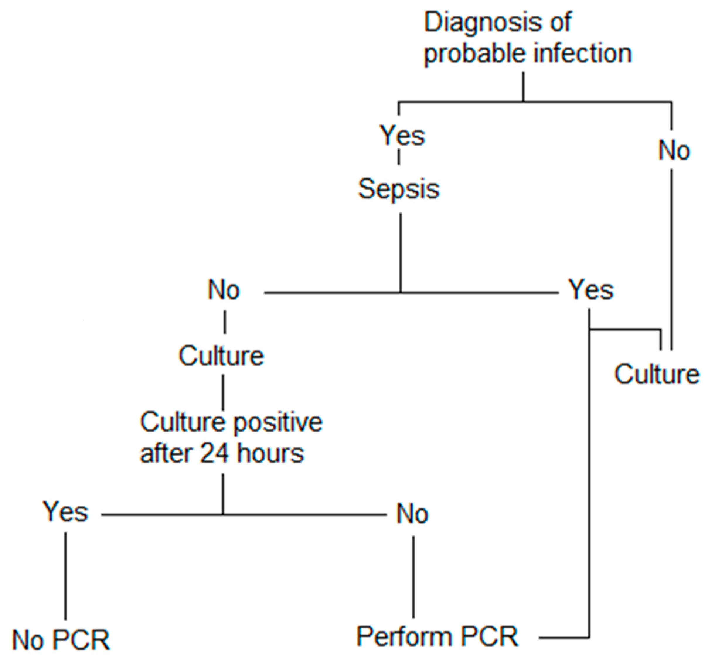
5.3. How Can We Use These Methods?

It seems clear that the incorporation of molecular biology into the diagnostic arsenal of microbiological tools for the diagnosis of PJI is not only possible, but desirable. However, if we consider the available techniques, the selection of one of them is important for the laboratory (and the hospital). All the previously described issues need to be considered, and possible options, from our personal point of view, are as follows: If the laboratory is a small–medium-size one without proper facilities for molecular diagnosis, a cartridge-based method is probably the best option. If the laboratory is a large one, with a specific molecular biology section, NGS and/or homemade broad-spectrum PCR can be selected, with some rapid diagnosis kits for some specific cases. In all cases, molecular biology will be complementary to conventional culture-based methods. The samples selected for processing will be selected with close relationships with multidisciplinary teams, looking for patients with a probable diagnosis of PJI with a high pretest index of suspicion. We have recently published our clinical experience with this approach [71], showing that the number of patients that can benefit from a molecular test is high, even in a routine setting and not an experimental one (Figure 1). We need more reports about the experience of the laboratories in the use of molecular tests for PJI to select the best protocol to obtain the best results with the incorporation of these methods in the routine setting.

6. Conclusions

Molecular-biology-based techniques for the diagnosis of PJI are here to stay. There is a lot of experience with many different tests that showed, in all cases, high specificity, with variable sensitivity [19,53,63,72]. However, the incorporation of these methods to the routine of microbiological diagnosis of these patients is not universal, and probably requires further studies about cost effectiveness and usefulness in routine practice. The incorporation of new methods will be of increasing interest, and further development of equipment and tools necessary to improve the use of complex methods will make them available for more laboratories [72]. However, these methods have limitations that are necessary to understand before a proper interpretation of the results can be performed. When this interpretation is carried out in the setting of multidisciplinary teams, it is extremely useful, and it can benefit the management of these complex patients and improve their outcome.

Author Contributions

Conceptualization: J.E. and L.S.-V.; Methodology, J.E., L.S.-V. and Á.A.; Software, J.E. and L.S.-V.; Validation, J.E., L.S.-V. and Á.A.; Formal Analysis, L.S.-V. and Á.A.; Investigation, J.E. and Á.A.; Resources, J.E.; Writing—Original Draft Preparation, J.E., L.S.-V. and Á.A.; Writing—Review and Editing, J.E., L.S.-V. and Á.A.; Visualization, J.E., L.S.-V. and Á.A.; Supervision, J.E.; Project Administration, J.E. All authors have read and agreed to the published version of the manuscript.

Funding

This research received no external funding.

Conflicts of Interest

JE has received research grants from Biomérieux and Laboratorios Leti, S.L., conference fees from Biomérieux and Heraeus, and funds for congresses from Biomérieux and Pfizer. LS received conference fees from Werfen, and funds for congresses from Vircell. There were no conflict for AA regarding this manuscript.

References

  1. Tande, A.J.; Patel, R. Prosthetic joint infection. Clin. Microbiol. Rev. 2014, 27, 302–345. [Google Scholar] [CrossRef] [PubMed] [Green Version]
  2. Kurtz, S.M.; Lau, E.; Watson, H.; Schmier, J.K.; Parvizi, J. Economic burden of periprosthetic joint infection in the United States. J. Arthroplast. 2012, 27, 61–65.e1. [Google Scholar] [CrossRef] [PubMed]
  3. Lora-Tamayo, J.; Murillo, O.; Iribarren, J.A.; Soriano, A.; Sánchez-Somolinos, M.; Baraia-Etxaburu, J.M.; Rico, A.; Palomino, J.; Rodríguez-Pardo, D.; Horcajada, J.P.; et al. A large multicenter study of methicillin-susceptible and methicillin-resistant Staphylococcus aureus prosthetic joint infections managed with implant retention. Clin. Infect. Dis. 2013, 56, 182–194. [Google Scholar] [CrossRef] [Green Version]
  4. Papadopoulos, A.; Ribera, A.; Mavrogenis, A.F.; Rodriguez-Pardo, D.; Bonnet, E.; Salles, M.J.; Dolores Del Toro, M.; Nguyen, S.; Blanco-García, A.; Skaliczki, G.; et al. Multidrug-resistant and extensively drug-resistant Gram-negative prosthetic joint infections: Role of surgery and impact of colistin administration. Int. J. Antimicrob. Agents 2019, 53, 294–301. [Google Scholar] [CrossRef]
  5. Lora-Tamayo, J.; Senneville, É.; Ribera, A.; Bernard, L.; Dupon, M.; Zeller, V.; Li, H.K.; Arvieux, C.; Clauss, M.; Uçkay, I.; et al. The Not-So-Good Prognosis of Streptococcal Periprosthetic Joint Infection Managed by Implant Retention: The Results of a Large Multicenter Study. Clin. Infect. Dis. 2017, 64, 1742–1752. [Google Scholar] [CrossRef]
  6. Kusejko, K.; Aunon, A.; Jost, B.; Natividad, B.; Strahm, C.; Thurnheer, C.; Pablo-Marcos, D.; Slama, D.; Scanferla, G.; Uckay, I.; et al. The Impact of Surgical Strategy and Rifampin on Treatment Outcome in Cutibacterium Periprosthetic Joint Infections. Clin. Infect. Dis. 2021, 72, e1064–e1073. [Google Scholar] [CrossRef]
  7. Benito, N.; Franco, M.; Ribera, A.; Soriano, A.; Rodriguez-Pardo, D.; Sorlí, L.; Fresco, G.; Fernández-Sampedro, M.; Dolores del Toro, M.; Guío, L.; et al. Time trends in the aetiology of prosthetic joint infections: A multicentre cohort study. Clin. Microbiol. Infect. 2016, 22, 732.e1–732.e8. [Google Scholar] [CrossRef] [Green Version]
  8. Benito, N.; Mur, I.; Ribera, A.; Soriano, A.; Rodríguez-Pardo, D.; Sorlí, L.; Cobo, J.; Fernández-Sampedro, M.; Del Toro, M.D.; Guío, L.; et al. The Different Microbial Etiology of Prosthetic Joint Infections according to Route of Acquisition and Time after Prosthesis Implantation, Including the Role of Multidrug-Resistant Organisms. J. Clin. Med. 2019, 8, 673. [Google Scholar] [CrossRef] [PubMed] [Green Version]
  9. Bellova, P.; Knop-Hammad, V.; Königshausen, M.; Mempel, E.; Frieler, S.; Gessmann, J.; Schildhauer, T.A.; Baecker, H. Sonication of retrieved implants improves sensitivity in the diagnosis of periprosthetic joint infection. BMC Musculoskelet. Disord. 2019, 20, 623. [Google Scholar] [CrossRef] [Green Version]
  10. Esteban, J. Microbiologial diagnosis of prosthetic joint infection: Is there a need for standardization? Enferm. Infecc. Y Microbiol. Clin. 2022, 40, 287–288. [Google Scholar] [CrossRef]
  11. Higgins, E.; Suh, G.A.; Tande, A.J. Enhancing Diagnostics in Orthopedic Infections. J. Clin. Microbiol. 2022, 60, e0219621. [Google Scholar] [CrossRef] [PubMed]
  12. Vicenti, G.; Bizzoca, D.; Nappi, V.; Pesce, V.; Solarino, G.; Carrozzo, M.; Moretti, F.; Dicuonzo, F.; Moretti, B. Serum biomarkers in the diagnosis of periprosthetic joint infection: Consolidated evidence and recent developments. Eur. Rev. Med. Pharmacol. Sci. 2019, 23, 43–50. [Google Scholar] [CrossRef] [PubMed]
  13. Solarino, G.; Bizzoca, D.; Moretti, L.; Vicenti, G.; Piazzolla, A.; Moretti, B. What’s New in the Diagnosis of Periprosthetic Joint Infections: Focus on Synovial Fluid Biomarkers. Trop. Med. Infect. Dis. 2022, 7, 355. [Google Scholar] [CrossRef] [PubMed]
  14. Bizzoca, D.; Moretti, L.; Gnoni, A.; Moretti, F.L.; Scacco, S.; Banfi, G.; Piazzolla, A.; Solarino, G.; Moretti, B. The Usefulness of Synovial Fluid Proteome Analysis in Orthopaedics: Focus on Osteoarthritis and Periprosthetic Joint Infections. J. Funct. Morphol. Kinesiol. 2022, 7, 97. [Google Scholar] [CrossRef]
  15. Yusuf, E.; Roschka, C.; Esteban, J.; Raglio, A.; Tisler, A.; Willems, P.; Kramer, T.S. The State of Microbiology Diagnostic of Prosthetic Joint Infection in Europe: An In-Depth Survey among Clinical Microbiologists. Front. Microbiol. 2022, 13, 906989. [Google Scholar] [CrossRef]
  16. Falces-Romero, I.; Rico-Nieto, A. Processing of osteoarticular samples for microbiological diagnosis: Results of a national multi-center survey. Enferm. Infecc. Y Microbiol. Clínica 2013, 31, 523–525. [Google Scholar] [CrossRef]
  17. Del Pozo, J.L.; Patel, R. Clinical practice. Infection associated with prosthetic joints. N. Engl. J. Med. 2009, 361, 787–794. [Google Scholar] [CrossRef] [Green Version]
  18. Esteban, J.; Sorli, L.; Alentorn-Geli, E.; Puig, L.; Horcajada, J.P. Conventional and molecular diagnostic strategies for prosthetic joint infections. Expert Rev. Mol. Diagn. 2014, 14, 83–96. [Google Scholar] [CrossRef]
  19. Trampuz, A.; Osmon, D.R.; Hanssen, A.D.; Steckelberg, J.M.; Patel, R. Molecular and antibiofilm approaches to prosthetic joint infection. Clin. Orthop. Relat. Res. 2003, 414, 69–88. [Google Scholar] [CrossRef]
  20. Marín, M.; Garcia-Lechuz, J.M.; Alonso, P.; Villanueva, M.; Alcalá, L.; Gimeno, M.; Cercenado, E.; Sánchez-Somolinos, M.; Radice, C.; Bouza, E. Role of universal 16S rRNA gene PCR and sequencing in diagnosis of prosthetic joint infection. J. Clin. Microbiol. 2012, 50, 583–589. [Google Scholar] [CrossRef]
  21. Gomez, E.; Cazanave, C.; Cunningham, S.A.; Greenwood-Quaintance, K.E.; Steckelberg, J.M.; Uhl, J.R.; Hanssen, A.D.; Karau, M.J.; Schmidt, S.M.; Osmon, D.R.; et al. Prosthetic joint infection diagnosis using broad-range PCR of biofilms dislodged from knee and hip arthroplasty surfaces using sonication. J. Clin. Microbiol. 2012, 50, 3501–3508. [Google Scholar] [CrossRef] [PubMed] [Green Version]
  22. Bémer, P.; Plouzeau, C.; Tande, D.; Léger, J.; Giraudeau, B.; Valentin, A.S.; Jolivet-Gougeon, A.; Vincent, P.; Corvec, S.; Gibaud, S.; et al. Evaluation of 16S rRNA gene PCR sensitivity and specificity for diagnosis of prosthetic joint infection: A prospective multicenter cross-sectional study. J. Clin. Microbiol. 2014, 52, 3583–3589. [Google Scholar] [CrossRef] [PubMed] [Green Version]
  23. Plouzeau, C.; Bémer, P.; Valentin, A.S.; Héry-Arnaud, G.; Tandé, D.; Jolivet-Gougeon, A.; Vincent, P.; Kempf, M.; Lemarié, C.; Guinard, J.; et al. First experience of a multicenter external quality assessment of molecular 16S rRNA gene detection in bone and joint infections. J. Clin. Microbiol. 2015, 53, 419–424. [Google Scholar] [CrossRef] [PubMed] [Green Version]
  24. Achermann, Y.; Vogt, M.; Leunig, M.; Wüst, J.; Trampuz, A. Improved diagnosis of periprosthetic joint infection by multiplex PCR of sonication fluid from removed implants. J. Clin. Microbiol. 2010, 48, 1208–1214. [Google Scholar] [CrossRef] [Green Version]
  25. Portillo, M.E.; Salvadó, M.; Sorli, L.; Alier, A.; Martínez, S.; Trampuz, A.; Gómez, J.; Puig, L.; Horcajada, J.P. Multiplex PCR of sonication fluid accurately differentiates between prosthetic joint infection and aseptic failure. J. Infect. 2012, 65, 541–548. [Google Scholar] [CrossRef]
  26. Esteban, J.; Alonso-Rodriguez, N.; del-Prado, G.; Ortiz-Perez, A.; Molina-Manso, D.; Cordero-Ampuero, J.; Sandoval, E.; Fernandez-Roblas, R.; Gomez-Barrena, E. PCR-hybridization after sonication improves diagnosis of implant-related infection. Acta Orthop. 2012, 83, 299–304. [Google Scholar] [CrossRef] [Green Version]
  27. Dubouix-Bourandy, A.; de Ladoucette, A.; Pietri, V.; Mehdi, N.; Benzaquen, D.; Guinand, R.; Gandois, J.-M. Direct detection of Staphylococcus osteoarticular infections by use of Xpert MRSA/SA SSTI real-time PCR. J. Clin. Microbiol. 2011, 49, 4225–4230. [Google Scholar] [CrossRef] [Green Version]
  28. Vasoo, S.; Cunningham, S.A.; Greenwood-Quaintance, K.E.; Mandrekar, J.N.; Hanssen, A.D.; Abdel, M.P.; Osmon, D.R.; Berbari, E.F.; Patel, R. Evaluation of the FilmArray Blood Culture ID Panel on Biofilms Dislodged from Explanted Arthroplasties for Prosthetic Joint Infection Diagnosis. J. Clin. Microbiol. 2015, 53, 2790–2792. [Google Scholar] [CrossRef] [Green Version]
  29. Metso, L.; Mäki, M.; Tissari, P.; Remes, V.; Piiparinen, P.; Kirveskari, J.; Tarkka, E.; Anttila, V.-J.; Vaara, M.; Huotari, K. Efficacy of a novel PCR- and microarray-based method in diagnosis of a prosthetic joint infection. Acta Orthop. 2014, 85, 165–170. [Google Scholar] [CrossRef]
  30. Järvinen, A.-K.; Laakso, S.; Piiparinen, P.; Aittakorpi, A.; Lindfors, M.; Huopaniemi, L.; Piiparinen, H.; Mäki, M. Rapid identification of bacterial pathogens using a PCR- and microarray-based assay. BMC Microbiol. 2009, 9, 161. [Google Scholar] [CrossRef]
  31. Prieto-Borja, L.; Rodriguez-Sevilla, G.; Aunon, A.; Perez-Jorge, C.; Sandoval, E.; Garcia-Canete, J.; Gadea, I.; Fernandez-Roblas, R.; Blanco, A.; Esteban, J. Evaluation of a commercial multiplex PCR (Unyvero i60((R))) designed for the diagnosis of bone and joint infections using prosthetic-joint sonication. Enferm. Infecc. Y Microbiol. Clin. 2017, 35, 236–242. [Google Scholar] [CrossRef] [PubMed]
  32. Borde, J.P.; Häcker, G.A.; Guschl, S.; Serr, A.; Danner, T.; Hübner, J.; Burrack-Lange, S.; Lüdke, G.; Helwig, P.; Hauschild, O.; et al. Diagnosis of prosthetic joint infections using UMD-Universal Kit and the automated multiplex-PCR Unyvero i60 ITI(®) cartridge system: A pilot study. Infection 2015, 43, 551–560. [Google Scholar] [CrossRef] [PubMed]
  33. Hischebeth, G.T.R.; Gravius, S.; Buhr, J.K.; Molitor, E.; Wimmer, M.D.; Hoerauf, A.; Bekeredjian-Ding, I.; Randau, T.M. Novel Diagnostics in Revision Arthroplasty: Implant Sonication and Multiplex Polymerase Chain Reaction. J. Vis. Exp. JoVE 2017, 130, 55147. [Google Scholar] [CrossRef] [PubMed] [Green Version]
  34. Renz, N.; Feihl, S.; Cabric, S.; Trampuz, A. Performance of automated multiplex PCR using sonication fluid for diagnosis of periprosthetic joint infection: A prospective cohort. Infection 2017, 45, 877–884. [Google Scholar] [CrossRef]
  35. Malandain, D.; Bémer, P.; Leroy, A.G.; Léger, J.; Plouzeau, C.; Valentin, A.S.; Jolivet-Gougeon, A.; Tandé, D.; Héry-Arnaud, G.; Lemarié, C.; et al. Assessment of the automated multiplex-PCR Unyvero i60 ITI® cartridge system to diagnose prosthetic joint infection: A multicentre study. Clin. Infect. Dis. 2018, 24, 83.e1–83.e6. [Google Scholar] [CrossRef] [Green Version]
  36. Morgenstern, C.; Cabric, S.; Perka, C.; Trampuz, A.; Renz, N. Synovial fluid multiplex PCR is superior to culture for detection of low-virulent pathogens causing periprosthetic joint infection. Diagn. Microbiol. Infect. Dis. 2018, 90, 115–119. [Google Scholar] [CrossRef]
  37. Renz, N.; Cabric, S.; Morgenstern, C.; Schuetz, M.A.; Trampuz, A. Value of PCR in sonication fluid for the diagnosis of orthopedic hardware-associated infections: Has the molecular era arrived? Injury 2018, 49, 806–811. [Google Scholar] [CrossRef]
  38. Sigmund, I.K.; Windhager, R.; Sevelda, F.; Staats, K.; Puchner, S.E.; Stenicka, S.; Thalhammer, F.; Holinka, J. Multiplex PCR Unyvero i60 ITI application improves detection of low-virulent microorganisms in periprosthetic joint infections. Int. Orthop. 2019, 43, 1891–1898. [Google Scholar] [CrossRef] [Green Version]
  39. Suren, C.; Feihl, S.; Cabric, S.; Banke, I.J.; Haller, B.; Trampuz, A.; von Eisenhart-Rothe, R.; Prodinger, P.M. Improved pre-operative diagnostic accuracy for low-grade prosthetic joint infections using second-generation multiplex Polymerase chain reaction on joint fluid aspirate. Int. Orthop. 2020, 44, 1629–1637. [Google Scholar] [CrossRef] [Green Version]
  40. Villa, F.; Toscano, M.; De Vecchi, E.; Bortolin, M.; Drago, L. Reliability of a multiplex PCR system for diagnosis of early and late prosthetic joint infections before and after broth enrichment. Int. J. Med. Microbiol. IJMM 2017, 307, 363–370. [Google Scholar] [CrossRef]
  41. Lausmann, C.; Kolle, K.N.; Citak, M.; Abdelaziz, H.; Schulmeyer, J.; Delgado, G.D.; Gehrke, T.; Gebauer, M.; Zahar, A. How reliable is the next generation of multiplex-PCR for diagnosing prosthetic joint infection compared to the MSIS criteria? Still missing the ideal test. HIP Int. 2020, 30, 72–77. [Google Scholar] [CrossRef] [PubMed]
  42. Lüdemann, M.; Sulastyanto, S.; Raab, P.; Schoen, C.; Rudert, M. Periprosthetic joint infection: Comparison of automated multiplex-PCR Unyvero i60 ITI cartridge system with bacterial culture and real-time PCR. Technol. Health Care 2022, 30, 1005–1015. [Google Scholar] [CrossRef] [PubMed]
  43. Graue, C.; Schmitt, B.H.; Waggoner, A.; Laurent, F.; Abad, L.; Bauer, T.; Mazariegos, I.; Balada-Llasat, J.-M.; Horn, J.; Wolk, D.; et al. 322. Evaluation of the BioFire® Bone and Joint Infection (BJI) Panel for the Detection of Microorganisms and Antimicrobial Resistance Genes in Synovial Fluid Specimens. Open Forum. Infect. 2020, 7, S233–S234. [Google Scholar] [CrossRef]
  44. Tarabichi, M.; Alvand, A.; Shohat, N.; Goswami, K.; Parvizi, J. Diagnosis of Streptococcus canis periprosthetic joint infection: The utility of next-generation sequencing. Arthroplast. Today 2018, 4, 20–23. [Google Scholar] [CrossRef] [Green Version]
  45. Indelli, P.F.; Ghirardelli, S.; Violante, B.; Amanatullah, D.F. Next generation sequencing for pathogen detection in periprosthetic joint infections. EFORT Open Rev. 2021, 6, 236–244. [Google Scholar] [CrossRef]
  46. Hantouly, A.T.; Alzobi, O.; Toubasi, A.A.; Zikria, B.; Al Dosari, M.A.A.; Ahmed, G. Higher sensitivity and accuracy of synovial next-generation sequencing in comparison to culture in diagnosing periprosthetic joint infection: A systematic review and meta-analysis. Knee Surg. Sport. Traumatol. Arthrosc. 2022. online ahead of print. [Google Scholar] [CrossRef]
  47. Ivy, M.I.; Thoendel, M.J.; Jeraldo, P.R.; Greenwood-Quaintance, K.E.; Hanssen, A.D.; Abdel, M.P.; Chia, N.; Yao, J.Z.; Tande, A.J.; Mandrekar, J.N.; et al. Direct Detection and Identification of Prosthetic Joint Infection Pathogens in Synovial Fluid by Metagenomic Shotgun Sequencing. J. Clin. Microbiol. 2018, 56, e00402-18. [Google Scholar] [CrossRef] [Green Version]
  48. Street, T.L.; Sanderson, N.D.; Atkins, B.L.; Brent, A.J.; Cole, K.; Foster, D.; McNally, M.A.; Oakley, S.; Peto, L.; Taylor, A.; et al. Molecular Diagnosis of Orthopedic-Device-Related Infection Directly from Sonication Fluid by Metagenomic Sequencing. J. Clin. Microbiol. 2017, 55, 2334–2347. [Google Scholar] [CrossRef] [Green Version]
  49. Huang, Z.; Li, W.; Lee, G.-C.; Fang, X.; Xing, L.; Yang, B.; Lin, J.; Zhang, W. Metagenomic next-generation sequencing of synovial fluid demonstrates high accuracy in prosthetic joint infection diagnostics: mNGS for diagnosing PJI. Bone Jt. Res. 2020, 9, 440–449. [Google Scholar] [CrossRef]
  50. Tarabichi, M.; Shohat, N.; Goswami, K.; Parvizi, J. Can next generation sequencing play a role in detecting pathogens in synovial fluid? Bone Jt. Res. 2018, 100-B, 127–133. [Google Scholar] [CrossRef]
  51. Thoendel, M.J.; Jeraldo, P.R.; Greenwood-Quaintance, K.E.; Yao, J.Z.; Chia, N.; Hanssen, A.D.; Abdel, M.P.; Patel, R. Identification of Prosthetic Joint Infection Pathogens Using a Shotgun Metagenomics Approach. Clin. Infect. Dis. 2018, 67, 1333–1338. [Google Scholar] [CrossRef] [PubMed] [Green Version]
  52. Liu, K.; Fu, J.; Yu, B.; Sun, W.; Chen, J.; Hao, L. Meta-analysis of sonication prosthetic fluid PCR for diagnosing periprosthetic joint infection. PLoS ONE 2018, 13, e0196418. [Google Scholar] [CrossRef] [PubMed] [Green Version]
  53. Esteban, J.; Gómez-Barrena, E. An update about molecular biology techniques to detect orthopaedic implant-related infections. EFORT Open Rev. 2021, 6, 93–100. [Google Scholar] [CrossRef] [PubMed]
  54. McNally, M.; Sousa, R.; Wouthuyzen-Bakker, M.; Chen, A.F.; Soriano, A.; Vogely, H.C.; Clauss, M.; Higuera, C.A.; Trebše, R. The EBJIS definition of periprosthetic joint infection. Bone Jt. Res. 2021, 103-B, 18–25. [Google Scholar] [CrossRef]
  55. Parvizi, J.; Tan, T.L.; Goswami, K.; Higuera, C.; Della Valle, C.; Chen, A.F.; Shohat, N. The 2018 Definition of Periprosthetic Hip and Knee Infection: An Evidence-Based and Validated Criteria. J. Arthroplast. 2018, 33, 1309–1314.e2. [Google Scholar] [CrossRef]
  56. Shohat, N.; Bauer, T.; Buttaro, M.; Budhiparama, N.; Cashman, J.; Della Valle, C.J.; Drago, L.; Gehrke, T.; Marcelino Gomes, L.S.; Goswami, K.; et al. Hip and Knee Section, What is the Definition of a Periprosthetic Joint Infection (PJI) of the Knee and the Hip? Can the Same Criteria be Used for Both Joints?: Proceedings of International Consensus on Orthopedic Infections. J. Arthroplast. 2019, 34, S325–S327. [Google Scholar] [CrossRef]
  57. Elkins, J.M.; Kates, S.; Lange, J.; Lange, J.; Lichstein, P.; Otero, J.; Soriano, A.; Wagner, C.; Wouthuyzen-Bakker, M. General Assembly, Diagnosis, Definitions: Proceedings of International Consensus on Orthopedic Infections. J. Arthroplast. 2019, 34, S181–S185. [Google Scholar] [CrossRef]
  58. Li, M.; Zeng, Y.; Wu, Y.; Si, H.; Bao, X.; Shen, B. Performance of Sequencing Assays in Diagnosis of Prosthetic Joint Infection: A Systematic Review and Meta-Analysis. J. Arthroplast. 2019, 34, 1514–1522.e4. [Google Scholar] [CrossRef]
  59. Schäfer, P.; Fink, B.; Sandow, D.; Margull, A.; Berger, I.; Frommelt, L. Prolonged bacterial culture to identify late periprosthetic joint infection: A promising strategy. Clin. Infect. Dis. 2008, 47, 1403–1409. [Google Scholar] [CrossRef] [Green Version]
  60. Portillo, M.E.; Corvec, S.; Borens, O.; Trampuz, A. Propionibacterium acnes: An Underestimated Pathogen in Implant-Associated Infections. BioMed Res. Int. 2013, 2013, 804391. [Google Scholar] [CrossRef]
  61. Portillo, M.E.; Salvadó, M.; Alier, A.; Martínez, S.; Sorli, L.; Horcajada, J.P.; Puig, L. Advantages of sonication fluid culture for the diagnosis of prosthetic joint infection. J. Infect. 2014, 69, 35–41. [Google Scholar] [CrossRef] [PubMed]
  62. Portillo, M.E.; Salvadó, M.; Trampuz, A.; Siverio, A.; Alier, A.; Sorli, L.; Martínez, S.; Pérez-Prieto, D.; Horcajada, J.P.; Puig-Verdie, L. Improved diagnosis of orthopedic implant-associated infection by inoculation of sonication fluid into blood culture bottles. J. Clin. Microbiol. 2015, 53, 1622–1627. [Google Scholar] [CrossRef] [PubMed] [Green Version]
  63. Oliva, A.; Miele, M.C.; Al Ismail, D.; Di Timoteo, F.; De Angelis, M.; Rosa, L.; Cutone, A.; Venditti, M.; Mascellino, M.T.; Valenti, P.; et al. Challenges in the Microbiological Diagnosis of Implant-Associated Infections: A Summary of the Current Knowledge. Front. Microbiol. 2021, 12, 750460. [Google Scholar] [CrossRef]
  64. Renz, N.; Mudrovcic, S.; Perka, C.; Trampuz, A. Orthopedic implant-associated infections caused by Cutibacterium spp.—A remaining diagnostic challenge. PLoS ONE 2018, 13, e0202639. [Google Scholar] [CrossRef] [PubMed] [Green Version]
  65. Parvizi, J.; Gehrke, T. Proceedings of the Second International Consensus Meeting on Musculoskeletal Infection, 1st ed.; Data Trace Publishing Company: Brooklandville, MD, USA, 2018; ISBN 978-57400-157-0. [Google Scholar]
  66. Miller, S.; Chiu, C.; Rodino, K.G.; Miller, M.B. Point-Counterpoint: Should We Be Performing Metagenomic Next-Generation Sequencing for Infectious Disease Diagnosis in the Clinical Laboratory? J. Clin. Microbiol. 2020, 58, e01739-19. [Google Scholar] [CrossRef] [Green Version]
  67. Tang, Y.; Zhao, D.; Wang, S.; Yi, Q.; Xia, Y.; Geng, B. Diagnostic Value of Next-Generation Sequencing in Periprosthetic Joint Infection: A Systematic Review. Orthop. Surg. 2022, 14, 190–198. [Google Scholar] [CrossRef]
  68. Tan, J.; Liu, Y.; Ehnert, S.; Nüssler, A.K.; Yu, Y.; Xu, J.; Chen, T. The Effectiveness of Metagenomic Next-Generation Sequencing in the Diagnosis of Prosthetic Joint Infection: A Systematic Review and Meta-Analysis. Front. Cell Infect. Microbiol. 2022, 12, 875822. [Google Scholar] [CrossRef]
  69. Iorio, R.; Viglietta, E.; Mazza, D.; Petrucca, A.; Borro, M.; Iolanda, S.; Simmaco, M.; Ferretti, A. Accuracy and Cost-Effectivenss of a Novel Method for Alpha Defensins Measurement in the Diagnosis of Periprosthetic Joint Infections. J. Arthroplast. 2021, 36, 3275–3281. [Google Scholar] [CrossRef]
  70. Torres, C.; Oyagüez, I.; Prieto, L.; Rodriguez, G.; Esteban, J. Costs Analysis Of Pcr Unyverotm I60-Iti Technique For Detecting Microorganisms In Patients With Suspected Chronic Infection At Musculoskeletal Implants. Value Health 2015, 18, A351. [Google Scholar] [CrossRef] [Green Version]
  71. Auñon, Á.; Coifman, I.; Blanco, A.; Garcia-Canete, J.; Parron-Cambero, R.; Esteban, J. Usefulness of a Multiplex PCR Assay for the Diagnosis of Prosthetic Joint Infections in the Routine Setting. Orthop. Surg. 2022, 14, 383–388. [Google Scholar] [CrossRef]
  72. Gatti, G.; Taddei, F.; Brandolini, M.; Mancini, A.; Denicolò, A.; Congestrì, F.; Manera, M.; Arfilli, V.; Battisti, A.; Zannoli, S.; et al. Molecular Approach for the Laboratory Diagnosis of Periprosthetic Joint Infections. Microorganisms 2022, 10, 1573. [Google Scholar] [CrossRef] [PubMed]
Figure 1. Potential scheme for the incorporation of molecular biology in PJI diagnosis in a cost-effective manner.
Prosthesis 05 00001 g001
Disclaimer/Publisher’s Note: The statements, opinions and data contained in all publications are solely those of the individual author(s) and contributor(s) and not of MDPI and/or the editor(s). MDPI and/or the editor(s) disclaim responsibility for any injury to people or property resulting from any ideas, methods, instructions or products referred to in the content.

