Article

Peptidomic Fingerprints of Stored UHT Milk Inoculated with Protease Extracts from Different Pseudomonas Strains Relative to aprX Expression and Visible Spoilage

by
Miguel Aguilera-Toro
1,*,
Søren Drud-Heydary Nielsen
1,
Martin Laage Kragh
2,
Yinghua Xiao
3,
Lisbeth Truelstrup Hansen
2,
Valentin Rauh
3,
Lars Wiking
1,
Nina Aagaard Poulsen
1 and
Lotte Bach Larsen
1
1
Department of Food Science, Aarhus University, Agro Food Park 48, 8200 Aarhus, Denmark
2
Research Group for Food Microbiology and Hygiene, National Food Institute, Technical University of Denmark, 2800 Kongens Lyngby, Denmark
3
Arla Innovation Center, Arla Foods Amba, 8200 Aarhus, Denmark
*
Author to whom correspondence should be addressed.
Dairy 2023, 4(1), 83-97; https://doi.org/10.3390/dairy4010005
Submission received: 11 November 2022 / Revised: 22 December 2022 / Accepted: 27 December 2022 / Published: 9 January 2023
(This article belongs to the Special Issue Thermal Processing of Milk and Milk Products)

Abstract

:
Lately, concern about the protease AprX produced by Pseudomonas has increased in the dairy industry due to its ability to survive UHT treatment and spoil UHT milk. Efficient prediction methods for UHT milk spoilage are currently lacking, mainly due to high diversity in proteolytic potential between Pseudomonas strains. The present study aimed to gain more insight into the variability between Pseudomonas strains regarding proteolytic potential by comparing their proteolytic capability with their aprX expression levels and differences in peptide formation. The variability in aprX expression levels in four Pseudomonas strains were related to physical stability, milk proteolysis and peptidomic cleavage patterns of milk proteins in a storage experiment of UHT milk inoculated with protease extracellular extracts and stored for 45 days at 20 °C. A positive relationship was observed between the relative expression of aprX and milk proteolysis during storage, with the strain Pseudomonas panacis DSM 18529 showing the highest level in both parameters. This strain was the only strain to show visual gelation, which occurred after 21 days. The peptide formation analysis showed a similar protein hydrolysis pattern between strains and high hydrolysis of αs1-caseins during long-term spoilage putatively due to the activity of AprX was observed.

1. Introduction

In recent years, the interest in ultra-high temperature (UHT) milk has increased due to its ability to retain the quality during long-distance transport and storage under non-refrigerated conditions; which is key to selling milk products at international markets, such as a growing Chinese dairy market [1,2]. UHT treatment can eliminate microorganisms, but there are some bacterial spores and enzymes that can survive the heat treatment, influencing the dairy product quality during storage. Among these, AprX, an alkaline zinc metalloprotease produced by Pseudomonas spp., has been pinpointed as one of the main causes of endogenous enzymatic spoilage in UHT milk [3,4,5]. In addition to its heat resistance, AprX relevance is due to the ability of Pseudomonas spp. to survive and grow in refrigerated milk, which makes this genus to most abundant in refrigerated raw milk [6].
AprX belongs to the serralysin family and has a molecular mass of 45–50 kDa [7]. The protease is encoded by the aprX gene, which is located in the aprX-lipA operon [8]. Current methods for spoilage prediction consist of the quantification of total bacterial count (TBC) and psychrotrophic bacteria counts (PBC), which are time consuming and unspecific for a correct assessment of the expected shelf-life prior to UHT processing [9]. New rapid methods for spoilage prediction, such as qPCR and ELISA, have been suggested as alternatives for the dairy industry [10]. The successful implementation of an accurate and rapid method for spoilage prediction would allow the reduction in food waste, environmental impact, customer complaints and product cost within the dairy industry [5,11].
The biggest challenge for the implementation of accurate prediction methods for Pseudomonas-mediated spoilage is the heterogeneity in proteolytic activity that exists among different Pseudomonas spp., with large variation observed even within the same species [7]. Dufour et al. [12] suggested that the level of aprX expression is the main driver of variability and thus the cause of strain variability relative to milk spoilage potential, but no study has been performed on the relationship between the level of aprX expression and proteolytic activity. In addition, variability can also be due to qualitative changes in the spoilage potential, such as variability regarding enzymatic protein preference and cleavage sites, which may influence proteolytic activity. In milk, AprX has been reported to have an effect on κ-casein comparable to chymosin, i.e., being able to cleave off the glycosylated C-terminal part thereby reducing the steric and electrostatic repulsion between micelles and enabling micellar aggregation and gelation [13,14]. However, the AprX enzyme has shown less specificity than chymosin by cleaving other peptide bonds in κ-casein and hydrolyzing β- and αs-caseins, which may also induce destabilization of casein micelles, further promoting milk gelation [10]. Several researchers have labelled AprX cleavage patterns as highly unspecific [15,16], with different protein hydrolysis patterns described in the literature, where both β- and κ-casein have been described as the preferred substrate for hydrolysis [14,16,17,18].
The aim of the present study was to gain more insight into the causes of proteolytic activity variability between Pseudomonas strains by comparing the spoilage potential of strains with their aprX expression variation and differences in peptide formation. UHT milk was inoculated with extracellular extracts from four commercially available strains of Pseudomonas and stored at 20 °C for 45 days. The strains comprised three dairy relevant and heat-resistant proteolytic enzyme producing strains, Pseudomonas panacis DSM 18529 [19], Pseudomonas weihenstephanensis DSM 29166 [20] and Pseudomonas fluorescens DSM 50120 [21]; as well as Pseudomonas fluorescens DSM 50090 representing a low or even non-proteolytic strain, since it has previously been reported to lack proteolytic activity [22]. Milk quality parameters including physical stability, pH and level of proteolysis were monitored during the storage experiment and related to aprX expression levels. Variability within specificities of proteases secreted by each strain towards milk proteins was deciphered by peptidomics.

2. Materials and Methods

2.1. Bacterial Strains and Preparation of Bacterial Extracellular Extracts

The selected Pseudomonas strains (P. panacis DSM 18529, P. weihenstephanensis DSM 26166, P. fluorescens DSM 50090 and P. fluorescens DSM 50120) were obtained at the German Collection of Microorganisms and Cell Cultures (DSMZ) and frozen in glycerol at -80 °C for long-term storage. Strains were reactivated on tryptone soy agar (TSA, Oxoid, UK) plates for 24 h at 30 °C and grown in tryptic soy broth (TSB, Oxoid, UK) for 24 h at 30 °C. To quantify cell density, the TSB cultures were diluted in maximum recovery diluent (Oxoid, UK), spread plated on TSA and enumerated following overnight incubation of agar plates at 30 °C (Table S1).
Preparation of bacterial extracellular extracts was carried out essentially as described by Zhang et al. [21]. To induce production of AprX, bacteria were harvested from 9 mL of TSB cultures (24 h, 30 °C) by centrifugation (10 min, 20 °C, 4000× g) and transferred to 9 mL minimal media (MM) containing 2% (v/v) commercial UHT milk (3.5% fat), and subsequently cultured for 24 h at 25 °C with gentle stirring. MM contained 2 g/L KH2PO4, 7 g/L K2HPO4, 0.2 g/L MgSO4·7H2O, 2 g/L (NH4)2SO4 and 8 g/L glycerol in MilliQ water. Bacterial cells were then removed by centrifugation (30 min, 4 °C, 10,800× g) and the supernatants containing the secreted extracellular proteases were recovered and concentrated by spin filtration (20 min, 4 °C, 4000× g) using Amicon Ultra Filters (10 kDa cut-off, Millipore). These concentrates were then dialyzed against 10 mM potassium phosphate buffer, pH 7, for 48 h using Spectra/Por®3 Dialysis Membranes (3.5 kD cut-off). The dialyzed supernatants were freeze dried (Cooling Trap pro Teflon, CoolSafeTM, Scanvac, Lynge, Denmark) and stored at −80 °C until further use. A standard extracellular extract was made for each strain by dissolving this freeze-dried material in 5 mL MilliQ water. Protein concentrations of the dissolved extracts were then determined by Dumas (Dumatherm, Gerhardt, Königswinter, Germany).

2.2. UHT Milk Preparation, Storage Experiment and Sampling

As substrate for the UHT milk storage test, fat free milk was obtained from commercial semi-skimmed UHT milk (1.5% fat) after extra skimming steps by centrifugation (30 min, 4 °C, 2600× g) and separation of the fat layer by pipetting. This centrifugation process was repeated three times to ensure complete fat removal. A volume of 100 mL of skimmed milk were transferred to a test glass bottle used for the storage experiment, from where pH measurements and milk aliquots for further analyses were drawn twice a week, corresponding to t = 0, 3, 7, 10, 13, 17, 21, 24, 28, 31, 35, 38, 42 and 45 days. In parallel, seven Falcon tubes containing each 10 mL of skimmed milk aliquots were prepared in order to enable weekly assessment of visual gelation and/or precipitate formation during the storage period at t = 3, 10, 17, 24, 41, 38 and 45 days after inoculation. This set-up (one 100 mL glass bottle and seven 10 mL Falcon tubes) was carried out in duplicate for each of the four extracellular extracts and the control (no extract added). Extracellular extracts were added to the 100 mL and 10 mL milk aliquots in volumes corresponding to 200 µg of extract protein per mL of milk. This concentration was estimated in a previous pilot experiment to be the necessary amount to observe proteolytic activity in 1 month (results not shown). To inhibit bacterial growth, 0.1% (w/v) sodium azide was added to all milk samples. Storage was carried out at 20 °C for 45 days. Milk pH was measured twice per week using a pH meter (PHM 92, Radiometer Copenhagen) by inserting the probe directly into the 100 mL bottle without stirring. Additionally, twice a week, a milk aliquot was drawn from the top and the bottom regions of the 100 mL bottle. These aliquots were stored at −20 °C until further analyses.

2.3. Determination of Physical Stability in Inoculated UHT Milk during Storage

Formation of gelation or sedimentation in the 10 mL Falcon tubes of inoculated UHT milk during storage was assessed by visual inspection and weight measurement as previously described [23,24]. Briefly, gelated or sedimented material were measured by calculating the difference in weight between the Falcon tube with milk and the leftover wet material after letting the tube rest upside down for 1 min, hence allowing decantation of free liquid fraction. Milk physical stability was expressed as the percentage of wet gelled and sedimented material, referred as precipitate in the following sections, relative to total milk weigh prior to decanting.

2.4. Determination of Proteolytic Activity in Inoculated UHT Milk during Storage

Proteolytic activity was measured by the fluorescamine assay, as described previously [25], on aliquots taken from the storage experiment twice a week. Aliquots were mixed with an equal volume of 24% trichloroacetic acid (TCA), precipitated on ice for 30 min and centrifuged (20 min, 4 °C, 20,000× g). The supernatants were recovered and diluted 1:2 with 1 mM HCl. Thirty-seven µL of each diluted supernatant was then mixed with 900 µL of 0.1 M Na-tetraborate and 300 µL of 0.2 mg/mL fluorescamine in dried acetone. Volumes of 250 µL from this mixture were transferred in triplicates to 96-microwell plates (white polystyrene microplates, Corning®, Nümbrecht, Germany). Na-tetraborate (0.1 M) mixed with the fluorescamine solution was used as blank. Absorbance was measured by a spectrophotometer (multi-mode microplate reader Synergy™ 2, BioTeK®, Winooski, VT, USA) using Ex 400 nm and Em 485 nm. Levels of free amino-terminals were expressed as L-leucine equivalents, calculated by the use of a standard curve based on leucine [25].

2.5. Peptidomics of Inoculated UHT Milk from Storage Experiment

Milk aliquots from day 0, 24 and 45 for each strain and control were used, and peptides were extracted from the UHT milk matrix according to Nielsen et al. [26]. Briefly the samples were added 20 mM triethylammonium bicarbonate (TEAB), reduced using 10 mM 1,4-dithioerythritiol and alkylated using 28 mM indole-3-acetic acid in 20 mM TEAB prior to precipitation of the proteins by adding ice-cold LC-MS grade acetonitrile (ACN). After centrifugation, the supernatants containing the peptides resulting from proteolysis were collected, lyophilized by Speedvac (Genevac EZ-2plus, BioLab, Ipswich, UK) at 35 °C and diluted with 0.1% trifloroacetic acid. These resolubilized supernatants were desalted using Pierce Peptide Desalting Spin Columns (Thermo Fisher Scientific, Waltham, MA, USA) according to the instructions of the manufacturer. Finally, the supernatants were lyophilized again, diluted with 2% ACN in 0.1% formic acid (FA), filtered with 10 kDa filters (Mini-Uniprep, Whatman, UK) and frozen at −20 °C until further analyzed.
Peptide separation was performed on a nanoElute (Bruker, Solna, Sweden), with a captive-spray ionization source connected to a timsTOF PRO 2 (Bruker, Bremen, Germany). A volume of 10 μL of each resolubilized peptide extract was loaded onto a C18 PepSep 15 column (1.9 × 150 mm, inner diameter of 75 µm, Bruker) at a column temperature of 50 °C. Peptides were eluted using 0.1% FA in water (solvent A) and 100% ACN, 0.1% FA (solvent B) with a flow rate of 400 nL/min. The 41 min gradient consisted of 2–17% solvent B for 22 min, 17–26% solvent B for 9 min, 26–37% solvent B for 5 min, 37–95% solvent B for 1 min and then 95% solvent B for 4 min. Spectra were collected in positive ionization mode using data-dependent acquisition. The mass spectrometer was set to scan masses between 100 and 1700 m/z.
The collected spectra were analyzed by PEAKS software (Bioinformatics Solutions Inc, Waterloo, ON, Canada) as previously described [26]. Peptides were searched against a custom made database of 566 milk proteins identified in UHT milk when searching a smaller set of UHT milk samples against the total bovine proteome, including isoforms obtained from SwissProt, as well as common genetic variants of the major milk proteins [26]. Potential post-translational modifications included were carbamidomethylation (C), deamidation (NQ), phosphorylation (STY), oxidation (M) and lactosylations (K). Analysis of amino acid residues present in the cleavage sites P1 and P1′ was performed as described by Nielsen et al. [27], with P1 referring to the residue before a cleavage site and P1′ referring to the residue after a cleavage site.

2.6. aprX mRNA Gene Expression

For the aprX mRNA gene expression analysis, TSB cultures of the four strains were prepared as described in Section 2.1. Bacteria pellets were separated by centrifugation (10 min, 20 °C, 4000× g) and resuspended in 5 mL of MM. A 1 mL aliquot of this solution was added to 9 mL of MM with 2% (v/v) UHT milk. Samples (n = 2 for each strain) were then incubated for 2 h at 25 °C with gentle stirring at 160 rpm. After incubation, a volume of 1.5 mL from each sample was added to pre-cooled (−20 °C) microcentrifuge tubes and centrifuged for 2 min at 9900× g. The supernatant was removed by decantation. Cell pellets were stored frozen at −20 °C until RNA extraction was performed on the following day using RNeasy Mini Kit (Qiagen, Sollentuna, Sweden) for total RNA isolation. Cell pellets were lyzed according to the manufacturer’s protocol with 600 µL RLT buffer (Qiagen, Sollentuna, Sweden) and 6 µL β-mercaptoethanol (Sigma-Aldrich, St. Louis, MO, USA) with two times mechanical disruption for 45 s (5 min break in between) (TissueLyser II, Qiagen) using 300 mg glass beads. The RNA concentration was quantified using Qubit 3.0 (Invitrogen, Carlsbad, CA, USA) with the Qubit RNA HS Assay Kit (Invitrogen). RNA samples were purified using TURBOTM DNase kit (2 U/μL) (Ambion, Life Technologies, Nærum, Denmark) to remove contaminating gDNA. Sample RNA was reverse-transcribed into cDNA using the High Capacity cDNA Reverse Transcription Kit (Life Technologies, Nærum, Denmark) with the following program: 25 °C for 10 min, 37 °C for 120 min, 85 °C for 5 min, and a cooling step to 4 °C. To quantify the mRNA levels of the rpoB and aprX genes, primers and probes were designed using CLC Genomics Workbench 22 (Qiagen) as presented in Table 1. Primer design was based on the strain’s genome sequences, which can be found in the NCBI Sequence Read Archive (SRA) under the project code PRJNA899805. All primers and probes were purchased from TAG Copenhagen A/S (Frederiksberg, Denmark). aprX expression was quantified by qPCR reaction using 3 μL of nuclease-free water, 10 μL of SsoAdvanced Universal Probes Supermix (Bio-Rad, Hercules, CA, USA), 0.8 μL each of 10 μM forward and reverse primers, 0.4 μL of 10 μM probe and 5 μL of sample cDNA. To quantify the expression of rpoB, a qPCR reaction was performed using 3.80 μL of nuclease-free water, 10 μL of Type-it HRM master mix (Qiagen), 0.8 μL of 10 μM forward primer, 0.4 μL of 10 μM reverse primer, and 5 μL of sample cDNA. The qPCR reactions were performed in optical tubes and caps (Agilent Technologies, Santa Clara, CA, USA) in a StrataGene Mx3000p qPCR System (Agilent Technologies) using a qPCR program, which consisted of 95 °C for 2 min, followed by 40 cycles of (95 °C for 5 s, 58 °C for 15 s, 72 °C for 15 s). The RT-qPCR was performed in technical duplicates based on two biological independent experiments with duplicates. The relative gene expression for each strain was calculated as 2−ΔΔCT [28] and relative quantities of aprX mRNA levels were normalized to rpoB mRNA levels and expressed relative to those in P. fluorescens 50090.

2.7. Statistical Analysis

Statistics were performed by using JASP version 0.16.3. ANOVA test was used to test for the presence of significant differences between strains/control and sampling zones (top and bottom of the bottle). Pairwise comparison with Tukey correction was used for assessing the significant differences between individual groups. p < 0.05 was considered as significant threshold.

3. Results

3.1. Physical Stability during Storage

The visual inspection of the inoculated UHT milk samples during the storage experiment revealed that only addition of extracellular extract from P. panacis DSM 18529 resulted in gelation during storage of UHT milk (Figure 1). The onset of this UHT milk gelation induced by P. panacis DSM 18529 extracellular extract occurred between 17 and 24 days of storage at 20 °C, with precipitate percentage increasing from 5.33 ± 0.08% to 28.30 ± 2.13% of the total weight. Precipitate formation in milk inoculated with P. panacis DSM 18529 extracellular extract continued to develop further through days 31 (60.44 ± 17.48%) and 38 (72.66 ± 13.72%), though with a larger standard deviation. At the end of the experiment, on day 45, precipitate formation in UHT milk with added P. panacis DSM 18529 extract was determined to be 49.86 ± 4.17%, thus almost half of the total sample weight. Precipitate formation by P. panacis DSM 18529 was significantly higher than the other strains as well as the control (p < 0.05). No significant differences in precipitate formation during storage were observed between the control and the UHT milk with added extracts from the other strains. The average precipitate formation in UHT milk inoculated with extracellular extract from P. weihenstephanensis DSM 29166, P. fluorescens DSM 50090, P. fluorescens DSM 50120 and control during the storage experiment were 6.39 ± 2.88%, 5.21 ± 2.32%, 4.69 ± 2.26% and 2.75 ± 0.86%, respectively.

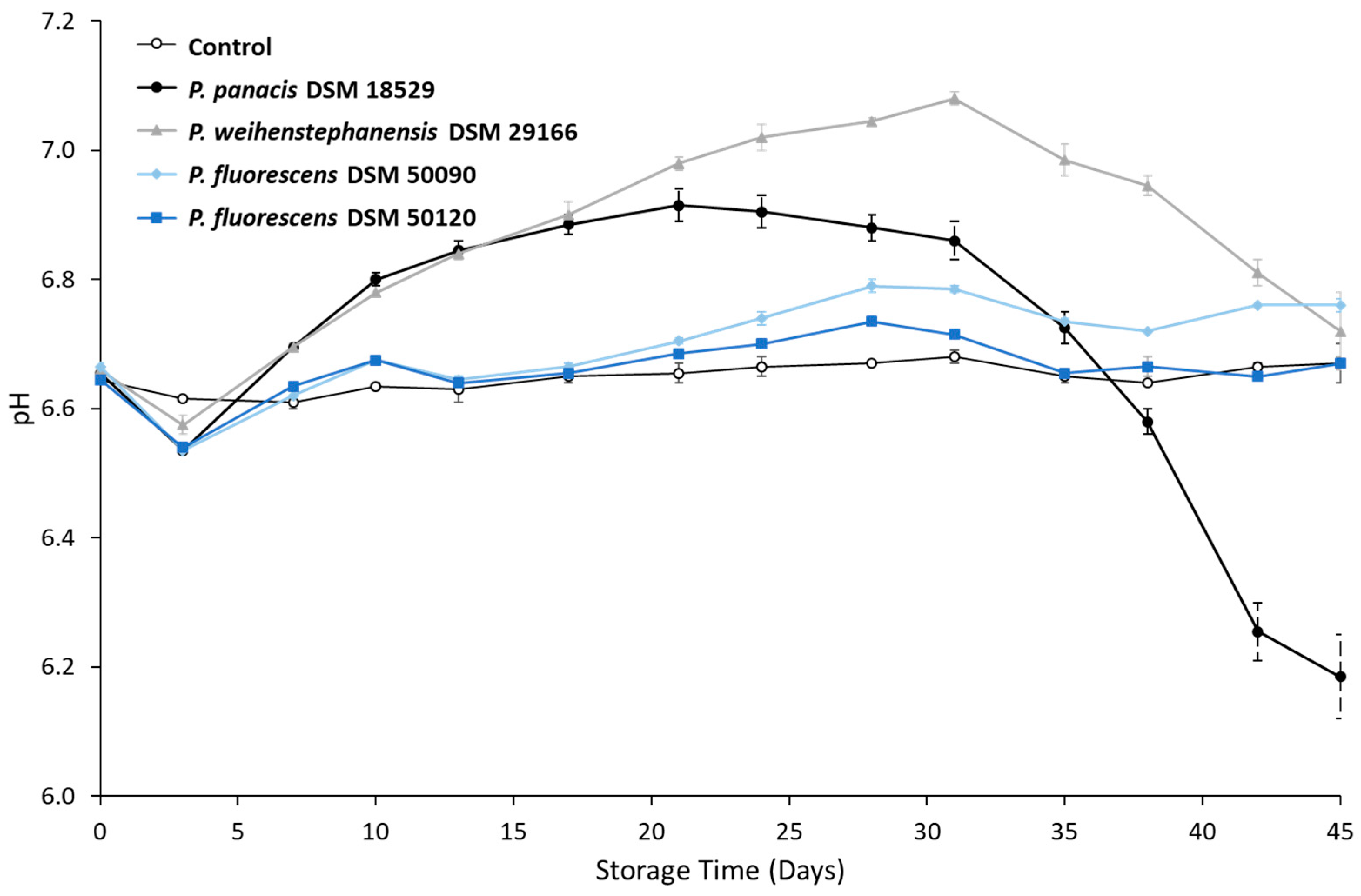
3.2. Development in pH during Storage

Developments in pH of the stored, inoculated UHT skimmed milk and the control during storage is shown in Figure 2. The control and UHT milk inoculated with extracellular extract from P. fluorescens DSM 50120 and P. fluorescens DSM 50090 showed a stable pH over storage time. UHT milk inoculated with extracellular extract from P. panacis DSM 18529 and P. weihenstephanensis DSM 29166 followed the same trend, where pH initially steadily increased over the first weeks and then decreased after 21 and 31 days, respectively. The UHT milk inoculated with P. panacis DSM 18529 extracellular extract reached a maximum pH value of 6.92 ± 0.03 on day 21 and a minimum pH value of 6.19 ± 0.07 on day 45. For the UHT milk inoculated with P. weihenstephanensis DSM 29166 extracellular extract, pH on day 0 was at its lowest with a pH value of 6.66 ± 0.00 and reached its maximum on day 31, with pH value of 7.08 ± 0.01. After 45 days, pH decreased back to pH 6.72 ± 0.06. The average pH of control and UHT milk inoculated with extracellular extracts from P. fluorescens DSM 50090 and P. fluorescens DSM 50120 was 6.64 ± 0.02, 6.70 ± 0.07 and 6.66 ± 0.04, respectively.

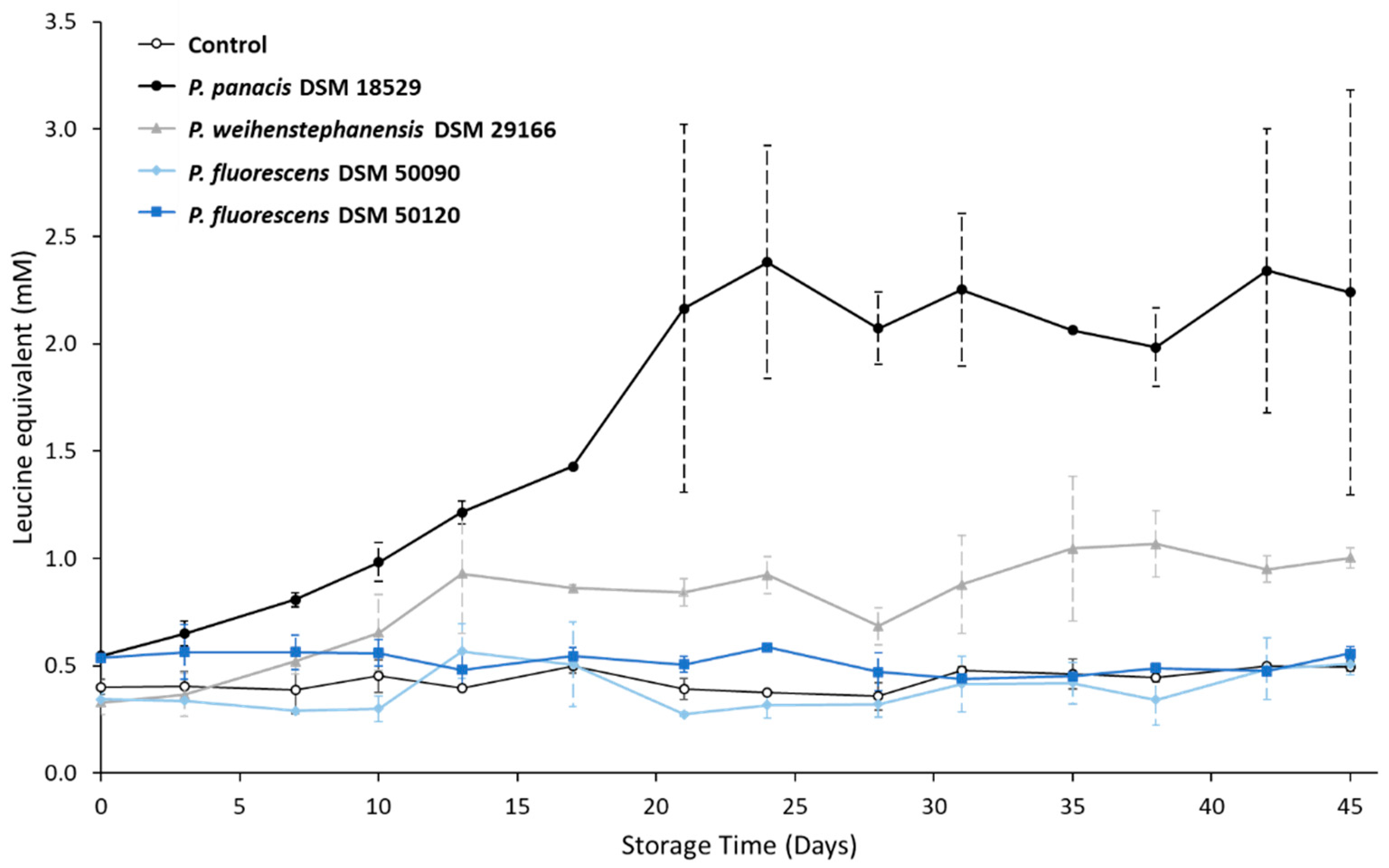
3.3. Development in Proteolysis Level during Storage

The development in the level of free N-terminals as a result of proteolysis in skimmed UHT milk samples during storage can be observed in Figure 3. UHT milk inoculated with extracellular extract from P. panacis DSM 18529 had the highest overall proteolytic activity (p < 0.05) as well as the highest increase in level of proteolysis degree over storage time, reaching high levels from 21 days onwards with a leucine equivalent of 2.16 ± 0.86 mM. The UHT milk inoculated with P. weihenstephanensis DSM 29166 extracellular extract was the second most proteolytic and exhibited a proteolytic level that was significantly (p < 0.05) higher than UHT milk inoculated with extracellular extract from P. fluorescens DSM 50090, P. fluorescens DSM 50120 and the control. There was no significant difference in the proteolytic activity between samples taken from the top or bottom layers of the bottles (results not shown).

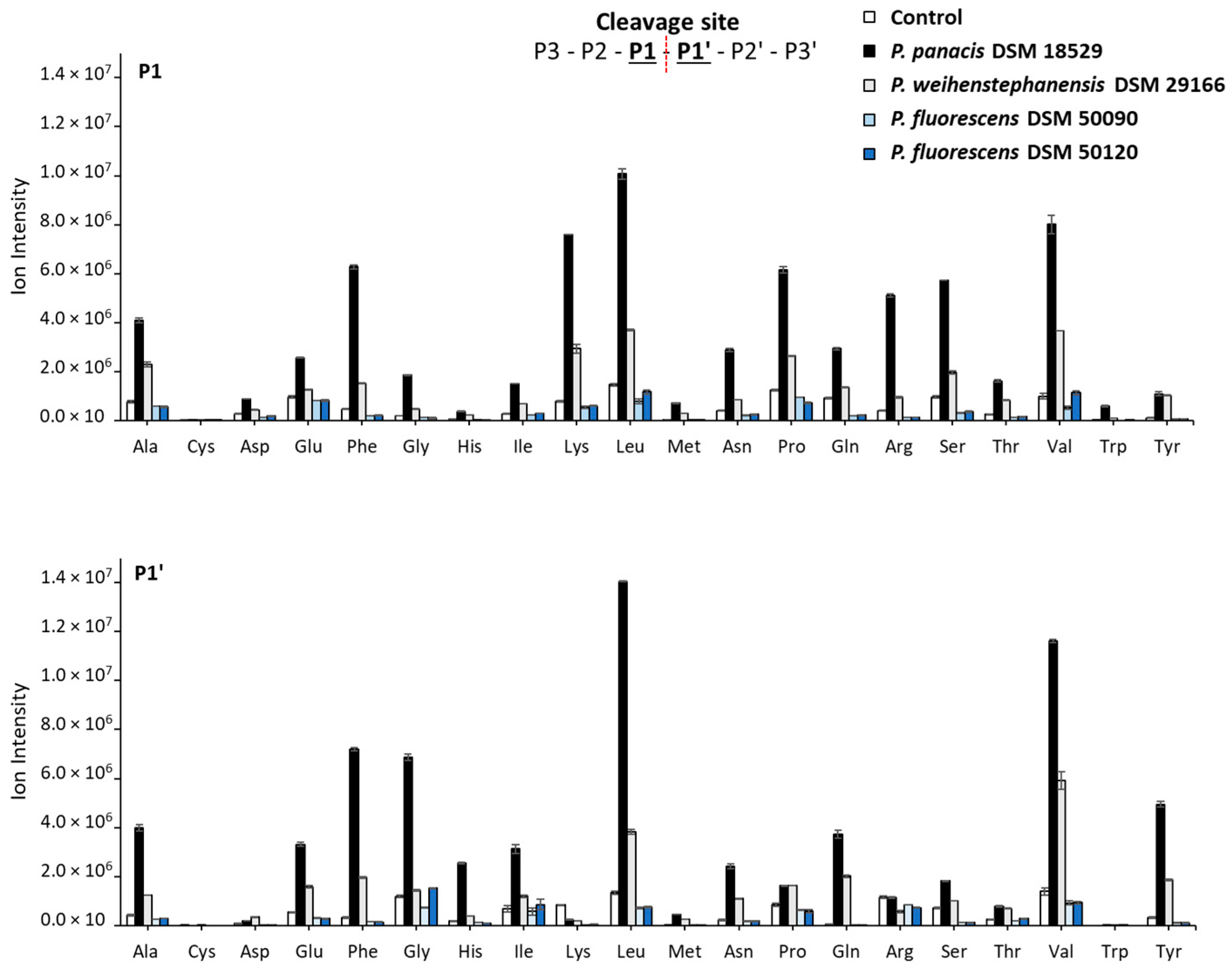
3.4. Generated Peptides and Sites Cleaved in the Major Milk Proteins by Peptidomic Analysis after 45 Days of Storage

The formation of peptides during storage was assessed by peptidomic analysis using nanoLC timsTOF Pro MS/MS and is overall expressed as number of peptides identified from major milk proteins, including β-, αs1-, αs2- and κ-casein and the whey protein, β-lactoglobulin relative distribution as well as relative ion intensities for identified peptides (Table 2 and Table S2). Looking at the relative number of peptides formed after the 45 days of storage showed β-casein to be the mother protein giving rise to most peptides across inoculations and control, with 45–50% of identified peptides from the major milk proteins being derived from β-casein, while only 9–13% came from κ-casein. Peptidomic results based on peptide ion intensities showed an even higher difference between β-casein (61–67%) and κ-casein (3–8%). Both the relative number of identified peptides and relative ion intensities followed the order of β-casein > αs1-casein > αs2-casein > κ-casein > β-lactoglobulin across inoculations. The biggest difference among strains was observed in the peptides obtained from αs1-casein in milk inoculated with extracellular extract from P. panacis DSM 18529. While it had a similar percentage of the number of peptides obtained from αs1-casein than the other strains and control (0–3% higher), it showed a very high ion intensity fraction of peptides derived from αs1-casein (8–11% higher than the other strains and control). Milk inoculated with extracellular extracts from both P. panacis DSM 18529 and P. weihenstephanensis DSM 29166 showed a lower relative percentage of κ-casein ion intensity than the other strains and control.
Using the peptidomic data to globally assess and rank the associated cleavage sites is shown in Figure 4. A cleavage site analysis based only on the five major mother proteins is separately reported in Figure S1. For skimmed UHT milk inoculated with P. panacis DSM 18529 extracellular extract the most frequent amino acid residues present at the P1 position of the cleavage sites for generated peptides were Leu > Val > Lys > Phe > Pro > Ser > Arg > Ala, while for P1′ they were Leu > Val > Phe > Gly > Tyr > Ala. For milk inoculated with P. weihenstephanensis DSM 29166 extracellular extract quite similar distribution was observed. Milk inoculated with extracellular extract from P. fluorescens DSM 50090, P. fluorescens DSM 50120 and the control all exhibited overall very low ion intensities of peptides generated and thereby low overall protein hydrolysis.

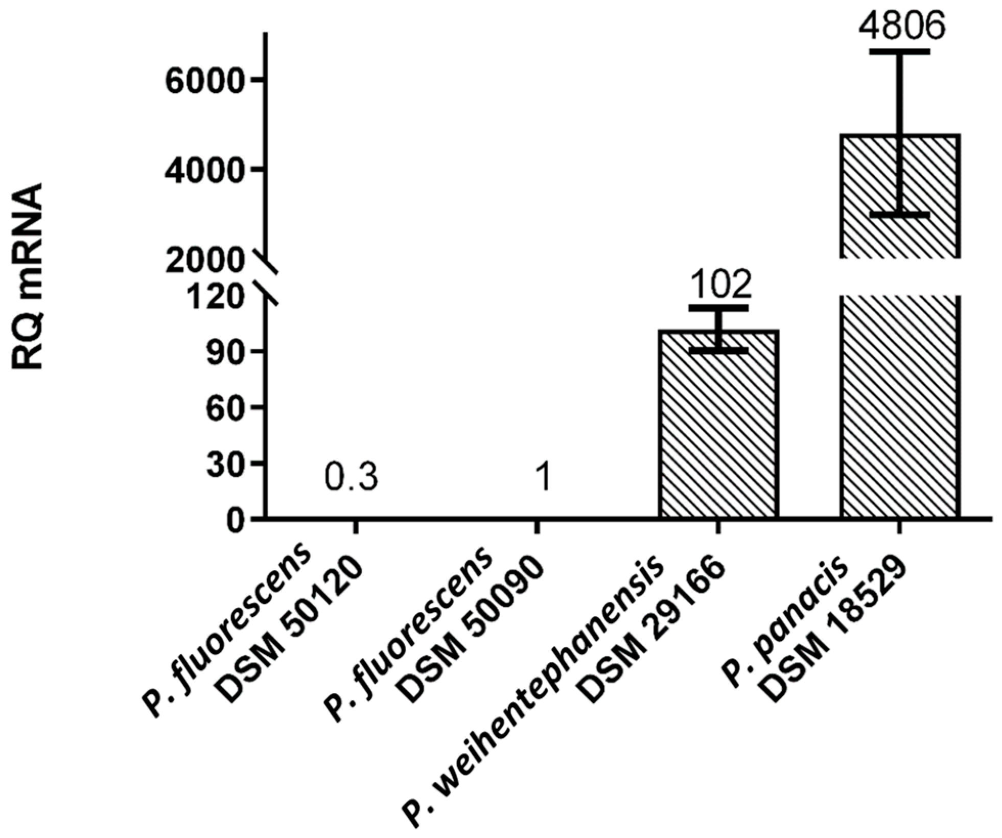
3.5. Expression Levels of aprX mRNA

Relative levels of aprX mRNA expression analyses of each strain after its sub-culturing for two hours in MM with 2% (v/v) UHT milk is shown in Figure 5. The aprX expression level was found to be highest for P. panacis DSM 18529, followed by P. weihenstephanensis DSM 29166, with its relative level being almost 50 times lower than P. panacis DSM 18529. Both P. fluorescens DSM 50090 and P. fluorescens DSM 50120 showed low expression levels of aprX mRNA, having relative quantities around 100 times lower than P. weihenstephanensis DSM 29166 and 4800 times lower than P. panacis DSM 18529.

4. Discussion

The present study investigated relationships between visible changes, proteolysis characterizations and aprX expression in UHT milk inoculated with different types of commercially available, but dairy-relevant Pseudomonas extracellular extracts. The changes in the stored UHT milk were assessed by quantifications of physical stability, milk compositional analyses, proteolysis and detailed state-of-the-art proteomics with the aim of getting more insight into the causes of variability regarding proteolytic activity among Pseudomonas strains through deciphering variability in peptide fingerprints, cleavage sites and aprX expression levels.

4.1. Relationship between Spoilage Potential, Milk Proteolysis and aprX Expression

Milk proteolysis, determined as level of free N-terminals generated in stored UHT milk inoculated with extracellular extracts from Pseudomonas strains, was observed to comply with the relative aprX expression levels measured from the same Pseudomonas strains cultured in MM with 2% (v/v) UHT milk. UHT milk inoculated with extracellular extract from P. panacis DSM 18529, which showed the highest aprX expression levels, was found to have the highest proteolytic activity and aprX expression based on both fluorescamine assay and peptidomic results. The same observation was true for P. weihenstephanensis DSM 29166 as the second highest proteolytic strain, having the second highest aprX expression levels. The two additional studied strains, P. fluorescens DSM 50090 and P. fluorescens DSM 50120 showed low expression of aprX and low proteolytic activity. This is in accordance with previous reports of protease expression being the main driver of milk proteolysis, with individual differences between proteases having a minor impact [12].
P. panacis DSM 18529 showed the highest overall proteolytic activity, being the only strain to produce visible spoilage after three weeks of storage. Gelation was observed at the same time that the milk proteolysis reached its maximum, around day 21. These results are in accordance with Baur et al. [19], where an isolated strain identified as P. panacis DSM 18529 was reported to produce milk spoilage after four weeks of storage when a protease induction step of ~30 h was used. In the present study, P. weihenstephanensis DSM 29166 induced increased proteolysis as measured by the fluorescamine assay (Figure 3) and peptide cleavage analysis (Figure 4), though no gelation was observed. P. weihenstephanensis DSM 29166 has previously been reported to be proteolytic and responsible for milk spoilage [20]. Differences in the spoilage potential may be due to storage times and conditions. Stoeckel et al. [20] showed that the proteases of P. weihenstephanensis DSM 29166 were able to spoil heat-treated milk after four months of storage at 20 °C when a four-day pre-storage of milk inoculated with bacteria was used. In the current experiment, up-concentration of protease-containing extracellular extracts was used in order to compensate for a shorter storage time. It seems, however, that in the case of P. weihenstephanensis DSM 29166, long times of pre-storage may be essential to induce the gelation onset. It has been reported that AprX production increases drastically at the end of the exponential phase and the beginning of the stationary phase of bacterial growth [29,30]. Incidentally, P. weihenstephanensis DSM 29166 showed the lowest bacterial concentration after 1 day of culture in TSB of the four studied strains (Table S2), which may indicate the necessity of a longer incubation time in order to reach the end of the exponential phase and beginning of the stationary phase. In addition, lower gene expression in this strain may also indicate a lower secretion of the extracellular protease per cell, which could reinforce the need for a longer incubation time. UHT milk inoculated with extracellular extract from P. fluorescens DSM 50090 and P. fluorescens DSM 50120 did not show any increase in milk proteolysis over storage in the present experiment. This was expected for P. fluorescens DSM 50090, as it has been reported previously to be a low or non-proteolytic strain [22]. On the other hand, Zhang et al. [21] showed that the onset of gelation by P. fluorescens DSM 50120 is dependent on the concentration of extracellular extract used. In their study, gelation occurred after three to four weeks, while lower concentrations of extracellular extracts showed an increased degree of proteolysis without gelation, even after six weeks at 20 °C. The low milk proteolysis observed for P. fluorescens DSM 50120 in the present study could be due to a less efficient up-concentration of protease extract than the one obtained by Zhang et al. [21]. However, expression of aprX was also the lowest in P. fluorescens DSM 50120, which indicates an overall lower secretion of AprX per cell. These results suggest the influence of additional factors up- or down-regulating the expression of aprX in P. fluorescens DSM 50120. Certain environmental factors such as temperature, medium composition, calcium presence and iron content have been previously described to influence AprX production [7,31,32].

4.2. pH Changes and UHT Milk Spoilage during Storage

P. panacis DSM 18529 was the only strain to induce gelation of UHT milk and also the one reaching the lowest pH (6.19 ± 0.07). Furthermore, extracellular extract from this strain was able to induce a precipitate formation of 49.86 ± 4.17% of the total weight after incubation for 45 days. pH increased for UHT milk inoculated with extracellular extracts from P. panacis DSM 18529 until day 21, which aligns with the onset of gelation, where a steep pH decrease was observed until the end of the storage experiment. The initial increase in pH was also observed for UHT milk inoculated with extracellular extracts from P. weihenstephanensis DSM 29166, a strain that also showed milk proteolysis. Anema [33] discussed that, while most studies report a decrease in pH in UHT milk during storage, some studies have highlighted a small pH increase in the first weeks of storage followed by a prolonged decrease. pH decrease for UHT milk storage has been previously reported to occur due to various potential contributions from dephosphorylation of casein micelles, breakdown of lactose, precipitation of calcium phosphate, Maillard reactions and proteolysis [34,35,36]. pH decrease was observed to occur in UHT milk inoculated with extracellular extract from P. panacis DSM 18529 after its maximum milk proteolysis was reached at day 21 (Figure 2 and Figure 3), but this relation was not observed for P. weihenstephanensis DSM 29166. Li et al. [37] discussed that pH increase during UHT milk storage might be related to Ca2+ balance disruption due to increases in temperature, while Pestana et al. [38] suggested that increases in pH for milk might be related to a lower association of whey proteins with the casein micelle. An additional potential hypothesis for the increase in pH at the beginning of the shelf-life storage might be related to the necessity of AprX to bind calcium to be functional. Gaur et al. [23] have shown that calcium chelators might effectively increase the pH of UHT milk during storage. Nevertheless, further studies would be necessary to confirm the cause of the observed pH increase.

4.3. Peptidomics: Identified Peptides and Cleavage Sites

Based on the information from both relative number of identified peptides and their relative ion intensities, protein hydrolysis by proteases secreted by the Pseudomonas strains was observed to follow the pattern β-casein > αs1-casein > αs2-casein > κ-casein > β-lactoglobulin. This order was observed for all strains, including P. panacis DSM 18529. These results are in accordance with the findings from other studies where peptide numbers were determined by LC-MS in UHT milk inoculated with either Pseudomonas cultures [14] or purified AprX [39]. This peptide hydrolysis pattern reflects milk protein composition, as β- and αs1-caseins represent the highest percentage of caseins, being around 35% for β-casein and 37% for αs1-casein, while αs2-casein accounts for 10% and κ-casein for 12% in bovine milk, generally speaking [6]. By analysis of number of peptides formed by extracellular extracts from Pseudomonas inoculated on casein isolates, Matéos et al. [16] observed comparable results, with number of peptides following the order of β-casein > αs1-casein > κ-casein. Stuknyte et al. [40], however, identified no major differences between the amount of peptides identified from β-casein, αs1-casein and κ-casein when analyzing the number of peptides coming from different types of casein isolates. Zhang et al. [21] analyzed the casein hydrolysis in AprX-inoculated milk by RP-HPLC, where κ-casein was observed to be the first casein to be hydrolyzed (95% of κ-casein hydrolyzed at gelation onset). In the present study, the major difference regarding peptide distribution between UHT milk inoculated with extracellular extract from P. panacis DSM 18529 and the other strains was a higher relative peptide ion intensity percentage derived from αs1-casein in UHT milk inoculated with extracellular extract from P. panacis DSM 18529. In addition, a lower relative peptide ion intensity percentage derive from κ-casein was observed in UHT milk inoculated with extracellular extract from P. panacis DSM 18529 and P. weihenstephanensis DSM 291666. This could be explained by a preferred cleavage of the caseinomacropeptide region of κ-casein, producing a higher quantity of glycosylated peptides, which are more difficult to detect when using mass spectrometry [10]. These findings suggest that, while κ–casein hydrolysis might be the main target for AprX at the onset of storage, long-term exposure to the protease, with concomitant destabilization of the casein micelles, may lead to more prominent cleavage of αs1-casein.
Some researchers have compared the proteolytic effect of AprX with chymosin [10,41], which has a high specificity for the peptide bond Phe105-Met106 of κ-casein. In addition, Reid et al. [42] and García-Risco et al. [43] reported the presence of para-κ-casein in milk of spoiled UHT milk samples. However, in Figure 4, it can be observed that, while there is a relatively high preference for Phe residues at the P1 position of cleavage sites for UHT milk inoculated with extracellular extracts from P. panacis DSM 18529 and P. weihenstephanensis DSM 291666, the preference for Met in P1′ is rather low. Individual analysis of preferred cleavage sites in κ-casein (Figure S1), showed for UHT milk inoculated with extracellular extract from P. panacis DSM 18529 a small preference for Phe and Met residues in P1 and P1′positions, respectively; however, with low ion intensities of the linked peptides. Datta and Deeth [13] have previously discussed that AprX specificity for the Phe105-Met106 bond is lower than that of chymosin, while Matéos et al. [16] have reported AprX to have generally low cleavage site specificity. Matéos et al. [16] reported that the presence of basic (Arg, Lys, His) or aromatic (Tyr, Phe, Trp) amino acids in P1 positions was correlated with a higher susceptibility for cleavage of the bond by AprX, while the presence of acidic (Asp, Glu, Ser) or aliphatic (Val, Ile, Pro) amino acids was unfavorable. For the P1′position, it was reported that presence of Val, Met, Phe, Tyr, His and Gln was favorable, while Pro, Trp, Asp, Ser, Lys and Arg residues were unfavorable. In the present study, preferred P1 residues for cleavage varied in UHT milk inoculate with extracellular extracts from both P. panacis DSM 18529 and P. weihenstephanensis DSM 29166, being the two strains that exhibited milk proteolysis, from those described by Matéos et al. [16]. Only some of the basic (Arg, Lys) and aromatic (Phe) amino acids showed high ion intensity, while some of the reported unfavorable amino acids by Matéos et al. [16] (Ser, Val, Pro) were also identified with high intensity. The high amount of Lys and Arg residues in the P1 cleavage site for peptides produced by P. panacis DSM 18529 extracellular extracts, which are mainly products of αs1- and β-caseins (Figure S1), could also be explained by plasmin activity, since plasmin is highly specific for cleavage after Lys and Arg. Fajardo-Lira et al. [44] described an increased plasmin activity in the presence of bacterial proteases as a result of plasmin release from the casein micelles. On the other hand, Frohbieter et al. [45] concluded that bacterial proteases instead had a direct stimulatory effect on the plasminogen activators. Regarding P1′, the observed results in this study for P. panacis DSM 18529 and P. weihenstephanensis DSM 291666 extracellular extracts matched the findings from Matéos et al. [16], and may indicate a high importance of specific residues in the P1′ site for AprX cleavage.

5. Conclusions

Expression of AprX mRNA was found to have a strong relation with milk proteolysis, reinforcing the idea that variations in spoilage by AprX are driven by quantitative differences rather than qualitative changes in the protein hydrolysis pattern.
While previous studies have highlighted κ-casein as a priority target for hydrolysis by AprX, the observed results showed that during long-term spoilage, αs1-caseins are the casein type that suffers the highest hydrolysis. P1′ amino acids at the cleavage site for proteases, including AprX, in the extracellular media from Pseudomonas are broad but very conserved between studies, which may indicate a high impact of these cleavage sites for protein hydrolysis and spoilage potential. In addition, a cooperative effect seems to exist between bacterial proteases and plasmin, even though the specific mechanism is still not elucidated.

Supplementary Materials

The following supporting information can be downloaded at: https://www.mdpi.com/article/10.3390/dairy4010005/s1, Table S1: Bacterial growth of the four studied strains, Table S2: Number of identified peptides and ion intensity of the four studied strains and control, Figure S1: Ion intensity of peptides obtained from the five most common proteins (β-casein, αs1-casein, αs2-casein, κ-casein and β-lactoglobulin) distributed according to their P1 and P1′ cleavage site for the four different strains and control after 45 days of storage.

Author Contributions

Conceptualization, M.A.-T., L.B.L., N.A.P. and L.W.; methodology, M.A.-T., S.D.-H.N., M.L.K., Y.X., V.R., L.B.L., N.A.P. and L.T.H.; validation, formal analysis and investigation, M.A.-T., S.D.-H.N. and M.L.K.; data curation, M.A.-T., S.D.-H.N. and M.L.K.; writing—original draft preparation, M.A.-T., L.B.L., N.A.P. and L.W.; writing—review and editing, M.A.-T., S.D.-H.N., M.L.K., Y.X., V.R., L.B.L., N.A.P., L.T.H. and L.W.; visualization, M.A.-T.; supervision, L.B.L., N.A.P., L.W., Y.X. and L.T.H.; project administration, L.B.L.; funding acquisition, L.B.L. All authors have read and agreed to the published version of the manuscript.

Funding

This research was founded by Milk Levy Fund, Danish Research Foundation, Arla Foods Amba, Aarhus University and Danish Technical University.

Institutional Review Board Statement

Not applicable.

Informed Consent Statement

Not applicable.

Data Availability Statement

Data is contained within the article or Supplementary Material.

Acknowledgments

The authors would like to thank all the involved lab technicians at the Aarhus University, Danish Technical University and Arla Innovation Center laboratories for their assistance with the performed analyses.

Conflicts of Interest

The authors declare no conflict of interest.

References

  1. Beldman, A.; Junfei, B.; Binbin, C.; Zhijun, C.; Xiangming, F.; Wen, D.; Huiyuan, G.; Pei, G.; Beizhong, H.; Dinghuan, H.; et al. White Paper. China Dairy, 1 January 2014; p. 1. [Google Scholar]
  2. Gooch, E.; Hoskin, R.; Law, J. China’s Dairy Supply and Demand. Livestock, Dairy, Poult. Outlook 2017, 282, 27. [Google Scholar] [CrossRef]
  3. Glück, C.; Rentschler, E.; Krewinkel, M.; Merz, M.; von Neubeck, M.; Wenning, M.; Scherer, S.; Stoeckel, M.; Hinrichs, J.; Stressler, T.; et al. Thermostability of peptidases secreted by microorganisms associated with raw milk. Int. Dairy J. 2016, 56, 186–197. [Google Scholar] [CrossRef]
  4. Machado, S.G.; Baglinière, F.; Marchand, S.; Van Coillie, E.; Vanetti, M.C.D.; De Block, J.; Heyndrickx, M. The Biodiversity of the Microbiota Producing Heat-Resistant Enzymes Responsible for Spoilage in Processed Bovine Milk and Dairy Products. Front. Microbiol. 2017, 8, 302. [Google Scholar] [CrossRef] [PubMed] [Green Version]
  5. Martin, N.; Torres-Frenzel, P.; Wiedmann, M. Invited review: Controlling dairy product spoilage to reduce food loss and waste. J. Dairy Sci. 2021, 104, 1251–1261. [Google Scholar] [CrossRef] [PubMed]
  6. Fox, P.F.; Uniacke-Lowe, T.; McSweeney, P.L.H.; O’Mahony, J.A. Enzymology of Milk and Milk Products; Springer: Berlin/Heidelberg, Germany, 2015; ISBN 9783319148915. [Google Scholar]
  7. Marchand, S.; Vandriesche, G.; Coorevits, A.; Coudijzer, K.; De Jonghe, V.; Dewettinck, K.; De Vos, P.; Devreese, B.; Heyndrickx, M.; De Block, J. Heterogeneity of heat-resistant proteases from milk Pseudomonas species. Int. J. Food Microbiol. 2009, 133, 68–77. [Google Scholar] [CrossRef] [PubMed]
  8. Woods, R.G.; Burger, M.; Beven, C.-A.; Beacham, I.R. The aprX–lipA operon of Pseudomonas fluorescens B52: A molecular analysis of metalloprotease and lipase production The GenBank accession numbers for the sequences reported in this paper are AF216700, AF216701 and AF216702. Microbiology 2001, 147, 345–354. [Google Scholar] [CrossRef] [PubMed] [Green Version]
  9. Maier, C.; Hofmann, K.; Huptas, C.; Scherer, S.; Wenning, M.; Lücking, G. Simultaneous quantification of the most common and proteolytic Pseudomonas species in raw milk by multiplex qPCR. Appl. Microbiol. Biotechnol. 2021, 105, 1693–1708. [Google Scholar] [CrossRef]
  10. Zhang, C.; Bijl, E.; Svensson, B.; Hettinga, K. The Extracellular Protease AprX from Pseudomonas and its Spoilage Potential for UHT Milk: A Review. Compr. Rev. Food Sci. Food Saf. 2019, 18, 834–852. [Google Scholar] [CrossRef] [Green Version]
  11. Kokkinidou, S.; Peterson, D.G. Control of Maillard-Type Off-Flavor Development in Ultrahigh-Temperature-Processed Bovine Milk by Phenolic Chemistry. J. Agric. Food Chem. 2014, 62, 8023–8033. [Google Scholar] [CrossRef]
  12. Dufour, D.; Nicodème, M.; Perrin, C.; Driou, A.; Brusseaux, E.; Humbert, G.; Gaillard, J.-L.; Dary, A. Molecular typing of industrial strains of Pseudomonas spp. isolated from milk and genetical and biochemical characterization of an extracellular protease produced by one of them. Int. J. Food Microbiol. 2008, 125, 188–196. [Google Scholar] [CrossRef]
  13. Datta, N.; Deeth, H. Age Gelation of UHT Milk—A Review. Food Bioprod. Process. Trans. Inst. Chem. Eng. Part C 2001, 79, 197–210. [Google Scholar] [CrossRef]
  14. Gaucher, I.; Tanguy, G.; Fauquant, J.; Jardin, J.; Rousseau, F.; Robert, B.; Madec, M.-N.; Gaucheron, F. Proteolysis of casein micelles by Pseudomonas fluorescens CNRZ 798 contributes to the destabilisation of UHT milk during its storage. Dairy Sci. Technol. 2011, 91, 413–429. [Google Scholar] [CrossRef] [Green Version]
  15. Datta, N.; Kelly, A. The Role of Proteases in the Stability of UHT-Treated Milk. In Agents of Change; Springer: Cham, Switzerland, 2021; pp. 311–347. ISBN 9783030554828. [Google Scholar]
  16. Matéos, A.; Guyard-Nicodème, M.; Baglinière, F.; Jardin, J.; Gaucheron, F.; Dary, A.; Humbert, G.; Gaillard, J.-L. Proteolysis of milk proteins by AprX, an extracellular protease identified in Pseudomonas LBSA1 isolated from bulk raw milk, and implications for the stability of UHT milk. Int. Dairy J. 2015, 49, 78–88. [Google Scholar] [CrossRef]
  17. Zhang, W.; Poojary, M.M.; Rauh, V.M.; Ray, C.A.; Olsen, K.; Lund, M.N. Quantitation of α-Dicarbonyls and Advanced Glycation Endproducts in Conventional and Lactose-Hydrolyzed Ultrahigh Temperature Milk during 1 Year of Storage. J. Agric. Food Chem. 2019, 67, 12863–12874. [Google Scholar] [CrossRef]
  18. Baglinière, F.; Tanguy, G.; Jardin, J.; Matéos, A.; Briard, V.; Rousseau, F.; Robert, B.; Beaucher, E.; Humbert, G.; Dary, A.; et al. Quantitative and qualitative variability of the caseinolytic potential of different strains of Pseudomonas fluorescens: Implications for the stability of casein micelles of UHT milks during their storage. Food Chem. 2012, 135, 2593–2603. [Google Scholar] [CrossRef]
  19. Baur, C.; Krewinkel, M.; Kutzli, I.; Kranz, B.; von Neubeck, M.; Huptas, C.; Wenning, M.; Scherer, S.; Stoeckel, M.; Hinrichs, J.; et al. Isolation and characterisation of a heat-resistant peptidase from Pseudomonas panacis withstanding general UHT processes. Int. Dairy J. 2015, 49, 46–55. [Google Scholar] [CrossRef]
  20. Stoeckel, M.; Lidolt, M.; Achberger, V.; Glück, C.; Krewinkel, M.; Stressler, T.; von Neubeck, M.; Wenning, M.; Scherer, S.; Fischer, L.; et al. Growth of Pseudomonas weihenstephanensis, Pseudomonas proteolytica and Pseudomonas sp. in raw milk: Impact of residual heat-stable enzyme activity on stability of UHT milk during shelf-life. Int. Dairy J. 2016, 59, 20–28. [Google Scholar] [CrossRef]
  21. Zhang, C.; Bijl, E.; Hettinga, K. Destabilization of UHT milk by protease AprX from Pseudomonas fluorescens and plasmin. Food Chem. 2018, 263, 127–134. [Google Scholar] [CrossRef] [Green Version]
  22. Maier, C.; Huptas, C.; Von Neubeck, M.; Scherer, S.; Wenning, M.; Lücking, G. Genetic Organization of the aprX-lipA2 Operon Affects the Proteolytic Potential of Pseudomonas Species in Milk. Front. Microbiol. 2020, 11, 1190. [Google Scholar] [CrossRef]
  23. Gaur, V.; Schalk, J.; Anema, S.G. Sedimentation in UHT milk. Int. Dairy J. 2018, 78, 92–102. [Google Scholar] [CrossRef]
  24. Akkerman, M.; Johansen, L.B.; Rauh, V.; Poulsen, N.A.; Larsen, L.B. Contribution of casein micelle size and proteolysis on protein distribution and sediment formation in UHT milk during storage. Int. Dairy J. 2021, 117, 104980. [Google Scholar] [CrossRef]
  25. Jansson, T.; Clausen, M.R.; Sundekilde, U.K.; Eggers, N.; Nyegaard, S.; Larsen, L.B.; Ray, C.; Sundgren, A.; Andersen, H.J.; Bertram, H.C. Lactose-Hydrolyzed Milk Is More Prone to Chemical Changes during Storage than Conventional Ultra-High-Temperature (UHT) Milk. J. Agric. Food Chem. 2014, 62, 7886–7896. [Google Scholar] [CrossRef] [PubMed]
  26. Nielsen, S.D.; Jakobsen, L.M.; Geiker, N.R.; Bertram, H.C. Chemically acidified, live and heat-inactivated fermented dairy yoghurt show distinct bioactive peptides, free amino acids and small compounds profiles. Food Chem. 2022, 376, 131919. [Google Scholar] [CrossRef]
  27. Nielsen, S.D.; Beverly, R.L.; Dallas, D.C. Milk Proteins Are Predigested Within the Human Mammary Gland. J. Mammary Gland. Biol. Neoplasia 2017, 22, 251–261. [Google Scholar] [CrossRef] [PubMed]
  28. Livak, K.J.; Schmittgen, T.D. Analysis of relative gene expression data using real-time quantitative PCR and the 2−ΔΔCT Method. Methods 2001, 25, 402–408. [Google Scholar] [CrossRef] [PubMed]
  29. Pinto, U.M.; Costa, E.D.; Mantovani, H.C.; Vanetti, M. The proteolytic activity of Pseudomonas fluorescens 07A isolated from milk is not regulated by quorum sensing signals. Braz. J. Microbiol. 2010, 41, 91–96. [Google Scholar] [CrossRef] [PubMed] [Green Version]
  30. Matselis, E.; Roussis, I. Proteinase and lipase production by Pseudomonas fluorescens. Proteolysis and lipolysis in thermized ewe’s milk. Food Control 1998, 9, 251–259. [Google Scholar] [CrossRef]
  31. Martins, M.; Pinto, U.; Riedel, K.; Vanetti, M.C.D. Milk-deteriorating exoenzymes from Pseudomonas fluorescens 041 isolated from refrigerated raw milk. Braz. J. Microbiol. 2015, 46, 207–217. [Google Scholar] [CrossRef] [Green Version]
  32. Maunsell, B.; Adams, C.; O’Gara, F. Complex regulation of AprA metalloprotease in Pseudomonas fluorescens M114: Evidence for the involvement of iron, the ECF sigma factor, PbrA and pseudobactin M114 siderophore. Microbiology 2006, 152, 29–42. [Google Scholar] [CrossRef] [Green Version]
  33. Anema, S.G. Age Gelation, Sedimentation, and Creaming in UHT Milk: A Review. Compr. Rev. Food Sci. Food Saf. 2018, 18, 140–166. [Google Scholar] [CrossRef]
  34. Al-Saadi, J.M.S.; Deeth, H.C. Cross-Linking of Proteins and Other Changes in UHT Milk during Storage at Different Temperatures. Aust. J. Dairy Technol. 2008, 63, 93–99. [Google Scholar]
  35. Ranvir, S.; Sharma, R.; Gandhi, K.; Nikam, P.; Mann, B. Physico-chemical changes during processing and storage of UHT milk. Indian J. Dairy Sci. 2021, 74, 39–47. [Google Scholar] [CrossRef]
  36. Akkerman, M.; Johansen, L.B.; Rauh, V.; Sørensen, J.; Larsen, L.B.; Poulsen, N.A. Relationship between casein micelle size, protein composition and stability of UHT milk. Int. Dairy J. 2021, 112, 104856. [Google Scholar] [CrossRef]
  37. Li, S.; Ye, A.; Singh, H. Physicochemical changes and age gelation in stored UHT milk: Seasonal variations. Int. Dairy J. 2021, 118, 105028. [Google Scholar] [CrossRef]
  38. Pestana, J.M.; Gennari, A.; Monteiro, B.W.; Lehn, D.N.; Souza, C.F.V. Effects of Pasteurization and Ultra-High Temperature Processes on Proximate Composition and Fatty Acid Profile in Bovine Milk. Am. J. Food Technol. 2015, 10, 265–272. [Google Scholar] [CrossRef] [Green Version]
  39. Baglinière, F.; Matéos, A.; Tanguy, G.; Jardin, J.; Briard-Bion, V.; Rousseau, F.; Robert, B.; Beaucher, E.; Gaillard, J.L.; Amiel, C.; et al. Proteolysis of ultra high temperature-treated casein micelles by AprX enzyme from Pseudomonas fluorescens F induces their destabilisation. Int. Dairy J. 2013, 31, 55–61. [Google Scholar] [CrossRef]
  40. Stuknytė, M.; Decimo, M.; Colzani, M.; Silvetti, T.; Brasca, M.; Cattaneo, S.; Aldini, G.; De Noni, I. Extracellular thermostable proteolytic activity of the milk spoilage bacterium Pseudomonas fluorescens PS19 on bovine caseins. J. Dairy Sci. 2016, 99, 4188–4195. [Google Scholar] [CrossRef]
  41. Recio, I.; García-Risco, M.R.; Ramos, M.; López-Fandiño, R. Characterization of peptides produced by the action of psychrotrophic proteinases on κ-casein. J. Dairy Res. 2000, 67, 625–630. [Google Scholar] [CrossRef]
  42. Reid, J.R.; Coolbear, T.; Ayers, J.S.; Coolbear, K.P. The action of chymosin on κ-casein and its macropeptide: Effect of pH and analysis of products of secondary hydrolysis. Int. Dairy J. 1997, 7, 559–569. [Google Scholar] [CrossRef]
  43. Ramos, M.; García-Risco, M.R.; López-Fandiño, R. Proteolysis, protein distribution and stability of UHT milk during storage at room temperature. J. Sci. Food Agric. 1999, 79, 1171–1178. [Google Scholar] [CrossRef]
  44. Fajardo-Lira, C.; Oria, M.; Hayes, K.; Nielsen, S. Effect of Psychrotrophic Bacteria and of an Isolated Protease from Pseudomonas fluorescens M3/6 on the Plasmin System of Fresh Milk. J. Dairy Sci. 2000, 83, 2190–2199. [Google Scholar] [CrossRef] [PubMed]
  45. Frohbieter, K.; Ismail, B.; Nielsen, S.; Hayes, K. Effects of Pseudomonas fluorescens M3/6 Bacterial Protease on Plasmin System and Plasminogen Activation. J. Dairy Sci. 2005, 88, 3392–3401. [Google Scholar] [CrossRef] [PubMed]
Figure 1. Skimmed UHT milk inoculated with extracellular media from the four Pseudomonas strains and the control after 45 days of storage at 20 °C. From left to right, 1–2: control UHT milk, 3–4: P. fluorescens DSM 50120, 5–6: P. weihenstephanensis DSM 29166, 7–8: P. fluorescens DSM 50090 and 9–10: P. panacis DSM 18529.
Dairy 04 00005 g001
Figure 2. Development in pH of skimmed UHT milk inoculated with the four studied bacterial extracellular extracts and the control during storage for 45 days at 20 °C. The measurements were carried out in biological replicates (n = 2).
Dairy 04 00005 g002
Figure 3. Development in levels of proteolysis assessed as level of free N-terminals in UHT milk inoculated with extracellular extracts, as well as the control, as a measure of proteolytic spoilage potential of the four Pseudomonas strains during storage for 45 days at 20 °C. Analysis performed on milk aliquots taken from the top of the milk bottles. The measurements were carried out from biological replicates (n = 2) and with technical replicates (n = 3).
Dairy 04 00005 g003
Figure 4. Total ion intensities of peptides distributed according to their P1 and P1′ cleavage sites per strain used to obtain the extracellular extract and control after 45 days of skimmed UHT milk storage based on extracted peptides subjected to nanoLC timsTOF Pro MS/MS peptidomics.
Dairy 04 00005 g004
Figure 5. Relative quantity of mRNA (RQ mRNA) for each strain after sub-culturing for two hours in MM with 2% (v/v) UHT milk. rpoB gene was used to normalize relative quantities of aprX mRNA. Results were expressed relative to P. fluorescens DSM 50090.
Dairy 04 00005 g005
Table 1. Primers and probes used in RT-qPCR to determine rpoB and aprX gene expression in Pseudomonas. Primers: forward (F), reverse (R) and probe (P).
Target Gene and StrainsSequence (5′–3′)Size (bp)Reference
rpoB
(Universal)
F: 5′-CAGCCGYTGGGTGGTAA-3′
R: 5′-CCGTTCACATCGTCCGA-3′
130[9]
aprX
P. panacis
DSM 18529
F: 5′-AAATCGATAGCTTCAGCC-3′
R: 5′-GAGGTCAGGAAGGTGTAG-3′
P: 5′-Fam-ACGGTGTAGGAGGGTTTGC-BHQ-1-3′
169This study
aprX
P. weihenstephanensis
DSM 29166
F: 5′-TGTGCAAAAGCTGTATGG-3′
R: 5′-GTGAGGTTGATTTTCTGG T -3′
P: 5′-Fam-CGAGGTGGCGCTATAGAAGT-BHQ-1-3′
195This study
aprX
P. fluorescens 
DSM 50090
F: 5′-TAAAGGGACAGCAGGACTAG-3′
R: 5′-GAGCAACACCAACCAGAA-3′
P: 5′-Fam-CCACCGTCCCATACCGAAAACA-BHQ-1-3′
248This study
aprX
P. fluorescens
DSM 50120
F: 5′-ATCTTACCTACACCTTCC-3′
R: 5′-CCACTTGTGTTGTACTTC-3′
P: 5′-Fam-TCAGCCAGTTCAGCAACC-BHQ-1-3′
265This study
Table 2. Relative distribution of the number of peptides and ion intensities of peptides identified as derived from the five major milk proteins by peptidomic analysis using nanoLC timsTOF Pro MS/MS of skimmed UHT milk inoculated with the four Pseudomonas strains and the control after 45 days of storage.
ControlP. panacis
DSM 18529
P. weihenstephanensis
DSM 29166
P. fluorescens
DSM 50090
P. fluorescens DSM 50120
Relative number of peptide
distribution (%)
β-casein4550474548
αs1-casein2325222525
αs2-casein1210131312
κ-casein139111111
β-lactoglobulin77755
Relative ion
intensity
distribution (%)
β-casein6567676162
αs1-casein1426171815
αs2-casein83999
κ-casein72378
β-lactoglobulin53456
Disclaimer/Publisher’s Note: The statements, opinions and data contained in all publications are solely those of the individual author(s) and contributor(s) and not of MDPI and/or the editor(s). MDPI and/or the editor(s) disclaim responsibility for any injury to people or property resulting from any ideas, methods, instructions or products referred to in the content.

