
Fluorescent Gold Nanoparticles in Suspension as an Efficient Theranostic Agent for Highly Radio-Resistant Cancer Cells
 , , , , and
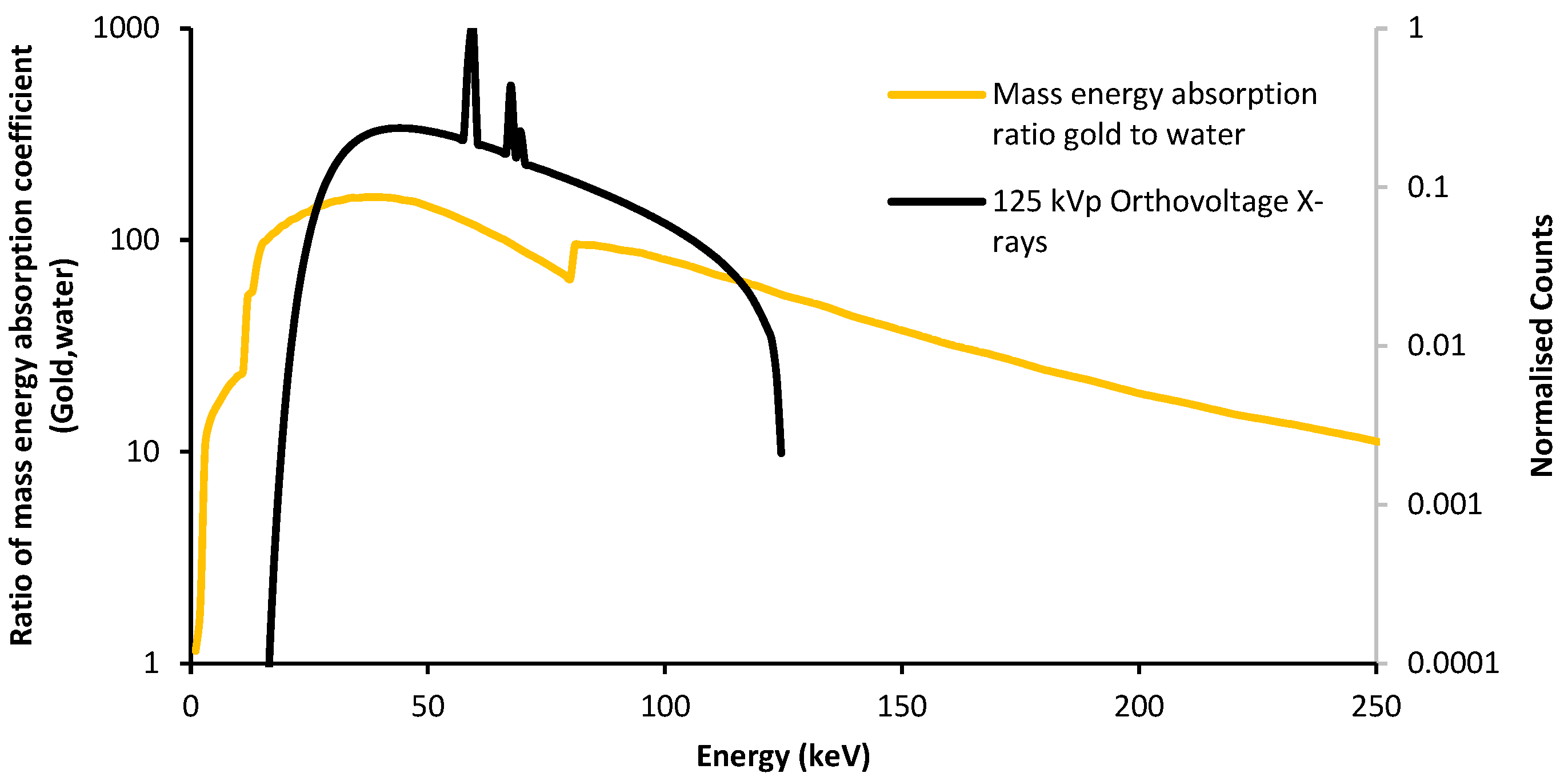
Abstract
:1. Introduction
2. Materials and Methods
2.1. Fluorescent Gold Nanoparticles Materials
2.1.1. L-Cys-DTPA Ligand Synthesis
2.1.2. Synthesis of [Au(L-Cys-DTPA)] Nanoparticles
2.1.3. Synthesis of [Au(L-Cys-DTPA)(Propargylamine)] Nanoparticles
2.1.4. Alexa Fluor Grafting on [Au(L-Cys-DTPA)(Propargylamine)] Nanoparticles
2.2. Transmission Electron Microscopy (TEM) and Energy Dispersive Spectroscopy (EDS)
2.3. Fourier-Transform Infrared Spectroscopy (FTIR)
2.4. UV-Vis Spectroscopy
2.5. Thermogravimetric Analysis
2.6. Absorption and Emission Spectra
2.7. Cell Culture
2.8. Cytotoxicity Assessment
2.9. Confocal Imaging
2.10. Flow Cytometry
2.11. Irradiation of Cell Culture and Clonogenic Cell Survival Assay
2.12. γ-H2AX Analysis
2.13. Injection into Rodent Postmortem
3. Results
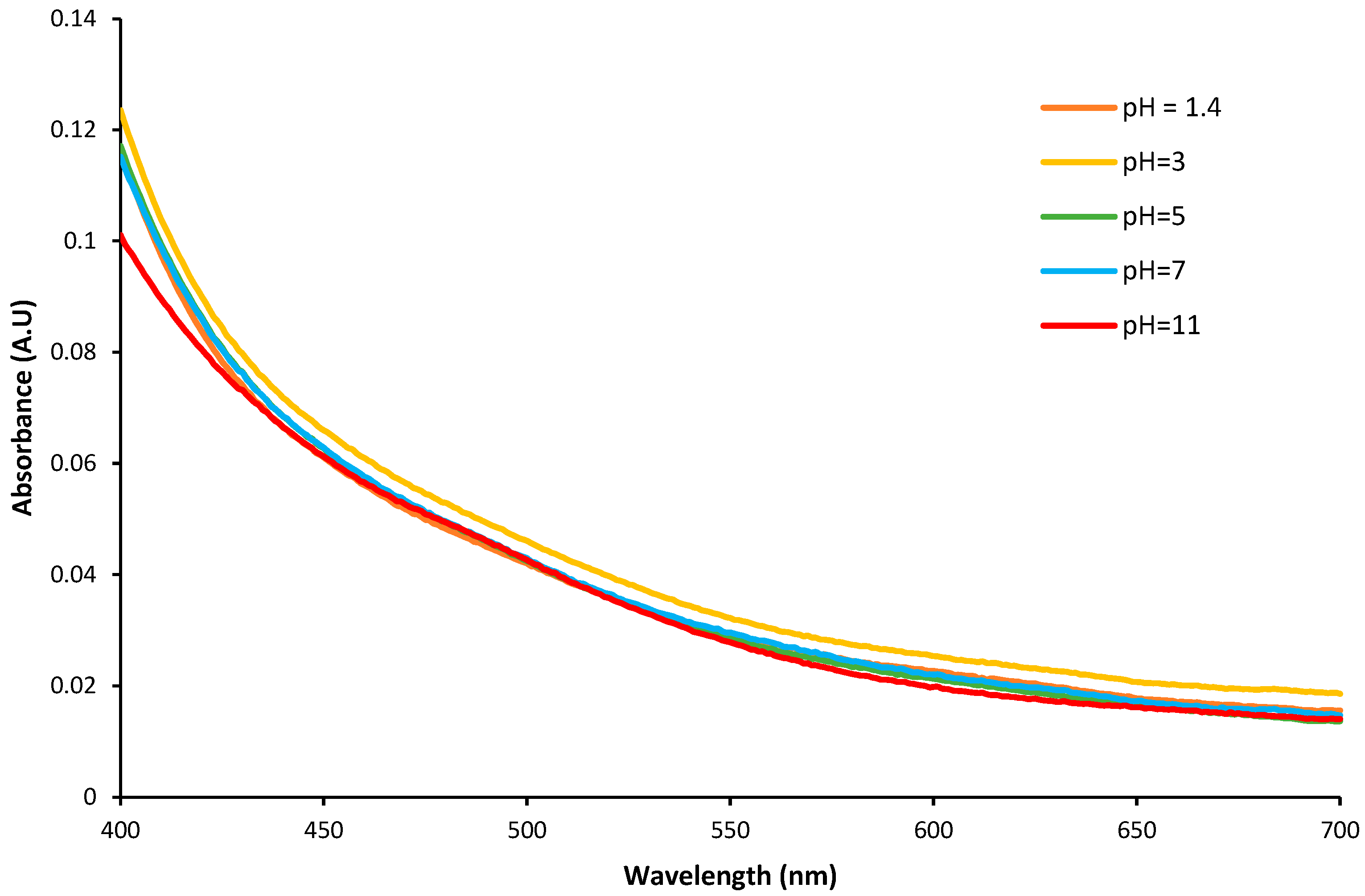
3.1. Study of the Stability of Colloidal Suspensions as a Function of pH
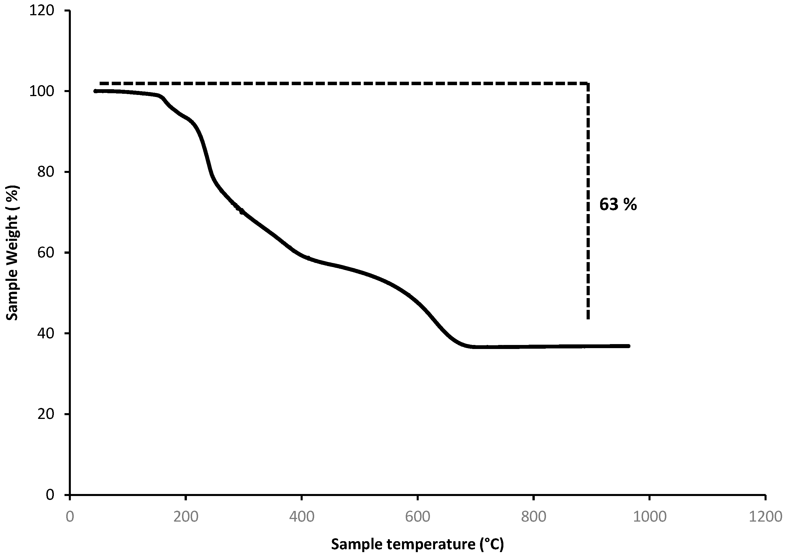
3.2. Thermogravimetric Analysis
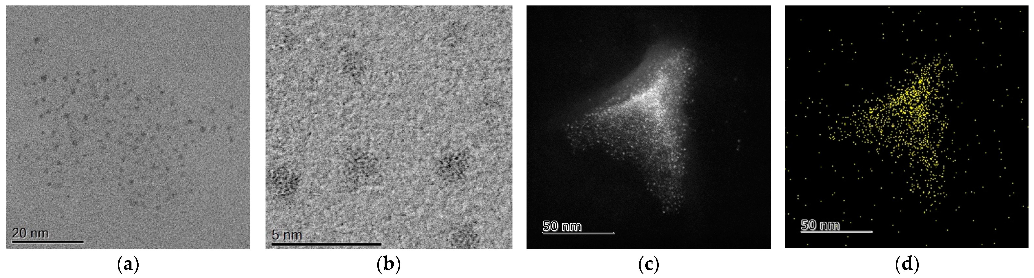
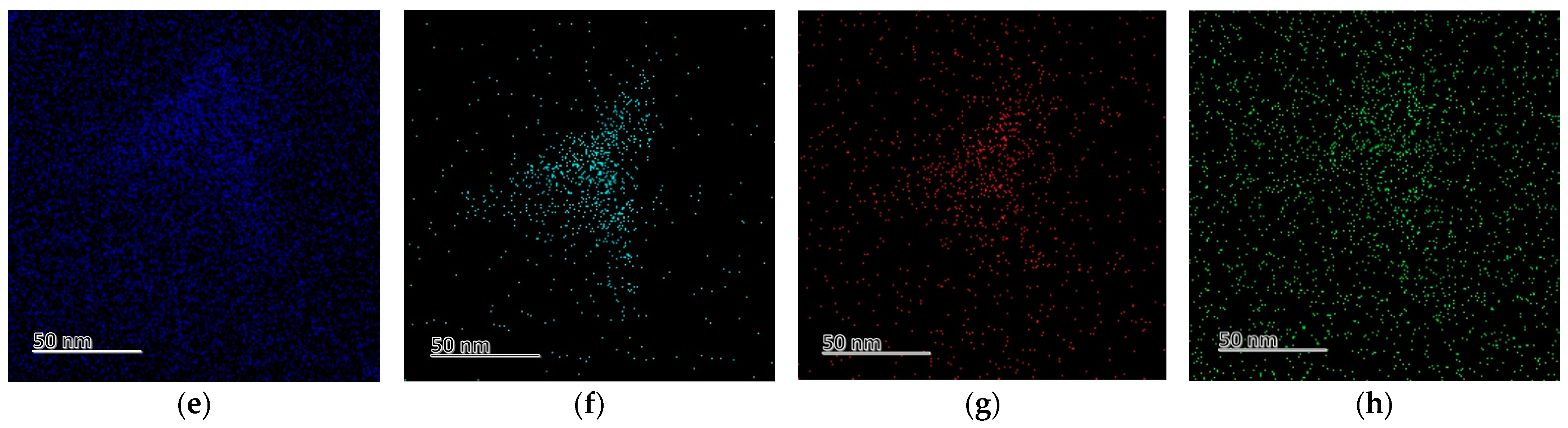
3.3. Transmission Electron Microscopy (TEM) and Energy Dispersive Spectroscopy (EDS)
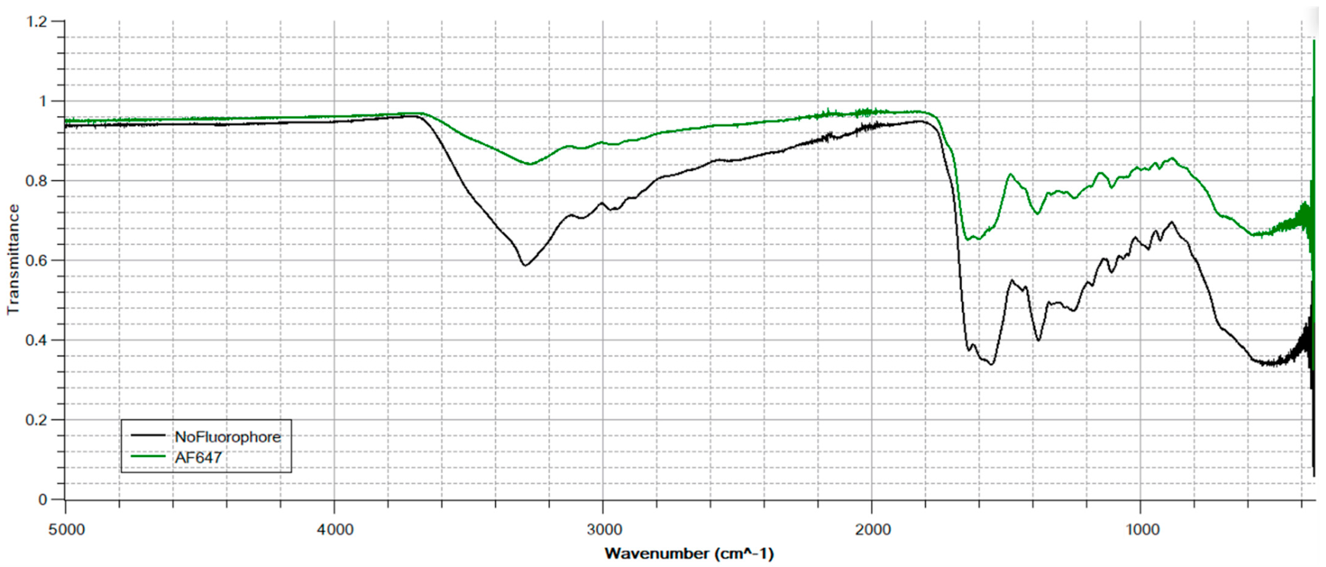
3.4. Fourier-Transform Infra-Red Spectroscopy (FTIR)
3.4.1. Bonding the Au Cores to a Ligand Coating
3.4.2. Grafting the Propargylamine Crosslinker to Ligand Coating
3.4.3. Click Reaction Bonding Crosslinker to AF647 Fluorophore
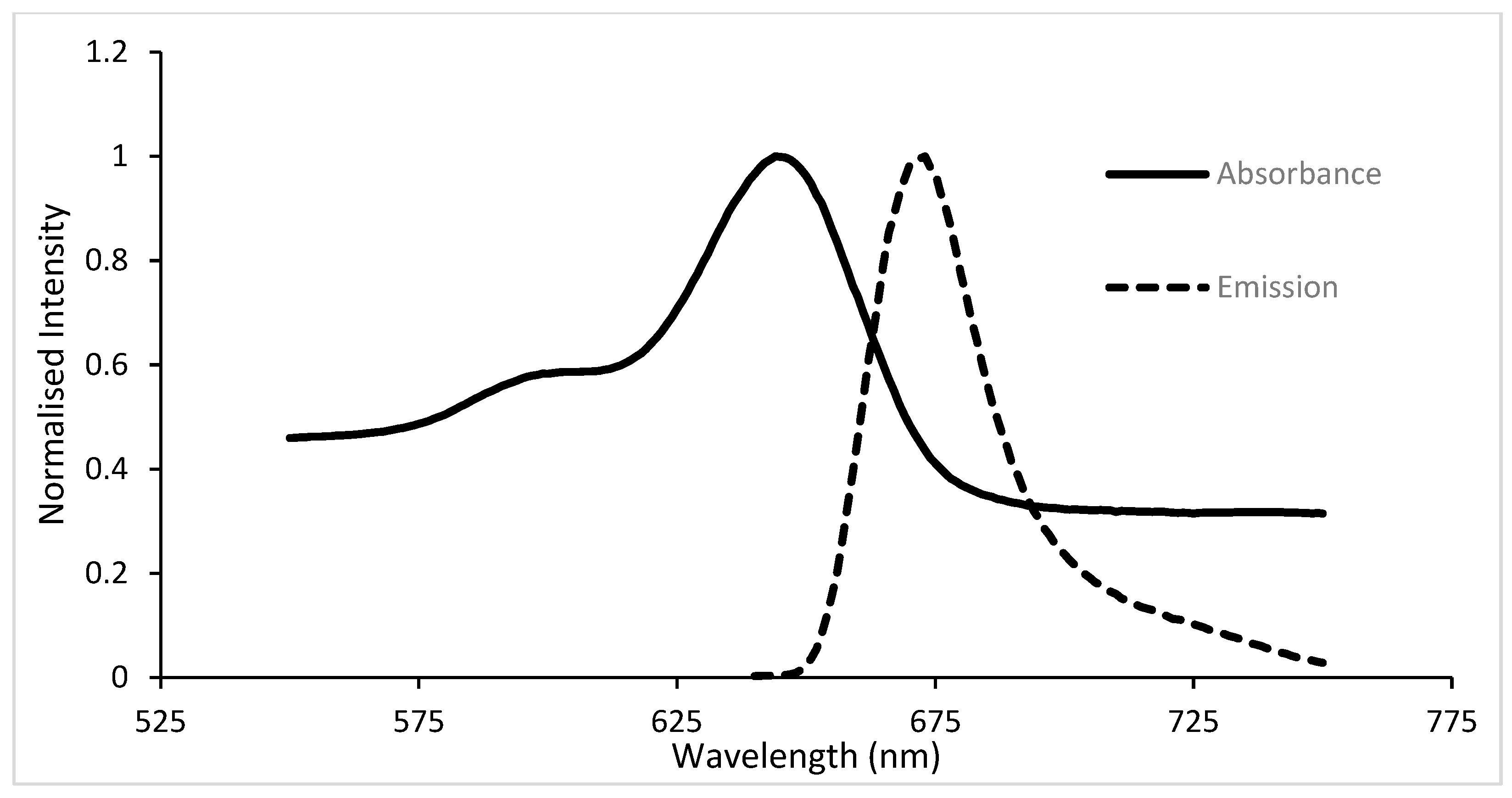
3.5. Absorption and Emission Spectra of Gold Suspension
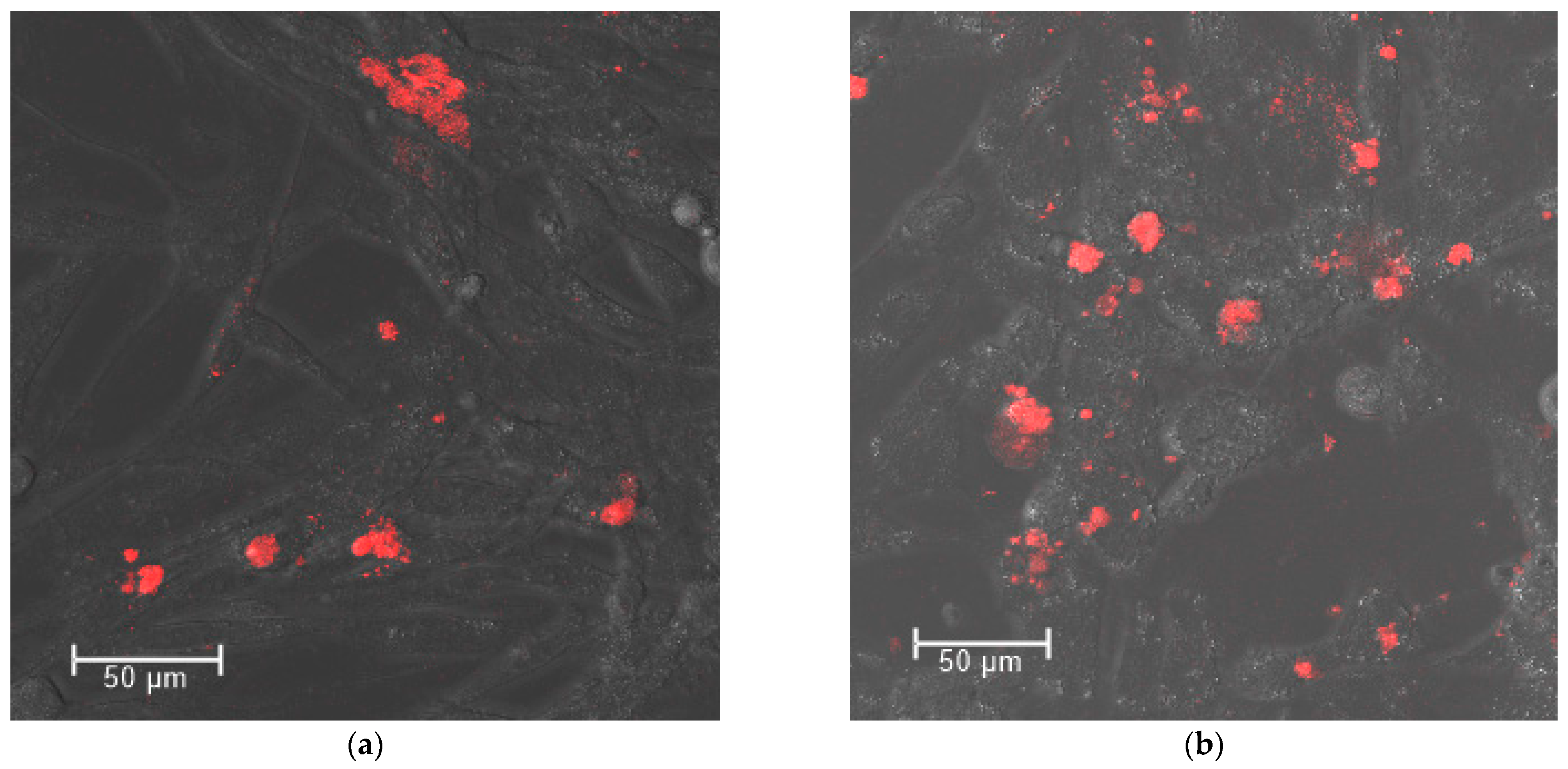
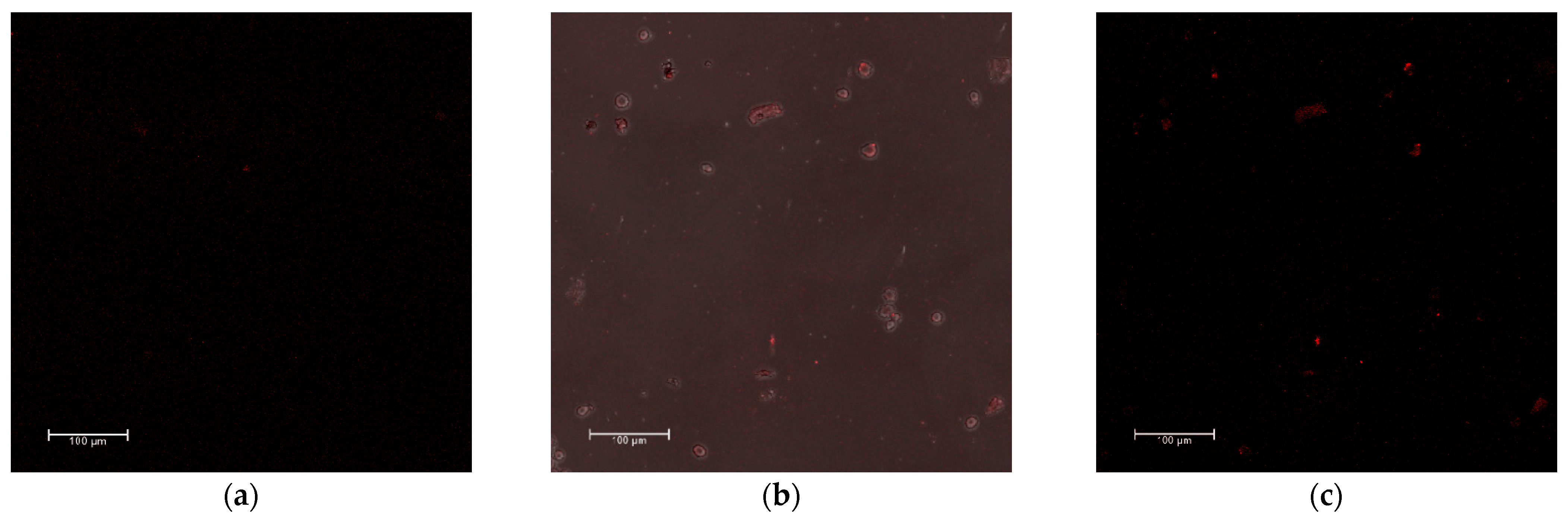
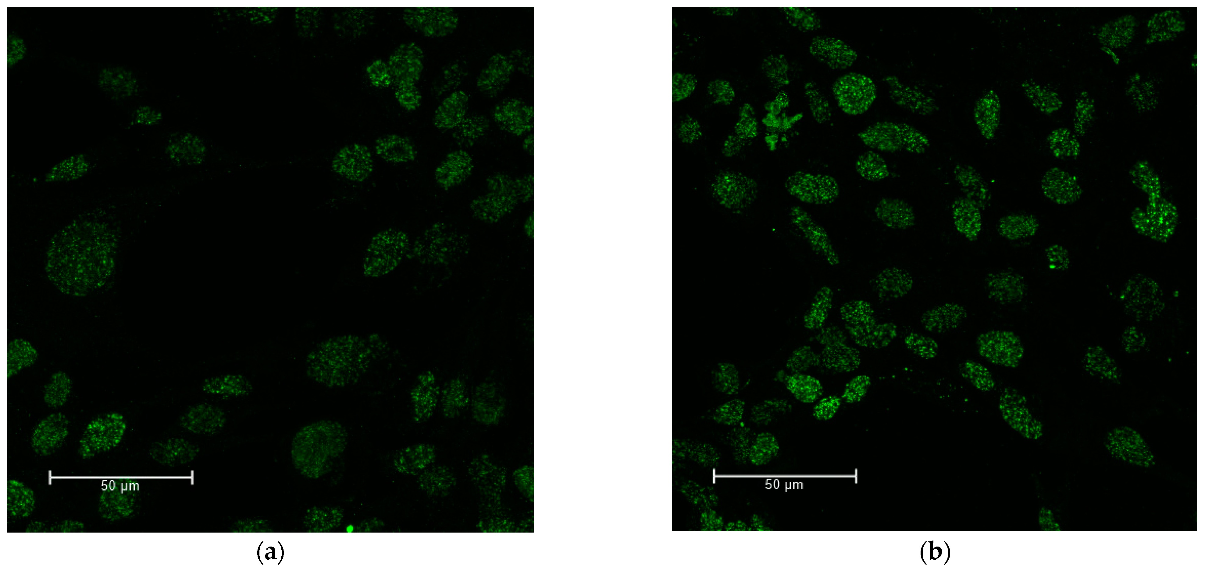
3.6. Confocal Imaging to Detect the Nanoparticles within 9LGS
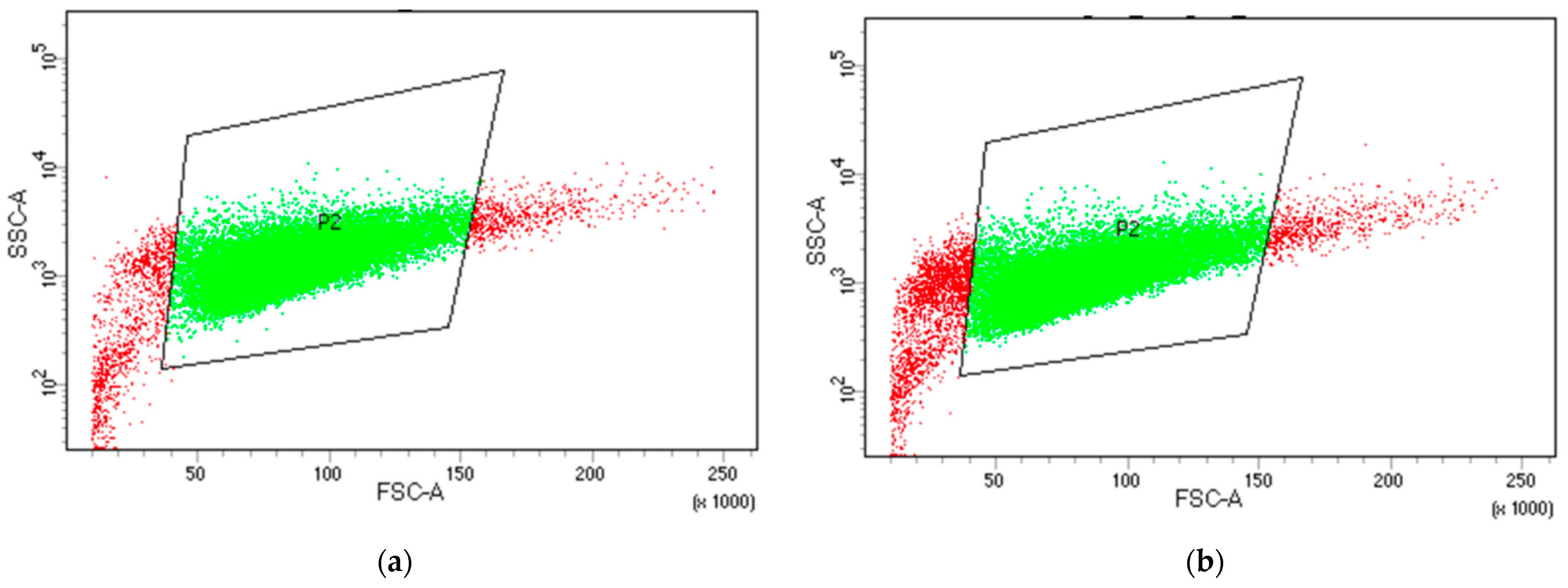
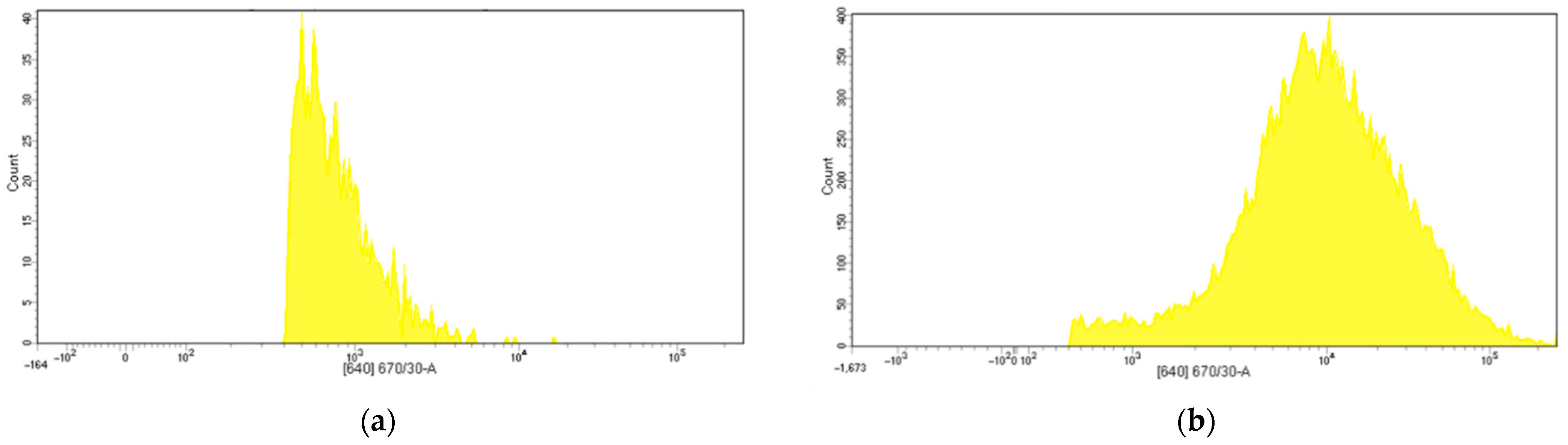
3.7. Flow Cytometry to Detect the Nanoparticle Uptake and Fluorescence within 9LGS
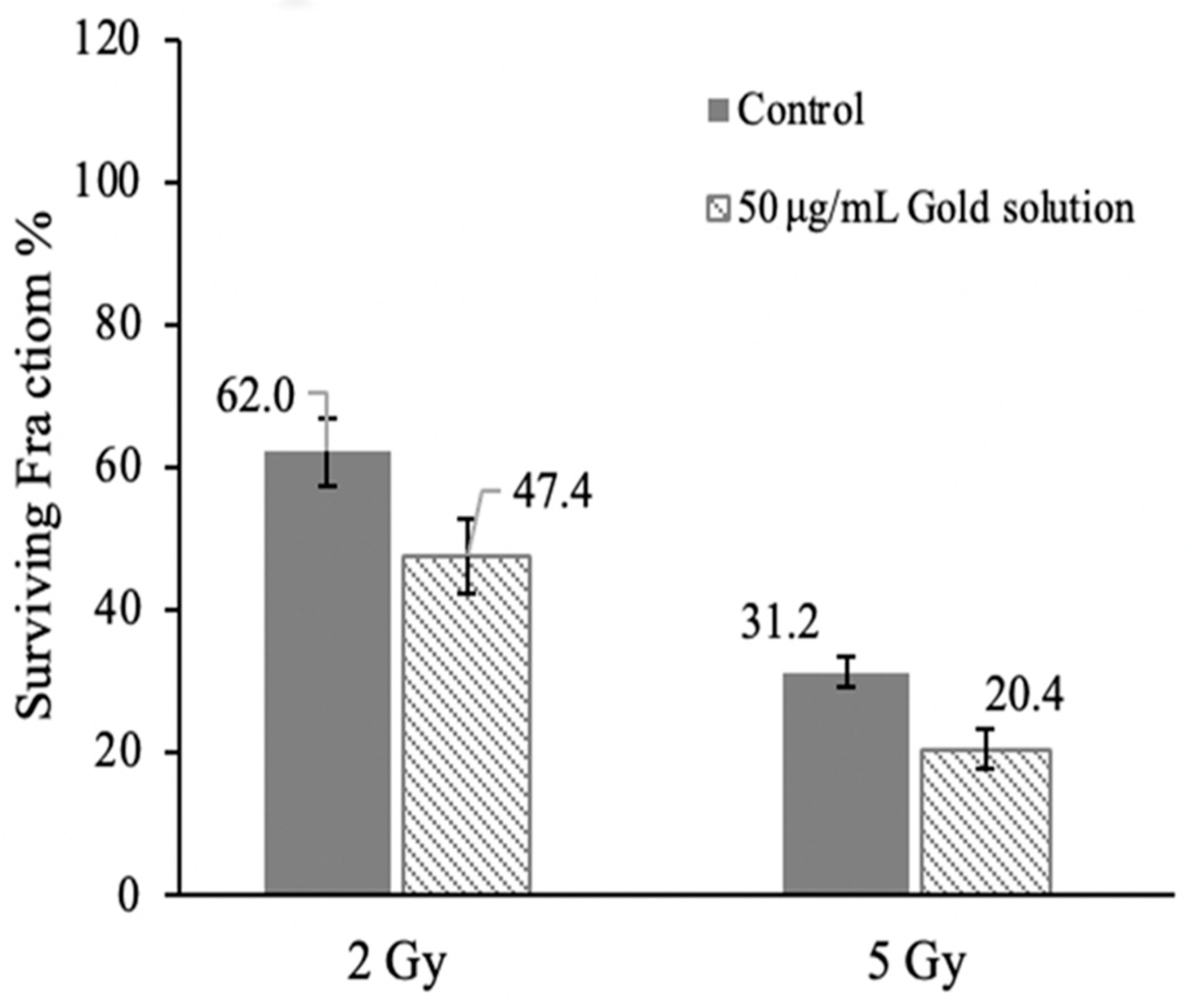
3.8. Gold Suspension Cytotoxicity and Orthovoltage Irradiation Survival
3.9. Double Strand Break Quantification in Irradiated 9LGS with AuNP Suspension
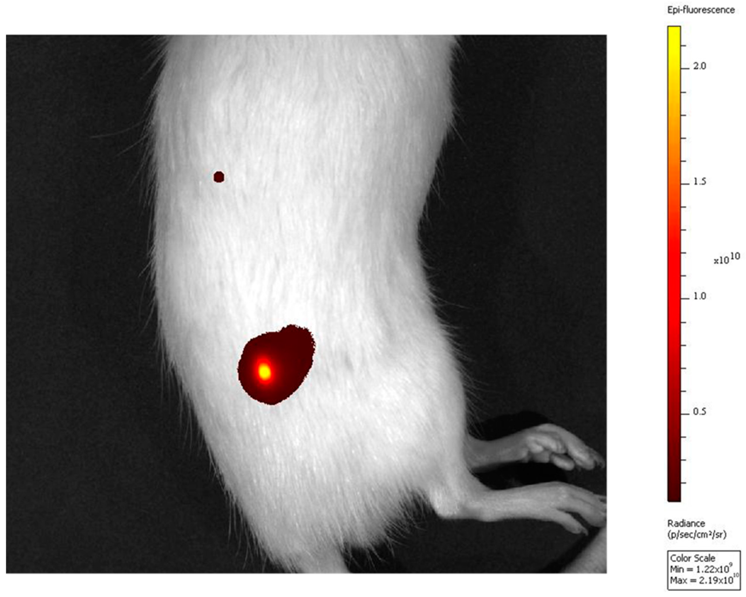
3.10. Post Mortem Injection of AuNP Suspension
4. Discussion
5. Conclusions
Supplementary Materials
Author Contributions
Funding
Institutional Review Board Statement
Informed Consent Statement
Data Availability Statement
Acknowledgments
Conflicts of Interest
References
- Choi, J.; Kim, G.; Cho, S.B.; Im, H.-J. Radiosensitizing high-Z metal nanoparticles for enhanced radiotherapy of glioblastoma multiforme. J. Nanobiotechnol. 2020, 18, 122. [Google Scholar] [CrossRef]
- Khochaiche, A.; Westlake, M.; O’Keefe, A.; Engels, E.; Vogel, S.; Valceski, M.; Li, N.; Rule, K.; Horvat, J.; Konstantinov, K.; et al. First extensive study of silver-doped lanthanum manganite nanoparticles for inducing selective chemotherapy and radio-toxicity enhancement. Mater. Sci. Eng. C 2021, 123, 111970. [Google Scholar] [CrossRef]
- Engels, E.; Westlake, M.; Li, N.; Vogel, S.; Gobert, Q.; Thorpe, N.; Rosenfeld, A.; Lerch, M.; Corde, S.; Tehei, M. Thulium Oxide Nanoparticles: A new candidate for image-guided radiotherapy. Biomed. Phys. Eng. Express 2018, 4, 044001. [Google Scholar] [CrossRef] [Green Version]
- Stewart, C.; Konstantinov, K.; McKinnon, S.; Guatelli, S.; Lerch, M.; Rosenfeld, A.; Tehei, M.; Corde, S. First proof of bismuth oxide nanoparticles as efficient radiosensitisers on highly radioresistant cancer cells. Phys. Med. 2016, 32, 1444–1452. [Google Scholar] [CrossRef] [Green Version]
- Brown, R.; Corde, S.; Oktaria, S.; Briggs, A.; Konstantinov, K.; Rosenfeld, A.; Lerch, M.; Tehei, M. Nanostructures, concentrations and energies: An ideal equation to extend therapeutic efficiency on radioresistant 9L tumour cells using Ta2O5 ceramic. Biomed. Phys. Eng. Express 2017, 3, 015018. [Google Scholar] [CrossRef]
- Bao, S.; Wu, Q.; McLendon, R.E.; Hao, Y.; Shi, Q.; Hjelmeland, A.B.; Dewhirst, M.W.; Bigner, D.D.; Rich, J.N. Glioma stem cells promote radioresistance by preferential activation of the DNA damage response. Nature 2006, 444, 756–760. [Google Scholar] [CrossRef]
- Teleanu, D.M.; Chircov, C.; Grumezescu, A.M.; Volceanov, A.; Teleanu, R.I. Impact of Nanoparticles on Brain Health: An Up to Date Overview. J. Clin. Med. 2018, 7, 490. [Google Scholar]
- Baranowska-Wójcik, E.; Szwajgier, D.; Oleszczuk, P.; Winiarska-Mieczan, A. Effects of Titanium Dioxide Nanoparticles Exposure on Human Health—A Review. Biol. Trace Elem. Res. 2020, 193, 118–129. [Google Scholar] [CrossRef] [Green Version]
- Svetlakova, A.S.; Brandt, N.N.; Priezzhev, A.V.; Chikishev, A.Y. Raman microspectroscopy of nanodiamond-induced structural changes in albumin. J. Biomed. Opt. 2015, 20, 047004. [Google Scholar] [CrossRef]
- Carnovale, C.; Bryant, G.; Shukla, R.; Bansal, V. Identifying Trends in Gold Nanoparticle Toxicity and Uptake: Size, Shape, Capping Ligand, and Biological Corona. ACS Omega 2019, 4, 242–256. [Google Scholar] [CrossRef] [Green Version]
- Chithrani, B.D.; Ghazani, A.A.; Chan, W.C.W. Determining the Size and Shape Dependence of Gold Nanoparticle Uptake into Mammalian Cells. Nano Lett. 2006, 6, 662–668. [Google Scholar] [CrossRef] [PubMed]
- Suk, J.S.; Xu, Q.; Kim, N.; Hanes, J.; Ensign, L.M. PEGylation as a strategy for improving nanoparticle-based drug and gene delivery. Adv. Drug Deliv. Rev. 2016, 99, 28–51. [Google Scholar] [CrossRef] [Green Version]
- Giesen, B.; Nickel, A.-C.; Garzón Manjón, A.; Vargas Toscano, A.; Scheu, C.; Kahlert, U.D.; Janiak, C. Influence of synthesis methods on the internalization of fluorescent gold nanoparticles into glioblastoma stem-like cells. J. Inorg. Biochem. 2020, 203, 110952. [Google Scholar] [CrossRef]
- Navarro, J.R.G.; Lerouge, F. From gold nanoparticles to luminescent nano-objects: Experimental aspects for better gold-chromophore interactions. Nanophotonics 2017, 6, 71–92. [Google Scholar] [CrossRef] [Green Version]
- Jeong, Y.; Kook, Y.-M.; Lee, K.; Koh, W.-G. Metal enhanced fluorescence (MEF) for biosensors: General approaches and a review of recent developments. Biosens. Bioelectron. 2018, 111, 102–116. [Google Scholar] [CrossRef] [PubMed]
- Luo, T.; Huang, P.; Gao, G.; Shen, G.; Fu, S.; Cui, D.; Zhou, C.; Ren, Q. Mesoporous silica-coated gold nanorods with embedded indocyanine green for dual mode X-ray CT and NIR fluorescence imaging. Opt. Express 2011, 19, 17030–17039. [Google Scholar] [CrossRef] [PubMed]
- Herranz, M.; Ruibal, A. Optical imaging in breast cancer diagnosis: The next evolution. J. Oncol. 2012, 2012, 863747. [Google Scholar] [CrossRef] [Green Version]
- Haume, K.; Rosa, S.; Grellet, S.; Śmiałek, M.A.; Butterworth, K.T.; Solov’yov, A.V.; Prise, K.M.; Golding, J.; Mason, N.J. Gold nanoparticles for cancer radiotherapy: A review. Cancer Nanotechnol. 2016, 7, 8. [Google Scholar] [CrossRef] [Green Version]
- Hirsjärvi, S.; Passirani, C.; Benoit, J.P. Passive and active tumour targeting with nanocarriers. Curr. Drug. Discov. Technol. 2011, 8, 188–196. [Google Scholar] [CrossRef]
- Barua, S.; Mitragotri, S. Challenges associated with Penetration of Nanoparticles across Cell and Tissue Barriers: A Review of Current Status and Future Prospects. Nano Today 2014, 9, 223–243. [Google Scholar] [CrossRef] [Green Version]
- Mesbahi, A. A review on gold nanoparticles radiosensitization effect in radiation therapy of cancer. Rep. Pr. Oncol. Radiother. 2010, 15, 176–180. [Google Scholar] [CrossRef] [PubMed]
- Engels, E.; Bakr, S.; Bolst, D.; Sakata, D.; Li, N.; Lazarakis, P.; McMahon, S.; Ivanchenko, V.; Rosenfeld, A.; Incerti, S.; et al. Advances in modelling gold nanoparticle radiosensitization using new Geant4-DNA physics models. Phys. Med. Biol. 2020, 65, 225017. [Google Scholar] [CrossRef] [PubMed]
- Butterworth, K.T.; McMahon, S.J.; Taggart, L.E.; Prise, K.M. Radiosensitization by gold nanoparticles: Effective at megavoltage energies and potential role of oxidative stress. Transl. Cancer Res. 2013, 2, 269–279. [Google Scholar]
- Gao, Q.; Zhang, J.; Gao, J.; Zhang, Z.; Zhu, H.; Wang, D. Gold Nanoparticles in Cancer Theranostics. Front. Bioeng. Biotechnol. 2021, 9, 647905. [Google Scholar] [CrossRef] [PubMed]
- Hein, C.D.; Liu, X.M.; Wang, D. Click chemistry, a powerful tool for pharmaceutical sciences. Pharm. Res. 2008, 25, 2216–2230. [Google Scholar] [CrossRef] [Green Version]
- Yi, G.; Son, J.; Yoo, J.; Park, C.; Koo, H. Application of click chemistry in nanoparticle modification and its targeted delivery. Biomater. Res. 2018, 22, 13. [Google Scholar] [CrossRef] [Green Version]
- Poludniowski, G.; Landry, G.; DeBlois, F.; Evans, P.M.; Verhaegen, F. SpekCalc: A program to calculate photon spectra from tungsten anode X-ray tubes. Phys. Med. Biol. 2009, 54, N433–N438. [Google Scholar] [CrossRef] [Green Version]
- Schneider, C.A.; Rasband, W.S.; Eliceiri, K.W. NIH Image to ImageJ: 25 years of image analysis. Nat. Methods 2012, 9, 671–675. [Google Scholar] [CrossRef]
- Hostetler, M.J.; Stokes, J.J.; Murray, R.W. Infrared Spectroscopy of Three-Dimensional Self-Assembled Monolayers: N-Alkanethiolate Monolayers on Gold Cluster Compounds. Langmuir 1996, 12, 3604–3612. [Google Scholar] [CrossRef]
- Shachat, N.; Bagnell, J.J. Reactions of Propargyl Alcohols and Propargylamines with Isocyanates. J. Org. Chem. 1963, 28, 991–995. [Google Scholar] [CrossRef]
- Fleming, D.A.; Thode, C.J.; Williams, M.E. Triazole Cycloaddition as a General Route for Functionalization of Au Nanoparticles. Chem. Mater. 2006, 18, 2327–2334. [Google Scholar] [CrossRef]
- Deng, X.; Eyster, T.; Elkasabi, Y.; Lahann, J. Bio-Orthogonal Polymer Coatings for Co-Presentation of Biomolecules. Macromol. Rapid Commun. 2012, 33, 640–645. [Google Scholar] [CrossRef]
- Aziz, S.G.; Elroby, S.A.; Alyoubi, A.; Osman, O.I.; Hilal, R. Experimental and theoretical assignment of the vibrational spectra of triazoles and benzotriazoles. Identification of IR marker bands and electric response properties. J. Mol. Model. 2014, 20, 2078. [Google Scholar] [CrossRef] [PubMed]
- Liang, Y.; Deng, X.; Senkevich, J.J.; Ding, H.; Lahann, J. Thermal and environmental stability of poly(4-ethynyl-p-xylylene-co-p-xylylene) thin films. Chin. Chem. Lett. 2015, 26, 459–463. [Google Scholar] [CrossRef]
- Lim, J.; Yang, H.; Paek, K.; Cho, C.-H.; Kim, S.; Bang, J.; Kim, B.J. “Click” synthesis of thermally stable au nanoparticles with highly grafted polymer shell and control of their behavior in polymer matrix. J. Polym. Sci. Part A Polym. Chem. 2011, 49, 3464–3474. [Google Scholar] [CrossRef]
- Rukmanikrishnan, B.; Muthusamy, S. Preparation and Properties of Polyimides Containing 1,2,3-Triazole Moieties. Adv. Polym. Technol. 2015, 37, 50–59. [Google Scholar] [CrossRef]
- Cheaburu, C.; Karavana, S.; Yılmaz, O. Functionalization of Chitosan by Click Chemistry; American Institute of Physics: New York, NY, USA, 2017; Volume 1918, p. 020009. [Google Scholar]
- Manta, P.; Nagraik, R.; Sharma, A.; Kumar, A.; Verma, P.; Paswan, S.K.; Bokov, D.O.; Shaikh, J.D.; Kaur, R.; Leite, A.F.V.; et al. Optical Density Optimization of Malaria Pan Rapid Diagnostic Test Strips for Improved Test Zone Band Intensity. Diagnostics 2020, 10, 880. [Google Scholar] [CrossRef]
- Subiel, A.; Ashmore, R.; Schettino, G. Standards and Methodologies for Characterizing Radiobiological Impact of High-Z Nanoparticles. Theranostics 2016, 6, 1651–1671. [Google Scholar] [CrossRef]
- Joh, D.Y.; Sun, L.; Stangl, M.; Al Zaki, A.; Murty, S.; Santoiemma, P.P.; Davis, J.J.; Baumann, B.C.; Alonso-Basanta, M.; Bhang, D.; et al. Selective targeting of brain tumors with gold nanoparticle-induced radiosensitization. PLoS ONE 2013, 8, e62425. [Google Scholar] [CrossRef] [Green Version]
- Klein, K.; Loza, K.; Heggen, M.; Epple, M. An Efficient Method for Covalent Surface Functionalization of Ultrasmall Metallic Nanoparticles by Surface Azidation Followed by Copper-Catalyzed Azide-Alkyne Cycloaddition (Click Chemistry). ChemNanoMat 2021, 7, 1330–1339. [Google Scholar] [CrossRef]
- van der Meer, S.B.; Loza, K.; Wey, K.; Heggen, M.; Beuck, C.; Bayer, P.; Epple, M. Click Chemistry on the Surface of Ultrasmall Gold Nanoparticles (2 nm) for Covalent Ligand Attachment Followed by NMR Spectroscopy. Langmuir 2019, 35, 7191–7204. [Google Scholar] [CrossRef] [PubMed]
- Liu, C.-J.; Wang, C.-H.; Chen, S.-T.; Chen, H.-H.; Leng, W.-H.; Chien, C.-C.; Wang, C.-L.; Kempson, I.M.; Hwu, Y.; Lai, T.-C.; et al. Enhancement of cell radiation sensitivity by pegylated gold nanoparticles. Phys. Med. Biol. 2010, 55, 931. [Google Scholar] [CrossRef] [PubMed]
- Chithrani, D.B.; Jelveh, S.; Jalali, F.; van Prooijen, M.; Allen, C.; Bristow, R.G.; Hill, R.P.; Jaffray, D.A. Gold nanoparticles as radiation sensitizers in cancer therapy. Radiat Res. 2010, 173, 719–728. [Google Scholar] [CrossRef] [PubMed]
- Mesbahi, A.; Jamali, F.; Garehaghaji, N. Effect of photon beam energy, gold nanoparticle size and concentration on the dose enhancement in radiation therapy. Bioimpacts 2013, 3, 29–35. [Google Scholar] [CrossRef] [PubMed]
- Liu, C.-J.; Wang, C.-H.; Chien, C.-C.; Yang, T.-Y.; Chen, S.-T.; Leng, W.-H.; Lee, C.-F.; Lee, K.-H.; Hwu, Y.; Lee, Y.-C.; et al. Enhanced X-ray irradiation-induced cancer cell damage by gold nanoparticles treated by a new synthesis method of polyethylene glycol modification. Nanotechnology 2008, 19, 295104. [Google Scholar] [CrossRef]


Disclaimer/Publisher’s Note: The statements, opinions and data contained in all publications are solely those of the individual author(s) and contributor(s) and not of MDPI and/or the editor(s). MDPI and/or the editor(s) disclaim responsibility for any injury to people or property resulting from any ideas, methods, instructions or products referred to in the content. |
© 2023 by the authors. Licensee MDPI, Basel, Switzerland. This article is an open access article distributed under the terms and conditions of the Creative Commons Attribution (CC BY) license (https://creativecommons.org/licenses/by/4.0/).
Share and Cite
Vogel, S.; O’Keefe, A.; Seban, L.; Valceski, M.; Engels, E.; Khochaiche, A.; Hollis, C.; Lerch, M.; Corde, S.; Massard, C.; et al. Fluorescent Gold Nanoparticles in Suspension as an Efficient Theranostic Agent for Highly Radio-Resistant Cancer Cells. J. Nanotheranostics 2023, 4, 37-54. https://doi.org/10.3390/jnt4010003
Vogel S, O’Keefe A, Seban L, Valceski M, Engels E, Khochaiche A, Hollis C, Lerch M, Corde S, Massard C, et al. Fluorescent Gold Nanoparticles in Suspension as an Efficient Theranostic Agent for Highly Radio-Resistant Cancer Cells. Journal of Nanotheranostics. 2023; 4(1):37-54. https://doi.org/10.3390/jnt4010003
Chicago/Turabian StyleVogel, Sarah, Alice O’Keefe, Léa Seban, Michael Valceski, Elette Engels, Abass Khochaiche, Carolyn Hollis, Michael Lerch, Stéphanie Corde, Christophe Massard, and et al. 2023. "Fluorescent Gold Nanoparticles in Suspension as an Efficient Theranostic Agent for Highly Radio-Resistant Cancer Cells" Journal of Nanotheranostics 4, no. 1: 37-54. https://doi.org/10.3390/jnt4010003