Article

N-Aryl Amino Acids as Potential Antibacterial Agents

by
Adejoke D. Osinubi
1,2,
Olayinka T. Asekun
1 and
Oluwole B. Familoni
1,*
1
Drug Design Research Group, Department of Chemistry, University of Lagos, Akoka-Yaba 101245, Lagos State, Nigeria
2
Department of Chemical Sciences, College of Science and Information Technology, Tai Solarin University of Education, Ijebu-Ode 120103, Ogun State, Nigeria
*
Author to whom correspondence should be addressed.
Reactions 2023, 4(2), 286-294; https://doi.org/10.3390/reactions4020017
Submission received: 7 April 2023 / Revised: 9 May 2023 / Accepted: 11 May 2023 / Published: 31 May 2023

Abstract

:
The resistance of bacteria to current antibiotic drugs and the re-occurrence of different ailments after several therapeutic protocols continue to be a cause for concern. Arylated amino acids are vital synthons to many compounds; they serve as essential building blocks in the synthesis of nitrogen heterocycles with various biological activities. This research reports on the synthesis of some N-aryl amino acids and evaluates their antibacterial activities. The N-aryl amino acids 3a3j were obtained by reacting different 4-substituted fluorobenzene 1a1d with different amino acids 2a2g via a metal-free base-induced aryl amination reaction of aryl halides. The antibacterial activities of the synthesized compounds were evaluated against eight bacterial strains (Four Gram-positive, Bacillus subtilis (ATCC 6633), Streptococcus pneumonia (ATCC 33400), Staphylococcus aureus (ATCC 25923), and Staphylococcus epidermidis (ATCC 14990), and four Gram-negative, Enterobacter cloacae (ATCC 43560), Escherichia coli (ATCC 25922), Proteus mirabilis (ATCC 43071), and Klebsiella oxytoca (ATCC 13182) using the agar well diffusion method with streptomycin as a reference drug. The biological screening indicates that the synthesized compounds 3a, 3e, and 3j have promising broad-spectrum antibacterial potential, as the N-aryl amino acid displayed activity that was comparable to the standard drug against Streptococcus pneumonia, Escherichia coli, and Proteus mirabilis.

1. Introduction

Alpha-Amino acids are the building blocks of life and are, therefore, of immense utility. Natural amino acid derivatives are of particular interest in drug discovery and organic synthesis, as well as in nutraceuticals, pharmaceuticals, agrochemicals, and materials science [1]. The N-arylation of amino acids is of interest because N-aryl amino acids are not only essential building blocks in the synthesis of fused nitrogen heterocycles with distinctive biological activities [2], but are also important motifs in many systems of physiological importance [3]. Most importantly, N-aryl amino acids are desirable compounds in the incorporation of functionalized amino acids in peptides and proteins for advances in chemical biology because they aid the development of novel procedures in studying protein structure and functions [4,5]. N-arylation reactions have been used in the introduction of diversity into bioactive molecules and the synthesis of anti-cancer agents with improved potencies, for example, prolinamides [6]. Furthermore, the presence of N-aryl amino acids moiety dates back to the time when the core structure of important bioactive molecules was formed, thus indicating them as scaffolds on which several other analogues can be built; some of these molecules include indolactam-V, a protein kinase C(PKC) activator and its analogue benzolactam V8, the antibacterilas methicillin and penicillin V (Figure 1), as well as tricyclic quinoxaline, which have anti-inflammatory, antiviral, antimalarial, anticancer, analgesic, antitubercular, antimicrobial, and antitumor properties [2,7,8]. Recently, Fabg et al. and Nowak et al. reported the synthesis of aryl amino containing acethydrazides, which possess fungicidal properties [9]. In addition to being components of important bioactive molecules, N-aryl amino acids have also been utilized as organocatalysts in various synthetic procedures. N-methylamino acid-derived organocatalysts were reported and shown to demonstrate high enantioselectivity in the reduction in aromatic ketimine with trichlorosilane [10]. They are prominently present in antimicrobial peptides, which have proven to have broad spectrum and high antibacterial activity, making them vital synthons in drug discovery and development. Antimicrobial resistance (AMR) has become a threat to health and development not just in the developing world but globally. It is one of the prevailing public threats with which humanity is confronted [11]. Due to membrane permeability barriers, active efflux reducing drug concentration at the target site [8], genetic changes, misuse/overuse of antimicrobials, and sanitation and hygiene, amongst others, AMR has continued to prevail, and as a result, bacteria such as Enterococcus faecium, Staphylococcus aureus, Klebsiella pneumoniae, Acinetobacter baumannii, Pseudomonas aeruginosa, and Enterobacter species have been designated in the pathogen priority category [12,13]. Some of these bacteria have become resistant to antibiotics such as ciprofloxacin and even carbapenem—a last-resort treatment commonly employed in the treatment of severe or high-risk bacterial infections [11,14].
Additionally, streptomycin and tetracyclines, for example, have an increasing incidence of resistance to Salmonella spp. of human and animal origin worldwide. These multidrug resistance usually results in increased morbidity, as well as mortality [15,16]. Drug-resistant parasites, as well as fungi, are not omitted, hence posing a significant threat to the public health systems of countries around the world due to the persistence and spread of infectious diseases [17,18]. As a result, there is a need for the development of new drugs to combat the effect of the resistance, which has become a major challenge in society [19].
In this study, therefore, we report the synthesis and antibacterial evaluation of N-aryl amino acids against eight bacterial strains: Bacillus subtilis (ATCC 6633), Streptococcus pneumoniae (ATCC 33400), Staphylococcus aureus (ATCC 25923), Staphylococcus epidermidis (ATCC 14990) Enterobacter cloacae (ATCC 43560), Escherichia coli (ATCC 25922), Proteus mirabilis (ATCC 43071), and Klebsiella oxytoca (ATCC 13182). The N-aryl amino acids were obtained from different 4-substituted fluorobenzene and different amino acids (Scheme 1), using a modified Ullman-type coupling reaction [20]. A synthesis report has been published. It is predicted that a new set of biologically active N-aryl amino acids that can serve as drug leads will be made available to help tackle the increasing incidence of antimicrobial resistance.

2. Experimental

2.1. Materials and Methods

Commercially available analytical grade reagents were purchased from Sigma and Merck and used without further purification; amino acids 2a, 2c, 2d, 2e, 2f, and 2g have an L configuration, while pipecolinic acid 2b is a racemic mixture DL. Nuclear magnetic resonance spectroscopy, high-resolution mass spectrometric analysis, and the cytotoxic activities of synthesized compounds were carried out at Soochow University, China, while infra-red spectroscopy and antibacterial analysis were performed at the University of Lagos and the University of Ibadan, respectively.
Glassware was flame-dried, and reactions were carried out under an inert (dry nitrogen gas) atmosphere. Reactions were monitored by thin-layer chromatography (TLC) on Merck silica gel 60 F254 precoated plates using an ethyl acetate/petroleum ether (1:2) solvent system, and visualized under a UV lamp (254 nm). Column chromatography was performed with silica gel (300–400 mesh) and solvents (ethyl acetate/petroleum ether (1:2)). Melting points were determined on an electrothermal digital melting point apparatus and were uncorrected. Infrared spectra were recorded on a Perkin Elmer Universal (ATR Spectrum 100) FT-IR spectrometer. 1H-NMR (400 MHz & 600 MHz), and 13C-NMR (150 MHz) spectra were recorded on a Varian-Inova (400 MHz or 600 MHz) spectrometer with CDCl3 or DMSO-d6 as solvent and tetramethylsilane (TMS) as internal standard. Chemical shifts (δ) are reported in parts per million (ppm), downfield from TMS. High-resolution mass spectra (m/z) were recorded on a Bruker Daltonics micro TOF–QIII (ESI) spectrometer in positive mode. Organic solutions were dried over anhydrous sodium sulphate (Na2SO4) or magnesium sulphate (MgSO4) and concentrated with a rotary evaporator at reduced pressure.
For the antibacterial assay, 8 bacterial strains, Bacillus subtilis (ATCC 6633), Streptococcus pneumoniae (ATCC 33400), Staphylococcus aureus (ATCC 25923), Staphylococcus epidermidis (ATCC 14990) Enterobacter cloacae (ATCC 43560), Escherichia coli (ATCC 25922), Proteus mirabilis (ATCC 43071), and Klebsiella oxytoca (ATCC 13182), were purchased from culture collection centers at the Nigerian Institute of Medical Research (NIMR).

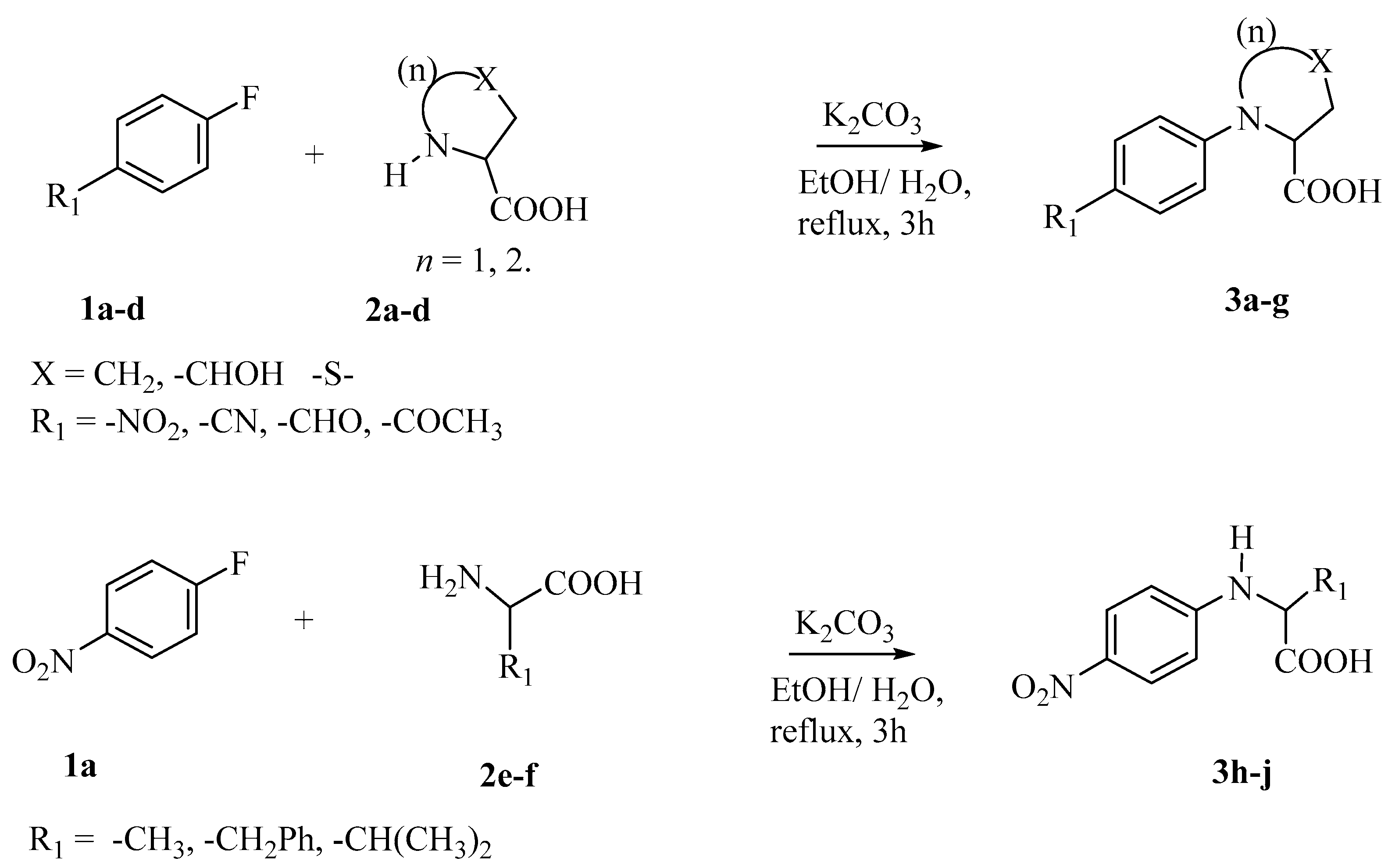
2.2. General Procedure for the Preparation of 3a3j [20,21]

To a clean round-bottomed flask charged with a condenser, amino acid 2ag (12 mmol) in C2H5OH/H2O (1:1) (20 mL) and K2CO3 (3.5 equiv.) was added. The mixture was then refluxed, with stirring for 10 min before p-substituted-fluorobenzene 1ad (10 mmol) was carefully added, stirred, and reflux for 3 h. The reaction medium was cooled to an ambient temperature and concentrated to reduce solvent volume. The concentrate was then diluted with CH2Cl2 (10 mL) and acidified with 6M HCl (10 mL). The aqueous layer was extracted into CH2Cl2 (20 mL × 3), and the combined organic layer was washed with saturated brine solution, dried with anhydrous Na2SO4, filtered, and concentrated in vacuo to obtain the corresponding N-(p-substituted phenyl)-amino acid adducts 3aj in 25–90% yield upon recrystallization in petroleum ether. Some of the products were purified by column chromatography on silica gel (petroleum ether/ethyl acetate; 2:1).

2.3. Evaluation of Antimicrobial Activities

The in vitro antibacterial activity of the synthesized compounds 3a3j was carried out against 8 bacterial strains, which include Gram-positive bacteria, Bacillus subtilis (ATCC 6633), Streptococcus pneumoniae (ATCC 33400), Staphylococcus aureus (ATCC 25923), and Staphylococcus epidermidis (ATCC 14990), and Gram-negative bacteria, Enterobacter cloacae (ATCC 43560), Escherichia coli (ATCC 25922), Proteus mirabilis (ATCC 43071), and Klebsiella oxytoca (ATCC 13182). The in vitro antibacterial activity was achieved using the agar well diffusion method. Streptomycin was used as positive control, while Dimethysulfoxide was the negative control. The bacteria were maintained on nutrient agar.
The agar well diffusion technique, as described by Adeniyi et al. [22], was used to determine the antibacterial activity of the synthesized compounds. For a sensitivity test, nutrient agar plates were seeded with 0.1 mL of an overnight culture of each bacterial strain (equivalent to 107–108 CFU/mL). The seeded plates were allowed to set, and a standard cork borer of 7 mm in diameter was used to cut uniform wells on the surface of the agar. Exactly 0.3 mL of the test compounds already dissolved in DMSO at a concentration of 20 mg/mL was then introduced into the wells and set in the incubator at 30 °C for 24 h. After 24 h, the diameter of the zone of inhibition (in mm), which is the clear area around each well, was measured.

2.4. Minimum Inhibitory Concentration (MIC) of the Synthesized Compounds

The minimum inhibitory concentrations (MICs) of the acids, and streptomycin were determined by a modification of the standard agar dilution method procedure, as previously described by Adeniyi et al. [23]. MICs were carried out for only selected compound(s), which displayed activity during the sensitivity test. For the MIC procedure, different dilutions of the compounds were prepared first at 20 mg/mL to give the final concentrations in the range of 20, 10, 5, 2.5, and 1.25 mg/mL. Two milliliters (2 mL) of each dilution was introduced onto the Mueller Hinton agar (MHA, Difco, France), poured into Petri dishes, and allowed to set. The agar was then streaked with the cultured bacterial strains incubated overnight (24 h), after which the plates were examined for the presence or absence of bacterial growth. The minimum concentration that inhibited microbial growth was regarded as the minimum inhibitory concentration (MIC) of the respective compounds.

3. Results and Discussion

The metal-free C–N coupling of para-substituted halobenzene and amino acids in the presence of a base resulted in the formation of different 4-substituted N-aryl amino acids with yields ranging from 25–90% (Table 1). The structures of compounds 3a to 3j are presented in Figure 2

Antibacterial Activity of Compounds 3aj

Table 2 reveals the zones of inhibition (ZI) of compounds 3aj at 20 mg/mL; the higher the ZI, the better the activity. At lower concentrations (10 mg/mL, 5 mg/mL, 2.5 mg/mL, and 1.25 mg/mL), only two (N-(4-Nitrophenyl)-L-proline 3a and N-(4-Nitrophenyl)-L-valine3j) of the compounds were able to sufficiently inhibit the growth of the selected bacteria. Compounds 3a and 3j displayed the best antibacterial activity against all the bacterial strains screened except against Bacillus subtilis, which was resistant to the test compounds. Interestingly, both compounds 3a and 3j had higher zones of inhibition than streptomycin against four out of the eight bacterial strains screened: Streptococcus pneumoniae, Staphylococcus aureus, Escherichia coli, and Proteus mirabilis. Additionally, amongst the four bacteria, Compound 3a displayed the highest zone of inhibition against Staphylococcus aureus. Furthermore, it was observed that 3a, 3d, 3i, and 3j with ZI of 12 mm, 16 mm, 18 mm, and 12 mm, respectively, were more potent against Proteus mirabilis and Streptococcus pneumoniae (ZI = 16, 16, 14, 18 mm, respectively) than streptomycin (8 and 8 mm, respectively). However, none of the compounds were able to inhibit the growth of Bacillus subtilis besides the standard drug. The best activity of all the N-aryl amino acids was observed against Proteus mirabilis and Streptococcus pneumoniae, where four of the amino acids displayed higher zones of inhibition (>8mm; >16mm, respectively) than streptomycin (ZI = 8 mm).
Surprisingly, the only N-aryl amino acid with sulfur embedded within its ring 3g was not able to inhibit the growth of as many bacterial strains compared to other compounds despite the biological significance of sulfur-containing molecules. The aforementioned amino acid was only able to inhibit the growth of four bacteria with lower zones of inhibition compared to other N-aryl amino acids in the series (Table 2). The least activity was recorded against Enterobacter cloacae across all compounds tested. The activity of these compounds can be attributed to the presence of the electron-withdrawing group, amine functionality, N-alkyl chain substituents, rigid proline ring, and hydroxyl functional group, the majority of which are present within the structure of the standard drug—streptomycin. The exceptional activities observed with compounds 3a and 3j can be attributed to the presence of the nitro functional group common to both, while 3c and 3d possess the cyano and the carbonyl functions (both of which are also present in streptomycin), respectively (Figure 3) [21,22]. The presence of the aromatic ring in the synthesized compounds plays a crucial role in the bioactivities of the N-aryl amino acids, as is observed with well-established drugs, such as the antibacterials methicillin and penicillin V (Figure 1), both of which possess a phenoxy group within their structures [8]. Therefore, it can be said that the N aryl amino acids 3a, 3d 3i, and 3j leveraged the additional effect of the aromatic ring, and hence, displayed better activity than the standard drug (which lacks an aromatic ring) against Streptococcus pneumoniae, Staphylococcus aureus, Escherichia coli, and Proteus mirabilis (Table 2).
Having obtained results from the sensitivity test, the minimum inhibitory concentration (Table 3) of the most active compounds was determined with concentrations of 20, 10, 5, 2.5, and 1.25 mg/mL. It was observed that 3j displayed the best antibacterial activity against Escherichia coli at a concentration of 1.25 mg/mL, while compounds 3a and 3c were both active at 2.5 mg/mL. Additionally, 3j was the most active against Klebsiella oxytoca at a concentration of 2.5 mg/mL, while other compounds were only active between 10–20 mg/mL; it is worth stating that compound 3a sufficiently inhibited the growth of seven out of the eight bacteria screened with a MIC of 5 mg/mL; this observation is in agreement with the work of Odusami et al. [2], 2019, who reported that N-(2-nitrophenyl) pyrrolidine-2-carboxylic acid displayed better antibacterial activity against all the Gram-negative bacterial strains they tested except for K. oxytoca. As for the 4-Cyano substituted moiety, it is probable that the presence of the cyano group in N-(4-Cyanophenyl)-L-proline 3c coupled with the effect of the aromatic ring enhanced its activity against Escherichia coli (2.5 mg/mL) compared to bacterial strains screened (Table 3), hence the reason for the significant activity.
Of all the bacterial strains screened, the test compounds were observed to be most active against Escherichia coli because the compounds 3a, 3b, 3c, 3d, 3e, 3f, 3h, and 3j inhibited its growth even at a concentration as low as 1.25 mg/mL (Table 3).
By comparison, it is correct to state that the N-aryl amino acid 3a N-(4-Nitrophenyl)-L-proline and 3j N-(4-Nitrophenyl)-L-valine (with lower inhibitory concentration and significant broad-spectrum antibacterial activity) seem better motifs in the search for compounds that can serve as drug leads for combating drug-resistant bacterial against Streptococcus pneumoniae, Escherichia coli, Proteus mirabilis, and Klebsiella oxytoca specifically. These findings are also in consonance with the work of Odusami et al., 2019 [2], who reported that N-(Nitrophenyl)cycloamino acids sufficiently inhibited the growth of Escherichia coli and Proteus mirabilis among other bacteria screened.

4. Conclusions

The findings of this study indicated that the ten N-aryl amino acids evaluated for their antibacterial activities showed significant antibacterial activity. Of all the compounds tested, N-(4-Nitrophenyl)-L-proline 3a was more potent than streptomycin against Escherichia coli. While compounds N-(4-Nitrophenyl)-L-proline 3a, N-(4-Nitrophenyl)-D, L-pipecolinic acid 3d, N-(4-Nitrophenyl)-L-alanine 3i, and N-(4-Nitrophenyl)-L-valine 3j were more potent than streptomycin against Streptococcus pneumoniae. The significant antibacterial activities of the N-aryl amino acids reported in this study can be attributed to the presence of the aromatic ring, as well as the effect of the amino acids, which are precursors in many important bioactive molecules; thus, they stand out as lead compounds for future drug development in combating bacterial resistance.

Author Contributions

All authors contributed to the study conception and design. Material preparation, data collection, and analysis were performed by A.D.O. The first draft of the manuscript was written by O.T.A. and O.B.F., and all authors commented on previous versions of the manuscript. All authors have read and agreed to the published version of the manuscript.

Funding

This work was funded, in part, by the University of Lagos Central Research Committee (CRC No. 2015/25), Nigerian Government TetFund IBR (CRC/TETFUND/No. 2018/016).

Conflicts of Interest

The authors declare that they have no known competing financial interests or personal relationships that could have influenced the work reported in this paper.

References

  1. Barrett, G.C. Amino Acids. In Amino Acids, Peptides, and Proteins; Davies, J.S., Ed.; Royal Society of Chemistry: Cambridge, UK, 2000; Volume 31, pp. 1–85. [Google Scholar]
  2. Odusami, J.A.; Ikhile, M.I.; Fotsing, M.C.; Olasupo, I.A.; Izunobi, J.U.; Bamgbade, E.O.; Fonkui, T.Y. Towards eradicating antibiotic-resistant bacteria: Synthesis and antibacterial activities of substituted N-(2-nitrophenyl) pyrrolidine-and piperidine-2-carboxylic acids. J. Chin. Pharm. Sci. 2019, 28, 704–715. [Google Scholar]
  3. Kitajima, H.I.R.O.S.H.I.; Sakashita, H.; Akahoshi, F.; Hayashi, Y. Preparation of Proline Derivatives as Dipeptidyl Peptidase IV (DPP-IV) Inhibitors and use Thereof as Drugs. PCT International Patent Application WO2002014271A1, 21 February 2002. [Google Scholar]
  4. Dougherty, D.A. Unnatural amino acids as probes of protein structure and function. Curr. Opin. Chem. Biol. 2000, 4, 645–652. [Google Scholar] [CrossRef] [PubMed]
  5. King, S.M.; Buchwald, S.L. Development of a Method for the N-Arylation of Amino Acid Esters with Aryl Triflates. Org. Lett. 2016, 18, 4128–4131. [Google Scholar] [CrossRef] [PubMed]
  6. Fischer, C.; Koenig, B. Palladium-and copper-mediated N-aryl bond formation reactions for the synthesis of biologically active compounds. Beilstein J.Org. Chem. 2011, 7, 59–74. [Google Scholar] [CrossRef] [PubMed]
  7. Ma, D.; Zhang, Y.; Yao, J.; Wu, S.; Tao, F. Accelerating effect induced by the structure of α-amino acid in the copper-catalysed coupling reaction of aryl halides with α-amino acids. Synthesis of benzolactam-V8. J. Am. Chem. Soc. 1998, 120, 12459–12467. [Google Scholar] [CrossRef]
  8. Chu, W.; Yang, Y.; Cai, J.; Kong, H.; Bai, M.; Fu, X.; Qin, S.; Zhang, E. Synthesis and bioactivities of new membrane-active agents with aromatic linker: High selectivity and broad-spectrum antibacterial activity. ACS Infect. Dis. 2019, 5, 1535–1545. [Google Scholar] [CrossRef] [PubMed]
  9. Fang, H.; Chen, Z.; Liu, Y.; Zhang, T.; Chang, J.; Li, Z.; Zhang, L.; Sui, J.; Ru, J.; Gu, Y.; et al. Discovery of Aryloxy-, Arylthio-, and Arylamino-Containing Acethydrazides as Fungicidal Agents. J. Agric. Food Chem. 2023, 71, 920–933. [Google Scholar] [CrossRef] [PubMed]
  10. Nowak, M.G.; Skwarecki, A.S.; Milewska, M.J. Amino Acid Based Antimicrobial Agents—Synthesis and Properties. ChemMedChem 2021, 16, 3513–3544. [Google Scholar] [CrossRef] [PubMed]
  11. Malkov, A.V.; Stončius, S.; MacDougall, K.N.; Mariani, A.; McGeoch, G.D.; Kočovský, P. Formamides derived from N-methyl amino acids serve as new chiral organocatalysts in the enantioselective reduction of aromatic ketimines with trichlorosilane. Tetrahedron 2006, 62, 264–284. [Google Scholar] [CrossRef]
  12. World Health Organization. Antimicrobial-Resistance. 2021. Available online: https://www.who.int/news-room/fact-sheets/detail/antimicrobial-resistance (accessed on 25 March 2022).
  13. World Health Organization. Global Priority List of Antibiotic-Resistant Bacteria to Guide Research, Discovery, and Development of New Antibiotics. 2017. Available online: http://www.quotidianosanita.it/allegati/allegato4135670.pdf (accessed on 27 April 2022).
  14. De Oliveira, D.M.; Forde, B.M.; Kidd, T.J.; Harris, P.N.; Schembri, M.A.; Beatson, S.A.; Paterson, D.L.; Walker, M.J. Antimicrobial resistance in ESKAPE pathogens. Clin. Microbiol. Rev. 2020, 33, e00181-19. [Google Scholar] [CrossRef] [PubMed]
  15. Vargas-Casanova, Y.; Rodríguez-Mayor, A.V.; Cardenas, K.J.; Leal-Castro, A.L.; Muñoz-Molina, L.C.; Fierro-Medina, R.; Rivera-Monroy, Z.J.; García-Castañeda, J.E. Synergistic bactericide and antibiotic effects of dimeric, tetrameric, or palindromic peptides containing the RWQWR motif against gram-positive and gram-negative strains. RSC Adv. 2019, 9, 7239–7245. [Google Scholar] [CrossRef] [PubMed]
  16. Pezzella, C.; Ricci, A.; DiGiannatale, E.; Luzzi, I.; Carattoli, A. Tetracycline and streptomycin resistance genes, transposons, and plasmids in Salmonella enterica isolates from animals in Italy. Antimicrob. Agents Chemother. 2004, 48, 903–908. [Google Scholar] [CrossRef] [PubMed]
  17. Partridge, S.R.; Kwong, S.M.; Firth, N.; Jensen, S.O. Mobile genetic elements associated with antimicrobial resistance. Clin. Microbiol. Rev. 2018, 31, e00088-17. [Google Scholar] [CrossRef] [PubMed]
  18. Tanwar, J.; Das, S.; Fatima, Z.; Hameed, S. Multidrug resistance: An emerging crisis. Interdiscip. Perspect. Infect. Dis. 2014, 2014, 541340. [Google Scholar] [CrossRef] [PubMed]
  19. Dadgostar, P. Antimicrobial resistance: Implications and costs. Infect. Drug Resist. 2019, 12, 3903. [Google Scholar] [CrossRef] [PubMed]
  20. Osinubi, A.D.; Izunobi, J.U.; Asekun, O.T.; Familoni, O.B.; Bao, X. Transition Metal-Free, Base-Induced Arylation of Amino Acids: Synthesis of N-(para-Substituted phenyl)amino-2-carboxylic acids. ChemistrySelect 2020, 5, 8644–8648. [Google Scholar] [CrossRef]
  21. Osinubi, A.; Izunobi, J.; Bao, X.; Asekun, O.; Kong, J.; Gui, C.; Familoni, O. Synthesis and in vitro anticancer activities of substituted N-(4′-nitrophenyl)-L-prolinamides. R. Soc. Open Sci. 2020, 7, 200906. [Google Scholar] [CrossRef] [PubMed]
  22. Adeniyi, B.A.; Odelola, H.A.; Oso, B.A. Antimicrobial potentials of Diospyros mespiliformis (Ebenaceae). Afr. J. Med. Med. Sci. 1996, 25, 221–224. [Google Scholar] [PubMed]
  23. Adeniyi, C.B.A.; Lawal, T.O.; Mahady, G.B. In vitro susceptibility of Helicobacter pylori to extracts of Eucalyptus camaldulensis and Eucalyptus torelliana. Pharm. Biol. 2009, 47, 99–102. [Google Scholar] [CrossRef] [PubMed]
Figure 1. Structure of some important bioactive molecules with the N-aryl amino acids’ core structure.
Reactions 04 00017 g001
Scheme 1. Synthesis of N-aryl amino acids 3aj.
Reactions 04 00017 sch001
Figure 2. Structures of 4-substituted N-aryl amino acids (3a3j).
Reactions 04 00017 g002
Figure 3. Structural relationship of streptomycin and the synthesized N-aryl amino acids.
Reactions 04 00017 g003
Table 1. Metal-free C–N coupling of para-substituted fluorobenzene and amino acids.
EntryR1-Ar-XAMINO ACIDProductYield
R1X (%)
1.NO2F2aN-(4-Nitrophenyl)-L-proline (3a)90
2.NO2F2bN-(4-Nitrophenyl)-D, L-pipecolinic acid (3b)70 a
3.CNF2aN-(4-Cyanophenyl)-L-proline (3c)69
4.CHOF2aN-(4-Formylphenyl)-L-proline (3d)65
5.NO2 (OH)F2dN-(4-Nitrophenyl)-L-hydroxyproline (3e)89
6.CH3COF2aN-(4-Acetylphenyl)-L-proline (3f)54
7.NO2 (S)F2cN-(4-Nitrophenyl)-L-thioproline (3g)35 b
8.NO2F2gN-(4-Nitrophenyl)-L-phenylalanine (3h)30 c
9.NO2F2eN-(4-Nitrophenyl)-L-alanine (3i)25 b
10.NO2F2fN-(4-Nitrophenyl)-L-valine (3j)50
a reaction time (rt): 12 h, b rt: 24 h, c rt: 8 h, general reaction time: 3 h.
Table 2. Zones of inhibition (ZI) (mm) of synthesized N-Aryl amino acids at 20 mg/mL.
Bacterial Strains3a
R1 = NO2
n = 3
3b
R1 = NO2
n = 4
3c
R1 = CN
n = 3
3d
R1 = CHO
n = 3
3e
R1 = NO2
R2 = OH
n = 3
3f
R1 = CH3CO
n = 3
3g
R1 = NO2,
Thioproline
n = 3
3h
R1 = NO2
n = 0
3i
R1 = NO2
n = 0
3j
R1 = NO2
n = 0
Strep
(a) Gram-positive
Bacillus subtilis----------40
Streptococcus pneumoniae1610-1668106141810
Staphylococcus aureus221016121412-12102016
Staphylococcus epidermidis81016161412-10121830
(b) Gram-negative
Enterobacter cloacae84-842-44416
Escherichia coli1268668668208
Proteus mirabilis126816686412188
Klebsiella oxytoca108-1264106101212
Streptomycin (Strep)—standard drug. The values coloured red displayed activities higher than the standard drug.
Table 3. Minimum inhibitory concentration (MIC) (mg/mL) of synthesized compounds (3a, 3b, 3c, 3d, 3e, 3f, 3h, and 3j).
Bacterial Strains3a3b3c3d3e3f3h3j
(a) Gram-positive
Bacillus subtilis--------
Streptococcus pneumoniae520>2010>2020>2020
Staphylococcus aureus52010>201010>2010
Staphylococcus epidermidis5>20101010101010
(b) Gram-negative
Enterobacter cloacae5>20>20>20>2020>2020
Escherichia coli2.552.51055101.25
Proteus mirabilis52010101020>205
Klebsiella oxytoca510>2020>2020102.5
Streptomycin (Strep)—standard drug. The values coloured red (1.25 mg/mL, 2.5 mg/mL and 5 mg/mL) in Table 3 indicates values for compounds which displayed good antibacterial activiities at concentrations lower than 10 mg/mL (this was the concentration at which the sensitivity testing was carried out and at which compounds 3a, 3b, 3c, 3d, 3e, 3f, 3j and 3h displayed activity) while the other compounds were only active at higher concentrations.
Disclaimer/Publisher’s Note: The statements, opinions and data contained in all publications are solely those of the individual author(s) and contributor(s) and not of MDPI and/or the editor(s). MDPI and/or the editor(s) disclaim responsibility for any injury to people or property resulting from any ideas, methods, instructions or products referred to in the content.

