Article

Physiological Responses and Adaptations of the Halophyte Atriplex halimus to Soil Contaminated with Cd, Ni, and NaCl

1
Department of Botany, St. John’s College, Dr. B.R. Ambedkar University, Agra 282002, India
2
Department of Microbiology, School of Life Sciences, Dr. B.R. Ambedkar University, Agra 282004, India
3
Department of Botany, Hislop College, Nagpur 440001, India
4
School of Life Sciences and the Environment, University of Trás-os-Montes e Alto Douro—UTAD, Quinta de Prados, 5000-801 Vila Real, Portugal
5
MARE—Marine and Environmental Sciences Centre/ARNET—Aquatic Research Network, Department of Life Sciences, University of Coimbra, Calçada Martim de Freitas, 3000-456 Coimbra, Portugal
*
Author to whom correspondence should be addressed.
Soil Syst. 2023, 7(2), 46; https://doi.org/10.3390/soilsystems7020046
Submission received: 15 February 2023 / Revised: 1 May 2023 / Accepted: 3 May 2023 / Published: 5 May 2023

Abstract

:
Soils contaminated with potentially toxic elements (PTEs) and salt manifest a large number of physical, chemical, and structural problems by various processes such as reduced water availability, water and air movement in soil space, water holding capacity of soil, as well as perilous effects on plant growth and physiology. Halophytes have the ability to grow in saline environments and are better adapted to accommodate environmental constraints including PTE ions. An experiment was designed to study the response of the halophyte Atriplex halimus to a range of salinities and different concentrations of Cd and Ni. Tolerance and soil remedial potential of the plant were quantified in terms of PTE uptake and portioning, plant biomass, root/shoot ratio, chlorophyll and anti-oxidative enzyme production, along with stress markers such as lipid peroxidation, proline, and glycine betaine. The plant was also evaluated for its potential to phytoremediate PTE contaminated soil. The results suggest that A. halimus can tolerate moderate concentrations of both the PTEs and salt. The species holds promise for bio-reclamation of saline and PTE-contaminated soil.

1. Introduction

Soil contamination by potentially toxic elements (PTEs) such as Pb, Cd, Hg, Zn, and Cu has been receiving increased attention worldwide in the past few decades. Their persistent nature is a severe threat to the environment, and affects the lives of plants, animals, and humans. Once they enter the body, they accumulate in vital organs for years causing severe health problems [1]. Effects of PTE poisoning can be toxic, carcinogenic, mutagenic, or teratogenic [2]. Unlike other contaminants, PTEs cannot be degraded by chemical or biological treatments, are eventually indestructible, and their toxic effects last longer.
Soil salinity is another major issue that adversely affects agricultural productivity and sustainability specially in arid and semi-arid areas. Like PTE pollution, salinity problems also result from both natural and mismanaged anthropogenic activities. Salinity and PTE toxicity in soil impact a considerable portion of agricultural areas globally by decreasing the growth and thus the yield of plants by affecting their metabolic activities such as respiration, carbon-dioxide assimilation, chlorophyll biosynthesis, cell membrane elasticity and permeability, water uptake, protein synthesis, reduction in root elongation and reduced stomatal openings with turgor loss, and inhibition of anti-oxidative enzyme activities.
Various clean-up technologies have been developed and used to remediate contaminated soils. Phytoremediation is counted among the safer, cleaner, cost-effective, and environment-friendly technologies. It uses numerous biological organisms viz. bacteria, cyanobacteria, yeast, fungi, algae, and higher plants as tools for the restoration/remediation of soil and water contaminated within organic or organic contaminants. Ideal phytoremedial plants should exhibit a deep root system, high shoot biomass production, and perennial growing habit as well as be able to cope with low fertility and poor soil structure [3]. Most plants explored for phytoremediation are glycophytes, but these cannot survive in saline soils polluted by PTEs such as in semi-arid areas. Halophytes, however, are a group of plants that can tolerate saline soils and can even complete their life cycles in such conditions. They have evolved a range of morphological, physiological, and biochemical strategies to proliferate in high-salt environments [4] and may be ideal for phytoremedial applications in such cases. In fact, low to moderate levels of NaCl are believed to stimulate plant growth in halophytes and play an important role in protection against PTE toxicity by mediating ion absorption, osmotic adjustment, and induction of antioxidant systems [5,6].
Cd and Ni are among PTEs of great concern to environment and human health [7]. Cd is one of the most phytotoxic PTEs even at low concentrations causing impaired absorption of nutrients [8] and photosynthetic and growth inhibition [9,10]. Ni is a micronutrient, [11] but at higher concentrations it may have deleterious effects similar to Cd and Pb [12]. PTEs also increase the production of reactive oxygen species (ROS) in plant cells, which can cause oxidative damages such as the peroxidation of membrane lipids and carbonylation of proteins. Plants control such damage by protective ROS trapping systems including antioxidant enzymes such as superoxide dismutase (SOD), guaiacol peroxidase (GPX), and catalase (CAT), which control ROS levels [7,10,12].
Studies with halophytes have shown their tolerance to high concentrations of toxic PTE ions which most plants do not survive, in particular, under saline conditions [10,13]. To a certain extent, tolerance to salt and PTEs relies on common physiochemical mechanisms [3,14,15], and therefore, halophytes are being explored as candidates for phytomanagement of PTE-contaminated soils. Recent studies have demonstrated that halophytes such as Mesembryanthemum crystallinum, Salicornia brachiata, and Atriplex nummularia accumulate large amounts of Cd, Ni, and Pb [12,16,17,18].
Atriplex species are a very interesting group of halophytes that can survive and complete their life cycle at high salinity levels. They are equipped with salt bladders which accumulate salt. These glandular structures are not specific to Na and Cl but also accumulate and excrete PTEs such as Cd, Zn, and Pb on the leaf surface as a possible metal detoxification [19] mechanism as in Atriplex halimus and Tamarix smyrnensis [5,20]. They also have numerous layers of specialized balloon-like, vesiculated hairs called trichomes on the outer surface of the leaf [21]. These differ from salt glands and perform a range of different functions such as storage of water and its absorption from the atmosphere along with salt secretion. These plants show high potential to tolerate and concentrate PTE in their tissues by triggering detoxification mechanisms [12,22].
The present study aims to evaluate the response of Atriplex halimus to a range of salinities and concentrations of Cd and Ni in order to assess its potential to remediate soil contaminated with these two metals. Partitioning of the metals between the root and shoot, biochemical parameters that indicate stress, as well as enzymatic antioxidant activity will also be evaluated to obtain a comprehensive picture of Cd and Ni tolerance mechanism in the plant.

2. Materials and Methods

2.1. Plant Material and Soil

Seeds of Atriplex halimus (commercially available and certified) were procured from the United States Department of Agriculture–Agriculture Research Service (USDA-ARS), Washington State University, USA. These were allowed to germinate in the dark after sterilization in 0.1% HgCl2 and soaking for 3 h in double distilled water. The experimental soil was collected from the botanical garden of St. John’s College, Agra. The topsoil of the area is a sandy loam made of 60–80% sand, 10–24% silt, and 8–16% clay. The soil has moderate water retaining capacity with pH 7.18 ± 0.51, electrical conductivity (EC) 1.04 ± 0.23 dSm−1, moisture content 8.32 ± 0.59%. The organic carbon content was 1.10 ± 0.1%, available P2O5 8.7 ± 0.3 mg kg−1, available nitrogen 143.14 ± 1.8 mg kg−1, and available K2O 271 ± 7.1 mg kg−1. Sodium (Na), calcium–magnesium (Ca + Mg), and potassium (K) content of the soil was 730 ± 5, 4.4 ± 0.5, and 23 ± 4 meq L−1, respectively. Non-perforated plastic pots were used for the experiment to avoid leaching of salt and PTEs, each pot was filled with 4 kg autoclaved and air-dried soil.

2.2. Experimental Design

A greenhouse pot experiment was set up in the botanical garden at St. John’s College, Agra (27.18° N 78.02° E), India. The salinization of pot soil (4 kg) was carried out by adding 100 mL kg−1 of 1, 3, or 5% NaCl water solution to pots as per treatment plan. These were left for 15 days to stabilize. The temperature was maintained at 27–30 °C. Germinated seedlings were transplanted (one per pot) in pots having 4 kg soil. Plants were treated with different doses of cadmium (Cd) and nickel (Ni) as aqueous solutions using CdCl2·5H2O and NiSO4·6H2O, respectively. The treatments selected were as follows: T0 (control [Cd0/Ni0/NaCl0]); T1 (1% NaCl); T2 (3% NaCl); T3 (5% NaCl); T4 (Cd 25 mg kg−1); T5 (Cd 50 mg kg−1); T6 (Cd 100 mg kg−1); T7 (Cd 25 mg kg−1 + 1% NaCl); T8 (Cd 25 mg kg−1 + 3% NaCl); T9 (Cd 25 mg kg−1 + 5% NaCl); T10 (Ni 50 mg kg−1); T11 (Ni 100 mg kg−1); T12 (Ni 200 mg kg−1); T13 (Ni 50 mg kg−1 + 1% NaCl); T14 (Ni 50 mg kg−1 + 3% NaCl); T15 (Ni 50 mg kg−1 + 5% NaCl). Three replicates of each treatment were maintained in a complete randomized block experimental design. The pots were watered with distilled water as and when required to minimize leaching of NaCl and/or PTEs. Each pot had a plastic tray under it. Any leachate collected was added to the pot at next watering. At the end of the study, i.e., 120 DAT (days after treatment), each plant was uprooted and washed in deionized water and divided into root and shoot parts and analyzed for metal content. Root length, shoot length, and dry biomass were also noted. The soil was tested to analyze the electrical conductivity (EC), sodium adsorption ratio (SAR), and residual metal content. In addition, leaves were plucked for testing chlorophyll, proline content, lipid peroxidation, glycine betaine, relative electrolyte leakage, and antioxidative enzyme activity of plants at 30, 60, and 120 DAT to monitor the adaptation and health of the test plants through the study period.

2.3. Growth and Physiochemical Analysis

2.3.1. Chlorophyll

Chlorophyll estimation was performed using acetone (80% v/v) [23]. Crushed fresh leaves (0.5 g) were dissolved in 10 mL acetone and incubated at 4 °C for 12 h in dark. The supernatant was collected after centrifugation (for 5 min at 4000 rpm and 4 °C) and its optical density was measured at two wavelengths (663, 645 nm) using a UV–Vis spectrophotometer (Perkin Elmer Lambda 25). The concentration of total chlorophyll was expressed in mg g−1 fresh weight.

2.3.2. Membrane Damage Rate (MDR)

Relative electrolytic leakage was estimated to determine the membrane damage rate [24]. One gram of fresh leaf tissue was suspended in 10 mL distilled water in a test tube which was then incubated at room temperature for 24 h. After 24 h, the initial electrical conductivity (EC1) was measured. The solution was then autoclaved at 121 °C for 15 min in order to release ions from the tissue and allowed to cool down before the final electrical conductivity (EC2) was measured.
REL = (EC1/EC2) × 100.

2.3.3. Lipid Peroxidation

Lipid peroxidation was estimated from 1 g fresh leaf tissue and expressed as malondialdehyde (MDA) content [25]. A total of 5 mL of 10% trichloroacetic acid (TCA) was used to prepare a leaf extract crushed in liquid nitrogen. This was followed by centrifugation at 10,000 rpm and 4 °C for 10 min. An amount of 2 mL of supernatant was taken in a fresh test tube to which 2 mL of 0.67% thiobarbituric acid (TBA) was added. The test tube was placed in a water bath at 100 °C, followed by incubation in an ice bath to stop the reaction. MDA content (µg g−1 FW) was measured at 532 nm absorbance by using 0.025% TBA in 10% TCA as control.

2.3.4. Proline

The estimation of proline content as a stress marker was conducted according to Bates et al. [26]. Three percent sulfosalicylic acid (10 mL) was used to prepare an extract from fresh leaves (500 mg) followed by centrifugation at 2000 rpm for 15–20 min to separate the supernatant. A reaction mixture was prepared by adding 2 mL of ninhydrin and glacial acetic acid each to a test tube containing 2 mL of supernatant. This was then placed in a water bath for 30 min. The reaction was stopped by placing the test tube in an ice bath. After this, 4 mL toluene was added, followed by vigorous shaking to get a red chromophore (toluene layer) in the upper part. The optical density of the chromophore layer was measured at 520 nm. Toluene was used as a control (reference blank). Proline content was expressed in µmol g−1 FW and calculated with a standard curve of proline.

2.3.5. Glycine Betaine

Glycine betaine analysis was performed as per Grieve and Grattan [27]. A total of 500 mg fresh leaves were powdered in liquid nitrogen and suspended in deionized water (20 mL). This was followed by mechanical shaking at 25 °C for 16 h. The sample was strained. An amount of 500 μL filtrate and 2 N sulfuric acid (1:1) were mixed and cooled in an ice bath for 1 h. After this 200 μL of I2-KI reagent (20% potassium iodide and 15.7% iodine) was added to the cooled samples and kept for incubation at 4 °C for 16 h. The samples were centrifuged for 15 min (at 10,000 rpm and 0 °C). In total, 9 mL of 1, 2-dichloroethane was used to dissolve the crystals (periodide). After 2 h, absorbance (365 nm) of the samples were measured. Glycine betaine content of the samples was expressed in g−1 fw.

2.3.6. Antioxidative Enzyme Assay

Fresh leaf samples were obtained for the analysis of antioxidative enzyme activity. Leaf extract was prepared in 10 mL of 50 mM phosphate buffer (pH 7.0) and 0.5 mM EDTA using 1 g crushed leaf sample (in liquid nitrogen). For ascorbate peroxidase (APX) analysis, 1 mM ascorbic acid was added in the extract at the time of crushing. The extract was filtered with cheesecloth (4 layered) and centrifuged at 16,000 rpm for 20 min at 4 °C. Supernatant was first used to analyze protein content of the extract and then processed further to measure the antioxidative enzymes of the plant. Bradford assay was used for protein estimation with bovine serum albumin (BSA) as standard.
Catalase (EC 1.11.1.6) activity was estimated according to Aebi [28]. An amount of 0.1 M phosphate buffer (pH adjusted to 7) was prepared and used to make reaction mixture with 50 µL enzyme extract and 0.5 mL 75 mM H2O2. Distilled water was used to make up the final volume to 3 mL. Decrease in absorbance (decomposed amount of H2O2) was calculated at 240 nm and used to measure the catalase activity with the help of a standard curve of H2O2.
The Nakano and Asada [29] method was used for ascorbate peroxidase (APX) estimation. Leaf extract (0.1 mL) + 1.5 mL phosphate buffer (0.1 M) + 0.5 mL ascorbic acid (3 mM) +0.1 mL H2O2 (3 mM) + 0.8 mL distilled water (to make final volume 3 mL) was used to prepare a reaction mixture. The decrease in absorbance (290 nm) was measured to calculate the amount of ascorbic acid (µmol) oxidized min−1 mg protein−1.
For superoxide dismutase (SOD) (EC 1.15.1.1), 560 nm wavelength was used to record the absorbance of the reaction mixture containing 0.2 mL 200 mM methionine, 0.1 mL 2.25 mM NBT, 0.1 mL 3 mM EDTA,1.5 mL 0.1 M phosphate buffer, 0.1 mL 1.5 M Na2CO3, 0.1 mL enzyme extract, and 0.9 mL of water [30]. A total of 2 M riboflavin (0.1 mL) was used to start the reaction under a 15 W fluorescent lamp, which was maintained for 15 min. Reaction mixture without enzyme extract served as control, and a non-irradiated complete reaction mixture served as blank. One unit of enzyme activity was taken as the amount of enzyme that decreased the absorbance by 50% in comparison to control.
The glutathione reductase (GR) (EC 1.6.4.2) assay was performed by using the Smith et al. method [31]. The reaction was started by adding 0.1 mL of 20.0 mM oxidized glutathione (GSSG) to 3 mL reaction mixture containing potassium phosphate buffer (pH 7.5), EDTA, DTNB, NADPH, enzyme extract, and distilled water. The activity was measured as total absorbance (increase in absorbance) at 412 nm and expressed as GSSG reduced min−1 mg protein−1.

2.3.7. Sodium Adsorption Ratio (SAR), Cd and Ni Analysis

One gram soil (in 5 mL distilled water) was used as soil–water extract to estimate Na+, Mg2+, and Ca2+ using a flame atomic absorption spectrophotometer [32]. Sodium adsorption ratio (SAR) was calculated from the following formula:
SAR = Na+/[(Ca2+ + Mg2+)/2]1/2
Soil, root, and shoot samples were digested using the wet method with HNO3 (69%, Merck) in combination with HCl (30%, Merck) for soil and H2O2 (30%, m/v; Merck) for plant material. The samples were analyzed for Cd and Ni (mg kg−1) using a flame atomic absorption spectrophotometer.

2.4. Statistical Analysis

The experiment and tests were performed in triplicate. Data were analyzed using one-way and two-way ANOVA (SigmaPlot 11.0).

3. Results

3.1. PTE Accumulation and Phytoremedial Potential of A. halimus

A. halimus showed greater accumulation of Cd in the roots (Table 1). At low concentration i.e., Cd25, an uptake of 6.41 mg kg−1 in roots and 2.61 mg kg−1 in shoots was observed at 120 DAT. Test plants accumulated Cd in a concentration-dependent manner. A further increase in Cd accumulation was obtained in NaCl treatments. Cd accumulation increased up to 6.89 and 7.41 mg kg−1 in roots and 4.75 and 6.97 mg kg−1 in shoots with 1% and 3% NaCl at 120 DAT. In correlation with the reduced physiology, 5% salt concentration was found to decrease Cd accumulation in both the root and shoot of the plant (6.02 and 3.85 mg kg−1, respectively).
Ni distribution showed a different pattern; with greater accumulation in shoot tissues (Table 1). Ni uptake in shoots was almost twice that of roots. Maximum Ni uptake occurred in shoots of the plant (9.42 mg kg−1) in 3% NaCl treatment. Ni uptake was found to increase with increasing concentration of the metal in soil i.e., 1.72, 2.52, and 4.5 mg kg−1 in roots and 5.56, 7.14, and 10.6 mg kg−1 in shoots for Ni50, Ni100, and Ni200 mg kg−1, respectively. The addition of salt to soil at lower doses increased Ni uptake, but in 5% salt treatment, it decreased sharply. Results of one-way ANOVA indicate significant statistical difference (p ≤ 0.001) in the mean Cd and Ni content in the root and shoot of A. halimus across the different PTE and salt treatments at the end of the study (120 DAT).
Phytoremediation characteristics of A. halimus for Cd accumulation at 25 mg kg−1 dose indicate that the roots are capable of accumulating more Cd than shoots. This indicates the phytostabilizing nature of A. halimus for the metal. Similar pattern of accumulation was also observed at higher concentrations i.e., 50 and 100 mg kg−1. Although the plant had low values of root uptake and transfer to shoot, the percentage of metal reduction from soil was satisfactory. The transportation of Cd from soil to root to shoot increased with increasing concentration of the metal. Transfer of Ni from root to shoot was more which indicates extraction and sequestration of the metal in above-ground parts. Clean-up % of Ni from soil by A. halimus was almost two times higher at low concentration. Around 2.8% reduction in soil Ni concentration was observed at Ni50 which increased up to 7.8% with 3% salt. Significant statistical difference (p ≤ 0.001) in the mean Cd and Ni content in soil due to different treatments was obtained at 120 DAT.

3.2. Potential of A. halimus for Restoration of Saline Soil

The desalinization potential of A. halimus was measured by comparing the electrical conductivity (EC) and sodium adsorption ratio (SAR) of soil at initial and final day of the study. EC was 1.04 dSm−1 for control soil; and in the ranges of 4.5–4.9 dSm−1 for 1% NaCl treatments, 8.1–8.7 dSm−1 for 3% NaCl treatments, and 10.1–10.3% dSm−1 for 5% NaCl treatments initially. These were reduced to 2.5–3.7, 4.2–5.1, and 9.4–9.6 dSm−1 for 1, 3, and 5% NaCl treatments, respectively, after 120 days. Around 50% reduction in soil EC was obtained in 1% NaCl treatment (T1). Thus, 1% NaCl treatments (T1, T7, T13) were reduced from slightly saline to non-saline and 3% NaCl treatments (T2, T8, T14) were reduced from slightly saline to somewhere between non to slightly saline as per the limits suggested by FAO for saline soils. Reduction in soil EC indicates reduction in salt content of soil due to its uptake and sequestration by A. halimus. The highest reduction in soil SAR was observed in Cd25 + 1% NaCl (52.6%) followed by 3% NaCl (51.9%) treatment. The lowest reduction in soil SAR was obtained with 5% NaCl (2.5%). Thus, the optimum concentration of salt for optimum growth and development of A. halimus was 3%. The data indicate that metal ions competed with salt ions for translocation to shoots.

3.3. Growth and Physiochemical Analysis

3.3.1. Dry Biomass and Root/Shoot Ratio

Dry biomass and root/shoot ratio of the test plants were measured at the end of the study i.e., 120 DAT (Table 1). A slight increase in biomass was observed in response to Cd25 (38.6 g) and Ni50 (37.7 g) treatments. As the PTE concentration increased, biomass decreased gradually. As the concentration of NaCl increased from 1 to 3%, a significant increase in the biomass of the plants was observed. Plants treated with 3% salt were the healthiest as compared to other salt treatments. On the other hand, salt at 5% dose in soil inhibited growth individually and in combination with PTEs. Still, all the plants survived. Similar trends were observed for root/shoot ratios. Lower values in 1 and 3% salt treatments (with or without metals) can be explained by longer shoots. Higher values were obtained in 5% NaCl treatments because of reduced growth of shoots. Two-way ANOVA indicates significant statistical difference (p ≤ 0.001) in the mean biomass, due to Cd and salt treatments (Table 2). The interaction between Cd and salt doses was also statistically significant (p ≤ 0.001). In Ni treatments, significant difference was obtained for salt treatments as well as Ni and salt interaction (p ≤ 0.001).

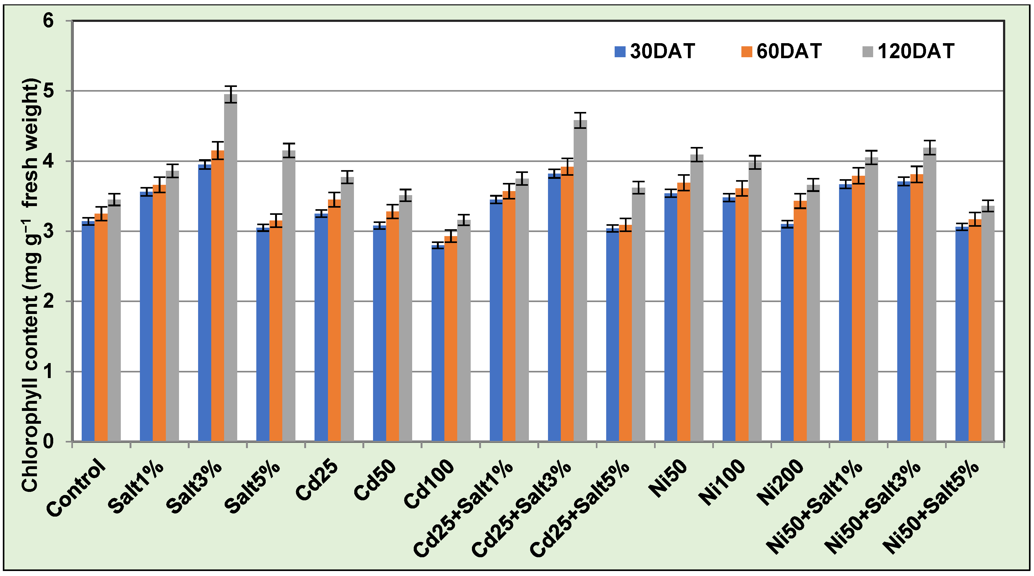
3.3.2. Chlorophyll Content

Total chlorophyll content of A. halimus leaves was found to increase as the study progressed (Figure 1). No significant reduction in chlorophyll content of Cd treated plant was observed (except for Cd100 mg kg−1). Surprisingly, plants treated with Cd25 mg kg−1 showed a slight increase in their chlorophyll content as compared to control, while plants treated with Ni showed significant increase in chlorophyll as compared to control at all testing days. Highest chlorophyll synthesis occurred in Ni50 followed by Ni100 mg kg−1. A. halimus developed no visible symptoms of toxicity in NaCl treatments up to 3% concentration. An increase in the photosynthetic activity was seen in treatments of Ni and Cd with 1 and 3% salt. Two-way ANOVA indicates significant statistical difference (p ≤ 0.001) in the mean chlorophyll content due to each PTE treatment and day of testing (Table 3). The interaction between treatments and day of testing, however, was not significant (p ≤ 0.001).

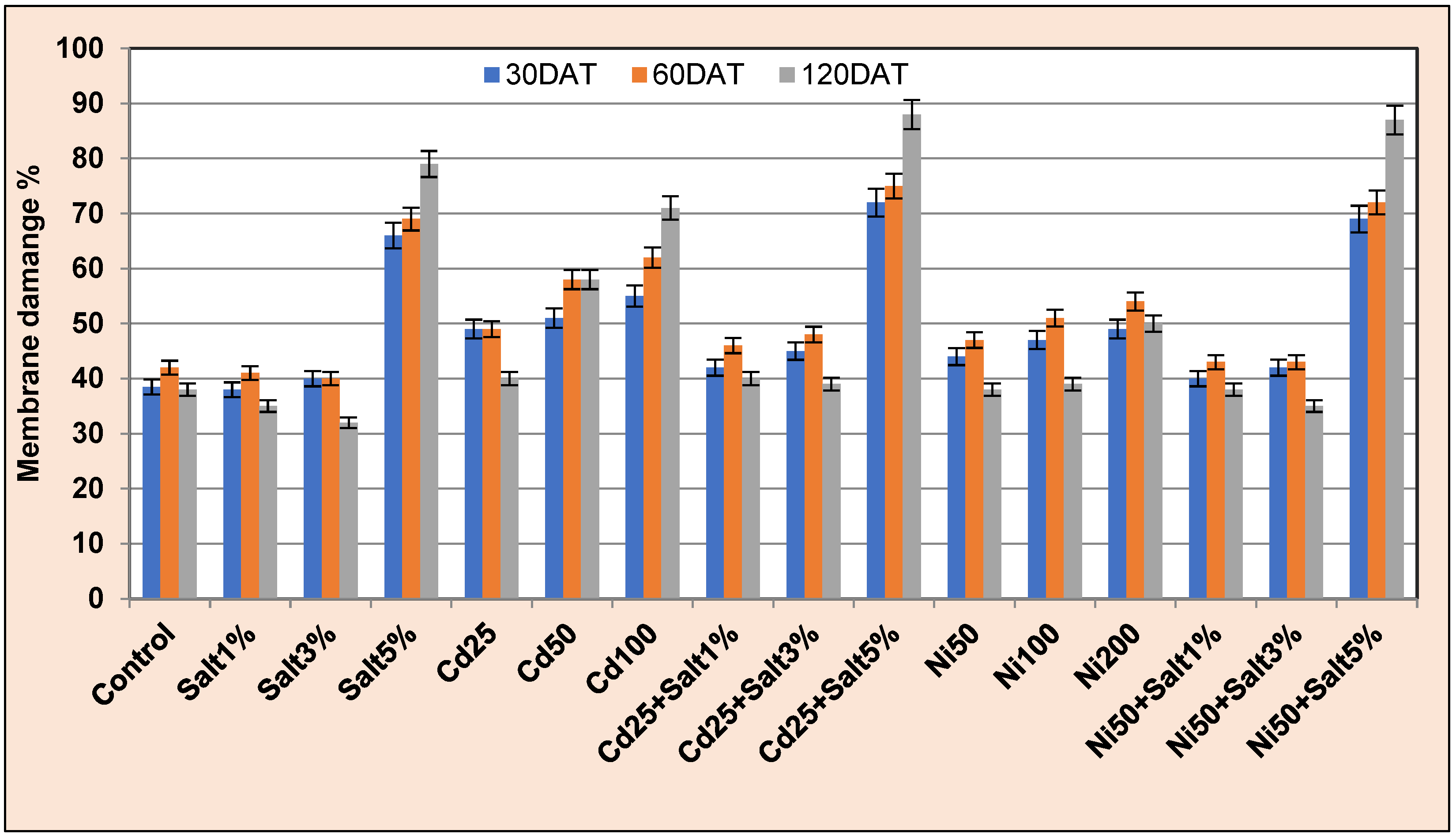
3.3.3. Membrane Damage Rate

Initially, A. halimus leaves showed significantly higher membrane damage rate as compared to control in all treatments (Figure 2). Lesser electrolyte leakage (MDR) was observed in most treatments as the experiment progressed. This indicates that plants became more tolerant in due course of time. Two-way ANOVA indicates statistically significant difference in mean MDR (p ≤ 0.001) due to different treatments and day of testing (Table 3). The interaction between treatment dose and testing day was also significant (p ≤ 0.001). This can be understood as the effect of treatments changing over time. Lower dose of PTEs showed membrane damage comparable to control at 120 DAT. Cd and Ni treatments with 1–3% salt exhibited a reduction in MDR after 60 DAT. Higher combination of salt and metal showed increased membrane damage till the end of study. It is clear that salt was beneficial for the plant but only up to certain doses.
Table 3. Two-way ANOVA of PTE and NaCl treatments for physiological parameters.
ParametersChlorophyllMembrane
Damage Rate
Lipid
Peroxidation
ProlineGlycine
Betaine
Sources of Variations
CdTreatments**********
Day after treatment**********
Treatment × DATNs**Ns****
NiTreatments**********
Day after treatment**********
Treatment × DATNs**Ns****
(** = difference of means significant at p ≤ 0.001; ns = not significant).

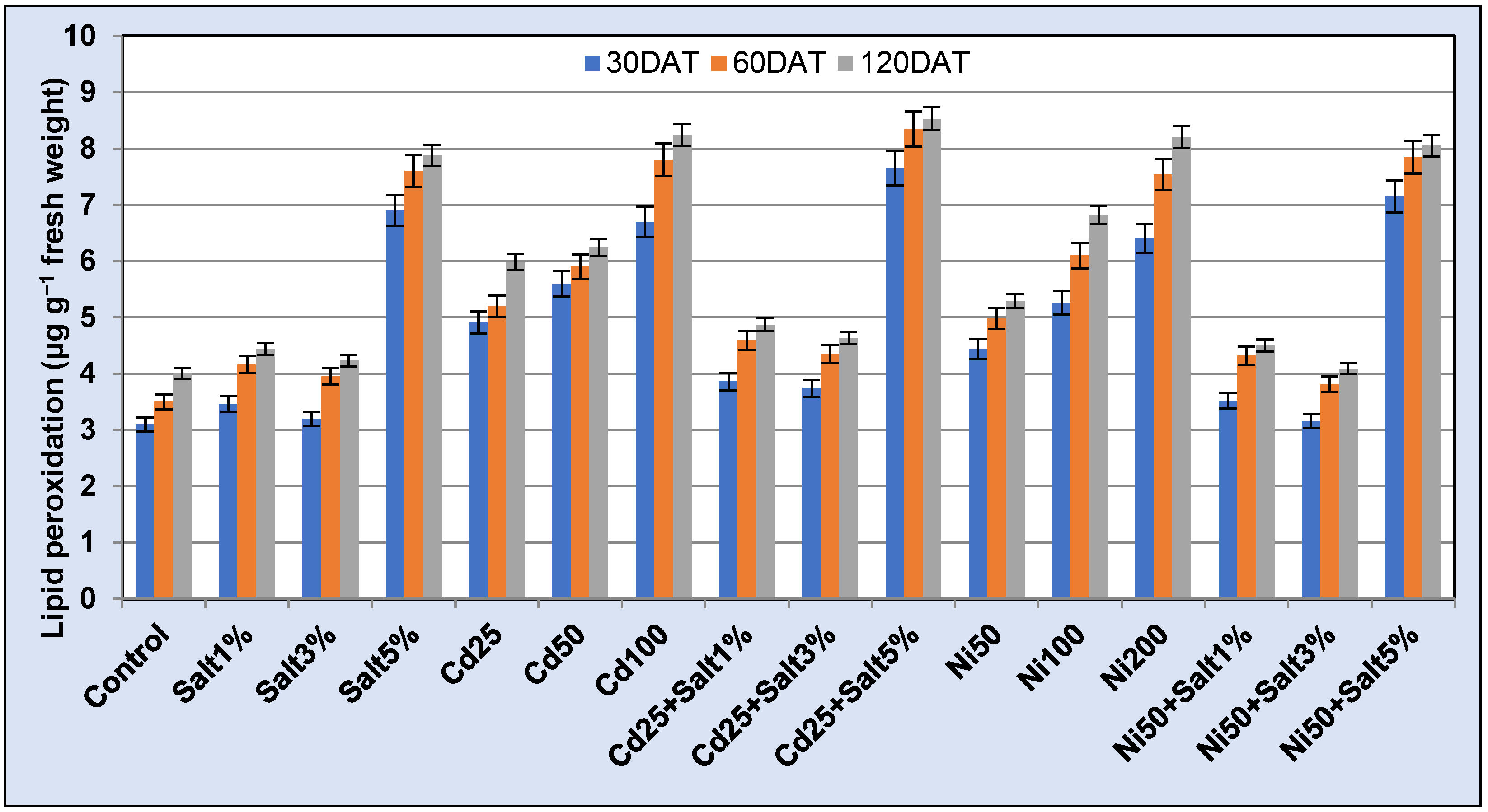
3.3.4. Lipid Peroxidation

Plants treated with only metal treatments exhibited a significant increase in lipid peroxidation expressed as malondialdehyde (MDA) content (Figure 3). It was found to increase with increasing metal concentrations. Maximum MDA content was (8.24 μg g−1 fw) for Cd 100 ppm. Least MDA was recorded in plants treated with 3% salt. The amount of MDA was found to decrease as the study progressed showing the adaptability of the plant to salt and Cd/Ni contamination in soil. At 120 DAT, all treatments showed MDA levels comparable to control. The difference in mean values due to the PTE and salt treatments (Table 3) as well as testing date was statistically significant (p ≤ 0.001).

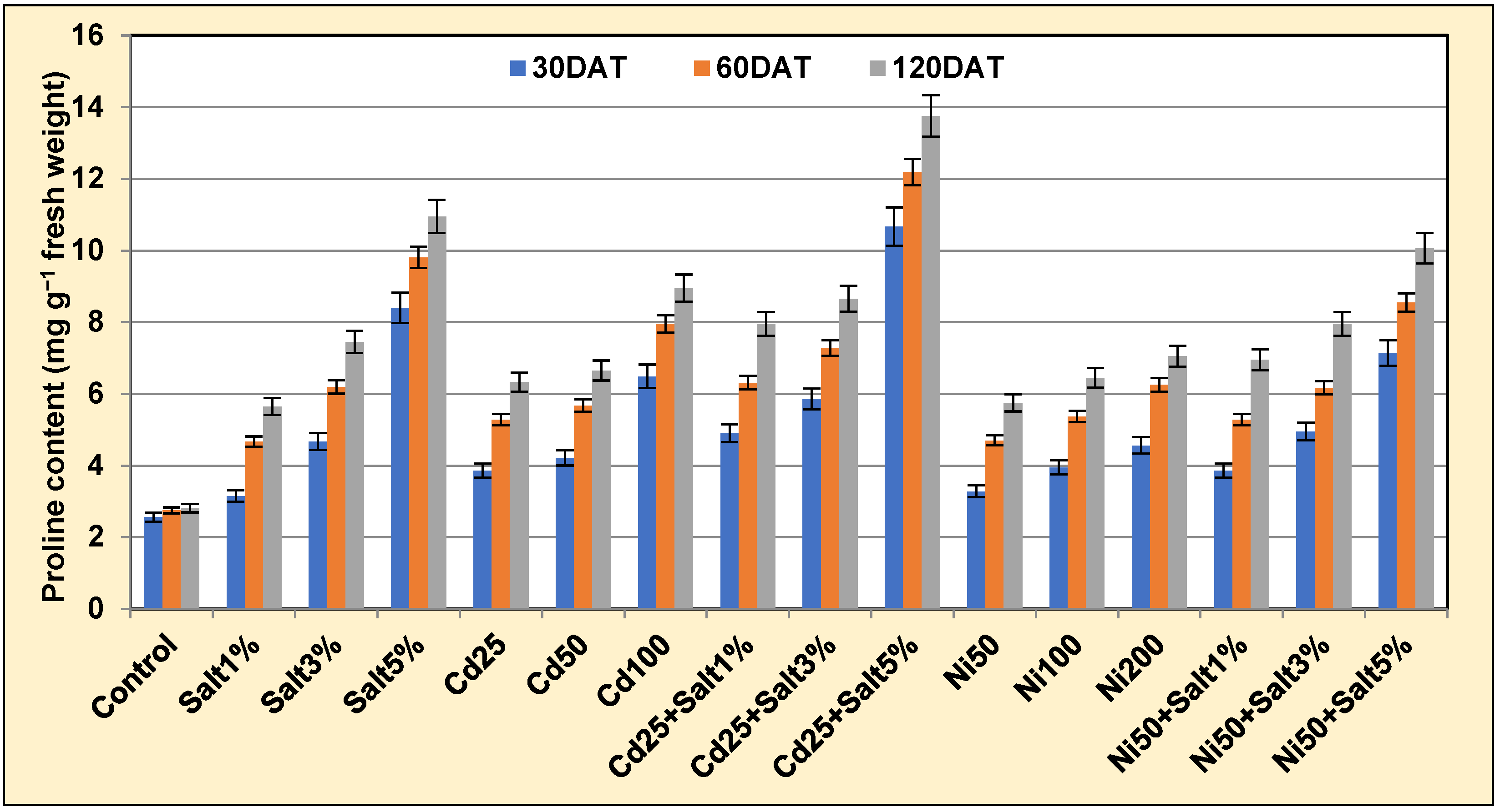
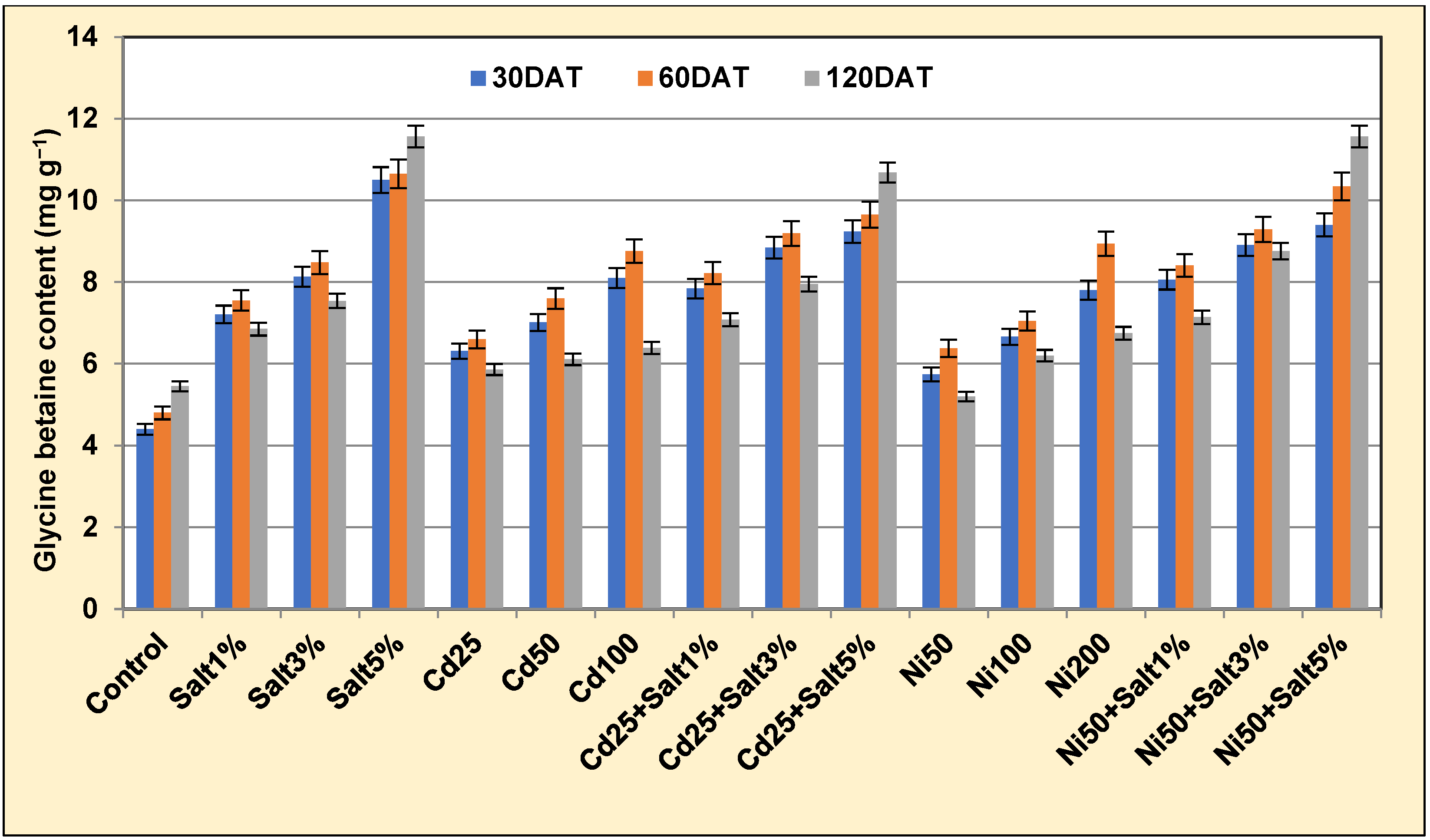
3.3.5. Proline and Glycine Betaine Content

Proline accumulation in test plants was found to increase with increasing concentration of salt and/or PTEs (Figure 4). Maximum proline was accumulated in plants treated with 5% salt with or without PTEs. Treatments with Cd and Ni caused higher levels of proline in test plants at all testing days. Lowest range of proline (5.28–6.95 mg g−1 fw) was observed in Ni50 + 1% NaCl.
Glycine betaine content of A. halimus was significantly influenced by the PTE treatments (Figure 5). Increased values were noticed till 60 DAT. Cd 100 and Ni200 mg kg−1 had almost similar impact on GB content. Lower doses of salt and salt + metal combinations showed gradual decrease in GB content after 60 DAT, while plants treated with 5% salt individually and in PTE combinations showed continuous increase in GB content till 120 DAT. ANOVA results indicate that difference in mean proline and glycine betaine content in response to Cd/Ni and NaCl treatments as well as testing dates was statistically significant (p ≤ 0.001) (Table 3). A statistically significant interaction between treatment dose and testing day (p ≤ 0.001) in the mean values was also obtained for proline. This translates to effect of treatment changing as the study progressed.

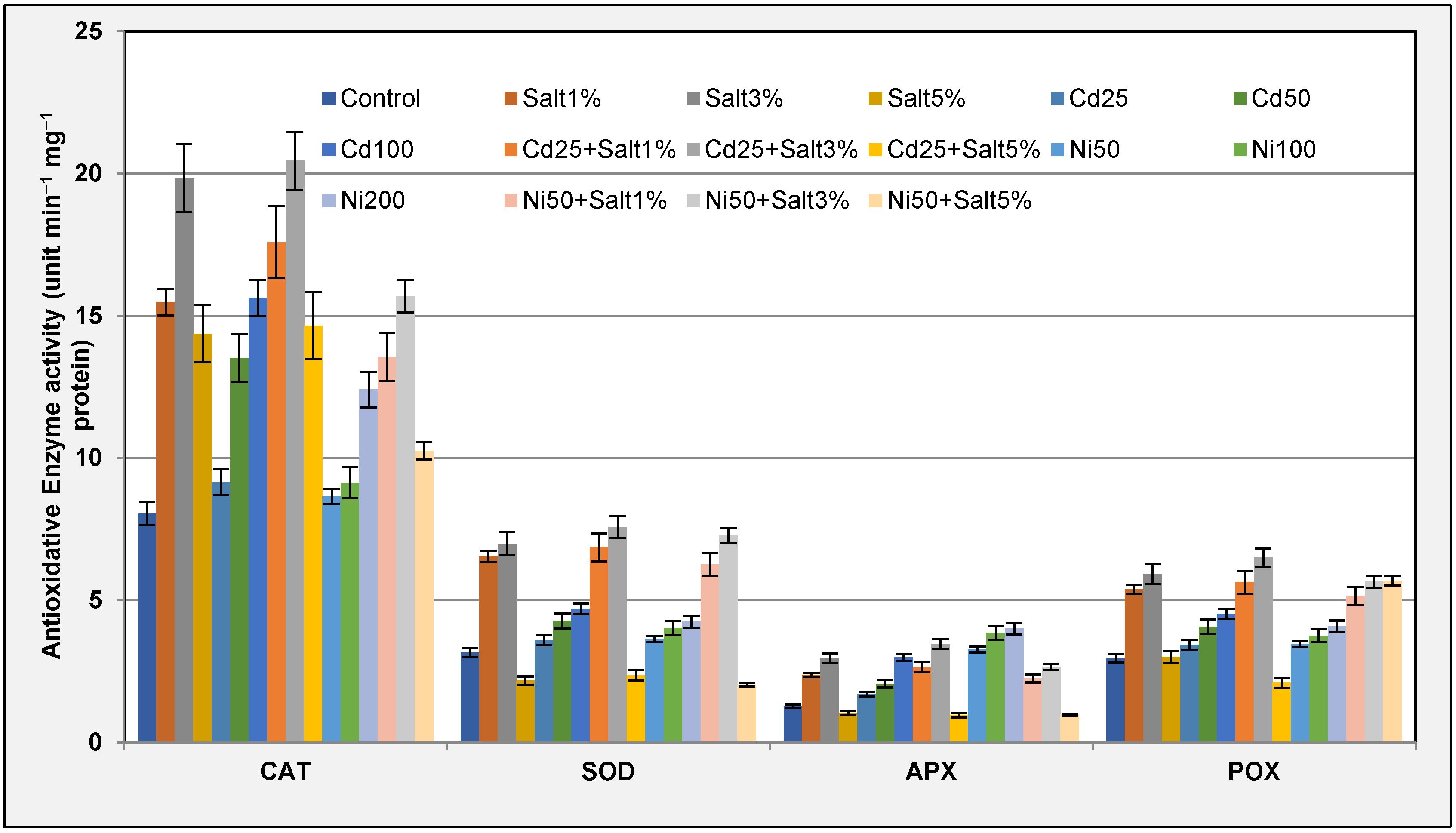
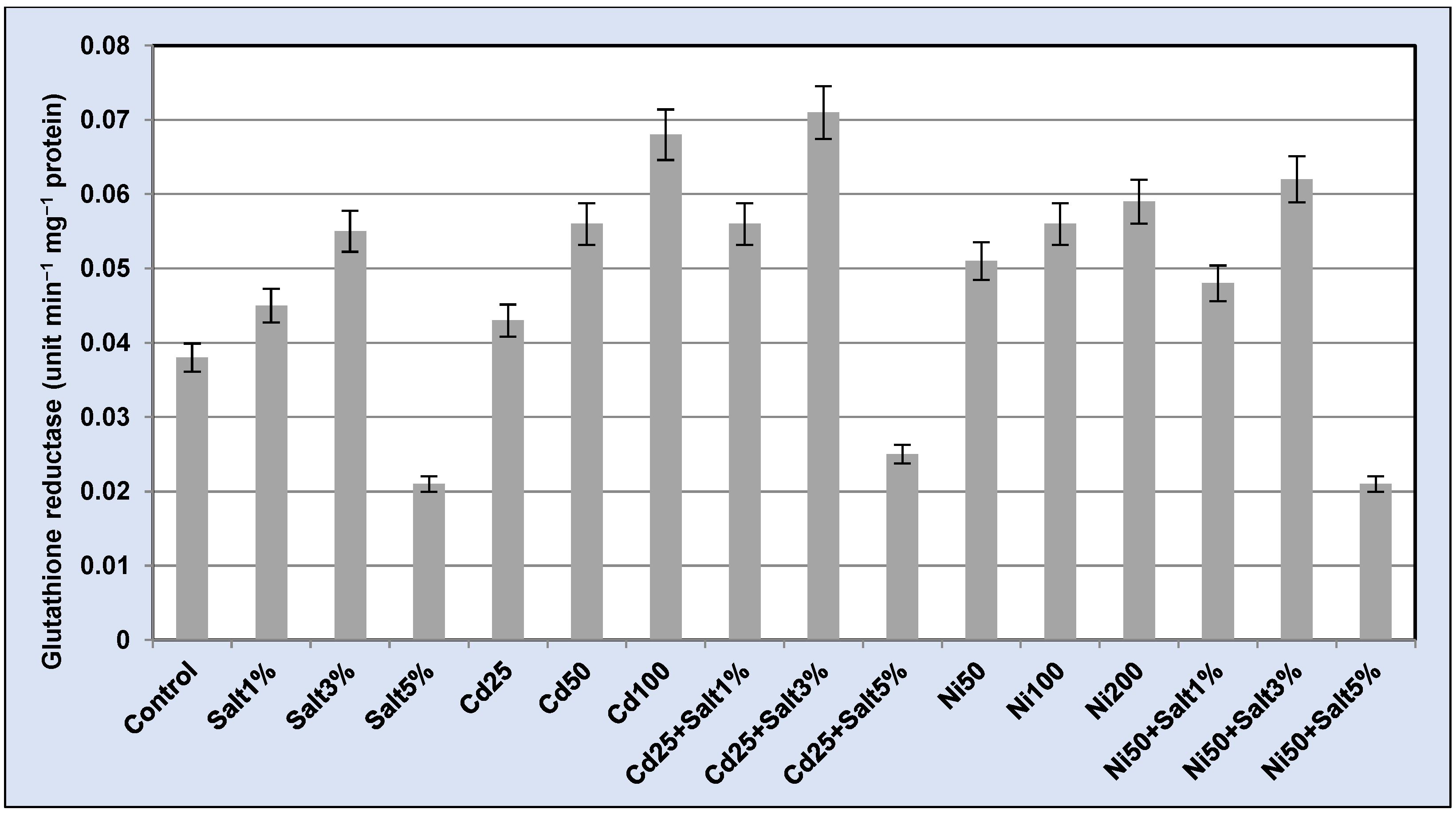
3.3.6. Antioxidative Enzymes

Antioxidative defense of the plant was expressed by a pool of antioxidative enzymes (CAT, APX, SOD, POX, and GR) (Figure 6 and Figure 7). Catalase activity was found to increase with increasing concentration of Cd and Ni. The order was Cd100 > Cd50 > Ni200 > Cd25 > Ni100 > Ni50 > control. Similar picture was obtained for SOD and POX as well. APX activity, however, showed slight variation. A minute increase was observed in Ni100 and Ni200 mg kg−1 treatments. The range of GR activity in plants under PTE treatments was 0.043–0.068 unit min−1 mg−1 protein for Cd.
Plants treated with 1 and 3% NaCl exhibited higher level of antioxidants as compared to control, but at 5% concentration of salt, a rapid decrease in the activity of all the enzymes was noticed. APX and SOD activity was noticed to be the most affected by 5% salt. Plants treated with both salt and PTEs showed further elevation in their antioxidative status. Highest CAT and SOD activity was found in plants treated with Cd 25 mg kg−1 + 3% salt. About 3.2-fold decrease in SOD was observed in plants treated with 5% salt as compared to the plants treated with 3% salt. GR activity showed positive correlation with APX. Mean values for enzyme activities (CAT, APX, SOD, and POX) were significantly different (p ≤ 0.001) in response to Cd and salt treatments; however, the interaction of Cd and salt treatment was not (Table 2). In Ni treatments, significant difference in mean enzyme activity (p ≤ 0.001) was obtained due to PTE and NaCl. The interaction Ni × NaCl was significant (p ≤ 0.001) only for CAT, APX, and POX.

4. Discussion

The species of Atriplex have been reported to accumulate Pb, Zn, Cd, Ni, Cu, and As [10,33,34,35]. It has been demonstrated that Pb and Zn in A. hortensis and A. rosea accumulated largely in by root tissues; with accumulation increasing in accordance with soil PTE concentrations [34]. This is in line with this investigation as higher accumulation of Cd was observed in roots of A. halimus. Cd concentration is usually higher in roots compared to above-ground tissues in most plants [36]. A. halimus has been reported to accumulate Ni, Pb, Zn, and As in roots and translocate small fractions to above-ground biomass [9,35,37]. It is interesting to note that Kahli et al. [10] observed a similar preferential partitioning of Cd in A. halimus roots at 50 μM Cd but the reverse at 20 μM Cd. Additionally, in a five-year study, where A. halimus was grown in mile tailings amended with pig slurry, the authors reported better translocation of Zn, Pb, Cd, and Cu to leaves and/or stem over roots [38]. This indicates that environmental factors may affect plant physiology and cause different patterns of PTE sequestration and accumulation within plant biomass.
On the other hand, Ni content was greater in the shoots of test plants at 120 DAT. Studies indicate that plants accumulate Ni predominantly in their aerial parts [39,40]. In 2003, Ni was added to the list of essential plant nutrients by US Department of Agriculture [41] which also explains its higher concentration in above-ground parts and lesser phytotoxicity at low to moderate concentrations.
In Cd (25, 50, and 100 mg kg−1) treatments, ~5.6, 6.4, and 3.5% clean-up of soil was obtained, while in Ni treatments (50, 100, and 200 mg kg−1) the clean-up was ~2.8, 1.8, and 0.8%. Thus, A. halimus demonstrated maximum phytoremedial potential at 50 mg kg−1 doses for both the PTEs. Phytoremediation potential of halophytes including Atriplex species for PTEs has been recorded previously [17,35,42,43]. Chenopodium botrys an annual halophyte may remove up to 180 g Cd ha−1, which is six times more than Cd removal by the hyperaccumulator Noccaea caerulescens [44]. Moderate level of Ni accumulation has been reported in Sporobolus virginicus shoots [17].
Halophytes have the ability to better adapt to many environmental constraints including toxic metal ions and could be more satisfactory for the extraction of PTEs from saline soils than glycophytes [5,45]. Milić et al. [46] found greater metal accumulation in aerial parts of Sesuvium portulacastrum (halophytes) in comparison to Brassica juncea (glycophyte) grown in a Pb amended growth matrix. Positive effect of the salinity on accumulation of Cd has been reported in A. halimus, A. nummularia, and A. canescens, especially at the low Cd doses [5,10]. Similar results were obtained in this study where the addition of salt to soil enhanced the uptake of both Cd and Ni in Atriplex halimus. Highest PTE uptake in root (Cd) and shoot (Cd & Ni) was observed with 3% salt. PTE uptake, however, seemed to take a toll on the desalinization ability of the test plant, which again strongly suggests that metal ions compete with Na+ ions for sequestration within the plants.
Toxic effects of PTEs generally show common side-effects such as reduction in plant biomass, height, pigment, sugar, protein, and carbohydrate concentration; inhibition of photosynthesis; and production of stress markers such as proline, MDA, and glycine betaine. Reduction in shoot length, specific leaf area, dry biomass, relative growth rate, and net photosynthetic rate due to increasing Cd concentration has been previously recorded in A. halimus [9]. Results of this study indicate that A. halimus was quite tolerant to moderate concentrations of both PTEs (Cd and Ni) and salt. The tolerance of A. halimus to Cd may be due to its greater sequestration in roots to alleviate the inhibition of photosynthesis and simultaneous increase in antioxidant enzymatic activity in leaves.
Significant increase in proline and glycine betaine accumulation under PTE stress has been observed previously in A. halimus [19]. PTE Slama et al. [47] observed increased accumulation of many osmotically agile compounds such as proline, trehalose, sucrose, polyols, and glycine betaine in halophytes. These compounds have been reported to play key roles for the stabilization of many proteins and their complexes, maintenance of membrane integrity, ROS scavenging and osmotic adjustments. In the present study, significantly higher contents of proline and glycine betaine were recorded in plants grown in PTE-contaminated soil. Plants showed highest production of proline and glycine betaine in response to Cd. Salt exposure also resulted in increased production of GB in A. halimus. Exposure to 5% salt caused the highest production of proline and glycine betaine. GB assists in salt tolerance by increasing the protection of thylakoid membranes of chloroplast, thereby strengthening the photosynthesis system of halophytes [48]. This could explain the increased production of chlorophyll in test plants treated with salt.
Cd, Ni, and Pb have been documented to exhibit adverse effects on membrane functionality of a cell and evoke a strong change in the compositions of lipids in cell membranes of plants [49]. These metals ultimately induce lipid peroxidation in plant cell which can be marked by excessive production of MDA content [50]. The test plant showed enhanced damage rate (MDR) and production of MDA under PTE exposure especially during the initial days of the study. Plants treated with NaCl in combination with PTEs exhibited a significant reduction in MDA content. since salt increased plant growth and improved their detoxification capability. Increased MDA content in plants after exposure to PTEs has been noted both in the absence and presence of salt in soil [10,35,51]. This suggests that halophytes plants have an effective antioxidant defence system which responds in proportion to stress [52].
Antioxidative mechanisms of plants are powered by many enzymatic scavengers (SOD, CAT, APX, etc.) and non-enzymatic compounds (ascorbic acid, proline, etc.). Fluctuations of their activity are generally used to forecast metal tolerance [53]. The results of the current investigation indicate that A. halimus showed increased production of antioxidative enzymes (CAT, SOD, APX, POX, and GR) in most PTE and salt treatments. Increased CAT and SOD activity in A. halimus leaves due to Cd and Pb has also been observed in previous studies [9,54]. El-Bakatoushi et al. studied the molecular and physiological mechanisms of Cd and Pb tolerance in Atriplex halimus from fourteen sites and found that among others, membrane transporter and ROS detoxification genes were consistently up-regulated in plants from polluted sites. They also detected overexpression of phytochelatin synthase responsible for producing phytochelatins, which bind to toxic PTEs forming complexes stored in cellular compartments [54]. This indicates the importance of using not just the right species of a plant but also the most suitable genotype.

5. Conclusions

Halophytic plant species are known to tolerate many abiotic stresses posed by their natural soil environment through efficient management of osmotic and oxidative disturbances. They are also able to take up appreciable amounts of potentially toxic elements from the soil and sequester them in salt glands, vacuoles, or trichomes to limit their toxic effects. In this context and taking into consideration the PTE accumulation patterns, biomass production and stress management strategies, Atriplex halimus holds promise for phytoremediation of Cd- and Ni-polluted sites with saline and non-saline environments. However, it would be prudent to note that every contaminated site is a unique microcosm where even a well-studied plant may behave differently due to environmental factors including soil chemistry and its own physiological responses. Molecular advances in the recent decades also indicate the importance of the correct genotype of the species being used for remedial purposes.

Author Contributions

Conceptualization: S.I., R.J.D., M.V. and M.S.P.; Software: S.I., H.K. and M.V.; Validation: R.J.D., P.J.C.F. and M.S.P.; Formal Analysis, S.I. and P.J.C.F.; Investigation, S.I., H.K. and M.V.; Resources: S.I., P.J.C.F. and M.S.P.; Writing—Original Draft Preparation: S.I. and R.J.D.; Writing—Review & Editing, R.J.D., P.J.C.F. and M.S.P.; Supervision: P.J.C.F. and M.S.P. All authors have read and agreed to the published version of the manuscript.

Funding

This study had the support of national funds through Fundação para a Ciência e Tecnologia, I. P (FCT), under the projects UIDB/04292/2020, UIDP/04292/2020, granted to MARE, and LA/P/0069/2020, granted to the Associate Laboratory ARNET.

Institutional Review Board Statement

Not applicable.

Informed Consent Statement

Not applicable.

Data Availability Statement

Not applicable.

Conflicts of Interest

The authors declare no conflict of interest.

References

  1. Sihlahla, M.; Mouri, H.; Nomngongo, P.N. Assessment of bioavailability and mobility of major and trace elements in agricultural soils collected in Port St Johns, Eastern Cape, South Africa using single extraction procedures and pseudo-total digestion. J. Environ. Health Sci. Eng. 2020, 18, 1615–1628. [Google Scholar] [CrossRef] [PubMed]
  2. Chaturvedi, R.; Favas, P.J.C.; Pratas, J.; Varun, M.; Paul, M.S. Harnessing Pisum sativum–Glomus mosseae symbiosis for phytoremediation of soil contaminated with lead, cadmium, and arsenic. Int. J. Phytoremediat. 2021, 23, 279–290. [Google Scholar] [CrossRef] [PubMed]
  3. Lutts, S.; Lefevre, I. How can we take advantage of halophyte properties to cope with heavy metal toxicity in salt-affected areas? Ann. Bot. 2015, 115, 509–528. [Google Scholar] [CrossRef] [PubMed]
  4. Grigore, M.N.; Ivanescu, L.; Toma, C. Halophytes: An Integrative Anatomical Study; Springer: New York, NY, USA, 2014. [Google Scholar]
  5. Manousaki, E.; Kalogerakis, N. Phytoextraction of Pb and Cd by the Mediterranean Saltbush (Atriplex halimus L.): Metal uptake in relation to salinity. Environ. Sci. Pollut. Res. 2009, 16, 844–854. [Google Scholar] [CrossRef]
  6. Han, R.M.; Lefevre, I.; Albacete, A.; Perez-Alfocea, F.; Barba-Espín, G.; Díaz-Vivancos, P.; Quinet, M.; Ruan, C.J.; Hernandez, J.H.; Cantero-Navarro, E.; et al. Antioxidant enzyme activities and hormonal status in response to Cd stress in the wetland halophyte Kosteletzkya virginica under saline conditions. Physiol. Plant. 2012, 147, 352–368. [Google Scholar] [CrossRef]
  7. Gill, S.S.; Khan, A.N.; Tuteja, N. Cadmium at high dose perturbs growth, photosynthesis and nitrogen metabolism while at low dose it up regulates sulfur assimilation and antioxidant machinery in garden cress (Lepidium sativum L.). Plant Sci. 2012, 182, 112–120. [Google Scholar] [CrossRef]
  8. Huang, C.; Zeng, G.; Huang, D.; Lai, C.; Xu, P.; Zhang, C.; Cheng, M.; Wan, J.; Hu, L.; Zhang, Y. Effect of Phanerochaete chrysosporium inoculation on bacterial community and metal stabilization in lead-contaminated agricultural waste composting. Bioresour. Technol. 2017, 243, 294–303. [Google Scholar] [CrossRef]
  9. Mesnoua, M.; Mateos-Naranjo, E.; Barcia-Piedras, J.M.; Pérez-Romero, J.A.; Lotmani, B.; Redondo-Gómez, S. Physiological and biochemical mechanisms preventing Cd-toxicity in the hyperaccumulator Atriplex halimus L. Plant Physiol. Biochem. 2016, 106, 30–38. [Google Scholar] [CrossRef]
  10. Kahli, H.; Sbartai, H.; Cohen-Bouhacina, T.; Bourguignon, J. Characterization of cadmium accumulation and phytoextraction in three species of the genus Atriplex (canescens, halimus and nummularia) in the presence or absence of salt. Plant Physiol. Biochem. 2021, 166, 902–911. [Google Scholar] [CrossRef]
  11. Chen, C.; Huang, D.; Liu, J. Functions and toxicity of nickel in plants: Recent advances and future prospects. Clean 2009, 37, 304–313. [Google Scholar] [CrossRef]
  12. Amari, T.; Ghnaya, T.; Abdelly, C. Nickel, cadmium and lead phytotoxicity and potential of halophytic plants in heavy metal extraction. S. Afr. J. Bot. 2017, 111, 99. [Google Scholar] [CrossRef]
  13. Chen, M.; Yang, Z.; Liu, J.; Zhu, T.; Wei, X.; Fan, H.; Wang, B. Adaptation mechanism of salt excluders under saline conditions and its applications. Int. J. Mol. Sci. 2018, 19, 3668. [Google Scholar] [CrossRef]
  14. Negrin, V.L.; Spetter, C.V.; Guinder, V.A.; Perillo, G.M.E.; Marcovecchio, J.E. The role of Sarcocornia perennis and tidal flooding on sediment biogeochemistry in a South American wetland. Mar. Biol. Res. 2013, 9, 703–715. [Google Scholar] [CrossRef]
  15. Cong, M.; Zhao, J.; Lu, J.; Ren, Z.; Wu, H. Homologous cloning, characterization and expression of a new halophyte phytochelatin synthase gene in Suaeda salsa. J. Oceanol. Limnol. 2016, 34, 1034. [Google Scholar] [CrossRef]
  16. Ghnaya, T.; Slama, I.; Messedi, D.; Grignon, C.; Abdelly, C. Cd-induced growth reduction in the halophyte Sesuvium portulacastrum is significantly improved by NaCl. J. Plant Res. 2007, 120, 309–316. [Google Scholar] [CrossRef]
  17. Eid, M.A. Halophytic plants for phytoremediation of heavy metals contaminated soil. J. Am. Sci. 2011, 7, 377–382. [Google Scholar]
  18. Chaturvedi, A.K.; Mishra, A.; Tiwari, V.; Jha, B. Cloning and transcript analysis of type 2 metallothionein gene (SbMT-2) from extreme halophyte Salicornia brachiata and its heterologous expression in E. coli. Gene 2012, 499, 280–287. [Google Scholar] [CrossRef]
  19. Lefevre, I.; Marchal, G.; Meerts, P.; Corréal, E.; Lutts, S. Chloride salinity reduces cadmium accumulation by the Mediterranean halophyte species Atriplex halimus L. Environ. Exp. Bot. 2009, 65, 142–152. [Google Scholar] [CrossRef]
  20. Kadukova, J.; Manousaki, E.; Papadantonakis, N.; Kalogerakis, N. Phytoextraction and phytoexcretion of Cd by the leaves of Tamarix smyrnensis growing on contaminated non-saline and saline soils. Environ. Res. 2008, 106, 326–332. [Google Scholar]
  21. Black, R.F. The leaf anatomy of Australian members of the genus Atriplex I. Atriplex vesicaria Heward and A. nummularia Lindl. Aust. J. Bot. 1954, 2, 269–286. [Google Scholar] [CrossRef]
  22. Sousa, A.I.; Caçador, I.; Lillebø, A.I.; Pardal, M.A. Heavy metal accumulation in Halimione portulacoides: Intra- and extra-cellular metal binding sites. Chemosphere 2008, 70, 850–857. [Google Scholar] [CrossRef] [PubMed]
  23. Lichtenthaler, H.K. Chlorophylls and Carotenoids: Pigments of Photosynthetic Biomembranes, Methods in Enzymology; Academic Press: Cambridge, MA, USA, 1987; Volume 148, pp. 350–382. [Google Scholar]
  24. Dionisio-Sese, M.L.; Tobita, S. Antioxidant responses of rice seedlings to salinity stress. Plant Sci. 1998, 135, 1–9. [Google Scholar] [CrossRef]
  25. Heath, R.L.; Packer, L. Photoperoxidation in isolated chloroplasts: I. Kinetics and stoichiometry of fatty acid peroxidation. Arch. Biochem. Biophys. 1968, 125, 189–198. [Google Scholar] [CrossRef] [PubMed]
  26. Bates, L.S.; Waldren, R.P.; Teare, I.D. Reclassification of Brevibacterium halotolerans DSM8802 as Bacillus halotolerans comb. nov. based on microbial and biochemical characterization and multiple gene sequence, rapid determination of free proline for water-stress studies. Plant Soil 1973, 39, 205–207. [Google Scholar] [CrossRef]
  27. Grieve, C.M.; Grattan, S.R. Rapid assay for determination of water-soluble quaternary ammonium compounds. Plant Soil 1983, 70, 303–307. [Google Scholar] [CrossRef]
  28. Aebi, H. Catalase In Vitro, Methods in Enzymology; Academic Press: Cambridge, MA, USA, 1984; Volume 105, pp. 121–126. [Google Scholar]
  29. Nakano, Y.; Asada, K. Hydrogen peroxide is scavenged by ascorbate specific peroxidase in spinach chloroplasts. Plant Cell Physiol. 1981, 22, 867–880. [Google Scholar]
  30. Dhindsa, R.; Plumb-Dhindsa, P.; Thorpe, T. Leaf senescence: Correlated with increased levels of membrane permeability and lipid peroxidation, and decreased levels of superoxide dismutase and catalase. J. Exp. Bot. 1981, 32, 93–101. [Google Scholar] [CrossRef]
  31. Smith, O.B.; Osafo, E.L.K.; Adegbola, A.A. Studies on the feeding value of agro-industrial by products: Strategies for improving the utilisation of cocoa-pod-based diets by ruminants. Anim. Feed Sci. Technol. 1988, 20, 189–201. [Google Scholar] [CrossRef]
  32. Rabhi, M.; Hafsi, C.; Lakhdar, A.; Hajji, S.; Barhoumi, Z.; Ham-Rouni, M.H.; Abdelly, C.; Smaoui, A. Evaluation of the capacity of three halophytes to desalinize their rhizosphere as grown on saline soils under non-leaching conditions. Afr. J. Ecol. 2009, 47, 463–468. [Google Scholar] [CrossRef]
  33. Vickerman, D.B.; Shannon, M.C.; Banuelos, G.S.; Grieve, C.M.; Trumble, J.T. Evaluation of Atriplex lines for selenium accumulation, salt tolerance and suitability for a key agricultural insect pest. Environ. Pollut. 2002, 120, 463–473. [Google Scholar] [CrossRef]
  34. Kachout, S.S.; Mansoura, A.B.; Mechergui, R.; Leclerc, J.C.; Rejeb, M.N.; Ouerghi, Z. Accumulation of Cu, Pb, Ni and Zn in the halophyte plant Atriplex grown on polluted soil. J. Sci. Food Agric. 2012, 92, 336–342. [Google Scholar] [CrossRef]
  35. Tapia, Y.; Diaz, O.; Pizarro, C.; Segura, R.; Vines, M.; Zúñiga, G.; Moreno-Jiménez, E. Atriplex atacamensis and Atriplex halimus resist As contamination in Pre-Andean soils [northern Chile]. Sci. Total Environ. 2013, 450–451, 188–196. [Google Scholar] [CrossRef]
  36. Ramos, J.; Naya, L.; Gay, M.; Abian, J.; Becana, M. Functional characterization of an unusual phytochelatin synthase, LjPCS3, of Lotus japonicas. Plant Physiol. 2002, 148, 536–545. [Google Scholar] [CrossRef]
  37. Amer, N.; Al Chami, Z.; Al Bitar, L.; Mondelli, D.; Dumontet, S. Evaluation of Atriplex halimus, Medicago lupulina and Portulaca oleracea for phytoremediation of Ni, Pb, and Zn. Int. J. Phytor. 2013, 15, 498–512. [Google Scholar] [CrossRef]
  38. Acosta, J.A.; Abbaspour, A.; Martínez, G.R.; Martínez-Martínez, S.; Zornoza, R.; Gabarrón, M.; Faz, A. Phytoremediation of mine tailings with Atriplex halimus and organic /inorganic amendments: A five-year field case study. Chemosphere 2018, 204, 71–78. [Google Scholar] [CrossRef]
  39. Bhatia, N.P.; Walsh, K.B.; Orlic, I.; Siegele, R.; Ashwath, N.; Baker, A.J.M. Studies on spatial distribution of nickel in leaves and stems of the metal hyperaccumulator Stackhousia tryonii Bailey using micro-PIXE and EDXS techniques. Funct. Plant Biol. 2004, 31, 1061–1074. [Google Scholar] [CrossRef]
  40. Yusuf, M.; Qazi, F.; Hayat, S.; Ahmad, A. Nickel: An overview of uptake, essentiality and toxicity in plants. Bull. Environ. Contam. Toxicol. 2011, 86, 1–17. [Google Scholar] [CrossRef]
  41. USDA. Heavy Metal Soil Contamination; Soil Quality-Urban Technical Note No.3; U.S. Department of Agriculture: Washington, DC, USA, 2003.
  42. Lutts, S.; Lefèvre, I.; Delpéré, C.; Kivits, S.; Dechamps, C.; Robledo, A.; Correal, E. Heavy metal accumulation by the halophyte species mediterranean saltbush. J. Environ. Qual. 2004, 33, 1271–1279. [Google Scholar] [CrossRef]
  43. Kachout, S.; Leclerc, J.C.; Mansoura, A.B.; Rejeb, M.N.; Ouerghi, Z. Effects of heavy metals on growth and bioaccumulation of the annual halophytes Atriplex hortensis and A. rosea. J. Appl. Sci. Res. 2009, 5, 746–756. [Google Scholar]
  44. Mazharia, M.; Homaeed, M. Annual halophyte Chenopodium botrys can phytoextract cadmium from contaminated soils. J. Basic Appl. Sci. Res. 2012, 2, 1415–1422. [Google Scholar]
  45. Zaier, H.; Ghnaya, T.; Lakhdar, A.; Baioui, R.; Ghabriche, R.; Mnasri, M.; Sghaier, S.; Lutts, S.; Abdelly, C. Comparative study of Pb phytoextraction potential in Sesuvium portulacastrum and Brassica juncea: Tolerance and accumulation. J. Hazard. Mater. 2010, 183, 609–615. [Google Scholar] [CrossRef]
  46. Milic, Y.; He, H.; Li, X.; Zhong, T.; Hui, L.; Li, S. Enhancement of Cd phytoextraction by two Amaranthus species with endophytic Rahnella sp. JN27. Chemosphere 2012, 103, 99–104. [Google Scholar]
  47. Slama, I.; Abdelly, C.; Bouchereau, A.; Flowers, T.; Savoure, A. Diversity, distribution, and roles of osmoprotective compounds accumulated in halophytes under abiotic stress. Ann. Bot. 2015, 115, 433–447. [Google Scholar] [CrossRef] [PubMed]
  48. Ashraf, M.; Foolad, M.R. Roles of glycine betaine and proline in improving plant abiotic stress resistance. Environ. Exp. Bot. 2007, 59, 206–216. [Google Scholar] [CrossRef]
  49. Gonnelli, C.; Galardi, F.; Gabbrielli, R. Nickel and copper tolerance in three Tuscan populations of Silene paradoxa. Plant Physiol. 2001, 113, 507–514. [Google Scholar] [CrossRef]
  50. Yang, M.F.; Song, J.; Wang, B.S. Organ-Specific Responses of Vacuolar H+-ATPase in the Shoots and Roots of C3 Halophyte Suaeda salsa to NaCl. J. Integr. Plant Biol. 2010, 52, 308–314. [Google Scholar] [CrossRef]
  51. Daud, M.K.; Quiling, H.; Lei, M.; Ali, B.; Zhu, S.J. Ultrastructural, metabolic and proteomic changes in leaves of upland cotton in response to cadmium stress. Chemosphere 2015, 120, 309–320. [Google Scholar] [CrossRef]
  52. Santos, D.; Duarte, B.; Caçador, I. Unveiling Zn hyperaccumulation in Juncus acutus: Implications on the electronic energy fluxes and on oxidative stress with emphasis on non-functional Zn-chlorophylls. J. Photochem. Photobiol. 2014, B140, 228–239. [Google Scholar] [CrossRef]
  53. Ishtiyaq, S.; Kumar, H.; Clement, O.; Varun, M.; Paul, M.S. Role of Secondary Metabolites in Salt and Heavy Metal Stress Mitigation by Halophytic Plants: An Overview. In Handbook of Bioremediation; Mirza, H., Prasad, M.N.V., Eds.; Academic Press: Cambridge, MA, USA; Elsevier: Amsterdam, The Netherlands, 2020; pp. 307–321. [Google Scholar]
  54. El-Bakatoushi, R.; Alframawy, A.M.; Tammam, A.; Youssef, D.; El-Sadek, L. Molecular and Physiological Mechanisms of Heavy Metal Tolerance in Atriplex halimus. Int. J. Phytor. 2015, 17, 789–800. [Google Scholar] [CrossRef]
Figure 1. Total chlorophyll content (mg g−1 fresh weight) in A. halimus leaves at successive days after treatment. (Each bar indicates mean ± SD; n = 3).
Soilsystems 07 00046 g001
Figure 2. Membrane damage rate (as percentage) in A. halimus leaves at successive days after treatment. (Each bar indicates mean ± SD; n = 3).
Soilsystems 07 00046 g002
Figure 3. Lipid peroxidation (μg g−1 fresh weight) in A. halimus leaves at successive days after treatment. (Each bar indicates mean ± SD; n = 3).
Soilsystems 07 00046 g003
Figure 4. Proline content (mg g−1 fresh weight) in A. halimus leaves at successive days after treatment. (Each bar indicates mean ± SD; n = 3).
Soilsystems 07 00046 g004
Figure 5. Glycine betaine content (mg g−1) in A. halimus leaves at successive days after treatment. (Each bar indicates mean ± SD; n = 3).
Soilsystems 07 00046 g005
Figure 6. Antioxidative enzyme activity (unit min−1 mg−1 protein) in A. halimus at end of study (120 days). (Each bar indicates mean ± SD; n = 3).
Soilsystems 07 00046 g006
Figure 7. Glutathione reductase activity (unit min−1 mg−1 protein) in A. halimus at end of study (120 days). (Each bar indicates mean ± SD; n = 3).
Soilsystems 07 00046 g007
Table 1. PTE accumulation and growth parameters of A. halimus at the end of study.
TreatmentsCd/Ni Content (mg kg−1)Growth Parameters
SoilRootShootDry Biomass (in g)Root/Shoot Ratio
T0ControlndNdnd37.5 ± 1.70.25
T1Salt1%ndNdnd51.2 ± 0.60.16
T2Salt3%ndNdnd55.6 ± 1.00.16
T3Salt5%ndNdnd32.4 ± 1.10.26
Cd content (mg kg−1)
T4Cd2523.6 ± 0.3 a6.41 ± 0.27 a2.61± 0.13 a38.6 ± 1.10.19
T5Cd5046.8 ± 0.7 b10.5 ± 0.33 b4.1 ± 0.18 b37.4 ± 0.60.19
T6Cd10096.5 ± 0.7 c16.7 ± 0.25 c8.0 ± 0.22 c36.8 ± 1.00.20
T7Cd25 + Salt1%22.5 ± 0.3 d6.89 ± 0.24 d4.75 ± 0.23 d42.5 ± 0.30.15
T8Cd25 + Salt3%21.8 ± 0.5 d7.41 ± 0.22 e6.97 ± 0.22 e49.8 ± 0.70.16
T9Cd25 + Salt5%22.9 ± 0.6 a6.02 ± 0.14 a3.85 ± 0.17 b29.9 ± 2.60.26
Ni content (mg kg−1)
T10Ni5048.6 ± 0.4 a1.72 ± 0.18 a5.56 ± 0.33 a37.7 ± 0.80.18
T11Ni10098.2 ± 0.1 b2.52 ± 0.27 b7.14 ± 0.27 b36.4 ± 0.50.18
T12Ni200198.4 ± 0.5 c4.5 ± 0.22 c10.6 ± 0.22 c36.1 ± 0.50.18
T13Ni50 + Salt1%46.8 ± 0.7 d1.92 ± 0.17 a7.12 ± 0.33 b53.5 ± 1.30.15
T14Ni50 + Salt3%46.1 ± 0.8 d3.45 ± 0.22 d9.42 ± 0.24 d56.2 ± 1.60.13
T15Ni50 + Salt5%49.2 ± 0.5 a1.24 ± 0.19 e2.42 ± 0.21 e30.2 ± 1.30.29
(n = 3; readings indicate mean value ± SD; nd = not detected; Different letters in the same column denote significant statistical difference (p ≤ 0.001) in mean PTE content).
Table 2. Two-way ANOVA of PTE and NaCl treatments for biomass and antioxidative enzymes.
ParametersBiomassCATSODAPXPOXGR
Sources of Variations
Cd****nsnsnsns
NaCl**********ns
Cd × NaCl****nsnsnsns
Ni***ns**ns
NaCl**********ns
Ni × NaCl***ns****ns
(** = difference of means significant at p ≤ 0.001; * = difference of means significant at p ≤ 0.05; ns = not significant).
Disclaimer/Publisher’s Note: The statements, opinions and data contained in all publications are solely those of the individual author(s) and contributor(s) and not of MDPI and/or the editor(s). MDPI and/or the editor(s) disclaim responsibility for any injury to people or property resulting from any ideas, methods, instructions or products referred to in the content.

