Article

Exploring the Diversity and Antibiogram of the Soil around a Tertiary Care Hospital and a University Precinct in Southern India: A Pilot Study

1
Department of Microbiology, Anatomy, Physiology and Pharmacology, School of Agriculture, Biomedicine and Environment, La Trobe University, Bundoora, VIC 3086, Australia
2
Department of Microbiology, JSS Hospital, Mysuru 570004, Karnataka, India
3
Centre for Cardiovascular Biology and Disease Research, School of Agriculture, Biomedicine and Environment, La Trobe University, Bundoora, VIC 3086, Australia
4
Pre-Clinical Critical Care Unit, Florey Institute of Neuroscience and Mental Health, University of Melbourne, Parkville, VIC 3052, Australia
5
Centre for Future Landscapes, La Trobe University, Bundoora, VIC 3086, Australia
6
Department of Microbiology, Faculty of Life Sciences, JSS Academy of Higher Education and Research, Mysuru 570015, Karnataka, India
*
Author to whom correspondence should be addressed.
Soil Syst. 2023, 7(2), 45; https://doi.org/10.3390/soilsystems7020045
Submission received: 16 February 2023 / Revised: 7 April 2023 / Accepted: 21 April 2023 / Published: 27 April 2023

Abstract

:
Soil contains an enormous diversity of microorganisms and can act as a reservoir of antibiotic resistance determinants. This study identified and compared the bacterial diversity and the antimicrobial resistance profile of clinically-relevant isolates around a newly developed hospital and university precinct. Eight soil samples were collected, genomic DNA was extracted and 16S rRNA gene sequencing was performed. Bacterial isolates cultured from the soil were identified using MALDI-TOF. Antibiotic sensitivity testing (AST) was performed on a subset of isolates. The soil from both precincts were similarly diverse. Phylum Proteobacteria was prevalent in all samples and was the most abundant in one of the hospital sites. Cyanobacteria was abundant in two hospital sites closer to a sewage treatment plant. Bacterial diversity was only significantly different between two of the hospital sites. A total of 22 Gram-negative organisms were isolated by culture. AST revealed that the soil isolates from both precincts exhibited low resistance. The unidentified bacteria closer to the hospital precinct with human interactions possibly hints at the role of anthropogenic activities on the soil microbial diversity. The abundance of Proteobacteria (causing majority of human infections) and Cyanobacteria nearer to the hospital premises, comprising more immunocompromised and immunocompetent individuals, is concerning.

1. Introduction

The soil microbiome plays an important role in the biogeochemical cycle and the biotransformation of nutrients and organic matter [1,2]. It is estimated that a single gram of soil contains approximately 1010 bacterial cells [3], of which less than 1% are readily culturable [4,5]. Soil harbors many antibiotic-producing bacteria, which makes it an ideal reservoir of distinct antibiotic resistance determinants [6,7,8,9]. The indiscriminate use of antibiotics over time has been a driving force for the selection and growing problem of antimicrobial resistance [10,11,12]. An unmet challenge remains to explore the microbial diversity, distribution and the origin of antibiotic resistance genes, specifically among environmental bacteria [13]. Antibiotic intake by humans, and their use in domesticated animals, results in discharge from their bodies without degradation, thus polluting different habitats, such as soil and waterbodies such as rivers [14]. Microplastics are another important source for the dissemination of antibiotic resistance genes in the environment, and they contribute to the overall burden of antibiotic resistance [15]. The use of consumer products such as food wraps or plastics in food packaging systems used in hospitals, and their improper disposal, exposes the environment to microplastics and potential microbial resistance selection pressure. Moreover, there is evidence that microplastics can act as a vehicle for the dissemination of antibiotic resistance genes deeper into the soil sub-surface or groundwater [16]. Although it is believed that antibiotic-resistant soil bacteria are not a threat to human health, they pose a danger by acting as reservoirs of antimicrobial resistance. It is also speculated that the mobilization and expression of these resistance determinants in pathogenic bacteria is a significant problem in the spread of antibiotic resistance [17].
Besides the soil habitat disseminating the antibiotic resistance determinants, horizontal gene transfer (HGT) describes the phenomenon of bacteria being able to exchange genetic material. Of concern for healthcare settings, this includes the ability to exchange genes that make them resistant to antibiotics. The co-existence of pathogenic and commensal bacteria in clinical environments, where antibiotic usage is high, has resulted in the emergence of resistant bacteria [18]. Thus, it is important to monitor the spread of antibiotic resistance in hospitals [19]. Antibiotic resistance determinants can be transferred to clinical strains through different modes of HGT (transformation, transduction and conjugation) [19,20]. The similarities identified between microorganisms in soil and clinical environments suggest their existence occurs within the same environment and results in the dissemination of antibiotic resistance genes [9].
The advent of metagenomics has paved ways to characterize the microbial diversity in the soil and the prevalence of resistance genes from the soil [9]. Exploring the microbial diversity in the soil within hospital precincts will help uncover the diversity of potential pathogenic bacteria near clinical settings. A comparison of the antibiotic resistance strains found in the soil near a hospital and clinical environment would provide insight into the potential factors that promote the dissemination of antibiotic resistance determinants and explore the relationships between antibiotic resistance genes in the environmental (soil) and clinical bacteria.
To the authors’ knowledge, no studies have evaluated the antimicrobial resistance of clinically-relevant isolates in the soil surrounding a hospital precinct. The main research question addressed in this study was to identify the different bacterial communities present in the soil of a southern Indian hospital, including the presence of putative human bacterial pathogens, and to compare bacterial isolates from the soil of a university precinct in the same city.

2. Materials and Methods

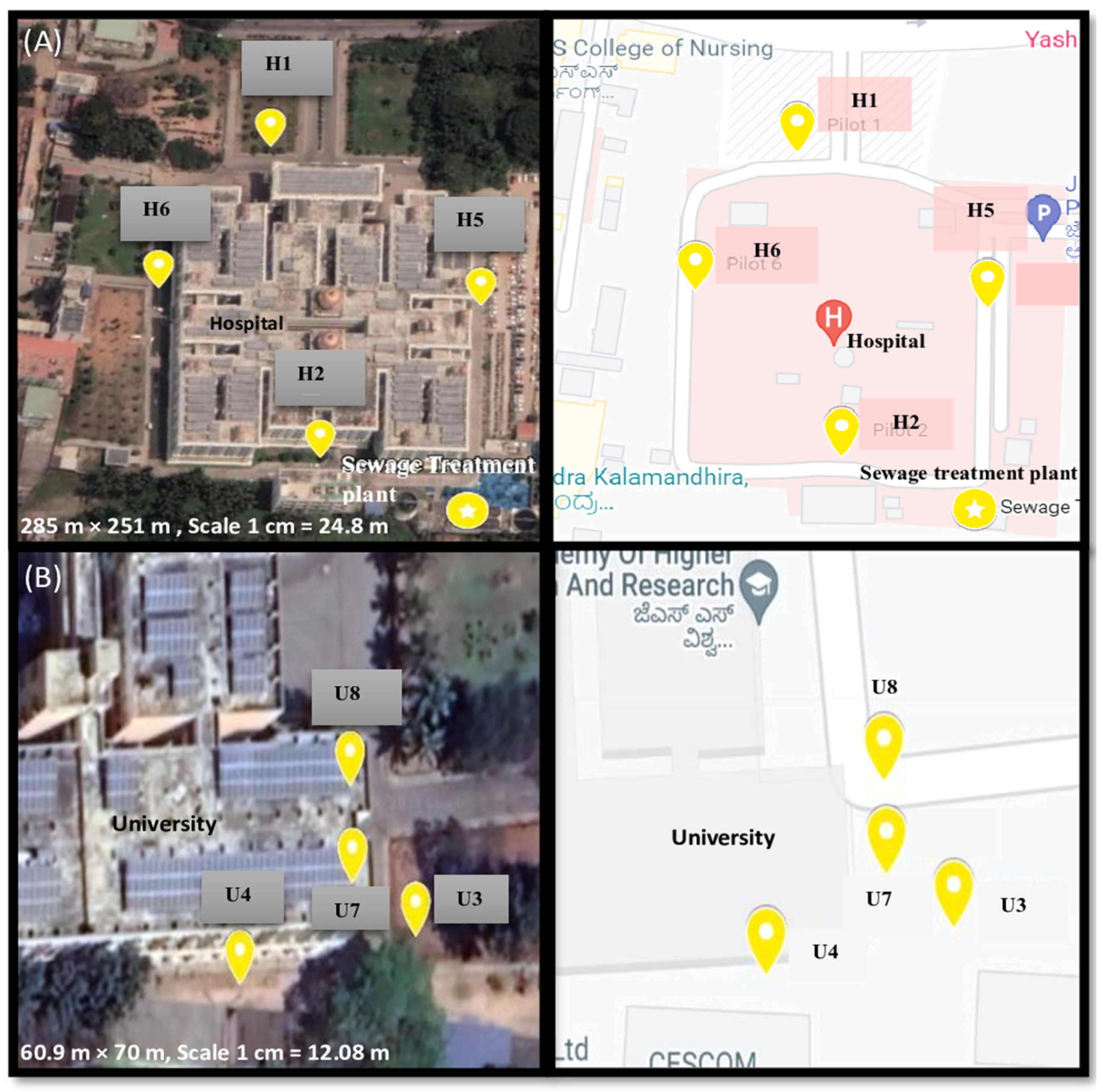
2.1. Sample Settings and Collection

Soil sampling sites for the study were the precincts of a major tertiary care (1800 bed) hospital established in 23 September 2013—Jagadguru Sri Shivarathreeshwara (JSS) Hospital and a University Medical College (JSS Academy of Higher Education and Research building)—within the metropolitan area (population 1.21 million) of Mysore, Karnataka, India. Permission for collection of soil samples from JSS Hospital and JSS University was obtained from the JSS Medical College and Institutional Ethical Committee on 31 October 2019 (JSSMC/IEC/3110/08NCT/ 2019–2020). Soil samples were collected over a 7-month period (30 July 2019 to 17 February 2020).
Figure 1 illustrates the eight sampling sites used in the study to assess the microbial diversity of the soil surrounding JSS Hospital versus JSS University, which were ~6 km apart. The soil samples were collected from the top 10 cm of the soil with a sterile spatula and glove (separate ones for every sampling site). The samples were collected from the gardens around both the precincts and the sampling locations (n = 4 from each precinct; hospital and university) were chosen with the following criteria in mind: building entrance and exit points, places with most human interactions and, for the hospital precinct, location of the nearby sewage treatment plant. In brief, hospital soil samples were collected from the gardens near the entrance (H1), the exit (H2), the lawns near the east gate (H5; closer to the parking area), and the west gate (H6). The university soil sampling sites focused on the gardens adjacent to a university building. They were collected from the east front garden (U3), the west side garden (U4), the north garden on the right of the entrance (U7) and the north garden on the left of the entrance (U8). Four sub-samples were obtained from each sampling spot. Briefly, a gram (1 g) of soil was collected along each of the 4 cardinal directions (north, south, west and east) from each sampling spot and placed into a sterile Nasco soil sampling bag. Next, the samples were pooled to become the representative soil sample from that site. Thus, a total of 4 g of soil was obtained from each sampling site, to establish each site. The same procedure was repeated for all soil sampling sites.

2.2. Genomic DNA Extraction and Sequencing

DNA was extracted directly from the soil samples collected at the eight locations. Genomic DNA extraction was carried out in triplicates using DNeasy® PowerSoil® Kits (Qiagen, Hilden, Germany, Cat No./ID: 12888-100) according to the instructions provided by the manufacturer. The genomic DNA extracted was stored at −80 °C until further use. The stored DNA samples were sequenced to analyse the microbial diversity and community composition in the soil. The samples were sequenced using the MiSeq Illumina Platform. The 16S rRNA PCR was performed to amplify the V1–V3 regions of the 16S rRNA gene. The concentration of extracted DNA was determined using Qubit 2.0 (Thermo Fisher Scientific, Waltham, MA, USA) and normalised to 5 ng/µL. PCR amplification was performed in two steps. A 25-cycle PCR was performed to amplify the V1–V3 regions of the 16S rRNA gene. The primers used were 27F(TCGTCGGCAGCGTCAGATGTGTATAAGAGACAGAGAGTTTGATCMTGGCTCAG) and 519R (GTCTCGTGGGCTCCCACATGTGTATAAGAGACAGGWATTACCGCGGCKGCTG) containing Illumina forward and reverse overhangs, respectively. The amplification was carried out using Platinum Taq Polymerase (Invitrogen, Waltham, MA, USA). The PCR products were cleaned according to the Illumina protocol, using AMPure XT beads (Beckman Coulter, Brea, CA, USA). The second PCR had 8 cycles and was performed using 5 µL of the PCR product from the first cycle with Illumina Nextera XT v2 primers (San Diego, CA, USA). This helped to index each sample according to the instructions provided by Illumina. The amplified DNA was cleaned using AMPure beads and quantified using QUBIT 3.0 (Thermo Fisher Scientific, Waltham, MA, USA). Each DNA library was normalised and pooled in equal quantities. Sequencing was performed on the Illumina MiSeq (Melbourne, Australia) using a MiSeq Reagent kit v3 (600 cycles) MS-102-3003.
The sequencing data was analysed using QIIME2 version 2021.11 (Flagstaff, AZ, USA). A quality check was performed and the good quality forward reads were retained for further processing. Reverse reads were removed due to low quality. To compare and identify the bacteria obtained in our study, the SILVA 138 database was used. The results were obtained as a qsv file and an OTU Excel file. Using QIIME2View, any bacteria with confidence levels <90% were removed. Further analysis was conducted using only bacteria with a confidence level >90%.
Statistical tests were performed using the R programming software (Version R4.2.1, R Foundation, Indianapolis, IN, USA). The different statistical tests, including Shannon Weiner diversity and Bray Curtis diversity, and ANOVA tests were used to identify the microbial diversity, compare them, and examine the similarities and differences between samples. Statistical significance was set at p < 0.05.

2.3. Identification and Antibiogram of Soil Isolates

For the culture-based approach, a gram (1 g) of soil from each of the 8 sampling sites was suspended in 10 mL normal saline (0.9% NaCl) and incubated at room temperature for 10 min. The spread plate technique was used to spread the different soil suspensions evenly over MacConkey agar plates for culture. MacConkey agar was used in this study, as this study focused on Gram-negative organisms for DNA transfer. The plates were incubated at 37 °C for 18–24 h and checked for any bacterial colony growth the following day. Additionally, the plates were incubated at 37 °C for 2 more days to identify any slow growing bacteria in the soil samples. Isolates obtained on MacConkey plates were further sub-cultured to obtain pure cultures. Our study primarily focused on Gram-negative bacteria—Pseudomonas spp., Acinetobacter spp., Klebsiella spp., and Escherichia coli. However, the other Enterobacteriaceae obtained were also identified during this study.
The diverse colonies obtained on the MacConkey agar plates were further sub-cultured to derive pure cultures. Next, to aid the identification of the microorganisms, Gram staining was performed as previously described [21] on each of the pure cultures obtained. Biochemical test reactions were also performed to aid the identification of the isolates. The following biochemical tests were used for the preliminary identification of organisms: Catalase (Slide) [22], Oxidase [23], Indole [24], Citrate utilization [25] and Triple Sugar Iron (TSI) test [26]. Molecular confirmation of the obtained isolates was made using Matrix Assisted Laser Desorption Ionization-Time of Flight (MALDI-TOF) mass spectrometry (VITEK MS V3.0) at the National Institute of Mental Health and Neurosciences, Bangalore, India using the database VITEK MS Knowledge Base V3.2.
Organisms identified and confirmed as Pseudomonas, Acinetobacter and Enterobacteriaceae family, namely, Klebsiella, E coli, Serratia marcescens and Enterobacter cloacae spp. cloacae were screened for Antibiotic Susceptibility Testing (AST). AST was performed using the Kirby-Bauyer disc diffusion method [27] and clinically-relevant antibiotics (Table 1), and an Automated VITEK 2 instrument (BioMerieux, St. Louis, MO, USA).
Quality control was performed on all the antibiotic discs used in the study. In brief, the disc diffusion method was conducted with an 18–24 h culture of each isolate of interest prepared by inoculating the required organism in Brain Heart Infusion Broth. The overnight cultures were then matched to a 0.5 McFarland standard. Lawn cultures were created by dipping sterile cotton swabs into the 0.5 McFarland tube and streaking them over the entire surface of the Muller Hinton Agar plates. Antibiotic discs described in Table 1 were placed on the inoculated Muller-Hinton Agar plates under aseptic conditions and incubated at 37 °C for 24 h to allow the bacteria to grow overnight. Areas of clear media surrounding the discs were measured to determine which antibiotics inhibited bacterial growth [27]. Results were reported as Susceptible (S), Intermediate (I) or Resistant (R), according to the Standard Clinical and Laboratory Standards Institute (CLSI) Guidelines, 2019.
The Automated VITEK 2 was performed in the following manner; 3.0 mL of sterile saline (0.45% to 0.5% NaCl, pH 4.5 to 7.0) was transferred aseptically into a clear plastic (polystyrene) test tube (12 mm × 75 mm). Using a sterile stick, morphologically similar colonies were transferred to the saline tube. A homogenous organism suspension with a density equivalent to 0.5–0.63 McFarland standard (for Gram-negative organisms) was prepared using the VITEK 2 DensiCHEK Plus (Biomerieux, Boston, MA, USA). In a second tube containing 3.0 mL of saline, 145µL of the above suspension was transferred for Antibiotic Susceptibility Testing. This tube was then placed in the cassette with a susceptibility card. The tube with the initial bacterial suspension could be used for the inoculation of an identification card (however, we did not perform identification using this method). In the case of the Automated VITEK 2, quality checks were performed on one card from every batch. For the Enterobacteriaceae family, an Escherichia coli ATCC 25922 standard was used, whereas for Pseudomonas and Acinetobacter, a Pseudomonas ATCC 27853 was used. AST was performed using AST-N280 (Enterobacteriaceae) and AST-N281 (Pseudomonas and Acinetobacter) cards in the Automated VITEK 2.

3. Results

3.1. Genomic DNA Extraction and Sequencing

The genomic DNA was extracted from each sampling site (in triplicates) and quantification revealed DNA yields between 6.0–47.7 ng/µL. A total of 54,522 reads were obtained from all the soil samples and the good quality forward reads were used further. The SILVA 138 database compared and identified the bacteria obtained in our study. The results were obtained as a qzv file, with 4557 identified bacteria with confidence levels ranging between 46 to 100%, and an OTU Excel file. Further analysis was conducted using only bacteria with a confidence level >90%.
The data containing all the DNA sequences have been deposited in the NCBI database with the following Accession number (Accession number GSE225419).

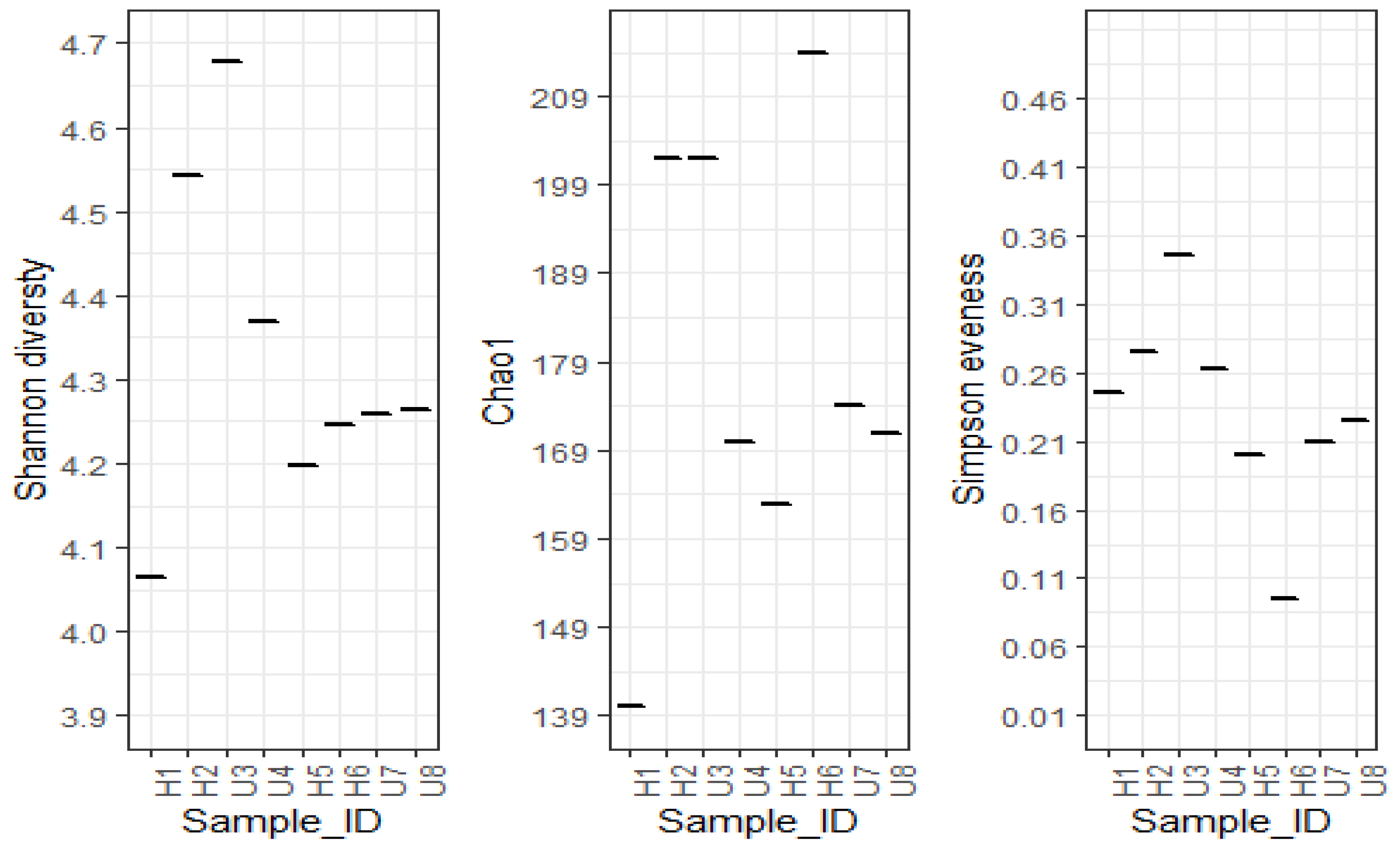
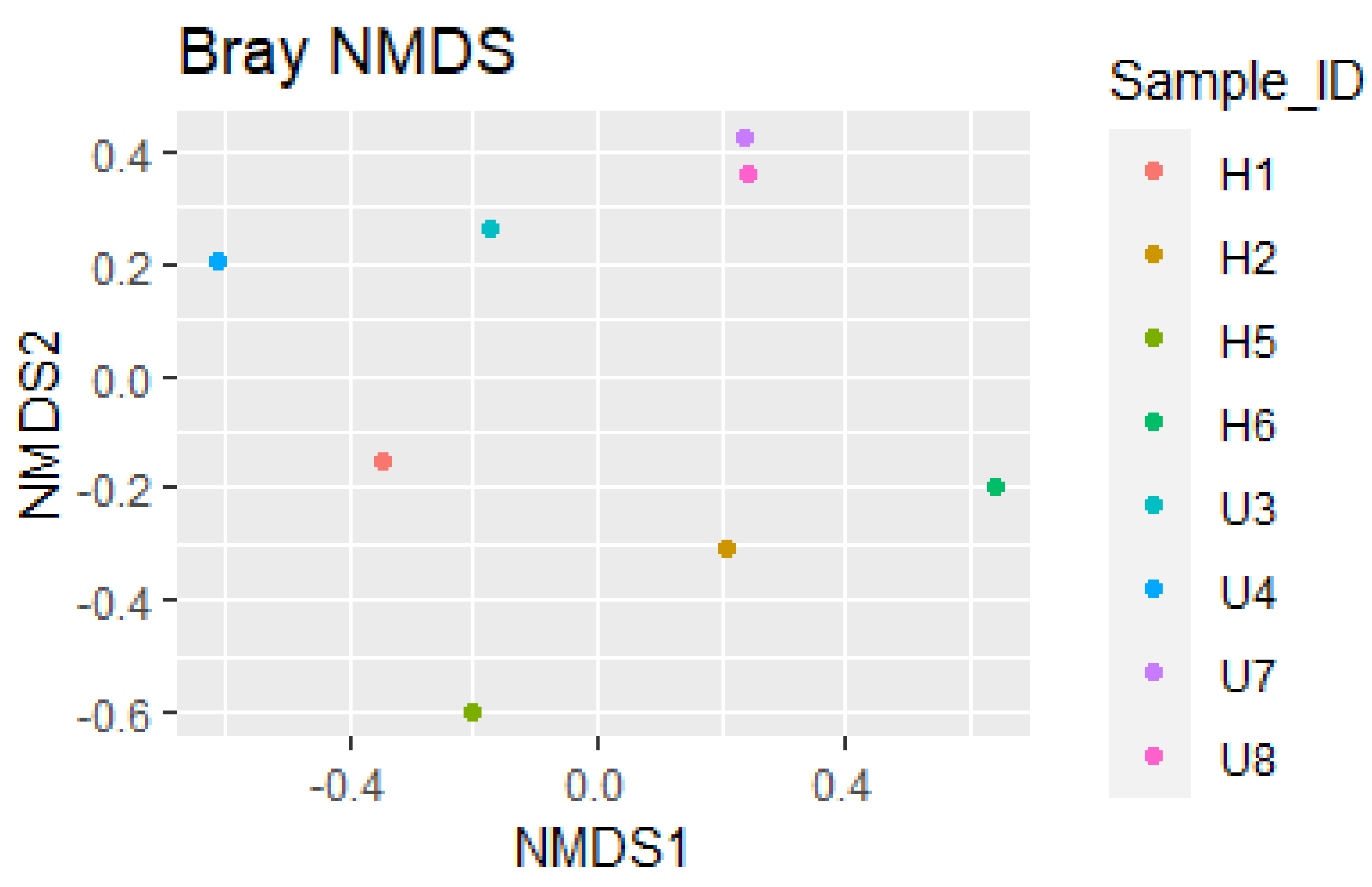
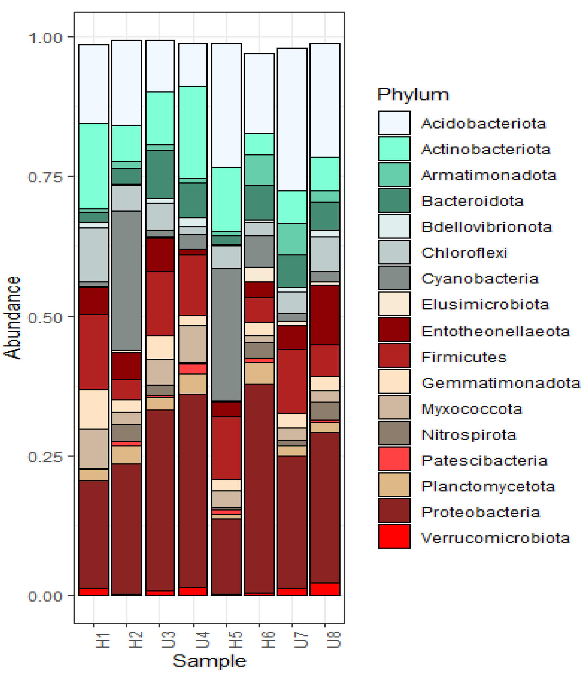
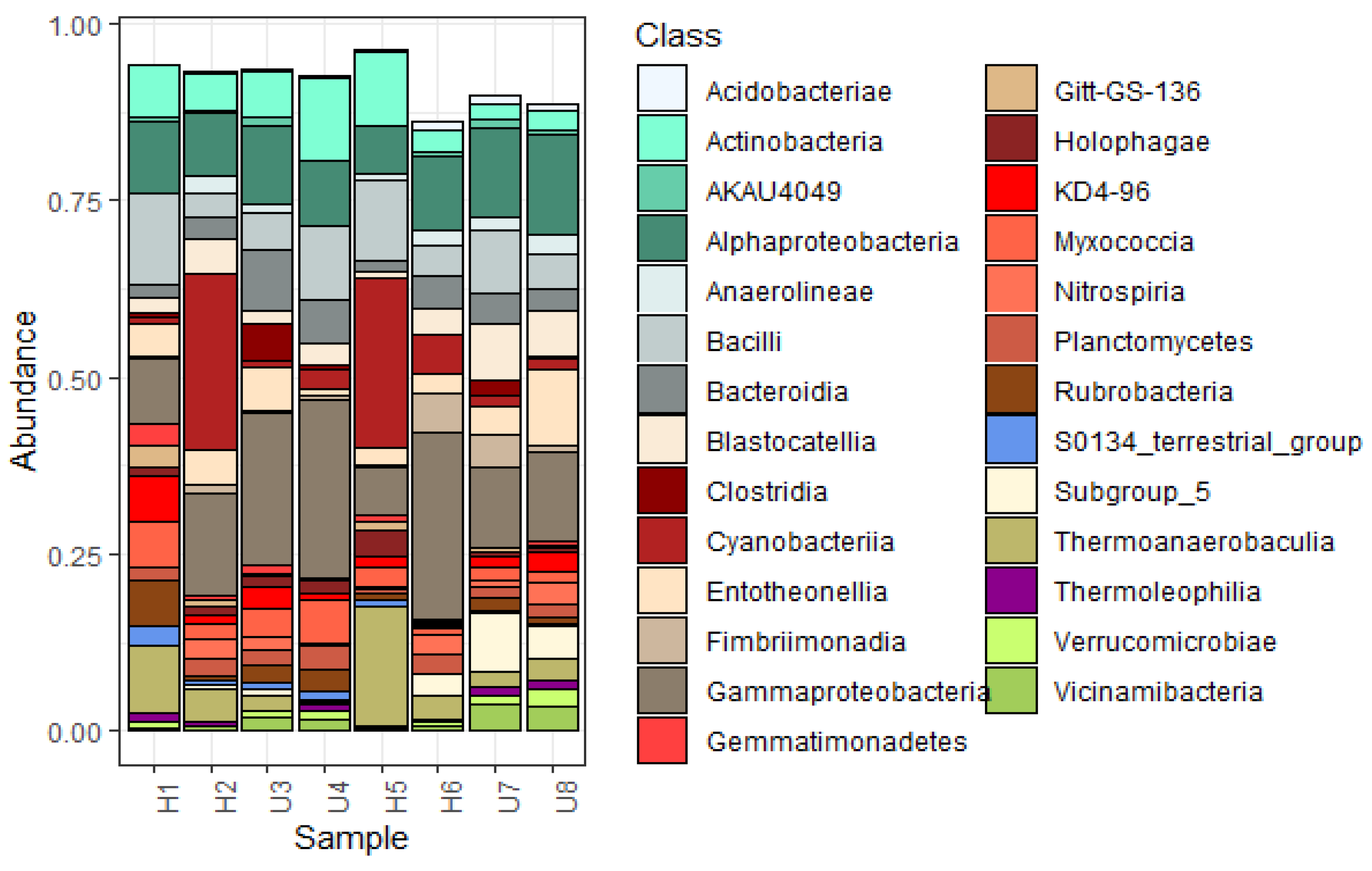
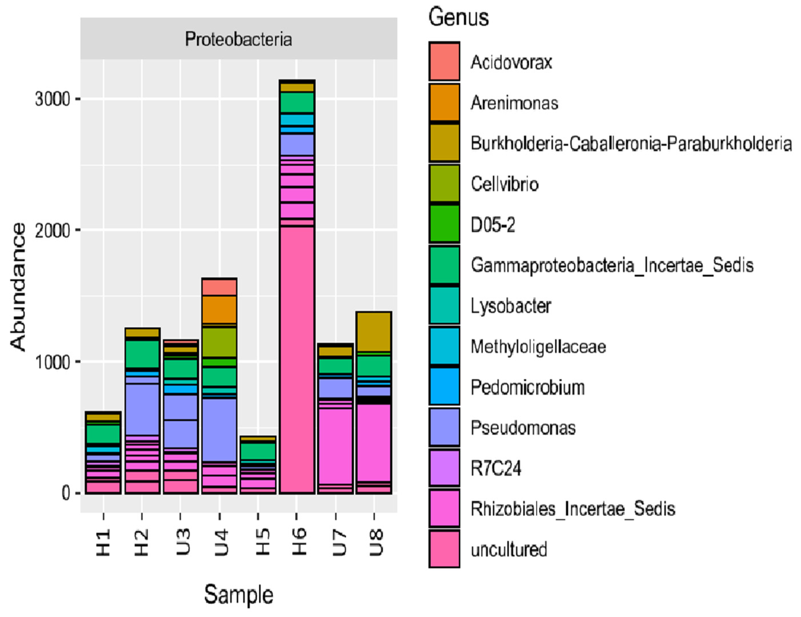
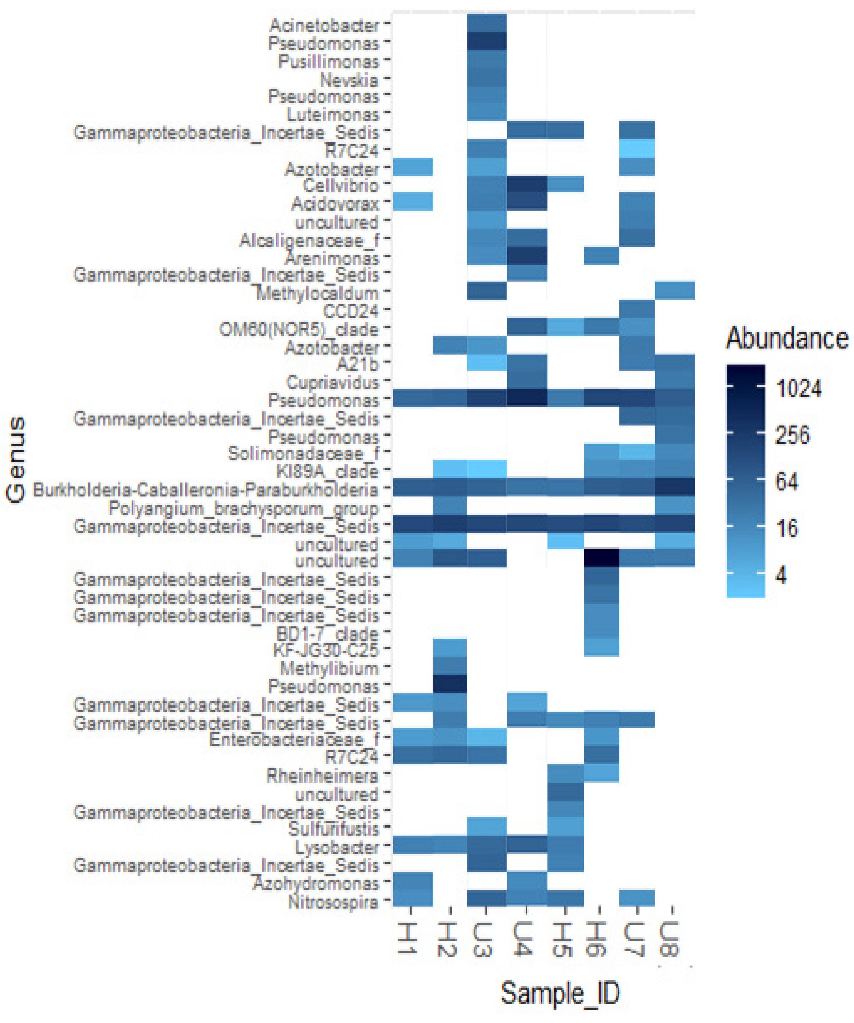
3.2. Analysis of Microbial Diversity and Community Composition

The Shannon index for all the samples ranged 4.0–4.69 and the Simpson’s index ranged 0.95–0.99, indicating large diversity in each of the eight samples collected (Figure 2 and Figure 3). Compared to all the samples used in this study, soil from the University U3 site was the most diverse. It had a Shannon index of 4.68 and Simpson’s index of 0.99 (Figure 2). Amongst all samples, hospital H1 site soil bacteria had the lowest Shannon index value (4.07). Bacteria from the hospital H6 sampling site had the lowest Simpson’s values amongst all samples, 0.95, although that is still diverse.
Bacterial soil communities from the H2, and U3 and U4 sites had Shannon diversity values of 4.54, 4.68 and 4.37, respectively, being more diverse than other samples (Figure 3). Bacterial soil communities from the H5 and H6, and U7 and U8 had very close values to each other, being 4.19, 4.24, 4.25 and 4.26, respectively (Figure 3). Thus, bacterial soil communities from these sites were less diverse than those from the H2, H3 and U4 study sites. Simpson’s evenness was the highest in U3 and the lowest in H5 (Figure 3). The overall bacterial diversity observed, in decreasing order of diversity were U3 > H2 > U4 > H6 > U7 > U8 > H5 > H1. This shows that the university and hospital sampling sites were similarly diverse. ANOVA tests were performed to analyse any significant variations in the sampling sites from the different locations (hospital and university). The results revealed that there were no statistically significant differences in the bacterial diversity among the sampling sites compared to the hospital and university buildings. The following results were obtained Shannon diversity, p = 0.39; Chao 1, p = 0.98; Simpson’s p = 0.24.
Beta diversity (a measure of the similarity or dissimilarity between two bacterial communities) was measured using the Bray Curtis dissimilarity calculation and was plotted using Non-Metric Multidimensional Scaling (NMDS). As illustrated in Figure 4, the locations of hospital soil from the H1, H2, H5 and H6 sites lacked any evident clustering patterns, indicating the overall diversities of the samples were different from each other. However, soil from the university U7 and U8 locations show some clustering, indicating the samples share similarity when compared to each other, but were not exactly the same. Hence, all the soil samples collected from the hospital and university were diverse from each other.
Table 2 summarizes the assessment of bacterial diversity between all sample sites. Only the H6 and H1 sites had a significantly different bacterial diversity (p = 0.027).
The relative abundance of different bacterial communities at the phylum level was depicted using abundance charts. A total of 32 phyla were identified in the 8 soil samples, and of these, 17 phyla were statistically diverse (p < 0.005) (Figure 5). The relative abundance of different phyla in each soil sampling spot was identified and compared using the abundance charts. Figure 5 shows that the most abundant phylum in all the samples was Proteobacteria (2.11). Next, in order of abundance were Acidobacteriota (1.29), Actinobacteriota (0.74), Firmicutes (0.72), Cyanobacteria (0.62), Bacteroidota (0.38), Chloroflexi (0.37), Entotheonellaeota (0.36), Myxococcota (0.29), Gemmatimonadata (0.24), Planctomycetota (0.18), Armatimonadata (0.17), Nitrospirota (0.12), Verrucomicrobiota (0.08), Bdellovibrionota (0.06), Patescibacteria (0.05), and Elusimicrobiota (0.05). Proteobacteria were most abundant in the H6 sampling site followed by two of the university sites (U4 and U3), and was lowest in the H5 sampling site. Nevertheless, a similar proportion of Proteobacteria were present in most samples.
A total of 86 different classes of bacteria were identified in this study, of which 27 classes were statistically diverse (p < 0.005), Figure 6. The five most abundant classes were Gammaproteobacteria (1.28), Alphaproteobacteria (0.83), Firmicutes (0.69) (Bacilli and Clostridia), Cyanobacteria (0.62), and Actinobacteria (0.49). The top 20 genera in phylum Proteobacteria identified in the samples are provided in Figure 7. It can be clearly observed that uncultured bacteria dominated the soil collected from H6, and this was a sampling site where considerable human interactions took place in the hospital precinct. Although uncultured bacteria were present in small proportions in other samples, Pseudomonas dominated in soil from the H2 (hospital) site and the U3, U4 and U7 (university) sites.
The Figure 8 heatmap shows that Pseudomonas, Burkholderia-Caballeronia-Paraburkholderia and Gammaproteobacteria Incertea Sedis were present in all the soil samples collected. The University Pilot 3 soil sample had the most diverse bacterial species when compared to the other samples. Clinically-relevant bacteria belonging to the Enterobacteriaceae family were present in soil from the H1 and H2 sampling sites (hospital), the U3 site (university) and the H6 site (hospital). It was noted that Pseudomonas was the most abundant organism and was present in all the samples collected.

3.3. Identification and Antibiotic Sensitivity Testing of Soil Isolates

A total of 22 organisms were selectively obtained on MacConkey Agar (differential media). Colony morphology varied from small circular, translucent, mucoid shiny colonies to flat smooth colonies. Lactose fermenting (pink) and non-lactose fermenting (pale coloured) colonies were obtained on MacConkey agar. These isolates were further sub-cultured and Gram-stained, followed by biochemical tests to aid identification, including the catalase, oxidase, indole, citrate and triple sugar iron tests (Table 3). As certain Gram-positive cocci such as Staphylococcus and Enterococcus have the ability to grow on MacConkey medium, Gram staining was performed to select the Gram-negative organisms and exclude Gram-positive cocci.
The cultured isolates were confirmed by MALDI-TOF analysis (Table 4). Among the Gram-negative organisms isolated in pure culture, Pseudomonas species accounted for 32%, Acinetobacter baumannii for 18%, Klebsiella pneumoniae (9%), Aeromonas salmonicida (9%), Enterobacter cloacae ssp. cloacae (5%), Alcaligenes faecalis spp. faecalis (5%), Escherichia coli (4%), Serratia marscesens (4%) and unidentified (14%). The location of isolation (sampling sites) and the organisms confirmed by MALDI-TOF analysis are provided in Table 4. Gram-negative human pathogens such as Pseudomonas aeruginosa, Klebsiella pneumoniae, Acinetobacter baumannii, Enterobacter cloacae ssp. cloacae and Aeromonas salmonicida were obtained from both the hospital and university sampling sites, while Escherichia coli and Serratia marcescens were obtained only from the university sampling site.
A total of 15 Gram-negative organisms were selected for the determination of their antibiotic resistance profiles. Antibiotic sensitivity testing (AST) was performed on 15 selected isolates of Pseudomonas, Acinetobacter (Table A1) and members of the Enterobacteriaceae family (Table A2) by the disc diffusion method and the Automated VITEK 2 (Table A3 and Table A4). Among these, included Pseudomonas aeruginosa (n = 2), Pseudomonas fluorescens (n = 1), Pseudomonas species (n = 3), Acinetobacter baumannii (n = 4), Klebsiella pneumoniae (n = 2), Escherichia coli (n = 1), Serratia marcescens (n = 1) and Enterobacter cloacae spp. cloacae (n = 1). The soil isolates obtained in this study by culture were mostly sensitive to all the antibiotics. Among the six isolates of the Pseudomonas species, one isolate was resistant to Aztreonam while three species of Pseudomonas exhibited an intermediate sensitivity (Pseudomonas aeruginosa, Pseudomonas fluorescens and Pseudomonas species) (Table A3). All the Pseudomonas isolates were intrinsically resistant to tigecycline. Among the six Pseudomonas species, one isolate of Pseudomonas (M4.2) was terminated in the Automated VITEK 2 (Table A3). This was a mucoid and very slow growing Pseudomonas strain. The disc diffusion test was conducted for this strain and the results from disc diffusion were considered for this isolate. All four Acinetobacter baumannii isolates obtained in our study exhibited intrinsic resistance to Aztreonam (Table A3). Among the Enterobacteriaceae family, 2 strains of Klebsiella pneumoniae were found to be resistant to Amoxyclav (Table A4). Overall, results of the antimicrobial assay of Pseudomonas and Acinetobacter, and Enterobacteriaceae are summarized in Table 5 and Table 6, respectively.

4. Discussion

Hospital precincts accommodate a number of individuals, including hospital staff, visitors, as well as immunocompetent and immunocompromised patients, and provides a favourable atmosphere for human interactions. Antibiotics are mainly prescribed in hospital settings and, hence, these environments serve as a hotspot of antimicrobial resistance [28]. This is the first study analysing the role of hospitals in bacterial diversity and community composition of the soil and the antimicrobial resistance profiles of the soil isolates around a hospital and university precinct. Analysis of soil samples collected from a southern Indian hospital and a university precinct, separated by ~6 km, identified that both precincts had soil exhibiting very diverse microbial communities.
Alpha diversity (indicating bacterial species diversity within the soil of each sampling site) was assessed using different indices, including Observed, Chao 1, Abundance-based Coverage Estimators (ACE), Shannon, Simpson’s, Inverse Simpson’s and Fischer. The Shannon index measured the species richness and evenness, and the Simpson’s index explains the relative abundance of different Operational Taxonomic Units (out’s). The observed species metric highlights only the uniquely identifioutOTU’s in the samples. Among all the samples, U3 was the most diverse (Shannon diversity, 4.68). This university sampling site was in a garden bed, where the soil would have experienced little disturbance. H2 had the second highest diversity in the hospital, and was the sampling site closest to the sewage treatment plant. Additionally, it was a little-disturbed area of the hospital premises (having less movement of people), and laboratories were close by, as was a sewage treatment plant (~77 m away). Our study revealed that the hospital and university samples were similarly diverse. Although they were diverse, the results were not statistically significant, which might be due to a smaller sampling size. Among the 32 phyla identified, 17 were statistically diverse and Proteobacteria was the most abundant, with the highest abundance in hospital H6 collected from the west gate. This site also contained a high proportion of unidentified bacteria. This is possibly because the west gate was closer to the emergency department of the hospital and had heavy patient and visitor traffic, providing increased opportunity for human interactions. Prolonged time spent by patients or visitors in the gardens could also be contributing to the exchange of bacteria between humans and soil. Here, people consume food, with likely variable hand hygiene practices, and this could contribute to the exchange of healthy and unhealthy bacteria between humans and soil. This can result in unhealthy or antibiotic resistant bacteria ending up in the soil and further possibilities of tracking these into the hospitals or having them taken up by other immunocompromised people. Gammaproteobacteria was the most abundant class in the study, with Pseudomonas being the most abundant organism identified and present in all the samples. Considering the relative abundance of different phyla and classes, it was observed that Sample H5 was different from the other samples. This could be due to different factors such as water availability, organic manure, differences in the amount of certain nutrients such as nitrogen and phosphorus present in the area, or due to an effect of pollutants present in the soil. Clinically-relevant bacteria belonging to the Enterobacteriaceae family were present in the soil from H1, H2 and H6 (hospital) and U3 (university). Their presence in the hospital precinct raises concern as these bacteria could have moved from the hospital to the soil via humans, or could pose a threat of moving from the soil to hospitals to infect humans. The study also isolated 22 Gram-negative organisms, with AST performed on 15 isolates. Of significance, one isolate of Pseudomonas (university isolate) was found to be resistant to Aztreonam, all four Acinetobacter baumannii isolates exhibited intrinsic resistance to Aztreonam and two strains of Klebsiella pneumoniae were resistant to Amoxyclav (one each from the hospital and university). The identified isolates are clinically-relevant bacteria capable of causing infection in humans and, hence, the low-level resistance developed by soil bacteria could be alarming.
We evaluated the different phyla present in the 8 samples and observed that the most notable difference was that Cyanobacteria were abundant in the H2 and H5 samples. A sewage treatment plant was located in reasonably close proximity to the samples (~77 m from H2, ~115 m from H5). In comparison, the other two hospital sampling sites (H1 and H6) did not even have 50% of the Cyanobacteria. This may be due to further distance from the sewage plant (~225 m and ~195 m, respectively). Proteobacteria was the most abundant in the H6 sampling site, which was the area mostly associated with the contact between many patients, staff and visitors. The H1 sampling site was the entrance area of the hospital that is commonly used by the public. Although people used the entrance to also exit the hospital, bacterial diversity was larger in H2 (exit gate) soil compared with H1 (entrance) soil. This finding may be highlighting the effect of human interactions on microbial diversity. However, the nearby presence of the sewage plant could be another possible reason accounting for the microbial dominance of H2 over H1.

4.1. Bacterial Diversity and Community Composition around a South Indian Hospital and University

Several studies have examined the microbial diversity in environments such as soil [29,30], water [31,32] and clinical settings [33,34]. In addition, many studies have investigated the soil microbiome (e.g., [35,36]) and human microbiome (e.g., [37,38]). Although these studies were performed in the soil, different environments were compared. For example, the soil microbiota and its resistome between a conventional and organic farming system [29], the waste soil and sediment microbiome from three rivers and one dumping site polluted by solid and liquid wastes from domestic, hospital and municipal premises [30], etc. However, to our knowledge, this is the first study exploring the microbial diversity in the soil around a hospital setting. The broad similarity of bacterial communities detected in the soil at both precincts could be explained by the fact that the hospital and university were situated only 6 km from each other and experienced the same climatic conditions. However, the differences in the relative abundances of different bacteria between the sampling sites may be attributable to different anthropogenic activities in the study environments. For example, there may have been an influence of the sewage treatment plant and laboratories at the hospital precinct (H2, H5). One of the sampling sites was also near a canteen in the hospital grounds (H5). The hospital entrance site (H1) experiences large numbers of people entering and exiting the hospital daily; many human-human as well as human-microbe interactions would be expected to occur at this site. One sampling site (H6) was close to the emergency department where many patients and visitors are either waiting or relaxing outside in the gardens.
Previously, the order of the relative abundances of the certain important bacteria in the soil was established: Proteobacteria (36.5%), Acidobacteria (30.9%), Actinobacteria (13.0%), Bacteroidetes (11.2%), Firmicutes (2.9%), Cyanobacteria (0.0%) and Verrucomicrobia (0.0%) [39]. Our findings were in general agreement with the top three dominant phyla—Proteobacteria, Acidobacteria and Actinobacteria in the soil sampling sites. However, after the top three, Firmicutes and Cyanobacteria dominated the soil, which differs from other studies [29,30,40]. Previous studies investigating the microbial diversity in soil and sediment samples associated with hospital waste [30], landfill leachate soil [40] and organic and conventional farming soils [29] identified Proteobacteria to be dominant in the soil. Proteobacteria comprises several known human pathogens and has been identified as a possible microbial marker of disease (e.g., a microbial signature of dysbiosis) in the human gastrointestinal tract [39,41]. Excessive growth of Proteobacteria has been associated with inflammatory bowel disease [42] and metabolic syndrome [39]. Among the four main phyla in the gastrointestinal microbiota, Proteobacteria is considered to be the most unstable [43]. The inability to resist the invasion and colonization by the microbial communities leads to the failure to maintain the low levels of commensal Proteobacteria. This favors the growth of exogenous pathogens [39]. Phyla such as Proteobacteria, Actinobacteria and Bacteroidetes are also known to have the ability to grow on antibiotics [44]. Our study observed Gammaproteobacteria and Alphaproteobacteria to be the most dominant classes of bacteria in our study sites. Other studies have also reported Gammaproteobacteria and Alphaproteobacteria to be dominant in aquatic environments such as the bacterial community of Arabian sea water [45], in low-oxygen zones of ocean water [46], and seasonally hypoxic estuaries [46].
Firmicutes are involved in anaerobic processes and help in methanogenic decomposition. Hence, these phyla are predominant in waste dumping sites [40]. Firmicutes and Actinobacteria have also been reported in high amounts in soils containing high amounts of compost [47]. Many different Bacillus species have been reported to be good de-nitrifiers [48].
A concerning and surprising finding was the abundance of Cyanobacteria in the soil sampling sites closest to the hospital sewage treatment plant. Cyanobacteria produce three toxins, namely, neurotoxins, hepatotoxins and lipopolysaccharide endotoxins, with reported gastrointestinal, respiratory and allergic reactions [49,50]. Acidobacteriota was the next dominant phyla identified in our study. They are known to help in the modulation of biogeochemical cycles and plant growth [51]. The presence of plants in the soil sampling site and surrounding area possibly points out their role in the study sampling sites.

4.2. Antibiotic Resistance Profile in Clinically Relevant Soil Bacteria

Soil is an ideal reservoir of distinct antibiotic resistance determinants [6,7,8,9]. Human- and animal-associated bacteria are introduced into the environment through different waste streams [28,52]. Human exposure to such sites facilitates the entry of these bacteria into the human body where they interact with the human microbiome, and under selective pressure may take up mobile genetic elements [52]. Environmental Gram-negative bacteria may act as reservoirs for emerging antibiotic resistance genes disseminating in human pathogens [53]. Pseudomonas and Acinetobacter species are well known for the acquisition of antibiotic resistance genes directly from the soil [54]. The concomitant presence of commensal bacteria and pathogens in clinical environments where antibiotic usage is high has resulted in bacteria developing resistance to most of the antibiotics developed [18]. A group of pathogens referred to as the ESKAPE pathogens—Enterococcus faecium, Staphylococcus aureus, Klebsiella pneumoniae, Acinetobacter baumannii, Pseudomonas aeruginosa and Enterobacter species—are responsible for nosocomial infections, and are capable of escaping the lethal action of antibiotics [55]. They acquire virulence and resistance determinants and develop resistant to multiple drugs [55].
This study mainly focused on clinically-relevant Gram-negative organisms such as Pseudomonas spp., Acinetobacter spp., Klebsiella spp. and Escherichia coli. To identify if the environmental isolates around a hospital setting contributed to the burden of antimicrobial resistance, our study analysed the antibiogram of the isolates from the soil around a hospital setting compared with a university precinct situated ~6 km away. Normally, when clinical isolates are examined from hospital settings such as an intensive care unit (ICU), they exhibit resistance to commonly used antibiotics. To this point, Mahendra, et al. [56] described the commonly prescribed antibiotics (penicillin group of antibiotics, lincosamide, cephalosporins, carbapenems, macrolides, polymyxin and quinolones), organisms and their resistance pattern obtained in a hospital setting in Mysuru, with special emphasis on respiratory infections. This study showed that resistance was exhibited against the most commonly used antibiotics, with the highest resistance to Piperacillin-tazobactam (60%) prescribed in the respiratory ICU setting [56]. Yet another study compared the antibiotic resistance genes in the soil between different land-use types in Great Britain. Significantly higher levels of antibiotic resistant bacteria were detected in agricultural land, further highlighting how geographical location and management practices followed in a particular area also influenced the presence of resistant bacteria [57]. Relative bacterial compositions and antibiotic resistance in soil and water studied at different sampling environments, such as a dairy water canal, a residential garden soil and a lake by hospitals displayed resistance [58]. Similarly, the influence of human beings in a hospital area witnessing the constant use of antibiotics needs to be monitored. Strict management practices need to be in place to avoid improper waste disposal or leaks in the sewage system. These results highlight the effect of antibiotic usage and the emergence of antibiotic resistance in hospital settings due to the overuse of antibiotics [59,60,61]. However, as demonstrated in our study, isolates from the hospital soil sampling sites were minimally resistant to antibiotics. The minimal resistance could possibly be due to the limited sampling size in this study. The Aztreonam resistance exhibited by Pseudomonas species could be suggestive of a slow uptake of resistance from the environment. Another notable resistance observed in our study was by Klebsiella pneumoniae against amoxyclav. K. pneumoniae accounts for human nosocomial infections and causes a range of infections such as pneumonia, urinary tract infections, skin and soft tissue infections, meningitis, septicemias and pyogenic infections of almost all tissues of the body [62]. High antibiotic usage in hospitals, and the human interactions (e.g., between patients, by-standers, visitors) around the hospital settings could be a reason for this resistance. It is speculated that anthropogenic activities contribute to the dissemination of antibiotic resistance between hospital and environmental pathogens. Moreover, there is evidence of the exchange of antibiotic resistant genes between environmental and clinical bacteria [63].
Several studies have reported the presence of antibiotic resistant bacteria in the hospital effluents or discharges released into the surrounding environment [64,65,66]. These studies shed light on the risks of antibiotic usage in hospitals reaching several other surrounding environments. The presence of highly resistant bacteria in such environments could be due to the over-use of antibiotics in hospital settings [67,68]. The transfer of antibiotic resistance genes from such environmental hotspots are a matter of concern. The presence of immunocompromised and immunocompetent patients in hospital surroundings enhances the chances of the uptake of these resistant bacteria or resistance determinants from the surroundings. It is speculated that anthropogenic activities also play a role in the mobilization of antibiotic resistant genes to pathogens [52]. There is also evidence of the transfer of resistance genes via food [69]. Patients and visitors of the hospital often consume food and spend time in/nearby these hospital gardens. Soils from these gardening sites were sampled and analysed in this study. Other factors such as poor hygiene and improper sanitary practices could also result in infection from environmental pathogens such as Pseudomonas aeruginosa or Acinetobacter baumannii [70], considered to be rampant opportunistic pathogens from the soil [71,72].
Our study identified Proteobacteria to be the most dominating phylum from the hospital environment soil. Proteobacterial species are likely the origin of antibiotic resistance genes [52]. Certain environmentally distributed super resistant microbes are capable of subsisting on antibiotics. The possibility of the exchange of these resistance determinants from these super resistant microbes to clinical pathogens should not be underestimated [44]. It is important to identify the taxa from which these resistance genes mobilized prior to the transfer to pathogens [52]. Antibiotic selective pressure on the taxa frequently associated with human or domesticated animal infections led to the emergence of these resistance genes clinically [52]. The origins of most antibiotic resistance genes are unknown, which possibly hints at their origin being environmental, because a majority of environmental species (including from soil) are not yet sequenced [73]. An overwhelming majority of unidentified organisms in our study also support this explanation.

5. Conclusions

This pilot study analysed the bacterial population and antibiogram of clinically-relevant Gram-negative microbes around a tertiary care hospital and university in southern India. Proteobacteria were the most abundant phyla, followed by Acidobacteriota, Actinobacteriota, Firmicutes, Cyanobacteria and Bacteriodota. The predominant classes of bacteria were Gammaproteobacteria, Alphaproteobacteria, Firmicutes, Cyanobacteria and Actinobacteria. The resistance observed in the soil isolates emphasizes the need to be more vigilant of the dissemination of antibiotic resistance from the soil to clinical settings. The majority of infections in humans are caused by Proteobacteria, and the abundance of Proteobacteria and Cyanobacteria nearer to the hospital premises comprising more immunocompromised and immunocompetent individuals is concerning compared to the university premises comprising healthier individuals. The presence of abundant Cyanobacteria in close proximity to the hospital sewage treatment plant also points to the role of anthropogenic activities on the bacterial diversity around hospital settings and to the importance of evaluating sensitive sites around a hospital. As this study was done with a limited number of samples, performing the study with more sampling sites would provide a deeper understanding of the effect of hospitals on soil microbes and the emergence of resistance.

Author Contributions

Conceptualization, S.K. (Shalini Kunhikannan), S.P., A.E.F., C.J.T., S.M.N., S.K. (Sumana Kumar) and A.E.S.; methodology, S.K. (Shalini Kunhikannan), C.R.S., J.R., S.P., A.E.F., C.J.T., S.M.N., S.K. (Sumana Kumar) and A.E.S.; software, S.K. (Shalini Kunhikannan), S.P., A.E.F. and A.E.S.; validation, S.P., A.E.F., C.J.T., S.M.N.., S.K. (Sumana Kumar) and A.E.S.; formal analysis, S.K. (Shalini Kunhikannan), S.P., A.E.F., C.J.T., S.M.N., S.K. (Sumana Kumar) and A.E.S.; investigation, S.K. (Shalini Kunhikannan), C.R.S. and J.R.; resources, S.P., A.E.F., C.J.T., S.M.N., S.K. (Sumana Kumar) and A.E.S.; data curation, S.K. (Shalini Kunhikannan) and A.E.S.; writing-original draft preparation, S.K. (Shalini Kunhikannan); writing—review and editing, C.R.S., J.R., S.P., A.E.F., C.J.T., S.M.N., S.K. (Sumana Kumar) and A.E.S.; visualization, S.K. (Shalini Kunhikannan), S.P., A.E.F., C.J.T., S.M.N., S.K. (Sumana Kumar) and A.E.S.; supervision, S.P., A.E.F., C.J.T.,S.M.N., S.K. (Sumana Kumar) and A.E.S.; project administration, S.P. and A.E.F.; funding acquisition, S.P. and A.E.F. All authors have read and agreed to the published version of the manuscript.

Funding

No outside funding was obtained for this study. This research was funded by La Trobe University (LTU), Bundoora, Victoria, Australia and the Jagadguru Sri Shivarathreeshwara (JSS) Academy of Higher Education and Research, Mysuru, Karnataka, India. S.K. (Shalini Kunhikannan) is a recipient of an LTU-JSS PhD Scholarship.

Institutional Review Board Statement

The study was conducted with permission from the JSS Medical College and approval was obtained from the Institutional Ethical Committee of JSS Academy of Higher Education and Research, Mysuru, Karnataka, India (JSSMC/IEC/3110/08NCT/2019-2020, accessed on 31 October 2019).

Informed Consent Statement

Not applicable.

Data Availability Statement

Data regarding nucleotide sequences are deposited in the NCBI database. Data that support the findings of this study are available from the corresponding author upon reasonable request.

Acknowledgments

We acknowledge the National Institute of Mental Health and Neuro Sciences (NIMHANS), Bangalore, Karnataka, India for their technical support for MALDI-TOF analysis of the soil bacterial isolates.

Conflicts of Interest

The authors declare no conflict of interest.

Appendix A

Table A1. The antibiotic sensitivity pattern exhibited by all the Pseudomonas species and Acinetobacter baumannii by the Kirby-Bauer disc diffusion method.
Sample IDOrganism IdentifiedLE5AT30CAZ30CL10COT25CPM30GEN10IMP10MRP10AK30MI30PIT100/10TGC
M2.1A. baumanniiS*S SSSSSSSSS
M3.4A. baumanniiS*S SSSSSSSSS
M4.4A. baumanniiS*S SSSSSSSSS
M7.3A. baumanniiS*S SSSSSSSS
M2.2Pseudomonas aeruginosaSISSSSSSSSSS*
M3.1P. aeruginosaSSSSRISSSSSS*
M3.2Pseudomonas spp.SSSS SSSSSSS*
M4.2Pseudomonas spp.SRSS RRRRSSS*
M4.3Pseudomonas spp.SRSSRSSSSS S*
M5.1Pseudomonas spp.SISS SSSSSSS*
M7.2P. fluorescensSISS SSSSS S*
S: Sensitive, R: Resistance, * Intrinsic resistance; LE, Levofloxacin; AT, Aztreonam; CAZ, Ceftazidime; CL, Colistin; COT, Cotrimoxazole; CPM, Cefepime; GEN, Gentamycin; IMP, Imipenem; MRP, Meropenem; AK, Amikacin; MI, Minocycline; PIT, Piperacillin/tazobactam; TGC, Tigecycline.
Table A2. The antibiotic sensitivity pattern exhibited by all the species belonging to the Enterobacteriaceae family by the Kirby-Bauer disc diffusion method.
Sample IDOrganism IdentifiedCL10COT25CPM30GEN10IMP10MRP10AK30MI30PIT100/10C30CTR30AMC30CIP5AMP10
M1.1K. pneumoniaeSSSSSSSSISSRSR
M3.5K. pneumoniaeSSSSSSIISSSRIR
M4.1Escherichia coliSSSSSSISSSSSSI
M3.3Serratia marcescens*SSSSSSSSSSRIR
M6.2Enterobacter cloacae ssp. cloacaeSSSSSSSSSSSRSR
S: Sensitive, R: Resistance, * Intrinsic resistance; CL, Colisitn; COT, Cotrimoxazole; CPM, Cefepime; GEN, Gentamycin; IMP, Imipenem; MRP, Meropenem; AK, Amikacin; MI, Minocycline; PIT, Piperacillin/tazobactam; C, Chloramphenicol; CTR, Ceftriaxone; AMC, Amoxyclav; CIP: Ciprofloxacin; AMP, Ampicillin.
Table A3. Antibiotic sensitivity pattern results of individual Pseudomonas and Acinetobacter species by Automated VITEK 2.
Sample IDOrganism IdentifiedLEATCAZCLCOTCPMGENIMPMRPAKMIPITTGCCIPTICSCFDOR
M2.1A. baumanniiS*SSSSSSSSSSSSSSS
M3.4A. baumanniiS*SSSSSSSSSSSSSSS
M4.4A. baumanniiS*SS*SSSSSSSSSSSS
M7.3A. baumanniiS*SSSSSSSSSSSSSSS
M2.2P. aeruginosaSISSSSSSSSSS*
M3.1P. aeruginosaSSSS*SSSSSSS*
M3.2Pseudomonas spp.SSSS*SSSSSSS*
M4.2Pseudomonas spp.TERMINATED
M4.3Pseudomonas spp.SRSS*SSSSSSS*
M5.1Pseudomonas spp.SISS*SSSSSSS*
M7.2P. fluorescensSISSSSSSSSSS*
S: Sensitive, R: Resistance, * Intrinsic resistance; LE, Levofloxacin; AT, Aztreonam; CAZ, Ceftazidime; CL, Colistin; COT, Cotrimoxazole; CPM, Cefepime; GEN, Gentamycin; IMP, Imipenem; MRP, Meropenem; AK, Amikacin; MI, Minocycline; PIT, Piperacillin/tazobactam; TGC, Tigecycline; CIP: Ciprofloxacin; TIC, Ticarcillin/clavulanic acid; SCF, Cefeperazone/sulbactam; DOR, Doripenem.
Table A4. Antibiotic sensitivity pattern results of individual Enterobacteriaceae family species by Automated VITEK 2.
Sample IDOrganism IdentifiedCLCOT CPM GENIMPMRPAKPITTGCCTRAMCCIPAMPSCFCXMCXM/AXTERT
M1.1K. pneumoniaeSSSSSSSSSSRS*SSSS
M3.5K. pneumoniaeSSSSSSSSSSRS*SSSS
M4.1Escherichia coliSSSSSSSSSSSSSSSSS
M3.3Serratia marcescens*SSSSSSSSS*S*S**S
M6.2Enterobacter cloacae ssp. cloacaeSSSSSSSSSS*S*S**S
S: Sensitive, R: Resistance, * Intrinsic resistance; CL, Colisitn; COT, Cotrimoxazole; CPM, Cefepime; GEN, Gentamycin; IMP, Imipenem; MRP, Meropenem; AK, Amikacin; PIT, Piperacillin/tazobactam; TGC, Tigecycline; AMC, Amoxyclav; CIP: Ciprofloxacin; AMP, Ampicillin; SCF, Cefoperazone/sulbactam; CXM, Cefuroxime; CXM/AXT, Cefuroxime auxetil; ERT, Ertapenem.

References

  1. Jansson, J.K.; Hofmockel, K.S. The soil microbiome—From metagenomics to metaphenomics. Curr. Opin. Microbiol. 2018, 43, 162–168. [Google Scholar] [CrossRef] [PubMed]
  2. Powlson, D.S.; Hirsch, P.R.; Brookes, P.C. The role of soil microorganisms in soil organic matter conservation in the tropics. Nutr. Cycl. Agroecosystems 2001, 61, 41–51. [Google Scholar] [CrossRef]
  3. Raynaud, X.; Nunan, N. Spatial ecology of bacteria at the microscale in soil. PLoS ONE 2014, 9, e87217. [Google Scholar] [CrossRef] [PubMed]
  4. Singh, B.K.; Millard, P.; Whiteley, A.S.; Murrell, J.C. Unravelling rhizosphere–microbial interactions: Opportunities and limitations. Trends Microbiol. 2004, 12, 386–393. [Google Scholar] [CrossRef] [PubMed]
  5. Vogel, T.M.; Simonet, P.; Jansson, J.K.; Hirsch, P.R.; Tiedje, J.M.; Van Elsas, J.D.; Bailey, M.J.; Nalin, R.; Philippot, L. TerraGenome: A consortium for the sequencing of a soil metagenome. Nat. Rev. Microbiol. 2009, 7, 252. [Google Scholar] [CrossRef]
  6. Riesenfeld, C.S.; Goodman, R.M.; Handelsman, J. Uncultured soil bacteria are a reservoir of new antibiotic resistance genes. Environ. Microbiol. 2004, 6, 981–989. [Google Scholar] [CrossRef]
  7. Donato, J.J.; Moe, L.A.; Converse, B.J.; Smart, K.D.; Berklein, F.C.; McManus, P.S.; Handelsman, J. Metagenomic analysis of apple orchard soil reveals antibiotic resistance genes encoding predicted bifunctional proteins. Appl. Environ. Microbiol. 2010, 76, 4396–4401. [Google Scholar] [CrossRef]
  8. D’Costa, V.M.; McGrann, K.M.; Hughes, D.W.; Wright, G.D. Sampling the antibiotic resistome. Science 2006, 311, 374–377. [Google Scholar] [CrossRef]
  9. Monier, J.-M.; Demanèche, S.; Delmont, T.O.; Mathieu, A.; Vogel, T.M.; Simonet, P. Metagenomic exploration of antibiotic resistance in soil. Curr. Opin. Microbiol. 2011, 14, 229–235. [Google Scholar] [CrossRef]
  10. Aslam, B.; Wang, W.; Arshad, M.I.; Khurshid, M.; Muzammil, S.; Rasool, M.H.; Nisar, M.A.; Alvi, R.F.; Aslam, M.A.; Qamar, M.U. Antibiotic resistance: A rundown of a global crisis. Infect. Drug Resist. 2018, 11, 1645. [Google Scholar] [CrossRef]
  11. Centers for Disease Control and Prevention. Office of Infectious Disease Antibiotic Resistance Threats in the United States, 2013. Apr, 2013. 2015. Available online: http://www.cdc.gov/drugresistance/threat-report-2013 (accessed on 28 November 2022).
  12. Ventola, C.L. The antibiotic resistance crisis: Part 1: Causes and threats. Pharm. Ther. 2015, 40, 277. [Google Scholar]
  13. Schmieder, R.; Edwards, R. Insights into antibiotic resistance through metagenomic approaches. Future Microbiol. 2012, 7, 73–89. [Google Scholar] [CrossRef]
  14. Martínez, J.L. Effect of antibiotics on bacterial populations: A multi-hierachical selection process. F1000Research 2017, 6, 51. [Google Scholar] [CrossRef] [PubMed]
  15. Liu, Y.; Liu, W.; Yang, X.; Wang, J.; Lin, H.; Yang, Y. Microplastics are a hotspot for antibiotic resistance genes: Progress and perspective. Sci. Total Environ. 2021, 773, 145643. [Google Scholar] [CrossRef] [PubMed]
  16. Yan, X.; Yang, X.; Tang, Z.; Fu, J.; Chen, F.; Zhao, Y.; Ruan, L.; Yang, Y. Downward transport of naturally-aged light microplastics in natural loamy sand and the implication to the dissemination of antibiotic resistance genes. Environ. Pollut. 2020, 262, 114270. [Google Scholar] [CrossRef]
  17. Peterson, E.; Kaur, P. Antibiotic Resistance Mechanisms in Bacteria: Relationships Between Resistance Determinants of Antibiotic Producers, Environmental Bacteria, and Clinical Pathogens. Front. Microbiol. 2018, 9, 2928. [Google Scholar] [CrossRef]
  18. Hancock, R.E. The end of an era? Nat. Rev. Drug Discov. 2007, 6, 28–29. [Google Scholar] [CrossRef]
  19. Evans, D.R.; Griffith, M.P.; Sundermann, A.J.; Shutt, K.A.; Saul, M.I.; Mustapha, M.M.; Marsh, J.W.; Cooper, V.S.; Harrison, L.H.; Van Tyne, D. Systematic detection of horizontal gene transfer across genera among multidrug-resistant bacteria in a single hospital. eLife 2020, 9, e53886. [Google Scholar] [CrossRef]
  20. Virolle, C.; Goldlust, K.; Djermoun, S.; Bigot, S.; Lesterlin, C. Plasmid transfer by conjugation in Gram-negative bacteria: From the cellular to the community level. Genes 2020, 11, 1239. [Google Scholar] [CrossRef]
  21. Panicker, V.; Nayak, P.; Krishna, R.; Sreenivaasan, N.; Thomas, J.; Sreedevan, V.; Anjaneyan, G.; Jagadeesan, S.; Lekshmi, S. Gram stain. J. Ski. Sex. Transm. Dis. 2023, 5, 60–61. [Google Scholar] [CrossRef]
  22. Reiner, K. Catalase test protocol. Am. Soc. Microbiol. 2010, 1–6. Available online: https://asm.org/getattachment/72a871fc-ba92-4128-a194-6f1bab5c3ab7/Catalase (accessed on 15 February 2023).
  23. Shields, P.; Cathcart, L. Oxidase test protocol. Am. Soc. Microbiol. 2010, 1–9. Available online: https://asm.org/getattachment/00ce8639-8e76-4acb-8591-0f7b22a347c6/oxidase-test-protocol-3229.pdf (accessed on 15 February 2023).
  24. MacWilliams, M.P. Indole test protocol. Am. Soc. Microbiol. 2012. Available online: https://asm.org/getattachment/200d3f34-c75e-4072-a7e6-df912c792f62/indole-test-protocol-3202.pdf (accessed on 15 February 2023).
  25. MacWilliams, M.P. Citrate test protocol. Am. Soc. Microbiol. Lab. Protocols. 2009, 3203. Available online: https://www.asmscience.org/content/education/protocol/protocol (accessed on 17 January 2020).
  26. Lehman, D. Triple sugar iron agar protocols. Microbe Libr. 2005. Available online: https://asm.org/ASM/media/Protocol-Images/Triple-Sugar-Iron-Agar-Protocols.pdf?ext=.pdf (accessed on 15 February 2023).
  27. Hudzicki, J. Kirby-Bauer disk diffusion susceptibility test protocol. Am. Soc. Microbiol. 2009, 15, 55–63. [Google Scholar]
  28. Kunhikannan, S.; Thomas, C.J.; Franks, A.E.; Mahadevaiah, S.; Kumar, S.; Petrovski, S. Environmental hotspots for antibiotic resistance genes. Microbiologyopen 2021, 10, e1197. [Google Scholar] [CrossRef]
  29. Armalytė, J.; Skerniškytė, J.; Bakienė, E.; Krasauskas, R.; Šiugždinienė, R.; Kareivienė, V.; Kerzienė, S.; Klimienė, I.; Sužiedėlienė, E.; Ružauskas, M. Microbial diversity and antimicrobial resistance profile in microbiota from soils of conventional and organic farming systems. Front. Microbiol. 2019, 10, 892. [Google Scholar] [CrossRef]
  30. De Mandal, S.; Mathipi, V.; Muthukumaran, R.B.; Gurusubramanian, G.; Lalnunmawii, E.; Kumar, N.S. Amplicon sequencing and imputed metagenomic analysis of waste soil and sediment microbiome reveals unique bacterial communities and their functional attributes. Env. Monit. Assess. 2019, 191, 778. [Google Scholar] [CrossRef]
  31. Tanentzap, A.J.; Fitch, A.; Orland, C.; Emilson, E.J.; Yakimovich, K.M.; Osterholz, H.; Dittmar, T. Chemical and microbial diversity covary in fresh water to influence ecosystem functioning. Proc. Natl. Acad. Sci. USA 2019, 116, 24689–24695. [Google Scholar] [CrossRef]
  32. Di Pippo, F.; Crognale, S.; Levantesi, C.; Vitanza, L.; Sighicelli, M.; Pietrelli, L.; Di Vito, S.; Amalfitano, S.; Rossetti, S. Plastisphere in lake waters: Microbial diversity, biofilm structure, and potential implications for freshwater ecosystems. Environ. Pollut. 2022, 310, 119876. [Google Scholar] [CrossRef] [PubMed]
  33. Chen, C.-H.; Lin, Y.-L.; Chen, K.-H.; Chen, W.-P.; Chen, Z.-F.; Kuo, H.-Y.; Hung, H.-F.; Tang, C.Y.; Liou, M.-L. Bacterial diversity among four healthcare-associated institutes in Taiwan. Sci. Rep. 2017, 7, 3602. [Google Scholar] [CrossRef]
  34. Yano, R.; Shimoda, T.; Watanabe, R.; Kuroki, Y.; Okubo, T.; Nakamura, S.; Matsuo, J.; Yoshimura, S.; Yamaguchi, H. Diversity changes of microbial communities into hospital surface environments. J. Infect. Chemother. 2017, 23, 439–445. [Google Scholar] [CrossRef] [PubMed]
  35. Cowan, D.; Lebre, P.; Amon, C.; Becker, R.; Boga, H.; Boulangé, A.; Chiyaka, T.; Coetzee, T.; de Jager, P.; Dikinya, O. Biogeographical survey of soil microbiomes across sub-Saharan Africa: Structure, drivers, and predicted climate-driven changes. Microbiome 2022, 10, 131. [Google Scholar] [CrossRef] [PubMed]
  36. Köberl, M.; Wagner, P.; Müller, H.; Matzer, R.; Unterfrauner, H.; Cernava, T.; Berg, G. Unraveling the complexity of soil microbiomes in a large-scale study subjected to different agricultural management in Styria. Front. Microbiol. 2020, 11, 1052. [Google Scholar] [CrossRef]
  37. Wang, T.H.M.P. Structure, function and diversity of the healthy human microbiome. Nature 2012, 486, 207–214. [Google Scholar] [CrossRef] [PubMed]
  38. Wang, B.; Yao, M.; Lv, L.; Ling, Z.; Li, L. The human microbiota in health and disease. Engineering 2017, 3, 71–82. [Google Scholar] [CrossRef]
  39. Shin, N.-R.; Whon, T.W.; Bae, J.-W. Proteobacteria: Microbial signature of dysbiosis in gut microbiota. Trends Biotechnol. 2015, 33, 496–503. [Google Scholar] [CrossRef] [PubMed]
  40. Song, L.; Wang, Y.; Tang, W.; Lei, Y. Bacterial community diversity in municipal waste landfill sites. Appl. Microbiol. Biotechnol. 2015, 99, 7745–7756. [Google Scholar] [CrossRef]
  41. Rizzatti, G.; Lopetuso, L.R.; Gibiino, G.; Binda, C.; Gasbarrini, A. Proteobacteria: A Common Factor in Human Diseases. BioMed Res. Int. 2017, 2017, 9351507. [Google Scholar] [CrossRef]
  42. Mukhopadhya, I.; Hansen, R.; El-Omar, E.M.; Hold, G.L. IBD—What role do Proteobacteria play? Nat. Rev. Gastroenterol. Hepatol. 2012, 9, 219–230. [Google Scholar] [CrossRef] [PubMed]
  43. Faith, J.J.; Guruge, J.L.; Charbonneau, M.; Subramanian, S.; Seedorf, H.; Goodman, A.L.; Clemente, J.C.; Knight, R.; Heath, A.C.; Leibel, R.L. The long-term stability of the human gut microbiota. Science 2013, 341, 1237439. [Google Scholar] [CrossRef] [PubMed]
  44. Dantas, G.; Sommer, M.O.; Oluwasegun, R.D.; Church, G.M. Bacteria subsisting on antibiotics. Science 2008, 320, 100–103. [Google Scholar] [CrossRef]
  45. Singh, S.; Ramaiah, N. Denaturing gradient gel electrophoresis profiling of bacterial communities composition in Arabian Sea. J. Environ. Biol. 2011, 32. [Google Scholar]
  46. Wright, J.J.; Konwar, K.M.; Hallam, S.J. Microbial ecology of expanding oxygen minimum zones. Nat. Rev. Microbiol. 2012, 10, 381–394. [Google Scholar] [CrossRef] [PubMed]
  47. Kim, S.; Samaddar, S.; Chatterjee, P.; Roy Choudhury, A.; Choi, J.; Choi, J.; Sa, T. Structural and Functional Shift in Soil Bacterial Community in Response to Long-Term Compost Amendment in Paddy Field. Appl. Sci. 2021, 11, 2183. [Google Scholar] [CrossRef]
  48. Gomes, J.; Khandeparker, R.; Bandekar, M.; Meena, R.M.; Ramaiah, N. Quantitative analyses of denitrifying bacterial diversity from a seasonally hypoxic monsoon governed tropical coastal region. Deep. Sea Res. Part II Top. Stud. Oceanogr. 2018, 156, 34–43. [Google Scholar] [CrossRef]
  49. Moe, C.L. Waterborne transmission of infectious agents. Man. Environ. Microbiol. 2007, 222–248. [Google Scholar]
  50. Wade, T.; Sams, E.; Brenner, K.; Haugland, R.; Chern, E.; Beach, M.; Wymer, L.; Rankin, C.; Love, D.; Li, Q. Environmental health: A global access science source [electronic resource]. Env. Health 2010, 9, 66. [Google Scholar] [CrossRef]
  51. Kalam, S.; Basu, A.; Ahmad, I.; Sayyed, R.; El-Enshasy, H.A.; Dailin, D.J.; Suriani, N.L. Recent understanding of soil acidobacteria and their ecological significance: A critical review. Front. Microbiol. 2020, 11, 580024. [Google Scholar] [CrossRef]
  52. Ebmeyer, S.; Kristiansson, E.; Larsson, D. A framework for identifying the recent origins of mobile antibiotic resistance genes. Commun. Biol. 2021, 4, 8. [Google Scholar] [CrossRef] [PubMed]
  53. Poirel, L.; Rodriguez-Martinez, J.-M.; Mammeri, H.; Liard, A.; Nordmann, P. Origin of plasmid-mediated quinolone resistance determinant QnrA. Antimicrob. Agents Chemother. 2005, 49, 3523–3525. [Google Scholar] [CrossRef]
  54. Bonomo, R.A.; Szabo, D. Mechanisms of multidrug resistance in Acinetobacter species and Pseudomonas aeruginosa. Clin. Infect. Dis. 2006, 43, S49–S56. [Google Scholar] [CrossRef] [PubMed]
  55. Rice, L.B. Federal funding for the study of antimicrobial resistance in nosocomial pathogens: No ESKAPE. J. Infect. Dis. 2008, 197, 1079–1081. [Google Scholar] [CrossRef] [PubMed]
  56. Mahendra, M.; Jayaraj, B.S.; Lokesh, K.S.; Chaya, S.K.; Veerapaneni, V.V.; Limaye, S.; Dhar, R.; Swarnakar, R.; Ambalkar, S.; Mahesh, P.A. Antibiotic Prescription, Organisms and its Resistance Pattern in Patients Admitted to Respiratory ICU with Respiratory Infection in Mysuru. Indian J. Crit. Care Med. 2018, 22, 223–230. [Google Scholar] [CrossRef]
  57. Osbiston, K.; Oxbrough, A.; Fernández-Martínez, L.T. Antibiotic resistance levels in soils from urban and rural land uses in Great Britain. Access Microbiol. 2021, 3, 000181. [Google Scholar] [CrossRef]
  58. Esiobu, N.; Armenta, L.; Ike, J. Antibiotic resistance in soil and water environments. Int. J. Environ. Heal. Res. 2002, 12, 133–144. [Google Scholar] [CrossRef]
  59. Kakolwa, M.A.; Woodd, S.L.; Aiken, A.M.; Manzi, F.; Gon, G.; Graham, W.J.; Kabanywanyi, A.M. Overuse of antibiotics in maternity and neonatal wards, a descriptive report from public hospitals in Dar es Salaam, Tanzania. Antimicrob. Resist. Infect. Control 2021, 10, 142. [Google Scholar] [CrossRef]
  60. Chang, Y.; Chusri, S.; Sangthong, R.; McNeil, E.; Hu, J.; Du, W.; Li, D.; Fan, X.; Zhou, H.; Chongsuvivatwong, V.; et al. Clinical pattern of antibiotic overuse and misuse in primary healthcare hospitals in the southwest of China. PLoS ONE 2019, 14, e0214779. [Google Scholar] [CrossRef]
  61. Sulis, G.; Daniels, B.; Kwan, A.; Gandra, S.; Daftary, A.; Das, J.; Pai, M. Antibiotic overuse in the primary health care setting: A secondary data analysis of standardised patient studies from India, China and Kenya. BMJ Glob. Health 2020, 5, e003393. [Google Scholar] [CrossRef]
  62. Paczosa, M.K.; Mecsas, J. Klebsiella pneumoniae: Going on the offense with a strong defense. Microbiol. Mol. Biol. Rev. 2016, 80, 629–661. [Google Scholar] [CrossRef] [PubMed]
  63. Forsberg, K.J.; Reyes, A.; Wang, B.; Selleck, E.M.; Sommer, M.O.; Dantas, G. The shared antibiotic resistome of soil bacteria and human pathogens. Science 2012, 337, 1107–1111. [Google Scholar] [CrossRef] [PubMed]
  64. Chowdhury, A.M.A.; Uddin, K.N. Analysis of the Occurrence of Antibiotic Resistant Bacteria in the Hospital’s Effluent and its Receiving Environment. Microbiol. Insights 2022, 15, 11786361221078211. [Google Scholar] [CrossRef]
  65. Jakobsen, L.; Sandvang, D.; Hansen, L.H.; Bagger-Skjøt, L.; Westh, H.; Jørgensen, C.; Hansen, D.S.; Pedersen, B.M.; Monnet, D.L.; Frimodt-Møller, N. Characterisation, dissemination and persistence of gentamicin resistant Escherichia coli from a Danish university hospital to the waste water environment. Environ. Int. 2008, 34, 108–115. [Google Scholar] [CrossRef]
  66. Prado, T.; Pereira, W.d.C.; Silva, D.; Seki, L.; Carvalho, A.D.A.; Asensi, M.D. Detection of extended-spectrum β-lactamase-producing Klebsiella pneumoniae in effluents and sludge of a hospital sewage treatment plant. Lett. Appl. Microbiol. 2008, 46, 136–141. [Google Scholar] [CrossRef]
  67. Richard, P.; Delangle, M.-H.; Merrien, D.; Barillé, S.; Reynaud, A.; Minozzi, C.; Richet, H. Fluoroquinolone use and fluoroquinolone resistance: Is there an association? Clin. Infect. Dis. 1994, 19, 54–59. [Google Scholar] [CrossRef]
  68. Blondeau, J.; Yaschuk, Y. Canadian ciprofloxacin susceptibility study: Comparative study from 15 medical centers. Canadian Ciprofloxacin Study Group. Antimicrob. Agents Chemother. 1996, 40, 1729–1732. [Google Scholar] [CrossRef]
  69. Dancer, S.J. Controlling hospital-acquired infection: Focus on the role of the environment and new technologies for decontamination. Clin. Microbiol. Rev. 2014, 27, 665–690. [Google Scholar] [CrossRef] [PubMed]
  70. Kalpana, P.; Trivedi, P.; Bhavsar, P.; Patel, K.; Yasobant, S.; Saxena, D. Evidence of Antimicrobial Resistance from Maternity Units and Labor Rooms: A Water, Sanitation, and Hygiene (WASH) Study from Gujarat, India. Healthcare 2022, 10, 648. [Google Scholar] [CrossRef]
  71. Gellatly, S.L.; Hancock, R.E. Pseudomonas aeruginosa: New insights into pathogenesis and host defenses. Pathog. Dis. 2013, 67, 159–173. [Google Scholar] [CrossRef]
  72. Dekic, S.; Hrenovic, J.; Durn, G.; Venter, C. Survival of extensively-and pandrug-resistant isolates of Acinetobacter baumannii in soils. Appl. Soil Ecol. 2020, 147, 103396. [Google Scholar] [CrossRef]
  73. Larsson, D.; Flach, C.-F. Antibiotic resistance in the environment. Nat. Rev. Microbiol. 2022, 20, 257–269. [Google Scholar] [CrossRef] [PubMed]
Figure 1. Location of soil sampling sites at the JSS Hospital and JSS University precincts in Mysore, India. (A) There were four hospital sampling sites, namely, H1, H2, H5 and H6. A sewage treatment plant was close to two of these sites: H2 and H5. (B) There were also four university sampling sites, namely, U3, U4, U7 and U8. JSS, Jagadguru Sri Shivarathreeshwara.
Soilsystems 07 00045 g001
Figure 2. Bacterial soil diversity from JSS Hospital and JSS University measured using different indices of alpha diversity (abundance and richness of species). Chao 1 and Abundance-based Coverage Estimators (ACE) indices reflect OTU (operational taxanomic unit) abundance in samples. Shannon, Simpson’s, InvSimpson and Fisher reflect variety of OTUs in samples. Soil sampled from the U3 site was the most diverse among all the samples. In contrast, soil sampled from the H6 site had the lowest bacterial diversity among all samples, but this was still high (Simpson’s diversity index = 0.95).
Soilsystems 07 00045 g002
Figure 3. Panel images depicting Shannon diversity, Chao1 and Simpson’s evenness. JSS Hospital sample sites = H1, H2, H5, H6; JSS University sample sites = U3, U4, U7, U8.
Soilsystems 07 00045 g003
Figure 4. Beta diversity of soil bacterial isolates from JSS Hospital and JSS University precincts calculated using Bray Curtis and plotted using Non-Metric Multidimensional Scaling (NMDS). Colored circles in the side legend represent the different soil sampling sites. JSS Hospital sites = H1, H2, H5, H6; JSS University sites = U3, U4, U7, U8.
Soilsystems 07 00045 g004
Figure 5. Relative abundance of different phyla in JSS Hospital and JSS University soil samples. Proteobacteria was the most abundant phylum. JSS Hospital sites = H1, H2, H5, H6; JSS University sites = U3, U4, U7, U8.
Soilsystems 07 00045 g005
Figure 6. Relative abundance of different bacterial classes in JSS Hospital and JSS University soil samples. Gammaproteobacteria was the most abundant class. Cyanobacteria was abundant in the hospital sampling sites H2 and H5. JSS Hospital sites = H1, H2, H5, H6; JSS University sites = U3, U4, U7, U8.
Soilsystems 07 00045 g006
Figure 7. Relative abundance of different bacterial genera in JSS Hospital and JSS University soil samples. Pseudomonas was relatively abundant in all the samples. Of note, soil from the hospital H6 site had a very large proportion of uncultured genus. JSS Hospital sites = H1, H2, H5, H6; JSS University sites = U3, U4, U7, U8.
Soilsystems 07 00045 g007
Figure 8. Heatmap showing the relative abundance of different genus at soil sample collection points from JSS Hospital and JSS University precincts. JSS Hospital sites = H1, H2, H5, H6; JSS University sites = U3, U4, U7, U8.
Soilsystems 07 00045 g008
Table 1. List of antibiotic discs used to determine the susceptibility of soil isolates of Pseudomonas, Acinetobacter and Enterobacteriaceae species via the Kirby-Bauer disc diffusion method.
Antibiotic Drugs Used for Disc Diffusion
Pseudomonas & Acinetobacter SpeciesEnterobacteriaceae
Levofloxacin (5 µg)Colistin (10 µg)
Aztreonam (30 µg)Cotrimoxazole (25 µg)
Ceftazidime (30 µg)Cefepime (30 µg)
Colistin (10 µg)Gentamycin (10 µg)
Cotrimoxazole (25 µg)Imipenem (10 µg)
Cefepime (30 µg)Meropenem (10 µg)
Gentamycin (10 µg)Amikacin (30 µg)
Imipenem (10 µg)Minocycline (30 µg)
Meropenem (10 µg)Piperacillin/tazobactum (100/10 µg)
Amikacin (30 µg)Chloramphenicol (30 µg)
Minocycline (30 µg)Ceftriaxone (30 µg)
Piperacillin/tazobactum (100/10 µg)Amoxyclav (30 µg)
Tigecycline (15 µg)Ciprofloxacin (5 µg)
Ampicillin (10 µg)
Table 2. Assessment for significant differences in bacterial diversity at the different soil sampling sites.
p ValueH1H2U3U4H5H6U7
H20.873------
U30.9990.991-----
U40.9820.9990.999----
H50.9580.9990.9990.999---
H60.027 *0.5760.1270.2990.393--
U70.9650.9990.9991.0001.0000.368-
U80.9830.9990.9991.0000.9990.2931.000
* p < 0.05, different from H1, JSS Hospital sites = H1, H2, H5, H6; JSS University sites = U3, U4, U7, U8.
Table 3. Gram staining and biochemical identification of the hospital and university soil culture isolates.
Soil Sample SiteSample IDLF/NLFGram StainingOxidaseIndoleCitrateTriple Sugar Iron
H1M1.1LFGNB--+A/A with gas no H2S
H2M2.1NLFGNcB--+K/no change no H2S
H2M2.2NLFGNB slender+-+K/no change no H2S
U3M3.1NLFGNB+-+K/no change no H2S
U3M3.2NLFGNB+-+K/no change no H2S
U3M3.3LFGNB--+K/A no H2S
U3M3.4NLFGNcB--+K/no change no H2S
U3M3.5LFGNB--+A/A with gas no H2S
U4M4.1LFGNB-+-A/A with gas no H2S
U4M4.2NLFGNB+-+K/no change no H2S
U4M4.3NLFGNB+-+K/no change no H2S
U4M4.4NLFGNcB--+K/no change no H2S
H5M5.1NLFGNB+-+K/no change no H2S
H5M5.2NLFGNB short+-+K/no change no H2S
H6M6.1NLFGNB+-+K/no change no H2S
H6M6.2LFGNB--+A/A with gas no H2S
U7M7.1NLFGNB+-+K/no change no H2S
U7M7.2NLFGNB+-+K/no change no H2S
U7M7.3NLFGNcB--+K/no change no H2S
U8M8.1NLFGNB short+-+K/no change no H2S
U8M8.2NLFGNB+-+K/no change no H2S
U8M8.3NLFGNB+-+K/no change no H2S
LF, Lactose Fermenting; NLF, Non-Lactose Fermenting; GNB, Gram-negative bacilli; GNcB, Gram-negative coccobacilli; A, Acidic slant; K, alkaline slant; H2S, hydrogen sulphide. JSS Hospital sites = H1, H2, H5, H6; JSS University sites = U3, U4, U7, U8.
Table 4. Confirmation of soil isolates obtained from the different locations around the hospital and university by MALDI–TOF.
Sampling SiteSample IDOrganism Identified
H1M1.1Klebsiella pneumoniae
H2M2.1Acinetobacter baumannii
H2M2.2Pseudomonas aeruginosa
U3M3.1Pseudomonas aeruginosa
U3M3.2Pseudomonas species
U3M3.3Serratia marcescens
U3M3.4Acinetobacter baumannii
U3M3.5Klebsiella pneumoniae
U4M4.1Escherichia coli
U4M4.2Pseudomonas species
U4M4.3Pseudomonas species
U4M4.4Acinetobacter baumannii
H5M5.1Pseudomonas species
H5M5.2Aeromonas salmonicida
H6M6.1Unidentified by VITEK
H6M6.2Enterobacter cloacae ssp. cloacae
U7M7.1Unidentified by VITEK
U7M7.2Pseudomonas fluorescens
U7M7.3Acinetobacter baumannii
U8M8.1Aeromonas salmonicida
U8M8.2Unidentified by VITEK
JSS Hospital sites = H1, H2, H5, H6; JSS University sites = U3, U4, U7, U8.
Table 5. Antibiogram of Pseudomonas and Acinetobacter baumannii.
Organism Identified LEATCAZCLCOTCPMGENIPMMRPAKMIPITTGCCIPTICSCFDOR
Pseudomonas
species, n = 6
S6266666666660
I0300000000000
R0100000000006
Acinetobacter
baumannii, n = 4
S40444444444444444
I00000000000000000
R04000000000000000
S, susceptible; I, intermediate; R, resistant. LE, Levofloxacin; AT, Aztreonam; CAZ, Ceftazidime; CL, Colistin; COT, Cotrimoxazole; CPM, Cefepime; GEN, Gentamycin; IMP, Imipenem; MRP, Meropenem; AK, Amikacin; MI, Minocycline; PIT, Piperacillin/tazobactam; TGC, Tigecycline; CIP: Ciprofloxacin; TIC, Ticarcillin/clavulanic acid; SCF, Cefoperazone/sulbactam; DOR, Doripenem.
Table 6. Antibiogram of members belonging to the family Enterobacteriaceae.
Organism Identified CLCOTCPMGENIMPMRPAKPITTGCCTRAMCCIPAMPSCFCXMCXM/AXTERT
Klebsiella pneumoniae n = 2S22222222220202222
I00000000000000000
R00000000002020000
Escherichia coli n = 1S11111111111111111
I00000000000000000
R00000000000000000
Serratia marsescens n = 1S01111111110101001
I00000000000000000
R10000000001010110
Enterobacter cloacae ssp. cloacae n = 1S11111111110101001
I00000000000000000
R00000000001010110
S, susceptible; I, intermediate; R, resistant. CL, Colisitn; COT, Cotrimoxazole; CPM, Cefepime; GEN, Gentamycin; IMP, Imipenem; MRP, Meropenem; AK, Amikacin; PIT, Piperacillin/tazobactam; TGC, Tigecycline; AMC, Amoxyclav; CIP: Ciprofloxacin; AMP, Ampicillin; SCF, Cefoperazone/sulbactam; CXM, Cefuroxime; CXM/AXT, Cefuroxime auxetil; ERT, Ertapenem.
Disclaimer/Publisher’s Note: The statements, opinions and data contained in all publications are solely those of the individual author(s) and contributor(s) and not of MDPI and/or the editor(s). MDPI and/or the editor(s) disclaim responsibility for any injury to people or property resulting from any ideas, methods, instructions or products referred to in the content.

