Case Report

The Durability and Efficacy of Cryopreserved Human Umbilical Cord Tissue Allograft for the Supplementation of Cartilage Defects Associated with the Sacroiliac Joint: A Case Series

1
Desert Pain Specialists, 72780 Country Club Drive, #C-300, Rancho Mirage, CA 92270, USA
2
Regenative Labs, 1700 W Main Street, Pensacola, FL 32502, USA
3
Western Orthopedics, 1830 Franklin Street, 450, Denver, CO 80218, USA
*
Author to whom correspondence should be addressed.
Reports 2023, 6(1), 12; https://doi.org/10.3390/reports6010012
Submission received: 16 December 2022 / Revised: 30 January 2023 / Accepted: 30 January 2023 / Published: 2 March 2023

Abstract

:
This Institutional Review Board (IRB)-approved retrospective observational protocol aims to report the safety and efficacy of birth tissue allografts applied in 38 patients with treatment-resistant sacroiliac (SI) joint pain. The research methodology consisted of an observational recording of the Numeric Pain Rating Scale (NPRS) and the Western Ontario and McMaster Universities Arthritis Index (WOMAC), which measure pain, stiffness, and physical function. No adverse events or adverse reactions were observed in the 38 patients. Statistically significant improvements in NPRS and WOMAC scores of the affected SI joint were reported after 90 days. The observational data suggests that Wharton’s jelly allograft applications are safe, minimally invasive, and efficacious. They may present an alternative to surgery for patients who fail conservative and procedural management of pain originating from chondral cartilage degeneration of the SI joint.

1. Introduction

Pain originating from the sacroiliac joint often mimics pain resulting from other anatomically juxtaposed structures, such as hip pathology, lumbar disc pathology, muscular strains, and nerve root injuries. SI joint pain can be multifocal in pathologic origin. However, axial loading and forcible rotation over time that causes deterioration of the articular cartilage protecting the joint is frequently observed in patients with SI pain. The sacroiliac joint is the largest in the body, with a surface area approaching 17.5 cm [1]. The SI joint has two component auricular cartilage facets, the sacral and the iliac. With age, sheering forces, and repetitive trauma, both the sacral and iliac facet cartilage exhibit morphological changes consistent with inhomogeneous extracellular matrix, fissuring, superficial irregularity of the articular surface, and chondrocyte clustering [2]. These degenerative changes constitute structural defects of the articular cartilage and may be primary pain generators. SI joint-mediated pain affects roughly 15% of the United States population, resulting in approximately 12 million physician visits yearly [3,4]. Treatment for SI joint pain and loss of income results in over 60 billion dollars in US healthcare costs annually [5]. The sacroiliac joint’s primary purpose is to lend stability and provide movement in three axes. Degenerative changes to the SI joint cartilage and trauma limit movement on the x-axis [6]. The resultant pain, functional impairment, and reduced quality of life can be disabling if not treated. Long-term symptomatic improvement in SI joint-related pain is not consistently supported in the medical literature, nor are the clinical benefits of intra-articular corticosteroids. Further, recent studies published in the Journal of Radiology suggest corticosteroids may cause more long-term harm to joint health than benefit [7].
Corticosteroids (CS), one of the most common injection treatments, have anti-inflammatory and immunosuppressive effects, which can not only lead to the reduction of vascular permeability but can also lead to the inhibition of inflammatory mediators that play a crucial role in the facilitation of immune cell infiltration [8,9]. While the exact mechanisms of corticosteroids are still unknown, it is theorized that the alteration of neutrophils inhibits them from reaching the inflammatory site [10]. Because of these side effects, corticosteroids have been clinically demonstrated to potentially worsen preexisting musculoskeletal conditions [11]. Current research suggests that CS injections should be used sparingly [12]. A controlled study involving 19 patients who had received CS (triamcinolone hexacetonide 40 mg/joint) to the SI joint bilaterally had transitory improvements in pain, stiffness, and spinal mobility, notable at one to three months after the injections. Nevertheless, by month 6, all 19 patients’ symptoms had returned to baseline, reversing any benefit recorded early in the study [13].
For those patients that exhaust all conservative and procedural options, SI fusion is a last-resort surgical intervention for refractory, symptomatic SI joint dysfunction. The procedure involves fusing the SI joint unilaterally or bilaterally through percutaneous or open technique. One to three dowels may be drilled through the joint to stabilize it. Screw fixation is another option, though reserved typically for traumatic fracture-related stabilization of the joint [14]. Although this intervention can relieve the symptomatic SI joint, the risk of complications is extremely high. Sacral incompetence fractures, instability of the contralateral SI joint, and stress on the hip joint below and the lumbar spine above prove common complications. One study concluded that 75% of SI fusion patients experienced adjacent segment degeneration within five years post-surgery [15]. While the surgery is a definitive approach to correcting the translational and painful movement of the SI joint, it does not correct the original cause of the pain as it does not contribute to the repair or regeneration of the degenerated SI joint articular cartilage. With unilateral SI fusion surgery approaching $35,000 in total expense, the need to investigate nonsurgical, durable alternatives to cortisone injections for refractory SI joint pain are imperative [16,17].
Wharton’s jelly (WJ), first discovered by Thomas Wharton in 1656, is a structural scaffolding tissue found in the umbilical cord, encompassing the two arteries and one vein [18]. WJ protects the umbilical cord’s vessels from external forces and allows for umbilical arterial and venous blood flow [19]. WJ has been reported to contain collagen types I and III, growth factors including insulin-like growth factor binding proteins 1,2,3,4 and 6, transforming growth factor alpha, and platelet-derived growth factor AA [19]. WJ was found to contain hyaluronic acid and immunomodulatory cytokines, including interleukin 6 receptor, interleukin 16, interferon gamma, and the anti-inflammatory cytokines, tumor necrosis factor receptor superfamily member 1A and 1B and interleukin 1 receptor antagonist [19]. It also contains proteoglycans and glycosaminoglycans. WJ and load-bearing articular cartilage share homologous cartilage extracellular matrix components and qualitative and quantitative characteristics [20]. These analogous biomechanical qualities make WJ an ideal allograft to supplement structural tissue defects in the articular cartilage of patients with refractory sacroiliac joint pathology. Clinical applications of WJ as a tissue supplement are increasingly being reported in the medical literature with positive functional quality of life and pain outcomes [21,22,23]. The application of WJ for structural tissue defects in load-bearing articular cartilage centers on supplementing the body’s chondrogenic response to injury instead of the fibrin-based response to injury. WJ supplementation promotes collagen-based articular cartilage scaffolding instead of fibrin-based scar tissue formation.
Addressing symptomatic articular cartilage defects in patients with SI joint-related complaints remains a clinical challenge. Most clinicians agree that nonsurgical intervention is the standard of care, yet current literature has yet to prove a reasonable and durable procedural alternative to steroid injections.

2. Case Presentation Section

All methods complied with the FDA and American Association of Tissue Banks (AATB) standards. The study was conducted in accordance with the Declaration of Helsinki and approved by the Institutional Review Board of the Institute of Regenerative and Cellular Medicine (IRCM-2022-311) on 12 January 2022. Informed consent was obtained from all patients. All data were analyzed by percent change analysis, as well as analysis of variance (ANOVA).

2.1. Material Collection and Preperation

After written consent, perinatal birth tissues were obtained from mothers following full-term Caesarian section deliveries. Prior to delivery, perinatal birth tissue donors completed a comprehensive medical, social, and blood test screening process. An independent certified laboratory tested all donations for infectious disease in accordance with Clinical Laboratory Improvement Amendments (CLIA) of 1988, 42 CFR part 493, and FDA regulations (Appendix A). The lab followed ISO 7 Certified Lab procedures in the processing of Wharton’s Jelly sample processing. Wharton’s jelly samples were aseptically dissociated from the rinsed umbilical cord. After dissociation, 100 mg of Wharton’s jelly was suspended in approximately 2 mL of sterile Sodium Chloride 0.9% solution (normal saline). The cryoprotectant dimethyl sulfoxide (DMSO) was added, and the suspension was frozen at −40 °C for storage until application day. The sample was not combined with cells, tissues, or articles other than the exceptions outlined in 21 CFR Part 1271.10(a)(3) (Human Cells, Tissues, and Cellular and Tissue-Based Product Regulation).

2.2. Study Population

This observational data collection study analyzed 38 (18 males and 20 females) adult patients who met predetermined inclusion and exclusion criteria at 14 clinics throughout the United States. Eligible patients included adults older than 18 with an articular cartilage structural tissue defect within the sacroiliac joint. The mean age was 71 ± 6.8 years, with a range of 52–86 years old. In total, 17 patients received a Wharton’s Jelly allograft application to the right SI joint, and 21 received a Wharton’s Jelly allograft application to the left SI joint.

2.3. Allograft Application

Fourteen clinics obtained umbilical cord tissue allografts. The application of the allografts was performed in a private medical setting. All patients underwent a professional medical history and physical examination and had exhausted over eight weeks of conservative management. All patients had radiographic evidence of cartilage degeneration of the symptomatic sacroiliac joint. All patients had failed prior systemic treatments that may have included in full or part Nsaid’s, muscle relaxants, physical therapy, pain medications, and diagnostic SI joint injections. All patients were examined on the day of Wharton’s Jelly allograft application to confirm symptoms site of application, and informed consent was obtained. Under sterile technique and with Fluoroscopic guidance, 2cc of Wharton’s jelly flowable allograft, or 100 mg of Wharton’s Jelly, was applied into the inferior third of the Sacroiliac joint after negative aspiration for blood. All patients were monitored for 30 min post-procedure. No patients experienced post-procedure complications and were all discharged home in stable condition.

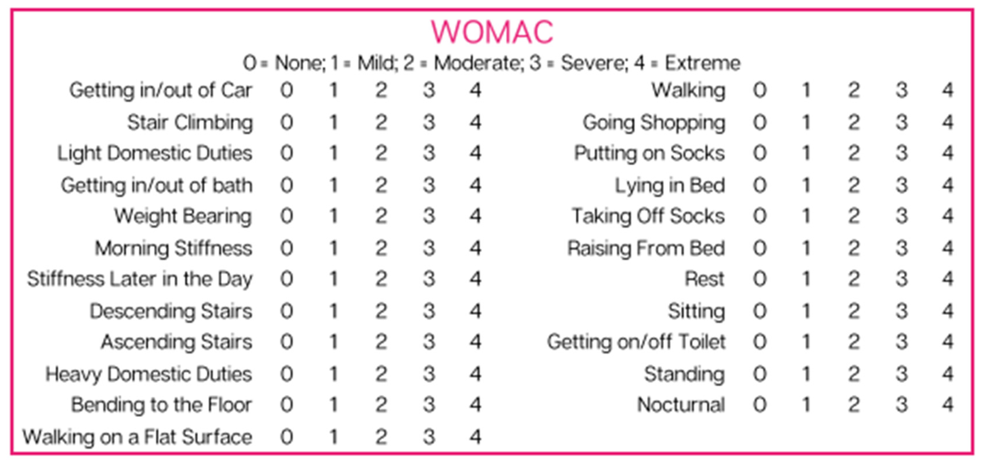
2.4. Questionnaire Composition

Patients were asked to fill out an initial questionnaire on the day of application consisting of NPRS and WOMAC scales (Figure 1 and Figure 2). Patients answered the same questionnaire 90 days after the initial allograft application. NPRS is a numerical pain scale employed as a subjective measurement of 0–10 for patients to rate their pain. A measurement of 0 indicates no pain, and 10 indicates the worst pain possible. WOMAC, measured on a scale of 0–4, is a combination of three questionnaires that measure pain, stiffness, and function. A measurement of 0 indicates that the patient is able to perform the activity with ease, and 4 indicates that the patient has extreme difficulty performing the activity. The total WOMAC score is calculated by adding each of the individual question scores together. The scores of these two scales were analyzed individually to allow for a more significant examination of the physical mobility of the affected joint. The reported scores for each patient can be found in Appendix B.

3. Results

Changes were recorded from baseline data obtained on the day of application to 90 days following the application for both NPRS and WOMAC. At baseline, the average NPRS score was 6.7 ± 2.1. At 90 days, the average NPRS score was 3.6 ± 2.3. Thirty-two out of the 38 patients (84%) reported lowered NPRS scores. Most patients also reported an improvement in their WOMAC scores. At baseline, the average total WOMAC score was 56.1 ± 18.8. At 90 days, the average total WOMAC score was 43.3 ± 22.7. Twenty-nine (76%) reported lowered WOMAC scores at the 90-day mark. The average scores and percent change of NPRS and WOMAC scales are reported in Table 1.
The percent change analysis found that NPRS scores improved by an average of 42%, and WOMAC scores improved by an average of 22% at the 90-day mark. An analysis of variance for WOMAC scores revealed that the average final WOMAC score was statistically significant compared to the average initial WOMAC score, as evidenced by a p-value of 0.009, as seen in Table 2.
Initial and final NPRS and WOMAC scores were also analyzed by gender, as seen in Table 3.
The percent change analysis shows that while females reported higher initial and final NPRS and WOMAC scores, males showed a higher percentage of improvement. On average, females improved by 37% on the NPRS and 22.9% on the WOMAC. Males improved by 57% on the NPRS and 27.4% on the WOMAC.
The presented data displays the change from baseline data obtained on the day of application to the 90-day point following the application. These changes are clinically and statistically significant measures of improvement in patients with symptomatic cartilage degeneration in the treatment of refractory sacroiliac dysfunction.

4. Discussion

The presented observational data reports the results following the application of Wharton’s Jelly birth tissue allografts in 38 patients with chronic, symptomatic, treatment-resistant SI joint dysfunction. This IRB-approved study protocol observed that the WJ applications in all 38 patients were both safe and efficacious. Patients evaluated retrospectively demonstrated statistically significant improvements in pain, stiffness, and physical mobility of the SI joints. Statistically significant differences between baseline and 90-day reported scores in NPRS and WOMAC were observed. The results presented in this case provide a foundation for the future prospective study of Wharton’s jelly allografts as applied for articular cartilage defects contributing to treatment-resistant SI joint dysfunction.
The observed results from this observational case report align with 2022 data reporting the observational safety and efficacy of Wharton’s jelly as applied to cartilage defects associated with knee osteoarthritis [24]. The systemic review on allogenic umbilical cord tissue applied for articular cartilage degeneration of the knee was presented by Gupta in 2022. He concluded that Wharton’s Jelly should be considered in the armamentarium of clinicians dealing with middle-aged patients suffering from OA-related cartilage degeneration of the knee and are not surgical candidates yet [22]. To the best of our knowledge, this is the first observational data reporting the safety and efficacy of Wharton’s Jelly allograft applications to load bearding articular cartilage degeneration of the SI joint. In theory, all SI joint interventions serve to ameliorate a local, focal defect in the structural and supporting cartilage of the SI joint. Having failed attempts to allay symptoms from the systemic process that is Osteoarthritis, addressing the specific degeneration of the SI joint cartilage has been one of either temporary symptom reduction with steroid injections, joint denervation by radiofrequency ablation, and, more recently, viscosupplementation with hyaluronic acid. Should these procedures fail, Si joint fusion is the inevitable option to eliminate painful joint translation or dysfunction. However, SI joint fusion may accelerate load redistribution and increase adjacent segment disease of the lower lumbar spine or the hip’s juxtaposed acetabular joint.
In a 2015 study, Lindsey et al. demonstrated that fusion of the SI joint resulted in increased adjacent segment lumbar motion at L5-S1 above normal flexion, extension, lateral bending, and axial rotation [25]. In a retrospective database study published by Schoell et al. in 2016, 469 patients were evaluated for fusion-related complications [17]. Overall, the complication rate was 13.2% at 90 days and 16.4% at six months, with up to a 5% incidence of novel lumbar pathology noted following the procedure documented more commonly in men.
While this observational data reports the safety and efficacy of Wharton’s jelly allografts as they are applied for structural tissue defects of the SI joint articular cartilage, it does have limitations. The study design was unblinded and observational. However, the primary outcomes of NPRS and WOMAC were patient-reported, eliminating the influence of an unblinded investigator. In addition, the potential for more statistically significant functional and pain improvements may have been noted if patients were offered a subsequent allograft application. Patients in this observational data repository were restricted to only one WJ allograft application. Future blinded, randomized, prospective studies, with serial applications of WJ allografts and follow-up over 6 and 12 months, may demonstrate more significant statistical improvement in NPRS and WOMAC as well as determine the overall durability of WJ allograft applications should a comparative study be performed with cortisone injections. A 1,3, and five-year longitudinal design might allow for a comparative analysis of patients that ultimately required SI fusion between those treated with conventional interventions and those who received WJ allograft applications.

5. Conclusions

In conclusion, this observational data collected on 38 patients with treatment-resistant pain and dysfunction of the SI joint reports that Wharton’s Jelly allografts demonstrated clinically and statistically significant improvements in function, joint mobility, and pain. No adverse events were reported in the observed WJ allograft application patient group. Based on the overall incidence of SI joint dysfunction, new literature cautioning the use of serial intra-articular cortisone injections, and the increased reports of SI fusion post-operative complications, alternatives to the current standard of care are warranted. Future randomized and prospective studies may further substantiate the efficacy and durability of WJ perinatal tissue allografts compared to cortisone injections, viscosupplementation, and, ultimately, SI joint fusion.

Author Contributions

Conceptualization, A.L. and T.B.; methodology, A.L.; validation, A.L.; formal analysis, A.L.; investigation, A.L., J.S. and S.A.T.; resources, A.L. and T.B.; data curation, A.L. and T.B.; writing—original draft preparation, A.L.; writing—review and editing, A.L., J.S. and S.A.T.; visualization, A.L.; supervision, T.B.; project administration, T.B.; funding acquisition, T.B. All authors have read and agreed to the published version of the manuscript.

Funding

This research received no external funding.

Institutional Review Board Statement

The study was conducted in accordance with the Declaration of Helsinki, and approved by the Institutional Review Board of the Institute of Regenerative and Cellular Medicine (protocol code IRCM-2022-311 and approved on 12 January 2022).

Informed Consent Statement

Informed consent was obtained from all subjects involved in the study.

Data Availability Statement

Data can be found in Appendix A and Appendix B.

Acknowledgments

The authors would like to thank Naomi Lambert for her contributions to reviewing the manuscript. The authors would also like to thank Renee Dodd for her contributions to data collection.

Conflicts of Interest

Albert Lai is associated with Desert Pain Specialists. Steve Traina is associated with Western Orthopedics. Jon Shou and Tyler Barrett are associated with Regenative Labs. Regenative Labs was involved in the design of the study, data analysis, and writing. Regenative Labs influenced the decision to publish.

Appendix A. Test Kits

  • HBcAb: Catalog number: 06P06, Abbott Laboratories, Abbott Park, IL, USA;
  • HbsAg: Catalog number: 06P02, Abbott Laboratories, Abbott Park, IL, USA;
  • HCV: Catalog number: 06P04, Abbott Laboratories, Abbott Park, IL, USA;
  • HIV1, HIV2, plus O: Catalog number: 06P01, Abbott Laboratories, Abbott Park, IL, USA;
  • HTLV-I/II: Catalog number: 06P07, Abbott Laboratories, Abbott Park, IL, USA;
  • RPR: Catalog number: 900025, Arlington Scientific, Springville, UT, USA;
  • HIV1, HCV, HBV, NAT: Catalog number: 303330, 303331, 303719, 303334, 303344;
  • WNV: Catalog number: 07001061190, Roche Diagnostics, Indianapolis, IN, USA.

Appendix B. Patient Reported Scores

GenderAgeApplication SiteInitial NPRSInitial Total WOMAC90-Day NPRS90-Day Total WOMAC
Patient 1M70R SI75917
Patient 2F71R SI343423
Patient 3F71L SI444423
Patient 4F73R SI652241
Patient 5M76L SI539230
Patient 6F72L SI869238
Patient 7M74L SI447132
Patient 8M72R SI930230
Patient 9F65R SI985584
Patient 10F52L SI868466
Patient 11F65L SI983481
Patient 12M72L SI930 330
Patient 13F52R SI868568
Patient 14M76R SI219112
Patient 15F78L SI875223
Patient 16M66L SI636423
Patient 17M71L SI864440
Patient 18M76L SI446344
Patient 19M77L SI646340
Patient 20F76L SI6549 68
Patient 21F74L SI539428
Patient 22M65R SI671445
Patient 23M71R SI1028444
Patient 24M75R SI1036131
Patient 25M69R SI545346
Patient 26F77L SI665435
Patient 27F74L SI761465
Patient 28F70L SI766874
Patient 29F65R SI584472
Patient 30M72R SI23002
Patient 31M65R SI760018
Patient 32F77R SI9921086
Patient 33F79L SI1092978
Patient 34F84L SI873312
Patient 35F72R SI564346
Patient 36M69R SI663560
Patient 37M75L SI845843
Patient 38F86L SI859459

References

  1. Raj, M.A.; Ampat, G.; Varacallo, M. Sacroiliac Joint Pain. In StatPearls [Internet]; StatPearls Publishing: Treasure Island, FL, USA, 2022. Available online: https://www.ncbi.nlm.nih.gov/books/NBK470299 (accessed on 4 September 2022).
  2. Kampen, W.U.; Tillmann, B. Age-related changes in the articular cartilage of human sacroiliac joint. Anat. Embryol. 1998, 198, 505–513. [Google Scholar] [CrossRef]
  3. Ou-Yang, D.C.; York, P.J.; Kleck, C.J.; Patel, V.V. Diagnosis and Management of Sacroiliac Joint Dysfunction. J. Bone Jt. Surg. 2017, 99, 2027–2036. [Google Scholar] [CrossRef] [PubMed]
  4. Dreyfuss, P.; Dreyer, S.J.; Cole, A.; Mayo, K. Sacroiliac Joint Pain. J. Am. Acad. Orthop. Surg. 2004, 12, 255–265. [Google Scholar] [CrossRef] [PubMed]
  5. Murray, C.W. Sacroiliac Joint Dysfunction: Evaluation and Management. Orthop. Nurs. 2011, 30, 126–131. [Google Scholar] [CrossRef] [PubMed]
  6. Calvillo, O.; Skaribas, I.; Turnipseed, J. Anatomy and pathophysiology of the sacroiliac joint. Curr. Rev. Pain 2000, 4, 356–361. [Google Scholar] [CrossRef] [PubMed]
  7. Kompel, A.J.; Roemer, F.W.; Murakami, A.M.; Diaz, L.E.; Crema, M.D.; Guermazi, A. Intra-articular Corticosteroid Injections in the Hip and Knee: Perhaps Not as Safe as We Thought? Radiology 2019, 293, 656–663. [Google Scholar] [CrossRef]
  8. Ostergaard, M.; Halberg, P. Intra-Articular Corticosteroids in Arthritic Disease: A guide to treatment. Biodrugs 1998, 9, 95–103. [Google Scholar] [CrossRef]
  9. Stone, S.; Malanga, G.A.; Capella, T. Corticosteroids: Review of the history, the effectiveness, and adverse effects in the treatment of joint pain. Pain Physician 2021, 24, S233–S246. [Google Scholar]
  10. Jüni, P.; Hari, R.; Rutjes, A.W.; Fischer, R.; Silletta, M.G.; Reichenbach, S.; da Costa, B.R. Intra-articular corticosteroid for knee osteoarthritis. Cochrane Database Syst. Rev. 2015, 2015, CD005328. [Google Scholar] [CrossRef] [Green Version]
  11. Stanbury, R.M.; Graham, E.M. Systemic corticosteroid therapy—Side effects and their management. Br. J. Ophthalmol. 1998, 82, 704–708. [Google Scholar] [CrossRef] [Green Version]
  12. Hanly, J.G.; Mitchell, M.; Macmillan, L.; Mosher, D.; Sutton, E. Efficacy of sacroiliac corticosteroid injections in patients with inflammatory spondyloarthropathy: Results of a 6 month controlled study. J. Rheumatol. 2000, 27, 719–722. [Google Scholar] [PubMed]
  13. Migliore, A.; Procopio, S. Effectiveness and utility of hyaluronic acid in osteoarthritis. Clin. Cases Miner. Bone Metab. 2015, 12, 31–33. [Google Scholar] [CrossRef] [PubMed]
  14. Heiney, J.; Capobianco, R.; Cher, D. A systematic review of minimally invasive sacroiliac joint fusion utilizing a lateral transarticular technique. Int. J. Spine Surg. 2015, 9, 40. [Google Scholar] [CrossRef] [PubMed] [Green Version]
  15. Ha, K.Y.; Lee, J.S.; Kim, K.W. Degeneration of Sacroiliac Joint After Instrumented Lumbar or Lumbosacral Fusion: A Prospective Cohort Study Over Five-Year Follow-up. Spine 2008, 33, 1192–1198. [Google Scholar] [CrossRef] [Green Version]
  16. Ackerman, S.J.; Polly, D.W., Jr.; Knight, T.; Schneider, K.; Holt, T.; Cummings, J. Comparison of the costs of nonoperative care to minimally invasive surgery for sacroiliac joint disruption and degenerative sacroiliitis in a United States Medicare population: Potential economic implications of a new minimally-invasive technology. Clin. Outcomes Res. 2013, 5, 575–587. [Google Scholar] [CrossRef] [Green Version]
  17. Schoell, K.; Buser, Z.; Jakoi, A.; Pham, M.; Patel, N.N.; Hsieh, P.C.; Liu, J.C.; Wang, J.C. Postoperative complications in patients undergoing minimally invasive sacroiliac fusion. Spine J. 2016, 16, 1324–1332. [Google Scholar] [CrossRef]
  18. Wampler, A.T.; Pace, L.H.; Thorne, J.M. Intra-Articular Injections of Wharton’s Jelly Allograft Combined with Amniotic Membrane Allograft are Safe and Effective for Relieving Knee Pain Associated with Osteoarthritis; A Retrospective Case Report. Elite Integr. Med. J. 2021, 4, 13. [Google Scholar] [CrossRef]
  19. Gupta, A.; El-Amin, S.F.; Levy, H.J.; Sze-Tu, R.; Ibim, S.E.; Maffulli, N. Umbilical cord-derived Wharton’s jelly for regenerative medicine applications. J. Orthop. Surg. Res. 2020, 15, 49. [Google Scholar] [CrossRef] [Green Version]
  20. Davis, J.M.; Purita, J.R.; Shou, J.; Barrett, T.C. Three-Dimensional Electron Microscopy of Human Umbilical Cord Tissue Allograft Pre and Post Processing: A Literature Comparison. J. Biomed. Res. Environ. Sci. 2022, 3, 934–940. Available online: https://www.jelsciences.com/articles/jbres1535.pdf. [CrossRef]
  21. Timmons, R.B.; Sugaya, K.; Bane, L.D. Homologous Use of Allogeneic Umbilical Cord Tissue to Reduce Knee Pain and Improve Knee Function. Life 2022, 12, 260. [Google Scholar] [CrossRef]
  22. Gupta, A.; Maffulli, N.; Rodriguez, H.C.; Carson, E.W.; Bascharon, R.A.; Delfino, K.; Levy, H.J.; El-Amin, S.F. Safety and efficacy of umbilical cord-derived Wharton’s jelly compared to hyaluronic acid and saline for knee osteoarthritis: Study protocol for a randomized, controlled, single-blind, multi-center trial. J. Orthop. Surg. Res. 2021, 16, 352. [Google Scholar] [CrossRef] [PubMed]
  23. Zhao, P.; Liu, S.; Bai, Y.; Lu, S.; Peng, J.; Zhang, L.; Huang, J.; Zhao, B.; Xu, W.; Guo, Q. hWJECM-Derived Oriented Scaffolds with Autologous Chondrocytes for Rabbit Cartilage Defect Repairing. Tissue Eng. Part A 2018, 24, 905–914. [Google Scholar] [CrossRef] [PubMed]
  24. Davis, J.M.; Sheinkop, M.B.; Barrett, T.C. Evaluation of the Efficacy of Cryopreserved Human Umbilical Cord Tissue Allografts to Augment Functional and Pain Outcome Measures in Patients with Knee Osteoarthritis: An Observational Data Collection Study. Physiologia 2022, 2, 109–120. [Google Scholar] [CrossRef]
  25. Lindsey, D.P.; Kiapour, A.; Yerby, S.A.; Goel, V.K. Sacroiliac Joint Fusion Minimally Affects Adjacent Lumbar Segment Motion: A Finite Element Study. Int. J. Spine Surg. 2015, 9, 64. [Google Scholar] [CrossRef] [PubMed] [Green Version]
Figure 1. Numeric Pain Rating Scale used by patients to assess pain level.
Reports 06 00012 g001
Figure 2. Western Ontario and McMaster Universities Arthritis Index used by patients to assess pain, stiffness, and function.
Reports 06 00012 g002
Table 1. Percent change analysis for initial and final NPRS and WOMAC scores.
ScaleAverage Initial ScoreAverage Final ScorePercent Change
NPRS6.73.642% decrease
WOMAC56.143.322% decrease
Table 2. Analysis of Variance for WOMAC scores.
ANOVA
Source of VariationSum of SquaresdfMean SquareF-valuep-value
WOMAC3069.5921013069.597.060820.00964significant
Table 3. Percent change analysis for females and males based on initial and final NPRS and WOMAC scores.
GenderAvg. Initial NPRSAvg. Final NPRS% Change NPRSAvg. Initial WOMACAvg. Final WOMAC% Change WOMAC
Females6.94.337% decrease66.451.222.9% decrease
Males6.32.757% decrease44.13227.4% decrease
Disclaimer/Publisher’s Note: The statements, opinions and data contained in all publications are solely those of the individual author(s) and contributor(s) and not of MDPI and/or the editor(s). MDPI and/or the editor(s) disclaim responsibility for any injury to people or property resulting from any ideas, methods, instructions or products referred to in the content.

