1. Introduction
Plasma cell neoplasms are characterized by increased monoclonal immunoglobulin secretion. These neoplasms can arise in either multiple lesions (multiple myeloma (MM)) or a single lesion (solitary plasmacytoma). Solitary plasmacytoma can be localized in the bone (solitary plasmacytoma of the bone (SPB)) or in other tissues (extramedullary (extraosseous) plasmacytoma (EMP)). EMP, a rare disease, accounts for 1–3% of all plasma cell proliferation [
1]. In approximately 80% of the cases, the EMPs are located in the upper airways, such as in the head and neck region [
2,
3], while the other locations are the gastrointestinal tract, lymph nodes, bladder, breast, thyroid, testes, and parotid glands [
3]. There have been only four reports of primary extramedullary plasmacytoma arising from the biliary tract [
4,
5,
6,
7] and five reports of secondary bile duct plasmacytoma with MM [
8,
9,
10,
11,
12]. An EMP occurs predominantly in males, with the average age at onset being from 55 to 60 years [
13]. Dores, G.M et al. [
14] reported a significant incidence of EMPs in Black men. Being a younger age at the onset of the EMP results in a better prognosis [
13,
15].
The EMP does not have apparent clinical features, while MM expresses systematic organ damage, such as hypercalcemia, renal insufficiency, anemia, and bone lesions. The symptoms of EMP depend on the location of the tumor. Approximately 20% of EMP patients have an M protein, which is mostly composed of IgA [
3]. The prognosis of an EMP is relatively good compared to that of MM, the extramedullary infiltration of MM, and plasmablastic lymphoma [
3,
8]. The progression to MM occurs in approximately 15–30% of EMP cases, resulting in worse clinical outcomes [
3,
16]. The morphological features of EMPs include the proliferation of monoclonal plasma cells, such as MM and SPB. The differential diagnoses of this condition are extranodal marginal zone lymphoma (MZL) of the mucosa-associated lymphoid tissue (MALT lymphoma), plasmablastic lymphoma, and extraosseous infiltrates of MM.
2. Case Presentation
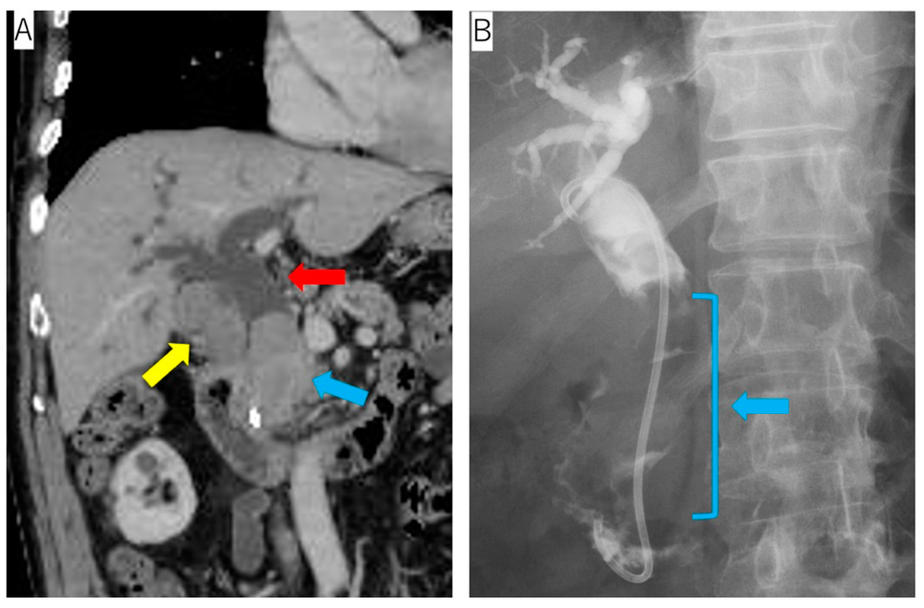
A 76-year-old man presented with jaundice during treatment for hypertension, diabetes, and hyperuricemia. Abdominal ultrasonography revealed a solid tumor in the gallbladder and common bile duct dilatation. Contrast-enhanced computed tomography (CT) detected an ischemic tumor of the gallbladder and the common bile duct and the dilation of the common bile duct. Endoscopic retrograde cholangiopancreatography (ERCP) revealed an expansive tumor in the common bile duct (
Figure 1).
The blood test findings indicated hyperbilirubinemia, mild anemia, high CRP levels, and high IL-2R levels. CEA and CA19-9 were within the normal limits. High IgA titers were also observed (
Table 1).
The bile cytology revealed the proliferation of degenerated atypical cells with poor epithelial cohesiveness. The immunohistochemical analyses of the cell blocks from the bile cytology specimens expressed positivity for CD45 LCA and vimentin and negativity for cytokeratin (CK) (AE1/AE3), CD3, CD20, CD79a, and CD30. Although a hematopoietic tumor was suspected, the details could not be determined.
After these tests were performed, endoscopic ultrasound-guided fine-needle aspiration (EUS-FNA) for the bile duct tumor was performed. The proliferation of the monoclonal atypical cells was observed with hematoxylin and eosin (H&E) staining. Immunohistochemically, these cells were positive for CD138 and negative for CD3, CD20, CD79a, and CK (AE1/AE3). The κ light chain was positive, while the λ one was negative. Genetic and chromosomal analyses for the EMP were not performed. The subsequent examination revealed no bone marrow involvement. Therefore, the diagnosis of a primary EMP localized in the biliary tract was confirmed.
The patient was administered bortezomib, dexamethasone, and lenalidomide (BLd). However, lenalidomide was discontinued because of the erythroderma that occurred as a severe adverse event after 7 days of administration, and the patient instead received three courses of a bortezomib and dexamethasone (Bd) regimen. The patient had diabetes mellitus as a comorbidity, and he required insulin therapy with dexamethasone, which forced him to reduce the dose of dexamethasone. Despite endoscopic biliary drainage being performed, it was difficult to control the biliary tract infection associated with obstructive jaundice due to the tumor. Bleeding from the tumor also occurred. After four rounds of chemotherapy, he underwent pancreaticoduodenectomy and cholecystectomy to control repeated cholangitis and bleeding from the bile ducts.
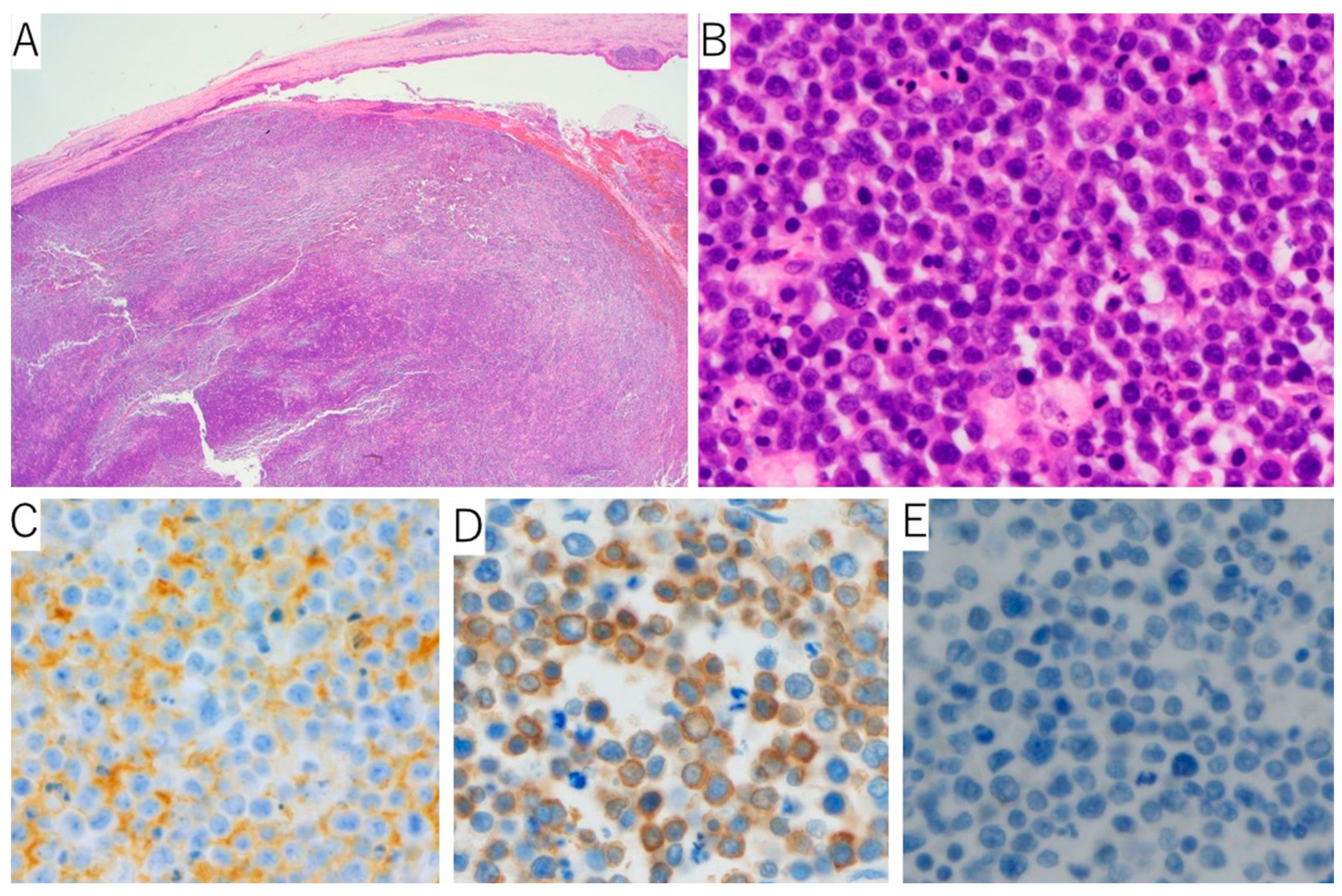
A whitish solid tumor filling in the bile duct and gallbladder was observed (
Figure 2). In the H&E sections, the monoclonal cells that looked like plasma cells were proliferating. The immunohistochemical staining revealed CD138 positivity, and immunoglobulin κ light chain
in situ hybridization was positive for most of the plasma cells, whereas that of the λ chain was negative (
Figure 3). After the surgery, the tumor’s progression rapidly became more similar to plasmablastic lymphoma, so systemic therapy, including cyclophosphamide, doxorubicin, vincristine, prednisone, and etoposide (EPOCH), was performed; however, the patient died of tumor bleeding, febrile neutropenia, and severe cholangitis after two cycles of the EPOCH regimen.
3. Discussion
EMP must be distinguished from an extranodal MZL of the MALT lymphoma, extraosseous infiltrates of MM, and plasmablastic lymphoma. MALT lymphoma is a low-grade B-cell lymphoma with prominent plasmacytic differentiation. A well-known pathological feature of MALT lymphoma is the presence of a lymphoepithelial lesion (LEL), which is the lymphocytic infiltration of the epithelial cells. LEL was not evident in this case. MM was ruled out because no bone marrow lesions were detectable and there were no clinical features of MM. Plasmablastic lymphoma commonly occurs in immunocompetent patients due to human immunodeficiency virus infection and iatrogenic immunosuppression. The EBV infection is strongly associated with plasmablastic lymphoma. In this case, the in situ hybridization of EBV-encoded RNA and CD56 were negative, which is unlike typical cases of plasmablastic lymphoma. Although the negative immunoexpression of CD20 and CD79a caused difficulty in making a histological diagnosis, positive CD45 LCA staining indicated hematopoietic malignancy. The final diagnosis was made according to the CD138 expression as well as immunoglobulin k light chain restriction.
The prognosis of an EMP is relatively good overall. Some case reports have mentioned the prognosis of primary EMP arising from the gall bladder. Kondo et al. reported a 53-year-old male who was confirmed to be alive for three years and seven months after being diagnosed [
4]. Hwang et al. described the case of a 63-year-old female patient with no evidence of the disease for eight years after surgery [
5]. Majerović et al. reported the case of a 69-year-old man who showed no sign of recurrence for two years [
6]. In contrast to these reports, the patient in this case had a poor prognosis as he survived for only seven months after the diagnosis was made. The diagnosis of the patient in this case report needed to be distinguished from plasmablastic lymphoma and extraosseous infiltrates of MM, which are both associated with aggressive courses. Clinically and per the immunohistochemistry, these diseases were excluded, as mentioned above.
Hughes et al. [
17] discussed the differences in the molecule patterns between 14 solitary plasmacytomas (four EMP and ten SPB) and 11 MMs. CD49d (Integrin α4), which are related to the promotion of plasma cell colonization, the prevention of cell apoptosis, and the development of cytotoxin resistance, and they showed a statistically significant difference between MM and solitary plasmacytomas. From this report, it might be suggestive that MM shows a poorer prognosis compared to EMP because the CD49d expression in MM was higher than that in EMP.
Although there are several reports which discuss the better prognosis of EMPs compared to MMs [
3,
15,
18], this case did not show a favorable prognosis. The prognosis of EMP usually worsens when it progresses to MM. In this case, the prognosis became poorer because the tumor progression was aggressive in a similar way to plasmablastic lymphoma. It is rare to have a poor prognosis without progression to MM.
Radiation therapy or surgery is generally the treatment of choice for localized solitary extramedullary plasmacytoma, but because the area occupied by the EMP extended into the intrahepatic bilateral bile ducts, gallbladder, and common bile duct, the irradiated area was considered to extend more into the liver, and radiation therapy was not chosen because of the high risk of severe liver failure and cholangitis. Pancreatoduodenectomy was forced to be chosen to control the local infection and bleeding, although it was a very strong invasive therapy.
Although immunotherapy with CD38-targeting antibodies might be an effective approach, this treatment was not covered as an induction therapy for MM by the national insurance in Japan at that time. Thus, the patient could not receive therapy using CD38-targeting antibodies.
4. Conclusions
EMP is difficult to diagnose because it’s symptoms vary with the organ from which the tumor originates. However, EMP must not be misdiagnosed as other plasma cell neoplasia such as extraosseous infiltrates of MM, plasmablastic lymphoma, or lymphomas with plasmacytic differentiation such as MALT lymphoma since their treatments and prognoses are totally different from those of EMP.
Author Contributions
E.S. carried out histopathological evaluations and drafted the manuscript. T.T. conceived the study and participated in the design and preparation of the manuscript. S.Y. reviewed the pathology. K.N., N.G., A.T. and Y.H. collected, evaluated, and interpreted clinical data. All authors have read and agreed to the published version of the manuscript.
Funding
The authors received no external funding for this research.
Institutional Review Board Statement
The ethical review and approval were waived for this study because Japanese national legislation that indicates ethics approval is not required for case report.
Informed Consent Statement
Written informed consent was obtained from the patient for the publication of this case report and any accompanying images.
Data Availability Statement
The data used in this case report are available upon reasonable request from the corresponding author.
Conflicts of Interest
The authors declare that they have no competing interest.
References
- Him, M.; Meier, M.; Mehta, V. Rare Presentation of Primary Extramedullary Plasmacytoma as Lip Lesion. Case Rep. Oncol. Med. 2017, 2017, 4296802. [Google Scholar] [CrossRef] [PubMed] [Green Version]
- Galieni, P.; Cavo, M.; Pulsoni, A.; Avvisati, G.; Bigazzi, C.; Neri, S.; Caliceti, U.; Benni, M.; Ronconi, S.; Lauria, F. Clinical outcome of extramedullary plasmacytoma. Haematologica 2000, 85, 47–51. [Google Scholar] [PubMed]
- McKenna, R.W.; Kyle, R.A.; Kuehl, W.M.; Harris, N.L.; Coupland, R.W.; Fend, F. Plasma cell neoplasms. In WHO Classification of Tumours of Haematopoietic and Lymphoid Tissues; Swerdlow, S.H., Campo, E., Harris, N.L., Jaffe, E.S., Pileri, S.A., Stein, H., Thiele, J., Eds.; International Agency of Research on Cancer: Lyon, France, 2017; pp. 241–258. [Google Scholar]
- Kondo, H.; Kainuma, O.; Itami, J.; Minoyama, A.; Nakada, H. Extramedullary plasmacytoma of maxillary sinus with later involvement of the gall bladder and subcutaneous tissues. Clin. Oncol. J. (R. Coll. Radiol.) 1995, 7, 330–331. [Google Scholar] [CrossRef]
- Hwang, D.W.; Lim, C.S.; Jang, J.Y.; Lee, S.E.; Yoon, S.O.; Jeon, Y.K.; Uk Lee, K.; Kim, S.W. Primary hematolymphoid malignancies involving the extrahepatic bile duct or gallbladder. Leuk. Lymphoma 2010, 51, 1278–1287. [Google Scholar] [CrossRef] [PubMed]
- Majerović, M.; Bogdanić, B.; Drinković, N.; Kinda, S.B.; Jakić-Razumović, J.; Gasparović, V. Extramedullary plasmacytoma imitating neoplasm of the gallbladder fossa after cholecystectomy. Coll. Antropol. 2012, 36, 331–333. [Google Scholar] [PubMed]
- Schuster, D.; Klosterhalfen, B.; Fiedler, C.; Prescher, A. Metastasis of medullary plasmocytoma as the cause of acute cholecystitis. Dtsch. Med. Wochenschr. 2007, 132, 612–615. [Google Scholar] [CrossRef] [PubMed]
- St Romain, P.; Desai, S.; Bean, S.; Jiang, X.; Burbridge, R.A. Extramedullary plasmacytoma of the gallbladder diagnosed by endoscopic ultrasound fine needle aspiration (EUS-FNA). J. Gastrointest. Oncol. 2015, 6, E7–E9. [Google Scholar] [CrossRef]
- Heckmann, M.; Uder, M.; Grgic, A.; Adrian, N.; Bautz, W.; Heinrich, M. Extraosseous manifestation of multiple myeloma with unusual appearance in computed tomography--case report. Rontgenpraxis 2008, 56, 249–253. [Google Scholar] [CrossRef]
- Fukatsu, H.; Hiramatsu, Y.; Takagi, S.; Morishita, H. Multiple myeloma involving the extrahepatic bile duct. Intern. Med. 2013, 52, 829–830. [Google Scholar] [CrossRef] [Green Version]
- Abughanimeh, O.; Qasrawi, A.; Abu Omar, M.; Bahaj, W.; Abu Ghanimeh, M. A Case of Multiple Myeloma Associated with Extramedullary Plasmacytoma of the Gallbladder Manifesting as Acute Cholecystitis. Cureus 2018, 10, e2688. [Google Scholar] [CrossRef] [PubMed]
- Abt, A.B.; Deppisch, L.M. Multiple myeloma involving the extrahepatic biliary system. J. Mt. Sinai. Hosp. N. Y. 1969, 36, 48–54. [Google Scholar] [PubMed]
- Lai, C.Y.; Hsieh, H.H.; Chen, H.K.; Chao, C.Y.; Hua, C.H.; Tai, C.J.; Bau, D.T.; Tsai, M.H.; Shih, L.C. Clinical Features of Head and Neck Solitary Extramedullary Plasmacytoma in Taiwan. In Vivo 2020, 34, 261–265. [Google Scholar] [CrossRef] [PubMed]
- Dores, G.M.; Landgren, O.; McGlynn, K.A.; Curtis, R.E.; Linet, M.S.; Devesa, S.S. Plasmacytoma of bone, extramedullary plasmacytoma, and multiple myeloma: Incidence and survival in the United States, 1992–2004. Br. J. Haematol. 2009, 144, 86–94. [Google Scholar] [CrossRef] [PubMed] [Green Version]
- Shih, L.Y.; Dunn, P.; Leung, W.M.; Chen, W.J.; Wang, P.N. Localised plasmacytomas in Taiwan: Comparison between extramedullary plasmacytoma and solitary plasmacytoma of bone. Br. J. Cancer 1995, 71, 128–133. [Google Scholar] [CrossRef] [PubMed] [Green Version]
- Merza, H.; Sarkar, R. Solitary extraosseous plasmacytoma. Clin. Case Rep. 2016, 4, 851–854. [Google Scholar] [CrossRef] [PubMed] [Green Version]
- Hughes, M.; Doig, A.; Soutar, R. Solitary plasmacytoma and multiple myeloma: Adhesion molecule and chemokine receptor expression patterns. Br. J. Haematol. 2007, 137, 486–487. [Google Scholar] [CrossRef] [PubMed]
- Goyal, G.; Bartley, A.C.; Funni, S.; Inselman, J.; Shah, N.D.; Marshall, A.L.; Ashrani, A.A.; Kapoor, P.; Durani, U.; Hashmi, S.K.; et al. Treatment approaches and outcomes in plasmacytomas: Analysis using a national dataset. Leukemia 2018, 32, 1414–1420. [Google Scholar] [CrossRef] [PubMed]
| Disclaimer/Publisher’s Note: The statements, opinions and data contained in all publications are solely those of the individual author(s) and contributor(s) and not of MDPI and/or the editor(s). MDPI and/or the editor(s) disclaim responsibility for any injury to people or property resulting from any ideas, methods, instructions or products referred to in the content. |
© 2022 by the authors. Licensee MDPI, Basel, Switzerland. This article is an open access article distributed under the terms and conditions of the Creative Commons Attribution (CC BY) license (https://creativecommons.org/licenses/by/4.0/).


 ,
, 


