Hypothesis

On the Potential Therapeutic Roles of Taurine in Autism Spectrum Disorder

by
Alberto Rubio-Casillas
1,2,*,
Elrashdy M. Redwan
3,4 and
Vladimir N. Uversky
5,*
1
Autlán Regional Hospital, Health Secretariat, Autlán 48900, Mexico
2
Biology Laboratory, Autlán Regional Preparatory School, University of Guadalajara, Autlán 48900, Mexico
3
Biological Science Department, Faculty of Science, King Abdulaziz University, Jeddah 21589, Saudi Arabia
4
Therapeutic and Protective Proteins Laboratory, Protein Research Department, Genetic Engineering and Biotechnology Research Institute, City for Scientific Research and Technology Applications, New Borg EL-Arab 21934, Egypt
5
Department of Molecular Medicine and USF Health Byrd Alzheimer’s Research Institute, Morsani College of Medicine, University of South Florida, Tampa, FL 33612, USA
*
Authors to whom correspondence should be addressed.
Neuroglia 2023, 4(1), 1-14; https://doi.org/10.3390/neuroglia4010001
Submission received: 20 November 2022 / Revised: 8 December 2022 / Accepted: 16 December 2022 / Published: 23 December 2022

Abstract

:
Contemporary research has found that people with autism spectrum disorder (ASD) exhibit aberrant immunological function, with a shift toward increased cytokine production and unusual cell function. Microglia and astroglia were found to be significantly activated in immuno-cytochemical studies, and cytokine analysis revealed that the macrophage chemoattractant protein-1 (MCP-1), interleukin 6 (IL-6), tumor necrosis factor α (TNF-α), and transforming growth factor β-1 (TGFB-1), all generated in the neuroglia, constituted the most predominant cytokines in the brain. Taurine (2-aminoethanesulfonic acid) is a promising therapeutic molecule able to increase the activity of antioxidant enzymes and ATPase, which may be protective against aluminum-induced neurotoxicity. It can also stimulate neurogenesis, synaptogenesis, and reprogramming of proinflammatory M1 macrophage polarization by decreasing mitophagy (mitochondrial autophagy) and raising the expression of the markers of the anti-inflammatory and pro-healing M2 macrophages, such as macrophage mannose receptor (MMR, CD206) and interleukin 10 (IL-10), while lowering the expression of the M1 inflammatory factor genes. Taurine also induces autophagy, which is a mechanism that is impaired in microglia cells and is critically associated with the pathophysiology of ASD. We hypothesize here that taurine could reprogram the metabolism of M1 macrophages that are overstimulated in the nervous system of people suffering from ASD, thereby decreasing the neuroinflammatory process characterized by autophagy impairment (due to excessive microglia activation), neuronal death, and improving cognitive functions. Therefore, we suggest that taurine can serve as an important lead for the development of novel drugs for ASD treatment.

1. Introduction

The term autism was created in 1911 by the psychiatrist Paul Eugen Bleuler (1857–1939), who employed the Greek word “self” to reflect one’s desire to retire into one’s thoughts [1]. Social difficulties, communication problems, an absence of social bonding behaviors, and the prevalence of repetitive behaviors are all symptoms of this disease [2]. Although several studies [3,4,5] have been dedicated to finding the genetic mechanisms of ASD, the majority of diagnosed cases are still unexplained [6]. It has been claimed that a range of gene–environment interactions, epigenetic changes, and environmental variables all have important and distinct roles in the genesis of ASD [7]. For example, prenatal, perinatal, and postnatal environment influences are considered to be responsible for 60% of the risk of ASD, whereas genetic features account for the remaining 35–40% [8]. A tendency toward increased cytokine release and unusual immune cell functions have been demonstrated in people with ASD in several investigations [9,10,11,12,13,14]. There is also proof that most of these abnormal immunological patterns are linked to the worsening of autistic behavior [14]. Even when the neurobiological origin of ASD is multifactorial, neuroinflammatory processes are thought to contribute to the formation of autistic behavioral alterations [15,16], and convincing research evidence indicates that microglial stimulation or disturbance can have a significant impact on neural development, leading to disorders in neuronal growth, such as ASD [17]. Basic research continues to be undertaken to identify the causes that promote the development of ASD. However, most of this research is oriented more toward the search for the causes of the disease than toward the development of effective medicines. As a result, children with ASD still do not have specific treatments capable of stopping the progressive neurodegeneration that some are affected by. This work provides the scientific rationale for the potential therapeutic utilization of taurine for patients with ASD.

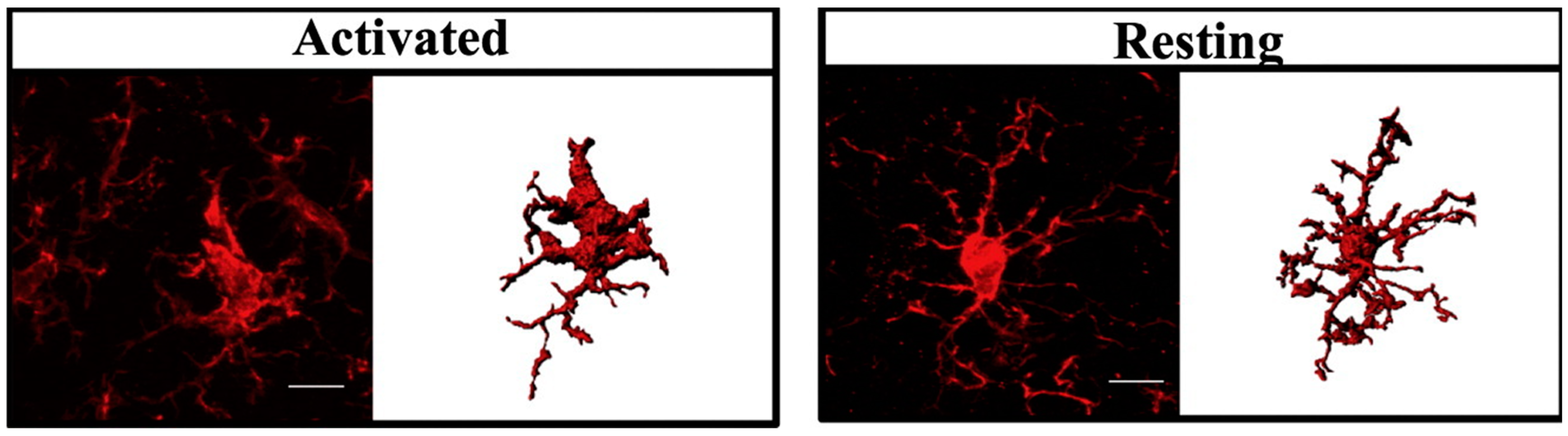
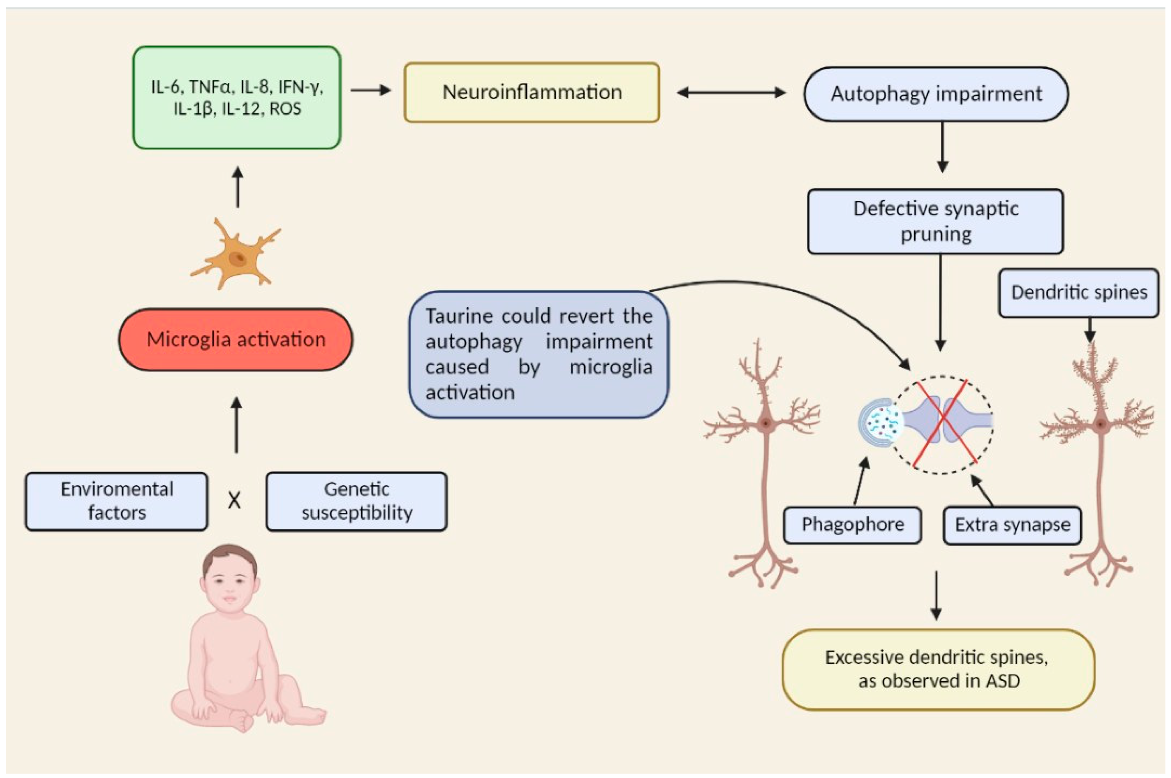
2. Microglial Activation in ASD

The central nervous system (CNS) contains a specialized population of macrophage-like cells known as microglia. Considered to be immune sentinels that are capable of orchestrating a potent inflammatory response, microglia can perform a variety of functions in various periods of life, both in normal and pathological conditions [18]. Microglia have a multitude of roles [19] throughout CNS maturation, such as phagocytic action during neuronal/synaptic development (most likely reflected in the elimination of repetitive neurons and synapses); this process is also known as “synaptic pruning” [17]. Appealing scientific proof implies that microglial activation or malfunction can have a significant impact on neuron maturation by reducing synaptic pruning, eventually leading to neurodevelopmental dysfunction, such as ASD [17].
Microglia, like macrophages, respond to CNS injuries by adopting distinct activated profiles [20]. When microglial cells are activated, they undergo phenotypical changes, migrate to the injured area, and proliferate (i.e., undergo microgliosis), as well as enhancing the synthesis of many proteins, including immunological intermediaries [17]. The standard, classically activated inflammatory and neurotoxic M1 macrophages, as well as the alternatively activated anti-inflammatory M2 macrophages, were described in [20,21,22].

3. M1 Polarization in ASD

Classically activated M1 macrophages are known to mediate excessive inflammatory responses, cytotoxicity, and tissue damage. They mediate the host’s defense against several bacterial, viral, and protozoal pathogens. Furthermore, aluminum from vaccines [23], bacterial lipopolysaccharide (LPS) [24,25], TNF-α [26], interferon γ (IFN-γ) [27], Aβ oligomers [27,28], and α-synuclein [29,30] are able to promote the M1 phenotype. “The induction of mitogen-activated protein kinase (ERK1/2 and p38), synthesis of MHC-II (major histocompatibility complex type II) cell membrane glycoprotein, the release of inflammatory cytokines (TNF-α, Interleukin-1 (IL-1), IL-6, and IL-12), and production of reactive oxygen species are all hallmarks of the classic M1 phenotype” [25].
Increased glutaminase, inducible nitric oxide (NO) synthase, (iNOS or NOS2), and inducible COX-2 (cyclooxygenase 2) synthesis leads to increased NO, glutamate, and prostaglandins release, respectively. Most of the factors released by microglia are neurotoxic for neuronal cell cultures [31]. The cytokines Interleukin 4 (IL-4) and IL-13, which are released from Th2 lymphocytes, can generate the alternative M2 profile in embryonic microglial cells [32]. In vitro, IL-4 reduces iNOS activity, TNF-α, and superoxide release in LPS and TNF-α-stimulated microglial cells, as well as protecting neurons against neuronal toxicity [33]. Microglial activation in human autistic brains was documented in several studies (for review see [17]). Active inflammatory mechanisms were detected within the brain cortex, white matter, and significantly in the cerebellum, according to neuropathological investigations of autistic brains [34].
Microglial and astroglial cells were significantly stimulated in immunological and cytochemical experiments, and cytokine profiles revealed both monocyte chemoattractant protein 1 (MCP-1, also known as C-C motif ligand 2, CCL2) and transforming growth factor β-1 (TGFB-1), which are produced by neuroglia cells, are abundant cytokines in the brain [34]. Furthermore, IL-6 was found to be very much elevated in the anterior cingulated cortex, and in the spinal fluid of individuals with ASD in recent studies [34,35]. It was claimed that increased IL-6 levels in the autistic brain induce instability of neuronal networks by affecting neural cell bonding and synapse establishment, and so contribute to ASD development [35]. Myeloid cells (which are a subgroup of leukocytes that includes dendritic cells (DCs), granulocytes, macrophages, and monocytes [36]) seem to make an important contribution to the pathophysiology of ASD in children, according to the research [37]. In the brains of children with ASD, immunohistochemistry indicated higher amounts of microglial cells in the parenchyma, an increased number of macrophages surrounding the vascular space, and heightened microglial and perivascular macrophage activation, as well as increased amounts of MCP-1 [34,38].
Growing amounts of monocytic leucocytes in the plasma and elevated cytokines released after toll-like receptor 4 (TLR4) stimulation in monocytes of individuals with ASD, together with the elevated concentrations of IL-1, IL-6, and IL-23, and correlations with behavioral evaluations have been found in studies examining peripheral myeloid activity, which are compatible with the study results in the brain [10,39,40]. Evidence has been provided of aberrant dispersion of dendritic cell density in children with ASD, similar to results in microglial cells and monocytes, thus implying that multiple lineages of the myeloid tissue are damaged in the disease [37].
A comprehensive investigation was conducted to find inflammatory signs that could be used for ASD diagnostics. In cultivated M1 and M2 macrophages obtained from individuals with ASD (n = 29) and normally developing persons (n = 30), the messenger RNA production of cytokines that involved TNF-α was assessed. TNF-α production in the M1 subtype was much greater in ASD individuals than in normal people, but this elevation was not detected in M2 macrophages [41].

4. Taurine Reprograms Macrophage M1 Polarization to the M2 Phenotype

Macrophage metabolism has historically been recognized to be highly malleable, reflecting pathologies related to distinct disease states [42,43,44]. In relatively recent times, ambient signals and the surrounding cytokine microenvironment were assumed to be in control of macrophage metabolic reactions, partially due to an old idea that all macrophages originated in the blood [45,46]. The M1 and M2 macrophages paradigm hypothesized that these cells in a resting phase of M0 may be transformed to M1 or M2 modes by exposing them to some specific cytokines [47]. However, a recent report outlined the fundamental metabolic and physiological distinctions between M1 and M2 macrophage polarization modes in laboratory settings [21].
M1 macrophages, which are stimulated by TNF-α or IFN-γ, exhibit a significant increase in the glycolytic metabolism to generate adenosine triphosphate (ATP) for phagocytic and microbicidal activities, while the mitochondrial tricarboxylic acid cycle (TCA cycle) is blocked. M2 macrophages, on the other hand, which are stimulated by IL-4 and IL-13, possess a working TCA circuit and increased mitochondrial oxidative phosphorylation (OXPHOS) capacity. Chemicals that reduce inflammation, such as steroids, IL-10, IL-13, and colony-stimulating factor 1 (CSF-1) are usually related to the M2 macrophage phenotype [21].
The reconfiguration of a macrophage from the M1 to M2 phenotype could be performed by focusing on their metabolism: according to the experimental evidence, reconnecting the metabolism to mitochondrial oxidative phosphorylation prevents the inflammation process (M1 state). As a result, modulation of the energy metabolism represents a promising option for the therapy of inflammatory disorders [48]. Taurine (an amino sulfonic acid, which is widely distributed in animal tissues, accounting for up to 0.1% of total human body weight) inhibits macrophage M1 polarization by suppressing mitophagy (mitochondrial autophagy), which reduces the expression of the M1 inflammatory genes, while raising the synthesis of the M2 markers (CD206 and IL-10), according to a recent study. Taurine also reconfigures the M1 macrophage power metabolism by preserving an elevated number of mitochondria that prevent the switch to glycolysis, which is essential for the M1 macrophages [48].
In this respect, taurine-chloramine inhibits macrophage release of inflammation promoters, such as macrophage inflammatory protein 2 (MIP-2, also known as chemokine CXC ligand 2, CXCL-2), MCP-1, MCP-2, TNF-α, IL-6, nitric oxide, nitrites, and prostaglandin E2 (PGE2) [49,50,51,52,53,54]. Taurine-chloramine also stimulates the synthesis of the nuclear factor erythroid 2-related factor 2 (NRF-2), which is a transcription factor that controls the production of important detoxification and antioxidant enzymes [55]. Importantly, these inflammation-promoting cytokines were shown to be significantly elevated in the brains of children with ASD. Since taurine administration was shown to inhibit the release of these cytokines, one can infer that taurine may have an important therapeutic action.

5. Taurine Decreases the Activation State of Microglia

Taurine (or 2-aminoethanesulfonic acid) is an amino acid, which is naturally derived from sulfur amino acids, such as methionine and cysteine, and which is not employed in protein biosynthesis. The enzyme cysteine sulfinic acid decarboxylase (CSD) synthesizes it from cysteine and methionine in the kidney, liver, and nerve cells, specifically in glial cells [56]. Taurine is three to four times more abundant in the growing brain compared with the adult brain [57], and its amount diminishes with age, implying that taurine exerts an essential function during neuronal maturation [58].
Anti-inflammatory effects are also known to be exerted by taurine [50,51,59,60,61]. In this regard, taurine was shown to decrease the amount of microglia-stimulated cells in elderly mice (Figure 1) [62]. In taurine-treated animals, stimulated microglia cells constituted 8.2% of the whole microglia, while in sodium chloride (NaCl)-treated animals, they constituted 37.8% [62].
Microglia undergo structural alterations as a consequence of brain inflammation. Here, resting microglia (phase I) have stick-formed cellular soma with tiny, bifurcated dendrites, while activated microglia (phase II) have enlarged cellular soma with extended and dumpy dendrites (Figure 2). The structure of microglial cells in taurine-treated and reference rats was also studied by Gebara and colleagues. Taurine therapy reduced the soma size, expanded the territorial projection area, and increased the number of microglia dendrites; these structural variations are associated with a lowered stimulated state of microglia [62].
As microglia activation was demonstrated to be reversed by taurine in the animal model, we hypothesize that this amino acid could revert some of the neurodegenerative processes in children with severe ASD.

6. Taurine Induces Autophagy

Lysosomes use an evolutionary preserved mechanism called autophagy to destroy extra or damaged cell organelles and proteins. When Ashford and Porter discovered in 1962 that cells could devour themselves, they made the first observation of the autophagy process [63]. Autophagy is a critical developmental mechanism that occurs during synaptic pruning. Both in human and animal models, deficiencies in autophagy have been linked to neurodevelopmental disorders such as ASD. Repeated early exposure to aluminum-containing vaccines during crucial immunological and neurological system development is among the environmental factors linked to ASD (for reviews, see [64,65,66,67,68]). Microglia, the main resident immune cells of the brain and spinal cord, constitute up to 15% of all CNS cells [69]. These “macrophage-like” cells are vital for maintaining brain homeostasis because they carry out key activities such as synaptic pruning and neurogenesis [69,70]. In reaction to harm brought on by either endogenous or exogenous stimuli, microglia can become activated [71].
The first report that aluminum from vaccines activated microglia cells in the animal model was delivered by Crépeaux and colleagues [72]. Subsequent work confirmed the presence of aluminum inside microglia in the brains of patients who died from ASD [66]. Furthermore, elevated levels of aluminum were found in human brain tissue samples of patients with sporadic Alzheimer’s disease, familial Alzheimer’s disease, ASD, and multiple sclerosis [73]. According to recent studies, autophagy exerts a significant role in maintaining brain homeostasis by controlling the activation degree of microglia [69,71]. Of note, it has been shown that autophagy is substantially dysfunctional in ASD brains and does not change normally over development, suggesting that both autophagy dysregulation and ASD etiology are involved (for reviews, see [64,65,66,67,68]).
Furthermore, it has been established that autophagy is a critical intracellular mechanism for macrophage and microglial polarization [74]. The promotion of M2 phenotypic polarization in microglia may result in a neuro-protective state as a result of increased microglial autophagy [75]. In conclusion, aluminum from vaccines (and other environmental/genetic factors) impairs autophagy by inducing excessive microglia activation, thus affecting normal neurodevelopment. In this regard, it is important to consider taurine as a potential therapeutic molecule for ASD as evidence has been provided that taurine induces autophagy (Figure 3) and reverts the M1 to the M2 polarization state [48].

7. Taurine Promotes Neurogenesis

Microglia cells are easily stimulated due to damage or immune stress [76]. Nitric oxide (NO), TNF-α, IL-1β, and Reactive Oxygen Species (ROS) are among the inflammatory substances secreted by activated microglia [77]. Furthermore, neuronal degeneration has been linked to the synthesis and accumulation of these substances [24,78,79,80], and it has been observed that microglia over-activation causes apoptosis [81]. One study, for example, collected autistic brain cells obtained from a brain tissue bank to examine their cellular and molecular alterations [82]. Three important brain sections, the hippocampus, the cerebellum, and the frontal cortex, were examined in six cases of autistic brains and six cases of non-autistic brains from deceased children aged 6 to 16. This analysis revealed that the three regions had a significant elevation in ER (endoplasmic reticulum) stress, as evidenced by the engagement of ER stress signals, such as serine/threonine-protein kinase/endoribonuclease IRE1, cyclic AMP-dependent transcription factor ATF-6-α (ATF6), and eukaryotic translation initiation factor 2-alpha kinase 3 (EIF2AK3, also known as RKR-like endoplasmic reticulum kinase (PERK)) [82]. It was also discovered that apoptosis rose in the three described areas, as evidenced by enhanced caspase 8 and PARP (poly (ADP-ribose) polymerase) breakdown [82].
We can deduct from this information that managing excessive oxidative stress is critical for preventing neuron death in the brains of some children who suffer from the most severe forms of ASD. Taurine has been demonstrated to be an essential component in the maturation of brain tissues [83] since its concentration is three to four times more elevated in the growing and newborn brains of mice than in adults [84]. Studies in monkeys that were fed with taurine-deficient dietary formulae revealed a significant deficiency in the cortical layer arrangement in the visual cortex [83]. Cats born from mothers with taurine deficiency had a reduced brain mass and altered cerebellum and visual cortex structure [85,86]. In taurine-deficient kittens, the number of pyramidal cells was diminished, and the neurons had poor ramification patterns [85,86]. Such findings highlight the significance of this amino acid for developing brains. Taurine has also been shown to promote or reestablish cellular division in neurons from human fetuses [87] and to affect neurotransmission [88]. Overall, these observations indicate that taurine is indispensable for adequate brain cell multiplication, growth, and specialization [89].
In this line, it was discovered that taurine significantly boosted the quantity of neuron progenitor cells (NPCs) collected from the hippocampal region of the aging mouse brain, and the increase in the NPCs stimulated by taurine was one of the greatest documented for any other molecule or situation in adult brain NPCs [90], far higher than that stimulated by melatonin, dopamine, or neuropeptide Y [91,92,93]. Furthermore, a seminal work demonstrated that taurine significantly enhances cell counts in vitro and neuron formation from fetal human NPCs [94].
Another study found that taurine stimulated NPC multiplication in the dentate gyri of developing brains, cultivated hippocampus progenitor cells, and hippocampus segments taken from mice brains. Taurine was also found to increase the generation of new synapses, which was a significant discovery [89]. Although such an increase persists during the lifetime in healthy people, a burst of synapsis production takes place throughout the initial brain maturation, termed “exuberant synaptogenesis” [95].

8. Conclusions

As mentioned in the introduction, the CNS contains a specialized population of macrophage-like cells known as microglia. Considered immune sentinels that are capable of orchestrating a potent inflammatory response, microglia can perform a variety of functions [18]. One such important microglia function is related to phagocytic action during neuronal/synaptic development (most likely represented by the elimination of repetitive neurons and synapses) [19]. Microglial activation or malfunction can have a significant impact on neuron maturation, leading to neurodevelopmental dysfunction, such as ASD [17]. Microglia, like macrophages, respond to CNS injuries by adopting distinct activated profiles [20].
When microglial cells are activated, they undergo phenotypical changes, that is, a change from the M2 to the M1 phenotype, known as M1 polarization [17]. M1 polarization has been involved in a variety of nervous system abnormalities, such as multiple sclerosis, Alzheimer’s disease [96], and ASD [42]. According to recent studies, increasing the M2 phenotype may improve cognitive performance [97]. It was found that transplanting natural IL-4 functional T cells into IL-4 knockout mice improves their behavior while transplanting M2-activated macrophages enhances mental performance in immunologically defective mice [98,99]. These findings reveal that M1 polarization has negative impacts on brain performance, whereas M2 polarization can counteract some of those consequences [14]. Results from these animal studies are encouraging, and we suggest that there may be a therapeutic opportunity to reprogram the metabolism of M1 macrophages that are over-activated in the ASD brain.
Irrespective of the origin, some children with ASD may have a severe inflammatory process in their brains, which interferes with appropriate neuronal maturation and leads to neuronal death [82]. As taurine promotes neurogenesis, synaptogenesis, and also reprograms the macrophage M1 polarization state to the M2 phenotype, we hypothesize that taurine administration could be useful to decrease neuroinflammation and neuron apoptosis, thus improving cognitive function. Regrettably, existing therapeutic interventions for ASD only treat the symptoms that come along with this disorder; they do not modify disease progression and do not produce adequate symptomatic relief for the main symptoms [100,101]. The concomitant mental symptomatology of some ASD patients involves a lack of concentration, stress, aggressiveness, restlessness, and self-damage, in conjunction with sensory, sleeping, and gastrointestinal disorders [102,103].
To date, there are only two specific drugs for ASD authorized by the US Food and Drug Administration (FDA) for alleviating psychological symptomatology: risperidone and aripiprazole [104,105]. Unfortunately, there are not many clinically effective treatments for the iconic ASD symptomatology [106,107].
Since a pioneering study in the 1980s demonstrated that taurine blocked aggressive behavior in mice [108], we suggest that taurine could also help treat irritability and aggressive conduct in some children with ASD, besides the other aforementioned important effects. Taurine is cheap, readily available, and does not produce severe side effects, making it an excellent candidate for ASD treatment. Although taurine has been used in clinical treatments, in 2015, a study in rats assessed for the first time its potential to protect the brain against oxidative damage caused by aluminum [109]. When compared to the control group, aluminum consumption significantly increased malondialdehyde (MDA) levels, while reducing the activities of superoxide dismutase, glutathione peroxidase (GSH-Px), Na+-K+-ATPase, and Mg2+-ATPase in the brain. The MDA content was substantially reduced after taurine supplementation, while the activities of the aforementioned enzymes were enhanced when the highest dose was used (800 mg/kg/day). The authors concluded that taurine administration can increase the activity of antioxidant enzymes and ATPase, which may be protective against aluminum-induced neurotoxicity [109]. Taurine deficiency is linked to a wide spectrum of clinical disorders, according to mounting evidence. Taurine is without a doubt one of the most important molecules in the body, despite being one of the few amino acids not employed in protein synthesis [110].
To investigate the probable function of taurine in ASD, serum was taken from 66 children with ASD (males: 45; females: 21, ages 1.5 to 11.5 years, average age, 5.2 ±1.6). Children with ASD did not significantly differ in taurine concentration from their siblings who were not impacted by the disease. However, 21 out of 66 ASD patients exhibited low serum taurine levels (<106 M). According to this research, taurine may serve as a reliable biomarker in a subpopulation of ASD children [111].
Of note, it has been demonstrated that aluminum exposure (281.4 mg/kg/day for 1 month) in rats, led to considerable decreases in the levels of taurine and GABA (gamma-aminobutyric acid) in the rat brain [110]. Taurine and GABA are inhibitory neurotransmitters, thus if neural inhibition is impaired by aluminum and other environmental factors, the glutamate pathway is hyperactivated through the mammalian target of rapamycin (mTOR) signaling, which results in impaired autophagy (characterized by an increased dendritic spine density with reduced developmental spine pruning). Overactive mTOR signaling may produce an excess of synaptic protein synthesis, which could indicate a common mechanism underlying ASD [112]. The mTOR pathway is one of the widely recognized regulators of autophagy [113]. mTOR is a serine/threonine protein kinase that consists of two subunits, mTORC1 and mTORC2. By phosphorylating ULK1 and ATG13, mTORC1 is an important negative regulator of autophagy that prevents this activity. When mTORC1 phosphorylates ULK1, its catalytic activity is suppressed, which prevents the start of autophagy [114]. In this regard, it is important to mention that taurine inhibits Akt/mTOR signaling and activates autophagy [115]. Interestingly, it has been demonstrated that the effects of mTOR blocking included a reduction in lesion size, a sharp decline in the production of cytokines and chemokines that promote inflammation, and a decrease in the M1-type microglia number [116].
Excessive glutamate release through the mTOR pathway can negatively impact the autophagy process [114]. In this regard, it is important to mention that astrocytes are responsible for the clearance and transport of glutamate, which is possible due to the presence of glutamate transport proteins (GLAST) and glutamate transporter 1 (GLT-1) on astrocytic membranes. Abnormalities of astroglia cells regarding glutamate metabolism may lead to behavioral impairments; for a review see [117].
Finally, in a recent work, taurine was administered to lipopolysaccharide (LPS)-treated mice and microglial (BV-2) cells. Taurine inhibited the LPS-induced increase in lysine demethylase 3a (KDM3a), a promoter of inflammation and microglia activation, improved the sociability of LPS-treated mice, inhibited microglia activation in the hippocampus, and reduced generation of brain inflammatory factors, such as interleukin-6, tumor necrosis factor-α, inducible nitric oxide synthase, and cyclooxygenase-2 [118].
Based on these compelling data, we hypothesize that oral taurine administration could restore levels of this amino acid to its basal state, thus restoring normal autophagy function. As taurine also promotes neurogenesis and synaptogenesis, it might improve cognitive function in severe cases by dampening the neurodegenerative process caused by apoptosis. All these neuro-protective effects lead us to propose taurine as an effective treatment for ASD. Information regarding our recommended therapeutic strategy can be found in Appendix A. Clinical trials should be performed to test the validity of our hypothesis.

Author Contributions

Conceptualization, A.R.-C. and V.N.U.; validation, A.R.-C., E.M.R. and V.N.U.; formal analysis, A.R.-C., E.M.R. and V.N.U.; investigation, A.R.-C., E.M.R. and V.N.U.; data curation, A.R.-C., E.M.R. and V.N.U.; writing—original draft preparation, A.R.-C. and V.N.U.; writing—review and editing, A.R.-C., E.M.R. and V.N.U.; visualization, A.R.-C.; supervision, A.R.-C. and V.N.U.; project administration, A.R.-C. All authors have read and agreed to the published version of the manuscript.

Funding

This research received no external funding.

Institutional Review Board Statement

Not applicable.

Informed Consent Statement

Not applicable.

Data Availability Statement

Not applicable.

Conflicts of Interest

The authors declare no conflict of interest. The funders had no role in the design of the study; in the collection, analyses, or interpretation of data; in the writing of the manuscript; or in the decision to publish the results.

Appendix A

Recommended Therapeutic Strategy

Experimental research showed that serum taurine concentrations (<106 μM) from 21 (10 females and 11 males) out of 66 children with ASD were remarkably lower, compared to the average taurine concentrations from their unaffected brothers and sisters (142.6 ± 10.4 and 150.8 ± 8.4 μM), leading the authors to suggest that taurine may be a valid biomarker in a subgroup of ASD patients [111]. Thus, low serum taurine levels could be used as a diagnostic tool and may further justify the therapeutic strategy we are recommending. Taurine treatment should be initiated in those ASD patients with low serum taurine concentrations.
Taurine treatment has been the subject of more than 30 peer-reviewed, human clinical experiments. Overall, the evidence indicates that supplementary taurine is rather safe [119]. There were no significant side effects recorded in any of the 30 trials analyzed, with the exception of some minor intestinal problems described in one study [120]. In the complete lack of a consistent trend of deleterious consequences in humans in reaction to oral route taurine, the Observed Safe Level (OSL) for taurine supplementation intakes in adults has been suggested to be up to 3 g per day [119].
In teenage cystic fibrosis sufferers, the lengthiest experimental period was 12 months at a concentration range between 500 and 1500 mg/day [121]. The median age of the patients in that research was 13.8 years, and they were given 30 mg/kg of body weight daily with no documented negative effects. To make therapy easier for children with ASD, we recommend giving them 30 mg/kg/day in a single dose. This concentration has been reported to be safe, and it is by far lower than the Observed Safe Level (OSL) of 3 g per day [119]. If no significant results are seen in the medium term, we suggest the concentration could be increased to 60 mg/kg/day.

References

  1. Blatt, G. Autism. In Encyclopedia Britannica; Encyclopsedia Britannica: Chicago, IL, USA, 2022. [Google Scholar]
  2. American_Psychiatric_Association. Diagnostic and Statistical Manual of Mental Disorders, 5th ed.; American Psychiatric Association: Arlington, TX, USA, 2013. [Google Scholar]
  3. Voineagu, I.; Wang, X.; Johnston, P.; Lowe, J.K.; Tian, Y.; Horvath, S.; Mill, J.; Cantor, R.M.; Blencowe, B.J.; Geschwind, D.H. Transcriptomic analysis of autistic brain reveals convergent molecular pathology. Nature 2011, 474, 380–384. [Google Scholar] [CrossRef] [PubMed] [Green Version]
  4. Lintas, C.; Sacco, R.; Persico, A.M. Genome-wide expression studies in autism spectrum disorder, Rett syndrome, and Down syndrome. Neurobiol. Dis. 2012, 45, 57–68. [Google Scholar] [CrossRef] [PubMed]
  5. Yrigollen, C.M.; Han, S.S.; Kochetkova, A.; Babitz, T.; Chang, J.T.; Volkmar, F.R.; Leckman, J.F.; Grigorenko, E.L. Genes controlling affiliative behavior as candidate genes for autism. Biol. Psychiatry 2008, 63, 911–916. [Google Scholar] [CrossRef] [PubMed] [Green Version]
  6. Abrahams, B.S.; Geschwind, D.H. Advances in autism genetics: On the threshold of a new neurobiology. Nat. Rev. Genet. 2008, 9, 341–355. [Google Scholar] [CrossRef] [Green Version]
  7. Muhle, R.; Trentacoste, S.V.; Rapin, I. The genetics of autism. Pediatrics 2004, 113, e472–e486. [Google Scholar] [CrossRef] [Green Version]
  8. Hallmayer, J.; Cleveland, S.; Torres, A.; Phillips, J.; Cohen, B.; Torigoe, T.; Miller, J.; Fedele, A.; Collins, J.; Smith, K.; et al. Genetic heritability and shared environmental factors among twin pairs with autism. Arch. Gen. Psychiatry 2011, 68, 1095–1102. [Google Scholar] [CrossRef]
  9. Ashwood, P.; Wills, S.; Van de Water, J. The immune response in autism: A new frontier for autism research. J. Leukoc. Biol. 2006, 80, 1–15. [Google Scholar] [CrossRef]
  10. Enstrom, A.M.; Onore, C.E.; Van de Water, J.A.; Ashwood, P. Differential monocyte responses to TLR ligands in children with autism spectrum disorders. Brain Behav. Immun. 2010, 24, 64–71. [Google Scholar] [CrossRef] [Green Version]
  11. Ashwood, P.; Krakowiak, P.; Hertz-Picciotto, I.; Hansen, R.; Pessah, I.; Van de Water, J. Elevated plasma cytokines in autism spectrum disorders provide evidence of immune dysfunction and are associated with impaired behavioral outcome. Brain Behav. Immun. 2011, 25, 40–45. [Google Scholar] [CrossRef] [Green Version]
  12. Ashwood, P.; Krakowiak, P.; Hertz-Picciotto, I.; Hansen, R.; Pessah, I.N.; Van de Water, J. Associations of impaired behaviors with elevated plasma chemokines in autism spectrum disorders. J. Neuroimmunol. 2011, 232, 196–199. [Google Scholar] [CrossRef]
  13. Ashwood, P.; Krakowiak, P.; Hertz-Picciotto, I.; Hansen, R.; Pessah, I.N.; Van de Water, J. Altered T cell responses in children with autism. Brain Behav. Immun. 2011, 25, 840–849. [Google Scholar] [CrossRef] [Green Version]
  14. Onore, C.E.; Careaga, M.; Babineau, B.A.; Schwartzer, J.J.; Berman, R.F.; Ashwood, P. Inflammatory macrophage phenotype in BTBR T+tf/J mice. Front. Neurosci. 2013, 7, 158. [Google Scholar] [CrossRef] [PubMed] [Green Version]
  15. Traetta, M.E.; Uccelli, N.A.; Zarate, S.C.; Gomez Cuautle, D.; Ramos, A.J.; Reines, A. Long-Lasting Changes in Glial Cells Isolated From Rats Subjected to the Valproic Acid Model of Autism Spectrum Disorder. Front. Pharmacol. 2021, 12, 707859. [Google Scholar] [CrossRef] [PubMed]
  16. Eissa, N.; Sadeq, A.; Sasse, A.; Sadek, B. Role of Neuroinflammation in Autism Spectrum Disorder and the Emergence of Brain Histaminergic System. Lessons Also for BPSD? Front. Pharmacol. 2020, 11, 886. [Google Scholar] [CrossRef] [PubMed]
  17. Takano, T. Role of Microglia in Autism: Recent Advances. Dev. Neurosci. 2015, 37, 195–202. [Google Scholar] [CrossRef] [PubMed]
  18. Czeh, M.; Gressens, P.; Kaindl, A.M. The yin and yang of microglia. Dev. Neurosci. 2011, 33, 199–209. [Google Scholar] [CrossRef] [PubMed]
  19. Boche, D.; Perry, V.H.; Nicoll, J.A. Review: Activation patterns of microglia and their identification in the human brain. Neuropathol. Appl. Neurobiol. 2013, 39, 3–18. [Google Scholar] [CrossRef] [PubMed]
  20. Varnum, M.M.; Ikezu, T. The classification of microglial activation phenotypes on neurodegeneration and regeneration in Alzheimer’s disease brain. Arch. Immunol. Ther. Exp. (Warsz) 2012, 60, 251–266. [Google Scholar] [CrossRef]
  21. Russell, D.G.; Huang, L.; VanderVen, B.C. Immunometabolism at the interface between macrophages and pathogens. Nat. Rev. Immunol. 2019, 19, 291–304. [Google Scholar] [CrossRef]
  22. Lee, K.Y. M1 and M2 polarization of macrophages: A mini-review. Med. Biol. Sci. Eng. 2019, 2, 1–5. [Google Scholar] [CrossRef]
  23. Danielsson, R.; Eriksson, H. Aluminium adjuvants in vaccines—A way to modulate the immune response. Semin. Cell Dev. Biol. 2021, 115, 3–9. [Google Scholar] [CrossRef] [PubMed]
  24. Chao, C.C.; Hu, S.; Molitor, T.W.; Shaskan, E.G.; Peterson, P.K. Activated microglia mediate neuronal cell injury via a nitric oxide mechanism. J. Immunol. 1992, 149, 2736–2741. [Google Scholar] [PubMed]
  25. Bhat, N.R.; Zhang, P.; Lee, J.C.; Hogan, E.L. Extracellular signal-regulated kinase and p38 subgroups of mitogen-activated protein kinases regulate inducible nitric oxide synthase and tumor necrosis factor-alpha gene expression in endotoxin-stimulated primary glial cultures. J. Neurosci. 1998, 18, 1633–1641. [Google Scholar] [CrossRef] [Green Version]
  26. Takeuchi, H.; Jin, S.; Wang, J.; Zhang, G.; Kawanokuchi, J.; Kuno, R.; Sonobe, Y.; Mizuno, T.; Suzumura, A. Tumor necrosis factor-alpha induces neurotoxicity via glutamate release from hemichannels of activated microglia in an autocrine manner. J. Biol. Chem. 2006, 281, 21362–21368. [Google Scholar] [CrossRef] [PubMed] [Green Version]
  27. Meda, L.; Cassatella, M.A.; Szendrei, G.I.; Otvos, L., Jr.; Baron, P.; Villalba, M.; Ferrari, D.; Rossi, F. Activation of microglial cells by beta-amyloid protein and interferon-gamma. Nature 1995, 374, 647–650. [Google Scholar] [CrossRef]
  28. Maezawa, I.; Zimin, P.I.; Wulff, H.; Jin, L.W. Amyloid-beta protein oligomer at low nanomolar concentrations activates microglia and induces microglial neurotoxicity. J. Biol. Chem. 2011, 286, 3693–3706. [Google Scholar] [CrossRef] [Green Version]
  29. Zhang, W.; Wang, T.; Pei, Z.; Miller, D.S.; Wu, X.; Block, M.L.; Wilson, B.; Zhang, W.; Zhou, Y.; Hong, J.S.; et al. Aggregated alpha-synuclein activates microglia: A process leading to disease progression in Parkinson’s disease. FASEB J. 2005, 19, 533–542. [Google Scholar] [CrossRef]
  30. Codolo, G.; Plotegher, N.; Pozzobon, T.; Brucale, M.; Tessari, I.; Bubacco, L.; de Bernard, M. Triggering of inflammasome by aggregated alpha-synuclein, an inflammatory response in synucleinopathies. PLoS ONE 2013, 8, e55375. [Google Scholar] [CrossRef] [Green Version]
  31. Fernandes, A.; Miller-Fleming, L.; Pais, T.F. Microglia and inflammation: Conspiracy, controversy or control? Cell. Mol. Life Sci. 2014, 71, 3969–3985. [Google Scholar] [CrossRef]
  32. Freilich, R.W.; Woodbury, M.E.; Ikezu, T. Integrated expression profiles of mRNA and miRNA in polarized primary murine microglia. PLoS ONE 2013, 8, e79416. [Google Scholar] [CrossRef]
  33. Zhao, W.; Xie, W.; Xiao, Q.; Beers, D.R.; Appel, S.H. Protective effects of an anti-inflammatory cytokine, interleukin-4, on motoneuron toxicity induced by activated microglia. J. Neurochem. 2006, 99, 1176–1187. [Google Scholar] [CrossRef] [PubMed]
  34. Vargas, D.L.; Nascimbene, C.; Krishnan, C.; Zimmerman, A.W.; Pardo, C.A. Neuroglial activation and neuroinflammation in the brain of patients with autism. Ann. Neurol. 2005, 57, 67–81. [Google Scholar] [CrossRef] [PubMed]
  35. Wei, H.; Zou, H.; Sheikh, A.M.; Malik, M.; Dobkin, C.; Brown, W.T.; Li, X. IL-6 is increased in the cerebellum of autistic brain and alters neural cell adhesion, migration and synaptic formation. J. Neuroinflammation 2011, 8, 52. [Google Scholar] [CrossRef] [PubMed] [Green Version]
  36. De Kleer, I.; Willems, F.; Lambrecht, B.; Goriely, S. Ontogeny of myeloid cells. Front. Immunol. 2014, 5, 423. [Google Scholar] [CrossRef] [PubMed] [Green Version]
  37. Breece, E.; Paciotti, B.; Nordahl, C.W.; Ozonoff, S.; Van de Water, J.A.; Rogers, S.J.; Amaral, D.; Ashwood, P. Myeloid dendritic cells frequencies are increased in children with autism spectrum disorder and associated with amygdala volume and repetitive behaviors. Brain Behav. Immun. 2013, 31, 69–75. [Google Scholar] [CrossRef] [Green Version]
  38. Morgan, J.T.; Chana, G.; Pardo, C.A.; Achim, C.; Semendeferi, K.; Buckwalter, J.; Courchesne, E.; Everall, I.P. Microglial activation and increased microglial density observed in the dorsolateral prefrontal cortex in autism. Biol. Psychiatry 2010, 68, 368–376. [Google Scholar] [CrossRef]
  39. Jyonouchi, H.; Geng, L.; Cushing-Ruby, A.; Quraishi, H. Impact of innate immunity in a subset of children with autism spectrum disorders: A case control study. J. Neuroinflammation 2008, 5, 52. [Google Scholar] [CrossRef] [Green Version]
  40. Sweeten, T.L.; Posey, D.J.; McDougle, C.J. High blood monocyte counts and neopterin levels in children with autistic disorder. Am. J. Psychiatry 2003, 160, 1691–1693. [Google Scholar] [CrossRef]
  41. Yamauchi, T.; Makinodan, M.; Toritsuka, M.; Okumura, K.; Kayashima, Y.; Ishida, R.; Kishimoto, N.; Takahashi, M.; Komori, T.; Yamaguchi, Y.; et al. Tumor necrosis factor-alpha expression aberration of M1/M2 macrophages in adult high-functioning autism spectrum disorder. Autism Res. 2021, 14, 2330–2341. [Google Scholar] [CrossRef]
  42. VanderVen, B.C.; Yates, R.M.; Russell, D.G. Intraphagosomal measurement of the magnitude and duration of the oxidative burst. Traffic 2009, 10, 372–378. [Google Scholar] [CrossRef]
  43. O’Neill, L.A.; Pearce, E.J. Immunometabolism governs dendritic cell and macrophage function. J. Exp. Med. 2016, 213, 15–23. [Google Scholar] [CrossRef] [PubMed]
  44. Van den Bossche, J.; O’Neill, L.A.; Menon, D. Macrophage Immunometabolism: Where Are We (Going)? Trends Immunol. 2017, 38, 395–406. [Google Scholar] [CrossRef]
  45. van Furth, R.; Cohn, Z.A. The origin and kinetics of mononuclear phagocytes. J. Exp. Med. 1968, 128, 415–435. [Google Scholar] [CrossRef] [Green Version]
  46. van Furth, R.; Cohn, Z.A.; Hirsch, J.G.; Humphrey, J.H.; Spector, W.G.; Langevoort, H.L. The mononuclear phagocyte system: A new classification of macrophages, monocytes, and their precursor cells. Bull. World Health Organ. 1972, 46, 845–852. [Google Scholar] [PubMed]
  47. Munder, M.; Eichmann, K.; Modolell, M. Alternative metabolic states in murine macrophages reflected by the nitric oxide synthase/arginase balance: Competitive regulation by CD4+ T cells correlates with Th1/Th2 phenotype. J. Immunol. 1998, 160, 5347–5354. [Google Scholar] [PubMed]
  48. Meng, L.; Lu, C.; Wu, B.; Lan, C.; Mo, L.; Chen, C.; Wang, X.; Zhang, N.; Lan, L.; Wang, Q.; et al. Taurine Antagonizes Macrophages M1 Polarization by Mitophagy-Glycolysis Switch Blockage via Dragging SAM-PP2Ac Transmethylation. Front. Immunol. 2021, 12, 648913. [Google Scholar] [CrossRef]
  49. Schuller-Levis, G.; Quinn, M.R.; Wright, C.; Park, E. Taurine protects against oxidant-induced lung injury: Possible mechanism(s) of action. Adv. Exp. Med. Biol. 1994, 359, 31–39. [Google Scholar] [CrossRef]
  50. Marcinkiewicz, J.; Grabowska, A.; Bereta, J.; Stelmaszynska, T. Taurine chloramine, a product of activated neutrophils, inhibits in vitro the generation of nitric oxide and other macrophage inflammatory mediators. J. Leukoc. Biol. 1995, 58, 667–674. [Google Scholar] [CrossRef]
  51. Marcinkiewicz, J.; Grabowska, A.; Bereta, J.; Bryniarski, K.; Nowak, B. Taurine chloramine down-regulates the generation of murine neutrophil inflammatory mediators. Immunopharmacology 1998, 40, 27–38. [Google Scholar] [CrossRef]
  52. Kontny, E.; Szczepanska, K.; Kowalczewski, J.; Kurowska, M.; Janicka, I.; Marcinkiewicz, J.; Maslinski, W. The mechanism of taurine chloramine inhibition of cytokine (interleukin-6, interleukin-8) production by rheumatoid arthritis fibroblast-like synoviocytes. Arthritis Rheum. 2000, 43, 2169–2177. [Google Scholar] [CrossRef]
  53. Barua, M.; Liu, Y.; Quinn, M.R. Taurine chloramine inhibits inducible nitric oxide synthase and TNF-alpha gene expression in activated alveolar macrophages: Decreased NF-kappaB activation and IkappaB kinase activity. J. Immunol. 2001, 167, 2275–2281. [Google Scholar] [CrossRef] [PubMed] [Green Version]
  54. Quinn, M.R.; Barua, M.; Liu, Y.; Serban, V. Taurine chloramine inhibits production of inflammatory mediators and iNOS gene expression in alveolar macrophages; a tale of two pathways: Part I, NF-kappaB signaling. Adv. Exp. Med. Biol. 2003, 526, 341–348. [Google Scholar] [CrossRef]
  55. Sun Jang, J.; Piao, S.; Cha, Y.N.; Kim, C. Taurine Chloramine Activates Nrf2, Increases HO-1 Expression and Protects Cells from Death Caused by Hydrogen Peroxide. J. Clin. Biochem. Nutr. 2009, 45, 37–43. [Google Scholar] [CrossRef] [PubMed] [Green Version]
  56. Ripps, H.; Shen, W. Review: Taurine: A “very essential” amino acid. Mol. Vis. 2012, 18, 2673–2686. [Google Scholar] [PubMed]
  57. Miller, T.J.; Hanson, R.D.; Yancey, P.H. Developmental changes in organic osmolytes in prenatal and postnatal rat tissues. Comp. Biochem. Physiol. Part A Mol. Integr. Physiol. 2000, 125, 45–56. [Google Scholar] [CrossRef]
  58. Banay-Schwartz, M.; Lajtha, A.; Palkovits, M. Changes with aging in the levels of amino acids in rat CNS structural elements. II. Taurine and small neutral amino acids. Neurochem. Res. 1989, 14, 563–570. [Google Scholar] [CrossRef]
  59. Menzie, J.; Prentice, H.; Wu, J.Y. Neuroprotective Mechanisms of Taurine against Ischemic Stroke. Brain Sci. 2013, 3, 877–907. [Google Scholar] [CrossRef] [Green Version]
  60. Kim, C.; Cha, Y.N. Taurine chloramine produced from taurine under inflammation provides anti-inflammatory and cytoprotective effects. Amino Acids 2014, 46, 89–100. [Google Scholar] [CrossRef]
  61. Reeta, K.H.; Singh, D.; Gupta, Y.K. Chronic treatment with taurine after intracerebroventricular streptozotocin injection improves cognitive dysfunction in rats by modulating oxidative stress, cholinergic functions and neuroinflammation. Neurochem. Int. 2017, 108, 146–156. [Google Scholar] [CrossRef]
  62. Gebara, E.; Udry, F.; Sultan, S.; Toni, N. Taurine increases hippocampal neurogenesis in aging mice. Stem Cell Res. 2015, 14, 369–379. [Google Scholar] [CrossRef]
  63. Ashford, T.P.; Porter, K.R. Cytoplasmic components in hepatic cell lysosomes. J. Cell Biol. 1962, 12, 198–202. [Google Scholar] [CrossRef] [PubMed] [Green Version]
  64. Boretti, A. Reviewing the association between aluminum adjuvants in the vaccines and autism spectrum disorder. J. Trace Elem. Med. Biol. 2021, 66, 126764. [Google Scholar] [CrossRef] [PubMed]
  65. Seneff, S.; Davidson, R.M.; Liu, J. Empirical Data Confirm Autism Symptoms Related to Aluminum and Acetaminophen Exposure. Entropy 2012, 14, 2227–2253. [Google Scholar] [CrossRef] [Green Version]
  66. Mold, M.; Umar, D.; King, A.; Exley, C. Aluminium in brain tissue in autism. J. Trace Elem. Med. Biol. 2018, 46, 76–82. [Google Scholar] [CrossRef]
  67. Tomljenovic, L.; Shaw, C.A. Do aluminum vaccine adjuvants contribute to the rising prevalence of autism? J. Inorg. Biochem. 2011, 105, 1489–1499. [Google Scholar] [CrossRef]
  68. Angrand, L.; Masson, J.D.; Rubio-Casillas, A.; Nosten-Bertrand, M.; Crépeaux, G. Inflammation and autophagy: A convergent point between Autism Spectrum Disorder (ASD)-related genetic and environmental factors. Focus on aluminum adjuvants. Toxics 2022, 10, 518. [Google Scholar] [CrossRef]
  69. Julg, J.; Strohm, L.; Behrends, C. Canonical and non-canonical autophagy pathways in microglia. Mol. Cell. Biol. 2021, 41, e00389-20. [Google Scholar] [CrossRef]
  70. Eshraghi, R.S.; Deth, R.C.; Mittal, R.; Aranke, M.; Kay, S.S.; Moshiree, B.; Eshraghi, A.A. Early Disruption of the Microbiome Leading to Decreased Antioxidant Capacity and Epigenetic Changes: Implications for the Rise in Autism. Front. Cell. Neurosci. 2018, 12, 256. [Google Scholar] [CrossRef] [Green Version]
  71. Su, P.; Zhang, J.; Wang, D.; Zhao, F.; Cao, Z.; Aschner, M.; Luo, W. The role of autophagy in modulation of neuroinflammation in microglia. Neuroscience 2016, 319, 155–167. [Google Scholar] [CrossRef]
  72. Crepeaux, G.; Eidi, H.; David, M.O.; Baba-Amer, Y.; Tzavara, E.; Giros, B.; Authier, F.J.; Exley, C.; Shaw, C.A.; Cadusseau, J.; et al. Non-linear dose-response of aluminium hydroxide adjuvant particles: Selective low dose neurotoxicity. Toxicology 2017, 375, 48–57. [Google Scholar] [CrossRef]
  73. Exley, C.; Clarkson, E. Aluminium in human brain tissue from donors without neurodegenerative disease: A comparison with Alzheimer’s disease, multiple sclerosis and autism. Sci. Rep. 2020, 10, 7770. [Google Scholar] [CrossRef] [PubMed]
  74. Zubova, S.G.; Suvorova, I.I.; Karpenko, M.N. Macrophage and microglia polarization: Focus on autophagy-dependent reprogramming. Front. Biosci. (Schol. Ed.) 2022, 14, 3. [Google Scholar] [CrossRef]
  75. Hu, K.; Gao, Y.; Chu, S.; Chen, N. Review of the effects and Mechanisms of microglial autophagy in ischemic stroke. Int. Immunopharmacol. 2022, 108, 108761. [Google Scholar] [CrossRef] [PubMed]
  76. Streit, W.J. Microglial response to brain injury: A brief synopsis. Toxicol. Pathol. 2000, 28, 28–30. [Google Scholar] [CrossRef] [PubMed]
  77. Banati, R.B.; Gehrmann, J.; Schubert, P.; Kreutzberg, G.W. Cytotoxicity of microglia. Glia 1993, 7, 111–118. [Google Scholar] [CrossRef] [PubMed]
  78. Dawson, V.L.; Dawson, T.M.; Uhl, G.R.; Snyder, S.H. Human immunodeficiency virus type 1 coat protein neurotoxicity mediated by nitric oxide in primary cortical cultures. Proc. Natl. Acad. Sci. USA 1993, 90, 3256–3259. [Google Scholar] [CrossRef] [Green Version]
  79. Bronstein, D.M.; Perez-Otano, I.; Sun, V.; Mullis Sawin, S.B.; Chan, J.; Wu, G.C.; Hudson, P.M.; Kong, L.Y.; Hong, J.S.; McMillian, M.K. Glia-dependent neurotoxicity and neuroprotection in mesencephalic cultures. Brain Res. 1995, 704, 112–116. [Google Scholar] [CrossRef]
  80. Jeohn, G.H.; Kong, L.Y.; Wilson, B.; Hudson, P.; Hong, J.S. Synergistic neurotoxic effects of combined treatments with cytokines in murine primary mixed neuron/glia cultures. J. Neuroimmunol. 1998, 85, 1–10. [Google Scholar] [CrossRef]
  81. Liu, B.; Wang, K.; Gao, H.M.; Mandavilli, B.; Wang, J.Y.; Hong, J.S. Molecular consequences of activated microglia in the brain: Overactivation induces apoptosis. J. Neurochem. 2001, 77, 182–189. [Google Scholar] [CrossRef]
  82. Dong, D.; Zielke, H.R.; Yeh, D.; Yang, P. Cellular stress and apoptosis contribute to the pathogenesis of autism spectrum disorder. Autism Res. 2018, 11, 1076–1090. [Google Scholar] [CrossRef]
  83. Sturman, J.A. Taurine in development. Physiol. Rev. 1993, 73, 119–147. [Google Scholar] [CrossRef] [PubMed]
  84. Agrawal, H.C.; Davis, J.M.; Himwich, W.A. Developmental changes in mouse brain: Weight, water content and free amino acids. J. Neurochem. 1968, 15, 917–923. [Google Scholar] [CrossRef] [PubMed]
  85. Sturman, J.A.; Moretz, R.C.; French, J.H.; Wisniewski, H.M. Taurine deficiency in the developing cat: Persistence of the cerebellar external granule cell layer. J. Neurosci. Res. 1985, 13, 405–416. [Google Scholar] [CrossRef] [PubMed]
  86. Palackal, T.; Moretz, R.; Wisniewski, H.; Sturman, J. Abnormal visual cortex development in the kitten associated with maternal dietary taurine deprivation. J. Neurosci. Res. 1986, 15, 223–239. [Google Scholar] [CrossRef] [PubMed]
  87. Chen, X.C.; Pan, Z.L.; Liu, D.S.; Han, X. Effect of taurine on human fetal neuron cells: Proliferation and differentiation. Adv. Exp. Med. Biol. 1998, 442, 397–403. [Google Scholar] [CrossRef]
  88. Kuriyama, K.; Ida, S.; Nishimura, C.; Ohkuma, S. Distribution and function of taurine in nervous tissues: An introductory review. Prog. Clin. Biol. Res. 1983, 125, 127–140. [Google Scholar]
  89. Shivaraj, M.C.; Marcy, G.; Low, G.; Ryu, J.R.; Zhao, X.; Rosales, F.J.; Goh, E.L. Taurine induces proliferation of neural stem cells and synapse development in the developing mouse brain. PLoS ONE 2012, 7, e42935. [Google Scholar] [CrossRef]
  90. Hernandez-Benitez, R.; Ramos-Mandujano, G.; Pasantes-Morales, H. Taurine stimulates proliferation and promotes neurogenesis of mouse adult cultured neural stem/progenitor cells. Stem Cell Res. 2012, 9, 24–34. [Google Scholar] [CrossRef] [Green Version]
  91. O’Keeffe, G.C.; Tyers, P.; Aarsland, D.; Dalley, J.W.; Barker, R.A.; Caldwell, M.A. Dopamine-induced proliferation of adult neural precursor cells in the mammalian subventricular zone is mediated through EGF. Proc. Natl. Acad. Sci. USA 2009, 106, 8754–8759. [Google Scholar] [CrossRef] [Green Version]
  92. Sotthibundhu, A.; Phansuwan-Pujito, P.; Govitrapong, P. Melatonin increases proliferation of cultured neural stem cells obtained from adult mouse subventricular zone. J. Pineal Res. 2010, 49, 291–300. [Google Scholar] [CrossRef]
  93. Thiriet, N.; Agasse, F.; Nicoleau, C.; Guegan, C.; Vallette, F.; Cadet, J.L.; Jaber, M.; Malva, J.O.; Coronas, V. NPY promotes chemokinesis and neurogenesis in the rat subventricular zone. J. Neurochem. 2011, 116, 1018–1027. [Google Scholar] [CrossRef] [PubMed]
  94. Hernandez-Benitez, R.; Vangipuram, S.D.; Ramos-Mandujano, G.; Lyman, W.D.; Pasantes-Morales, H. Taurine enhances the growth of neural precursors derived from fetal human brain and promotes neuronal specification. Dev. Neurosci. 2013, 35, 40–49. [Google Scholar] [CrossRef]
  95. Huttenlocher, P.R.; Dabholkar, A.S. Regional differences in synaptogenesis in human cerebral cortex. J. Comp. Neurol. 1997, 387, 167–178. [Google Scholar] [CrossRef]
  96. Mikita, J.; Dubourdieu-Cassagno, N.; Deloire, M.S.; Vekris, A.; Biran, M.; Raffard, G.; Brochet, B.; Canron, M.H.; Franconi, J.M.; Boiziau, C.; et al. Altered M1/M2 activation patterns of monocytes in severe relapsing experimental rat model of multiple sclerosis. Amelioration of clinical status by M2 activated monocyte administration. Mult. Scler. 2011, 17, 2–15. [Google Scholar] [CrossRef] [PubMed]
  97. Derecki, N.C.; Privman, E.; Kipnis, J. Rett syndrome and other autism spectrum disorders—Brain diseases of immune malfunction? Mol. Psychiatry 2010, 15, 355–363. [Google Scholar] [CrossRef] [Green Version]
  98. Derecki, N.C.; Cardani, A.N.; Yang, C.H.; Quinnies, K.M.; Crihfield, A.; Lynch, K.R.; Kipnis, J. Regulation of learning and memory by meningeal immunity: A key role for IL-4. J. Exp. Med. 2010, 207, 1067–1080. [Google Scholar] [CrossRef] [Green Version]
  99. Derecki, N.C.; Quinnies, K.M.; Kipnis, J. Alternatively activated myeloid (M2) cells enhance cognitive function in immune compromised mice. Brain Behav. Immun. 2011, 25, 379–385. [Google Scholar] [CrossRef] [Green Version]
  100. Wong, H.H.; Smith, R.G. Patterns of complementary and alternative medical therapy use in children diagnosed with autism spectrum disorders. J. Autism Dev. Disord. 2006, 36, 901–909. [Google Scholar] [CrossRef]
  101. Hanson, E.; Kalish, L.A.; Bunce, E.; Curtis, C.; McDaniel, S.; Ware, J.; Petry, J. Use of complementary and alternative medicine among children diagnosed with autism spectrum disorder. J. Autism Dev. Disord. 2007, 37, 628–636. [Google Scholar] [CrossRef]
  102. Lai, M.C.; Lombardo, M.V.; Baron-Cohen, S. Autism. Lancet 2014, 383, 896–910. [Google Scholar] [CrossRef]
  103. Summers, J.; Shahrami, A.; Cali, S.; D’Mello, C.; Kako, M.; Palikucin-Reljin, A.; Savage, M.; Shaw, O.; Lunsky, Y. Self-Injury in Autism Spectrum Disorder and Intellectual Disability: Exploring the Role of Reactivity to Pain and Sensory Input. Brain Sci. 2017, 7, 140. [Google Scholar] [CrossRef] [PubMed]
  104. Matson, J.L.; Sipes, M.; Fodstad, J.C.; Fitzgerald, M.E. Issues in the management of challenging behaviours of adults with autism spectrum disorder. CNS Drugs 2011, 25, 597–606. [Google Scholar] [CrossRef] [PubMed]
  105. LeClerc, S.; Easley, D. Pharmacological therapies for autism spectrum disorder: A review. Pharm. Ther. 2015, 40, 389–397. [Google Scholar]
  106. Sheldrick, R.C.; Carter, A.S. State-Level Trends in the Prevalence of Autism Spectrum Disorder (ASD) from 2000 to 2012: A Reanalysis of Findings from the Autism and Developmental Disabilities Network. J. Autism Dev. Disord. 2018, 48, 3086–3092. [Google Scholar] [CrossRef]
  107. Xu, G.; Strathearn, L.; Liu, B.; Bao, W. Prevalence of Autism Spectrum Disorder Among US Children and Adolescents, 2014–2016. JAMA 2018, 319, 81–82. [Google Scholar] [CrossRef] [PubMed] [Green Version]
  108. Mandel, P.; Gupta, R.C.; Bourguignon, J.J.; Wermuth, C.G.; Molina, V.; Gobaille, S.; Ciesielski, L.; Simler, S. Effects of taurine and taurine analogues on aggressive behavior. Prog. Clin. Biol. Res. 1985, 179, 449–458. [Google Scholar]
  109. Qiao, M.; Liu, P.; Ren, X.; Feng, T.; Zhang, Z. Potential protection of taurine on antioxidant system and ATPase in brain and blood of rats exposed to aluminum. Biotechnol. Lett. 2015, 37, 1579–1584. [Google Scholar] [CrossRef]
  110. Wenting, L.; Ping, L.; Haitao, J.; Meng, Q.; Xiaofei, R. Therapeutic effect of taurine against aluminum-induced impairment on learning, memory and brain neurotransmitters in rats. Neurol. Sci. 2014, 35, 1579–1584. [Google Scholar] [CrossRef]
  111. Park, E.; Cohen, I.; Gonzalez, M.; Castellano, M.R.; Flory, M.; Jenkins, E.C.; Brown, W.T.; Schuller-Levis, G. Is Taurine a Biomarker in Autistic Spectrum Disorder? Adv. Exp. Med. Biol. 2017, 975, 3–16. [Google Scholar] [CrossRef]
  112. Tang, G.; Gudsnuk, K.; Kuo, S.H.; Cotrina, M.L.; Rosoklija, G.; Sosunov, A.; Sonders, M.S.; Kanter, E.; Castagna, C.; Yamamoto, A.; et al. Loss of mTOR-dependent macroautophagy causes autistic-like synaptic pruning deficits. Neuron 2014, 83, 1131–1143. [Google Scholar] [CrossRef] [Green Version]
  113. Huang, H.; Kang, R.Y.; Wang, J.; Luo, G.X.; Yang, W.; Zhao, Z.D. Hepatitis C virus inhibits AKT-tuberous sclerosis complex (TSC), the mechanistic target of rapamycin (MTOR) pathway, through endoplasmic reticulum stress to induce autophagy. Autophagy 2013, 9, 175–195. [Google Scholar] [CrossRef] [PubMed] [Green Version]
  114. Zachari, M.; Ganley, I.G. The mammalian ULK1 complex and autophagy initiation. Essays Biochem. 2017, 61, 585–596. [Google Scholar] [PubMed] [Green Version]
  115. Wang, Z.; Lan, R.; Xu, Y.; Zuo, J.; Han, X.; Phouthapane, V.; Luo, Z.; Miao, J. Taurine Alleviates Streptococcus uberis-Induced Inflammation by Activating Autophagy in Mammary Epithelial Cells. Front. Immunol. 2021, 12, 631113. [Google Scholar] [CrossRef]
  116. Li, D.; Wang, C.; Yao, Y.; Chen, L.; Liu, G.; Zhang, R.; Liu, Q.; Shi, F.D.; Hao, J. mTORC1 pathway disruption ameliorates brain inflammation following stroke via a shift in microglia phenotype from M1 type to M2 type. FASEB J. 2016, 30, 3388–3399. [Google Scholar] [CrossRef] [PubMed] [Green Version]
  117. Gzielo, K.; Nikiforuk, A. Astroglia in Autism Spectrum Disorder. Int. J. Mol. Sci. 2021, 22, 11544. [Google Scholar] [CrossRef] [PubMed]
  118. Liu, K.; Zhu, R.; Jiang, H.; Li, B.; Geng, Q.; Li, Y.; Qi, J. Taurine inhibits KDM3a production and microglia activation in lipopolysaccharide-treated mice and BV-2 cells. Mol. Cell. Neurosci. 2022, 122, 103759. [Google Scholar] [CrossRef]
  119. Shao, A.; Hathcock, J.N. Risk assessment for the amino acids taurine, L-glutamine and L-arginine. Regul. Toxicol. Pharmacol. 2008, 50, 376–399. [Google Scholar] [CrossRef]
  120. Jeejeebhoy, F.; Keith, M.; Freeman, M.; Barr, A.; McCall, M.; Kurian, R.; Mazer, D.; Errett, L. Nutritional supplementation with MyoVive repletes essential cardiac myocyte nutrients and reduces left ventricular size in patients with left ventricular dysfunction. Am. Heart J. 2002, 143, 1092–1100. [Google Scholar] [CrossRef]
  121. Colombo, C.; Battezzati, P.M.; Podda, M.; Bettinardi, N.; Giunta, A. Ursodeoxycholic acid for liver disease associated with cystic fibrosis: A double-blind multicenter trial. The Italian Group for the Study of Ursodeoxycholic Acid in Cystic Fibrosis. Hepatology 1996, 23, 1484–1490. [Google Scholar] [CrossRef]
Figure 1. Taurine reduced the number of microglia and indicators of microglia activation. (A) Histogram displaying the total number of cells in the dentate gyrus of the hippocampus. *** Bilateral Student’s t-test p < 0.001. Each value represents the mean ± SEM (standard error of the mean). (B) Confocal micrographs of the hippocampal sections immunostained for MHC-II (major histocompatibility complex II molecules). Reproduced from Gebara E, Udry F, Sultan S, & Toni N. Taurine increases hippocampal neurogenesis in aging mice. Stem cell research. 2015; 14 (3), 369–379 (Ref. [62]). This figure is reproduced from an open access article distributed under the terms of the Creative Commons CC-BY license, which permits unrestricted use, distribution, and reproduction in any medium, provided the original work is properly cited.
Neuroglia 04 00001 g001
Figure 2. Taurine reverted the structural alterations of activated microglia cells to the basal state. Confocal micrographs immunostained for the ionized calcium-binding adaptor molecule 1 (Iba1) and 3D reconstruction of microglia in activated (left) and resting state (right). Reproduced from Gebara E, Udry F, Sultan S, & Toni N. Taurine increases hippocampal neurogenesis in aging mice. Stem cell research. 2015; 14 (3), 369–379 (Ref. [62]). This figure is reproduced from an open access article distributed under the terms of the Creative Commons CC-BY license, which permits unrestricted use, distribution, and reproduction in any medium, provided the original work is properly cited.
Neuroglia 04 00001 g002
Figure 3. Autism spectrum disorder (ASD) results from exposure to environmental factors + genetic susceptibility. After that exposure, microglia cells in the brain become activated and induce a neuro-inflammatory state characterized by autophagy impairment, which consists of defective synaptic pruning (the extra synapses are not removed by microglia cells, so the neurons from ASD brains have an excessive amount of dendritic spines. The “intense world syndrome,” which describes the autistic brain as hyper-reactive with a hyper-connectivity of local neural circuits, is similar to this phenotype. Due to a greater number of synaptic connections and increased spine density, such complex connections are characterized by heightened neuronal information processing and storage inside the brain microcircuits (for review see [68]). Modified with permission from Angrand, L.; Masson, J.-D.; Rubio-Casillas, A.; Nosten-Bertrand, M.; Crépeaux, G. Inflammation and Autophagy: A Convergent Point between Autism Spectrum Disorder (ASD)-Related Genetic and Environmental Factors: Focus on Aluminum Adjuvants. Toxics 2022, 10, 518. https://doi.org/10.3390/toxics10090518 [68]. Created with BioRender https://biorender.com/ (acceded on 5 December 2022). This figure is reproduced from an open access article distributed under the terms of the Creative Commons CC-BY license, which permits unrestricted use, distribution, and reproduction in any medium, provided the original work is properly cited.
Neuroglia 04 00001 g003
Disclaimer/Publisher’s Note: The statements, opinions and data contained in all publications are solely those of the individual author(s) and contributor(s) and not of MDPI and/or the editor(s). MDPI and/or the editor(s) disclaim responsibility for any injury to people or property resulting from any ideas, methods, instructions or products referred to in the content.

