Article

A Plasma-Based Decontamination Process Reveals Potential for an in-Process Surface-Sanitation Method

1
Leibniz Institute for Plasma Science and Technology, 17489 Greifswald, Germany
2
Naval School of Technology, 18445 Kramerhof, Germany
3
ROSOMA GmbH Rostocker Sondermaschinenbau, 18069 Rostock, Germany
*
Author to whom correspondence should be addressed.
Plasma 2022, 5(3), 351-365; https://doi.org/10.3390/plasma5030027
Submission received: 18 August 2022 / Revised: 31 August 2022 / Accepted: 1 September 2022 / Published: 6 September 2022

Abstract

:
Methods, which use an indirect plasma treatment for the inactivation of microorganisms in foods, claim a vastly growing field of research. This paper presents a method that uses plasma-processed air (PPA) as a sanitizer. In addition to a sanitation concept for the decontamination of produce in the value chain, the presented method offers a possible application as an “in-process” surface sanitation. PPA provides antimicrobial-potent species, which are predominantly reactive nitrogen species (RNS); this has an outstanding groove penetration property. In an experimental approach, surfaces, made from materials, which are frequently used for the construction of food-processing plants, were inoculated with different microorganisms. Listeria monocytogenes (ATCC 15313), Staphylococcus aureus (ATCC 6538), Escherichia coli (ATCC 10538), Salmonella enterica subsp. enterica serovar Typhimurium (ATCC 43971), and Salmonella enterica subsp. enterica serovar Enteritidis (ATCC 13076) are all microorganisms that frequently appear in foods and possess the risk for cross-contamination from the plant to the produce or vice versa. The contaminated samples were treated for various treatment times (1–5 min) with PPA of different antimicrobial potencies. Subsequently, the microbial load on the specimens was determined and compared with the load of untreated samples. As a result, reduction factors (RF) up to several log10-steps were obtained. Although surface and the bacterial strain showed an influence on the RF, the major influence was seen by a prolongation of the treatment time and an increase in the potency of the PPA.

1. Introduction

Hygiene is a significant element in the food production chain, i.e., the outcoming produce has to be within microbial specifications given by a national or international legislator [1,2]. Behind that background, poor hygienic production environments and inadequate sanitation will result in healthcare-associated infections and foodborne diseases, as well as high production losses of food [3,4]. Specifically, foods, such as fish, seafood, or fresh-cut salad, embrace a group of produce, which is sensitive in handling and vulnerable for microbial infections in the whole value chain [5]. With the wide variety of potential human pathogens, solely the human pathogen Listeria monocytogenes will be generically addressed in that introduction. For instance, isolates of Listeria monocytogenes, which were originally verifiable, harbored on cold-smoked fish, were found in patients diseased from a serious outbreak of listeriosis in Sweden in 1994. As pointed out in an interview, all of them ate cold rainbow trout or salmon. Primarily, the L. monocytogenes strains were found at the plant of a single producer. They were isolated from rainbow trout residues, which were found in the packing machine [6]. Throughout Europe, 94,625 confirmed salmonellosis cases appeared, which is a notification rate of 21.2 cases per 100,000 population. For Salmonella, the pathogen was most frequently detected in poultry meat, such as broiler meat or turkey meat [7]. Behind that background, cross-contaminations between the produce or its primary products and various food-processing plants, such as dicing machines, and vice versa frequently appear and are a main source of food-borne illnesses [8,9,10]. Therefore, the inactivation of human and animal pathogens in between the value chain is of great interest for producers and consumers. Most common is a post-production cleaning step in the production environment with sanitizers, such as foams or liquids, applied by a cleaner, which varies in its design [11,12,13]. Adversely, widespread use of disinfectants may create selective pressure for the development of resistance mechanisms by mutation or acquisition of genetic material, such as plasmids or transposons [14,15]. Here, the proton motive force-driven multi-drug efflux pump is one well-studied adaption mechanism, conferring resistance to a diverse range of chemicals [16]. Although disinfectants (i.e., chemicals with a lethal activity against microorganisms) are used to sanitize the production environments, microorganisms not only withstand cleaning and disinfection [14]. However, once bacteria are attached or internalized, no effective method exists to remove or destroy the contamination [17,18,19]. Because no particular characteristics of L. monocytogenes have been robustly linked to persistence, characteristics in the environment are likely to be a more critical determinant of persistence, a conclusion also reached by Carepentier and Cerf [20]. In crevices and corners, outlasting microorganisms bear the potential for a serious and continuous source of contamination, which is usually forced by a hygienic design in food-processing plants. Nevertheless, due to process-related inaccuracies and conditions during plant manufacturing, corners, holes, edges, and gaps could frequently appear, even for plants that provide a high-quality hygienic design. The usually used sanitizers put a disadvantage on the cleaning process because of their missing grove penetration properties, which additionally supports a microorganism growth in difficult-to-access cavities. Behind that background, sanitation methods, including a plasma-processed air (PPA) decontamination step, seem to be advantageous. Due to the gaseous state of PPA, it has the property to access hard-to-reach cavities. A vast number of patents [21] and publications [22,23,24,25,26,27], focusing on indirect plasma treatments, mirror the relevance of the method as a sanitation concept for produce and surfaces.
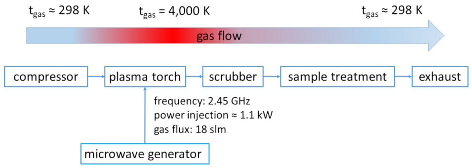
The present paper suggests an indirect microwave plasma-based sanitation method, which applies an indirect treatment via PPA. The PPA is made from ambient air that was carried over a plasma torch. Subsequently, the heat dissipates until the PPA is carried to a treatment chamber. In addition to the application as a tool for decontamination of produce, a PPA-sanitation step offers the possibility for in-process surface decontamination, i.e., a decontamination of produce and surfaces in the food-processing plants at the same time. This step cannot replace a conventional cleaning step but reduces microorganism growth and, thus, supports food safety.

2. Materials and Methods

2.1. Plasma Process

Plasma opens the possibility for various treatment regimes. Parameters, such as the process gas, the pressure, or the applied power density are highly variable and are easily adapted to different produce or different processing technologies. In the presented context, air seems to be very efficient reactant for the microbiological inactivation. Moreover, air plasmas are excellent sources of reactive oxygen and nitrogen species, such as O, O2, O3, OH, NO, and NO2 [28,29]. Schnabel et al. controlled the composition of PPA by mass spectroscopy and showed that 2.7% of the compressed air, which is used as a working gas, is transformed into reactive and antimicrobial-potent species. This paper also summarizes the exact composition of PPA, which was identically produced in the presented experiments [19].
The single-stage atmospheric plasma source, which was used throughout the experiments, is a microwave-driven (2.45 GHz) pulsed plasma generator. It has a power injection of approx. 1.1 kW. Accordingly, the gas temperature is about 4000 K using a gas flux of 18 slm air as a process gas (dry ambient air, dew point: 3 °C). A period of the pulse rate (on/off) has a duration of 10 s, whereas a 5 s ignition phase is implemented. In principle, the presented indirect plasma treatment of solids such as produce or surfaces is segmented into two sub steps (Figure 1). First, in the pre-treatment (PT) the PPA is generated and collected in an ordinary laboratory glass bottle (Carl Roth, Karlsruhe, Germany). In the presented experiments, three different timeframes for a PT were applied (5 s, 15 s, and 50 s), whereas the dimension of the timeframes only reflects the ignition of the plasma torch. For instance, a 5 s pre-treatment embraces a whole pulse period (which has a duration of 10 s), i.e., a 50 s PT includes 10 periods. In the second sub step, the freshly generated PPA was carried over a conduit system into a decontamination chamber where the technical specimens were placed. The so-called post-treatment (POT) has timeframes of 1 min, 3 min, and 5 min. Since the PPA was carried over high-grade steel pipes into the treatment chamber, it reaches the chamber at a temperature of approximately 26 °C. The combinations for all PT and POT to different treatment regimens are summarized in Table 1.

2.2. Technical Specimens and Their Characterization

For the investigation of a broad spectrum of materials that are common in food-processing plant manufacturing, the materials polyethylene (PE), glass (G), and high-grade steel (HGS) were used as surfaces for microbial contamination. The specimens were cut into a rectangular shape with various dimensions. The specimens were sterilized in a sterilizer for every experiment. The roughness of the specimens was determined by scanning probe microscopy (SPM). The SPM measurements were carried out on a DI CP II SPM (Veeco, Plainview, NY, USA), which was mounted on a vibration-free object table (TS-150, TableStable, Zwillikon, Switzerland). The setup was standing on an optical bench encased by an additional acoustic protection. The AFM was equipped with a linearized piezo scanner, on which the specimens were mounted on a metal sample holder. The samples were measured using cantilevers with nominal spring constants of k = 0.1–0.6 N·m−2 in contact mode. The pictures were taken with a scanning speed of 0.5 Hz by a picture size of 25 µm2 and a set point = 8 N·m2. Pictures were edited with the freeware Gwyddion (Czech Metrology Institute, Brno, Czech Republic). Table 2 summarizes the basic surface parameters of the specimens.

2.3. Microbiology and Microbial Preparation of the Technical Specimens

For the recent experiments the microorganisms Listeria monocytogenes, Staphylococcus aureus, Escherichia coli, Salmonella enterica subsp. enterica serovar Typhimurium, and Salmonella enterica sups. enterica serovar Enteritidis were used. Table 3 summarizes the basic information of all investigated microorganisms.
For the contamination of the technical specimens, a bacterial colony-forming unit (cfu) of a microbial strain was suspended in nutrient solutions of 20 mL in a conical glass flask. The nutrition solutions were aligned to the microorganism (see Table 3). The culture was incubated at 37 °C (for all microorganisms) for 4 h on a shaker (80 rpm). Hereafter, the specimens were submerged in a diluted (1:30) microbial suspension of 1.3 mL in an Eppendorf tube (1.5 mL). Subsequently, the specimens were cultured overnight at 37 °C on a shaker (80 rpm).
Immediately before the decontamination step, all specimens were washed in PBS and subsequently stored in a fresh Eppendorf tube (1.5 mL). For the decontamination, the specimens were kept upright in a tray, which were laid into the decontamination chamber. Subsequently the PPA was carried into the decontamination chamber and the technical specimens were treated for various POT times (see Table 1).
Hereafter, the inoculated specimens underwent a determination of the total viable count of the particular microorganism. Therefore, the treated specimens were shaken in a nutrient solution, which depends on the microorganism, for 15 min. Based on that solution, a serial dilution was prepared (1:10–1:1000). Further, 50 µL of each dilution was plated out on an agar plate and was stored at 37 °C overnight. The day after, cfus grown on the plates were counted and were used for the determination of the RF. For simplicity, Salmonella enterica subps. enterica serovar Enteritidis and Salmonella enterica subsp. enterica serovar Typhimurium are referred to as Enteritidis and Typhimurium in the remainder of the text.

2.4. Theoretical Experimental Handling

As mentioned above, the data points embrace three repetitions under the same conditions (N = 3). By the measurements, most values variate in between a narrow range of cfus depending on the performed experiments with some outliers to high and extremely high counts of cfu. Those extremely high counts infrequently appear on agar plates with a dense growth, which were uncountable and, thus, were rejected for the calculation of any statistical moment or RFs. Behind the background of the distribution of the values of a single N in the experiments, a log10-normal distribution is assumed. All statistical moments such as the geometric mean, the multiplicative standard deviation, and t-tests were computed based on an underlying log-normal distribution [30].
Every RF was calculated with respect to a reference, which is stored and handled in the same way as the PPA-treated samples. The RFs were calculated as follows:
RF = log 10 cfu ref log 10 cfu samp
where cfuref is the count of cfus obtained from the references and cfusamp the count of the cfus on PPA-treated samples. The detection limit of this procedure was one cfu/mL. Since inactivation kinetic using an RF was presented in this work, the lowest detectable value is in the range of the number of microorganisms (in log10 (cfu)) of the inoculated sample in the initial state. For a straightforward method, the number of microorganisms as log10 (cfu) used for the inoculation of the samples was used as the detection limit. On the other hand, this approach appears virtually impossible for the representations, since all of the figures embrace values that were obtained from different samples with diverse microorganisms, materials, or treatment times. Thus, the detection limit was set to log10 (cfu) = 5, which is within the standard variation of the cfu counts of the references.
Based on SPM measurements, the effective area of every technical sample was determined (data not shown). The uncertainties of the surface areas for every technical sample vary in a narrow range. Thus, due to an intuitive data representation, the uncertainties of the surface areas in the technical samples were not considered, i.e., the cfu, obtained from the technical samples were not normalized by the effective area.

3. Results

In the current experiments, technical specimens, which are frequently forming surfaces of food-processing equipment, were inoculated artificially. As a representation of the broad spectrum of microorganisms that could occur in a food-production chain, the specimens were contaminated with E. coli, S. aureus, Typhimurium, Enteritidis, and L. monocytogenes. After an incubation time of approx. 12 h (overnight) at 37 °C, the specimens were plasma treated with various regimes. Table 1 summarizes the applied regimes, which are divided into a pre-treatment and, hereafter, a post-treatment. For a more detailed description of the plasma processes, see the Experimental section of the current paper.

3.1. Characterization of the Technical Specimens

For the characterization of the technical specimens, SPM measurements were applied. The focus for the measurements was to gain insight about the relevant surface parameters. Table 2 summarizes the parameter area, relative area, and roughness.

3.2. RFs Depending on the Microorganism

Figure 2 opens an overview of all experiments and shows the inactivation kinetics of several microorganisms. The series of experiments embraces five different microorganisms (L. monocytogenes, E. coli, S. aureus, Typhimurium, Enteritidis), which were cultured on the surfaces of three different materials (PE, G, and HGS). Subsequently, all inoculated specimens underwent a two-step plasma-treatment. The treatment is subdivided into two interdependent plasma processes. The first process, the PT, generates the PPA, which lasts up to 50 s. Hereafter, the PPA is carried into a decontamination chamber, where the second process takes place. Actually, the second process comprises the treatment of produce. In that so-called POT, the samples are exposed to the PPA for up to 5 min. Since the PPA is composed of various chemically active compounds, such as NOX or, rather rarely, oxygen radicals, PPA possesses strong potential to inactivate surface-hosted microorganisms. Due to an increased PT, a strong decrease in proliferation for all bacteria was observed in all experiments.
It is obvious that an RF of minimum two log10 steps is obtained for all microorganisms after a 5 min post-treatment in the presence of PPA from a 50 s pre-treatment (Figure 2c). For that treatment regime, RFs frequently run below the detection limit. Enteritidis is on PE (POT ≤ 3 min), S. aureus, E. coli, and Enteritidis on G (POT: 5 min, all of them), plus on HGS S. Enteritidis and Typhimurium (POT: ≤3 min), together with L. monocytogenes (POT: 5 min), responded that way to PPA treatment. In addition to high RFs, which lie below the detection limit, the highest RF was determined on G for Enteritidis after a POT time of 5 min and a PT of 50 s (RF: 3.06 ± 0.74). The lowest RF was reached on G for L. monocytogenes (RF: 2.01 ± 0.72). Lower RF with a relatively potent disinfectant, such as PPA generated in a 50 s pre-treatment time, was observed for L. monocytogenes on PE and G (PE: 2.43 ± 0.41). Table 4 summarizes the highest RF for every microorganism and for various treatment regimes. Only E. coli shows a so-called tailing behavior [31], which is an increase in the RF to its maximum after a short POT (≤1 min) that stays constant over the residual POT.
As one can see from Table 4, the highest RF for all microorganisms is obtained after the most powerful post-treatment (5 min) based on a potent PPA, producing pre-treatment (50 s, Figure 2c) either on HGS (L. monocytogenes and Typhimurium), PE (Enteritidis), or on G (S. aureus, E. coli, and Enteritidis). In contrast, most of the lowest RFs for the tested microorganisms (L. monocytogenes, S. aureus, or E. coli, and Typhimurium) were obtained after a soft treatment (PT: 5 s and POT: 1 min) on PE or HGS, respectively (Figure 1), whereas Enteritidis revealed its lowest RF after a slightly stronger treatment with a longer PPA exposure (POT: 3 min) and a more potent PPA itself (PT: 15 s) on an HGS surface (Figure 2b).

3.3. RFs Depending on the Surface Material

Figure 2 additionally emphasizes the dependency of the RFs for the monitored microorganisms from their substrates, which are the materials PE, G, and HGS. As described briefly above, the foundations of Figure 2 are the experiments (and the resulting RFs), which are summarized in Table 3. Consequently, the highest RFs were obtained after the strongest treatment (PT: 50 s and POT: 5 min) on every material and fall below the detection limit (Figure 2c). In no particular order, RFs below the detection limit were obtained on G for Enteritidis, E. coli, and S. aureus after a PT of 50 s and a POT of 5 min, on HGS for Typhimurium and Enteritidis (PT: 50 s, POT: ≤3 min), together with L. monocytogenes (PT: 50 s, POT: 5 min). All RFs exceeded a limit of 2 log10 steps, with a minimum of RF: 2.01 ± 0.72 for L. monocytogenes on PE. Noteworthily, on PE, generally, the lowest RFs were obtained. As a great exemption, some RFs obtained on G and HGS by semi-strong or strong post-treatments (1 min < treatment ≤ 5 min) and a strong pre-treatment (50 s) fall below the ones obtained for PE (E. coli on HGS POT: 1 min, L. monocytogenes on G POT: 5 min, and Typhimurium on G POT: 5 min). Table 5 summarizes the effect of a PPA treatment on microorganisms hosted on the various materials.

3.4. RFs Depending on the Pre-Treatment

Further, also visible from Figure 2 is the PT-dependent RF progress over the monitored POT time. As described in the previous sections, the foundation of Figure 2 is also the experimental set that underlies the preceding graphs and tables. In contrast, this presentation emphasizes the length of the PT treatment, i.e., the antimicrobial strength of the PPA. As is obvious from the other representations, a 50 s PT leads to the strongest RF for every microorganism on every monitored surface material. When the PT time is increased, especially G and HGS surfaces showed a pronounced growth in the RF over the POT time. Consequently, Figure 2 shows very distinct inactivation kinetics for S. aureus, E. coli, and Enteritidis on G as well as Typhimurium, Enteritidis and L. monocytogenes on HGS, in terms of a higher RF for a longer PT time. All RFs for the mentioned microorganisms run below the detection limit. The inactivation kinetics found on PE for the microorganisms, which are not explicitly mentioned above, strongly differ from the later ones in terms of lower RFs, around approx. 2.5. The inactivation kinetics for L. monocytogenes and Typhimurium found on G did not fall below the detection limit (Figure 2c). Enteritidis showed varying inactivation kinetics on PE. Here, a POT ≤ 1 min reveals a sharp increase in the RF, which runs below the detection limit after a POT of 3 min. Table 1 summarizes the relevant values for RF in the dependency of the PT time.
As is represented by the data, no pronounced trend for the RFs influenced by the roughness is obvious. All microorganisms show the highest RFs on G, whereas the RFs on HGS and PE are not significantly different. For different roughness and surface chemistries, only the variance in the RFs differs on the various surfaces. Additionally, microorganisms were divided into a Gram-positive and Gram-negative group, which shows no significant difference in their RF. For that classification, G and HGS frequently show RFs, which run below the detection limit (Gram-negative on G: Enteritidis and E. coli and Gram-positive on G: S. aureus, Gram-negative on HGS: Enteritidis and Typhimurium as well as Gram-positive on HGS: L. monocytogenes). Table 6 outlines the range of treatment outcomes using extreme values

4. Discussion

How did PPA decontamination occur? The property of PPA for decontamination lies in the production of RNS, which was proved in several studies [22,23,27,
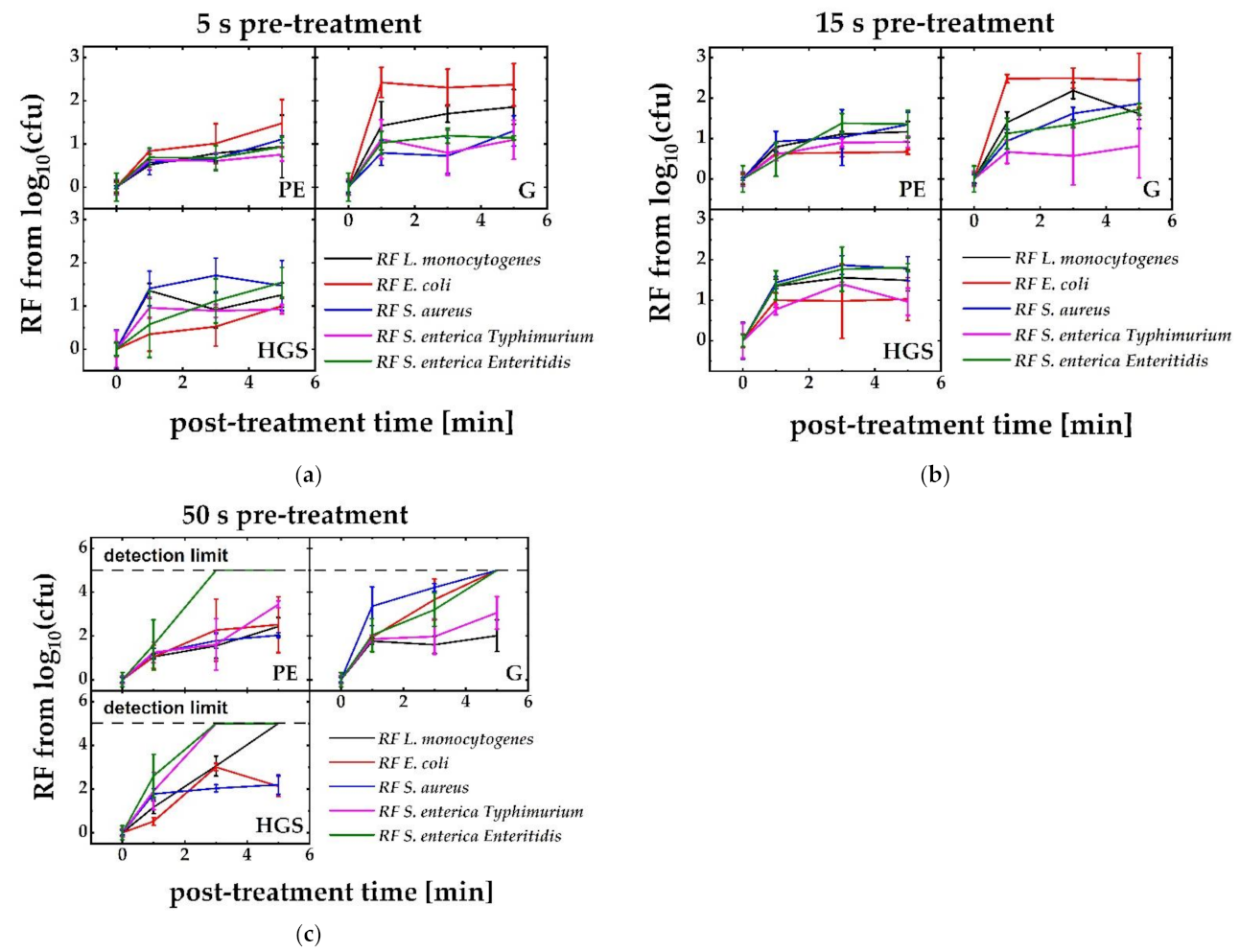
32]. When the bacteria, which are being used in the experiments, undergo an overtreatment in regimes with very potent PPA, the RFs fall below a detection limit. This behavior was found on every surface material for distinct microorganisms. Specifically, Enteritidis reacts in a similar manner to an overtreatment and falls below the detection limit on all materials. However, maximum RFs that are generally obtained in the experiments lie around four log10 steps. In comparison, other authors describe higher RF, up to 5.5 log10 steps [19], with the very same setup for P. fluorescence, 6.5 log10 steps on more complex surfaces, such as eggshells [33], with a resistive barrier discharge, or 6–9 log10 steps for S. aureus, with a cascaded dielectric barrier discharge on PET foils [28]. At first glance, the presented method of an indirect plasma-treatment seemed to be less effective than other plasma-based methods for a surface decontamination. We simply did not reach the RFs as described in the literature. However, it has to be mentioned that the other processes need treatment times of up to 60 min [33] or prefer a direct treatment that uses plasma ignition directly on the surface [28,34]. These methods are time consuming and, thus, not applicable in a commercial value chain or highly impartible for sensitive materials, such as food. Moreover, technically, plasma sources described in the literature are frequently run by inert gases or a mixture of gases, such as O2 or H2 [35]. On the contrary, PPA just needs compressed ambient air for its production, which additionally lowers the costs. Thus, the usage of indirect plasma methods for the sanitation is under active research, because it allows a fast and safe sterilization of different materials from glass over polymers to human tissue [24,36,37]. Of course, Schnabel et al. [38] used the same setup as we did, but since every single experiment is somehow unique in terms of repairs or maintenance of the plasma source and preferences in handling by the users, variation in a log10 step is reasonable. Additionally, the initial, on-growing concentration of microorganisms, directly on the surface, could be different in the experiments among the discussed publications.
The kinetics of an antimicrobial inactivation of different microorganisms on various surface materials appear very different. At a first, microbiologic appearance, it can be addressed to the cell wall structure of the cell, i.e., is the bacteria Gram-negative or Gram-positive? In the presented experiments, S. aureus and L. monocytogenes are Gram-positive bacteria, whereas the two Salmonellas and E. coli are Gram-negative. Because of hydrolysis in the cell wall, Gram-negative bacteria are more sensitive against acidification of their surrounding media, which is obvious in the experiments as Enteritidis seems to be very sensitive to PPA treatment. The RFs of the Gram-negative bacteria Enteritidis reliably run below the detection limit for intense treatment regimes. Only a treatment with less potent PPA (a PT of 5 s and 15 s) revealed higher RFs for the Gram-positive S. aureus than for any other Gram-negative bacteria. Because the graphs of the inactivation kinetics running in parallel and the RF for S. aureus frequently lie in the error tolerance of Enteritidis, these findings likely appear to be negligible. Due to a minor response to a PPA treatment of Gram-staining distinguishable bacteria, the composition of the cell wall seemed not to have a dominant role in our presented experiments. For all overtreatments, every single set of experiments only showed RFs below the detection limit for a maximum three microorganisms. For instance, the RFs of S. aureus, E. coli, and Enteritidis fall below the detection limit when G surfaces undergo PPA treatment. That is, the RFs of two Gram-negative and one Gram-positive species lie below the detection limit after a POT of 5 min. Since we have only a group of three bacteria, one cannot assume an effect of Gram-distinguishable bacteria. Thus, coincidence is within the realms of possibility. Nevertheless, Schnabel et al. [29] described Gram-negative pathogens as being more sensitive to PPA treatments, a finding that we can support, at least for the PPA-sensitive species Enteritidis. The author addresses this effect to a higher susceptibility against acidic surroundings that destabilize the membrane [19,39]. Schnabel et al. assigned the effect to the diffusion properties in the bacterial cell of the major gas components NO and NO2. Minor components, such as CO2, H2O, and HNO2, may play a subsidiary role [40].
However, a detailed representation of the steps leading to an inactivation of the bacteria is still under research. Resistances against nitric oxides are unknown up to date, which might because intracellular nitric oxides are signaling and defense molecules [41,42,43]. Various authors [41,44,45] highlighted enterobacterial flavohaemoglobin (Hmp) for NO detoxification. Evidentially, Hmp holds a central role when nitrosative stress overcomes a bacterium [41]. The ambiguous effectivity of nitric oxides and their energetically excited species (which also embraces RNS) may lower the bacteria’s ability to develop highly effective defense strategies, which is the case for oxidative stress [46,47]. Notably, prokaryotes and eukaryotes both possess specific forms of the enzyme superoxide dismutase (SOD), which is occasionally lacking in a few anaerobe forms [48]. The SOD catalyzes the dismutation of superoxide radicals, which might seriously damage the cell [49,50]. Behind that background, a treatment with RNS may be favorable. This advantage becomes more substantial, since reactive oxygen species are consumed by organic contaminations, which are logically present during food processing. Another notable aspect behind the discussed background is a possible viable but non-culturable state (VBNC state) in many bacteria, which is supposed to be a surviving strategy that is comparable to the sporulation or dormancy of other bacteria [51,52]. Under these circumstances, bacteria do not proliferate on or in nutrient media and, thus, are not detectable via ordinary proliferation assays. Moreover, the metabolic activity widely remains unchanged [53,54]; solely the synthesis of several macromolecules and the cell breath are affected [55] and the cells retain their virulence, which means other tests than proliferation assays are desirable to determine RFs. The microorganisms, which are used in the experiments, show such a behavior. VBNC states are known for L. monocytogenes [56,57], Salmonella [58,59], S. aureus [60], and E. coli [61,62]. Specifically, a low pH value tends into the focus for a trigger for a VBNC state [63,64].
The obtained inactivation kinetics were not proportional to time, which indicates a multi-phase kinetic, a phenomenon that is also described by others [65]. Frequently, experiments reveal a high RF after a short POT (approx. 1 min), which stays constant over the residual treatment time. This behavior is called tailing [66]. On the technical specimens, the graphs show only tailing in the case of E. coli, i.e., a maximum inactivation rate for E. coli was reached after one minute POT and a PT of 15 s. For RFs running in a global tailing, Schnabel et al. [19] suggested a rough surface that supports a strong linkage to the surface that negatively influences the recovery process and the detection in the used proliferation assay; this is an explanation that cannot be suitable for the presented data. In our experiments, tailing behavior was rather related to a pre-treatment time than to a specific material. Additionally, a general behavior of E. coli, which is independent from the substrate and the treatment, cannot be subjected in the case of the presented study. This may be in correlation with the limited penetration depth of plasma of a few nanometers. Therefore, stacked bacteria or surface layers may gradually slow down the inactivation process [29], which is, again, an effect that is independent from the surface material. Gusbeth et al. [67] described the deviation from the first-order kinetics by a different sensitivity of pelagic bacteria, spores, and bacteria hosted in biofilms. This explanation is still possible, because a 15 s pre-treatment can be strong enough to harm microorganisms in the suggested way and gently enough not to cover the effect by an overtreatment. However, the effect was not directly addressable via the conducted experiments.
Since no strict dependency from the surface material was observed, no surface variations, such as roughness or surface potential, seem to be meaningful for bacterial colonization. The colonization of surface embraces various steps, such as protein deposition on the surface, cell attachment or the ability of the cell to build a biofilm [68,69,70,71]. Behind that background, our findings support the thesis that the colonization and the resilience against PPA treatments are predominantly governed by the bacterial properties that vary from strain to strain but, abundantly, also from cell to cell or from their contemporary life cycle, which gives the kinetic graphs huge variations and might explain the observed inconsistency.
How do we see an “in-process” sanitation step? It is simply a cleaning step, which can be conducted between two processing steps or during the process itself. As the processing of food needs secure sanitation steps in terms of a reduction in microbial load, a conventional method for sanitation is the use of a sanitizer after the production when the plants stop running. Thereby, the frequency of a sanitation step depends on the produce. For instance, dryer produce normally does not frequently need a cleaning procedure. Ordinarily, dry produce, with a few exceptions, such as wheat grain [72], is not that susceptible to contamination, such as humid produces, which offers a good substrate for microbial growth. Especially in meat and fish processing, a spreading of microorganisms appears more likely due to deficient sanitation. As a result, frequent cleaning steps are necessary to guarantee safe produce, which fulfill microbial specifications and prevent foodborne illnesses for consumers. Behind that background, an in-process cleaning step, which inhibits the growth of microorganisms during the process, would be desirable, since continuous production will lower the costs of produce. Such a process will save resources in terms of time, materials, such as sanitizers or basic materials, and work force. PPA appears to be highly suitable for an in-process sanitation step. Generally, plasma-based surface sanitation is well described for medical devices [73], in high-vacuum environments [74] and in food processing [24], as a vast number of patents [75] and publications [36,40,76,77] proves. Schnabel et al. [29,40] and Fröhling et al. [78] described indirect plasma-based sanitation methods, which also embrace PPA treatment. These methods are particularly suitable for the treatment of foods. PPA has accessibility to caves, holes, and cavities, which cannot be eliminated by a hygienic design. Additionally, PPA is easy to produce by a plasma source, which is addable to any enclosed food-processing plant. The cleaning step itself can be conducted between the processes without any interventions by an operator. Thus, such a cleaning step is highly suitable for automated processes.

5. Conclusions

An indirect quasi-thermal plasma treatment is a promising technology for the food industry. It acts rapidly and beside nitrous compounds in the exhaust gases, which is contradictable by a scrubber column and it does not leave any toxic residuals on the processed parts. Additionally, the temperature rise can be kept at an acceptable level [79]. The cocktail of antimicrobial-potent plasma species generated via a microwave-driven plasma source makes quasi-thermal plasma suited for the decontamination of more sensitive surfaces in food-processing settings [80,81,82,83,84]. The groove penetration properties of the presented method make it an ideal tool for a quick sanitation step, either to replace or to support common sanitation steps. The antimicrobial efficacy of the PPA method is, for the tested microorganisms, comparable to other sanitation methods, such as, for instance, the use of chlorine dioxide, which has the disadvantage to attack seals and various synthetic materials. Due to easy handling, it can be integrated into established sanitation strategies, such as a CIP scenario.
In addition to these more general interpretations of the results, dependency of the inactivation efficiency due to the used microorganisms, the various treatment regimes, and of the surface was detected. Behind that background, it became obvious that the influence of the controlled substrate materials is very low and is, for some cases, negligible. For the recent data, there is no significant difference for the inactivation of Gram-positive or Gram-negative microorganisms detected. The major effect, which increases the inactivation efficiency, is the prolongation of the PT and the POT. The chemistry, which is underlying an indirect PPA treatment, should be subjected to further research efforts.

Author Contributions

Conceptualization, T.W., U.S.; investigation, T.W. and U.S.; validation, T.W., U.S., and M.A.; formal analysis, T.W. and M.A.; writing original draft, T.W. and U.S.; writing review editing, T.W., U.S., and M.A.; methodology, T.W.; M.A. and J.S.; supervision, J.E. and F.T.; funding acquisition, J.E. and F.T. All authors have read and agreed to the published version of the manuscript.

Funding

The research have been founded by the Federal Ministry for Economic Affairs and Climate Action, grant number 16KN017432.

Institutional Review Board Statement

Not applicable.

Informed Consent Statement

Not applicable.

Data Availability Statement

The data that support our findings is available for download at zenodo/doi 10.5281/zenedo.7006632.

Conflicts of Interest

The authors declare no conflict of interest.

References

  1. Garcia Martinez, M.; Fearne, A.; Caswell, J.A.; Henson, S. Co-regulation as a possible model for food safety governance: Opportunities for public–private partnerships. Food Policy 2007, 32, 299–314. [Google Scholar] [CrossRef]
  2. Holah, J.T. Industrial Monitoring: Hygiene in Food Processing. In Biofilms—Science and Technology; Melo, L.F., Bott, T.R., Fletcher, M., Capdeville, B., Eds.; Springer: Dordrecht, The Netherlands, 1992; pp. 645–659. [Google Scholar]
  3. Bruhn, C.M. Consumer concerns: Motivating to action. Emerg. Infect. Dis. 1997, 3, 511–515. [Google Scholar] [CrossRef] [PubMed]
  4. Powell, D.A.; Jacob, C.J.; Chapman, B.J. Enhancing food safety culture to reduce rates of foodborne illness. Food Control 2011, 22, 817–822. [Google Scholar] [CrossRef]
  5. Novotny, L.; Dvorska, L.; Lorencova, A.; Beran, V.; Pavlik, I. Fish: A potential source of bacterial pathogenes for human beings. Vet. Med. 2004, 49, 343–358. [Google Scholar] [CrossRef]
  6. Tham, W.; Ericsson, H.; Loncarevic, S.; Unnerstad, H.; Danielsson-Tham, M.L. Lessons from an outbreak of listeriosis related to vacuum-packed gravad and cold-smoked fish. Int. J. Food Microbiol. 2000, 62, 173–175. [Google Scholar] [CrossRef]
  7. EFSA. The European Union summary report on trends and sources of zoonoses, zoonotic agents and food-borne outbreakes in 2015. EFSA J. 2015, 12, 173–175. [Google Scholar]
  8. LundÉN, J.M.; Autio, T.J.; SjÖBerg, A.M.; Korkeala, H.J. Persistent and Nonpersistent Listeria monocytogenes Contamination in Meat and Poultry Processing Plants. J. Food Prot. 2003, 66, 2062–2069. [Google Scholar] [CrossRef]
  9. LundÉN, J.M.; Autio, T.J.; Korkeala, H.J. Transfer of Persistent Listeria monocytogenes Contamination between Food-Processing Plants Associated with a Dicing Machine. J. Food Prot. 2002, 65, 1129–1133. [Google Scholar] [CrossRef]
  10. Rivera-Betancourt, M.; Shackelford, S.D.; Arthur, T.M.; Westmoreland, K.E.; Bellinger, G.; Rossman, M.; Reagan, J.O.; Koohmaraie, M. Prevalence of Escherichia coli O157:H7, Listeria monocytogenes, and Salmonella in Two Geographically Distant Commercial Beef Processing Plants in the United States. J. Food Prot. 2004, 67, 295–302. [Google Scholar] [CrossRef] [PubMed]
  11. Jackson, L.S.; Al-Taher, F.M.; Moorman, M.; DeVries, J.W.; Tippett, R.; Swanson, K.M.J.; Fu, T.-J.; Salter, R.; Dunaif, G.; Estes, S.; et al. Cleaning and Other Control and Validation Strategies to Prevent Allergen Cross-Contact in Food-Processing Operations. J. Food Prot. 2008, 71, 445–458. [Google Scholar] [CrossRef]
  12. Poulis, J.A.; de Pijper, M.; Mossel, D.A.A.; Dekkers, P.P.A. Assessment of cleaning and disinfection in the food industry with the rapid ATP-bioluminescence technique combined with the tissue fluid contamination test and a conventional microbiological method. Int. J. Food Microbiol. 1993, 20, 109–116. [Google Scholar] [CrossRef]
  13. Mettler, E.; Carpentier, B. Variations over Time of Microbial Load and Physicochemical Properties of Floor Materials after Cleaning in Food Industry Premises. J. Food Prot. 1998, 61, 57–65. [Google Scholar] [CrossRef]
  14. De Oliveira, F.A.; Neto, O.C.; dos Santos, L.M.R.; Ferreira, E.H.R.; Rosenthal, A. Effect of high pressure on fish meat quality—A review. Trends Food Sci. Technol. 2017, 66, 1–19. [Google Scholar] [CrossRef]
  15. Abdallah, M.; Chataigne, G.; Ferreira-Theret, P.; Benoliel, C.; Drider, D.; Dhulster, P.; Chihib, N.-E. Effect of growth temperature, surface type and incubation time on the resistance of Staphylococcus aureus biofilms to disinfectants. Appl. Microbiol. Biotechnol. 2014, 98, 2597–2607. [Google Scholar] [CrossRef] [PubMed]
  16. Sundheim, G.; Langsrud, S.; Heir, E.; Holck, A.L. Bacterial resistance to disinfectants containing quaternary ammonium compounds. Int. Biodeterior. Biodegrad. 1998, 41, 235–239. [Google Scholar] [CrossRef]
  17. Itoh, Y.; Sugita-Konishi, Y.; Kasuga, F.; Iwaki, M.; Hara-Kudo, Y.; Saito, N.; Noguchi, Y.; Konuma, H.; Kumagai, S. Enterohemorrhagic Escherichia coli O157:H7 Present in Radish Sprouts. Appl. Environ. Microbiol. 1998, 64, 1532–1535. [Google Scholar] [CrossRef]
  18. Lang, M.M.; Ingham, B.H.; Ingham, S.C. Efficacy of novel organic acid and hypochlorite treatments for eliminating Escherichia coli O157:H7 from alfalfa seeds prior to sprouting. Int. J. Food Microbiol. 2000, 58, 73–82. [Google Scholar] [CrossRef]
  19. Schnabel, U.; Schmidt, C.; Stachowiak, J.; Bösel, A.; Andrasch, M.; Ehlbeck, J. Plasma processed air for biological decontamination of PET and fresh plant tissue. Plasma Process. Polym. 2018, 15, 185–192. [Google Scholar] [CrossRef]
  20. Carpentier, B.; Cerf, O. Review—Persistence of Listeria monocytogenes in food industry equipment and premises. Int. J. Food Microbiol. 2011, 145, 1–8. [Google Scholar] [CrossRef] [PubMed]
  21. Denes, A.R.; Somers, E.B.S.; Wong, A.C.L.; Denes, F.S. Plasma-Aided Treatment of Surfaces against Bacterial Attachment and Biofilm Deposition. U.S. Patent 6096564A, 1 August 2000. [Google Scholar]
  22. Baier, M.; Gorgen, M.; Ehlbeck, J.; Knorr, D.; Herppich, W.B.; Schluter, O. Non-thermal atmospheric pressure plasma: Screening for gentle process conditions and antibacterial efficiency on perishable fresh produce. Innov. Food Sci. Emerg. Technol. 2014, 22, 147–157. [Google Scholar] [CrossRef]
  23. Andrasch, M.; Stachowiak, J.; Schlüter, O.; Schnabel, U.; Ehlbeck, J. Scale-up to pilot plant dimensions of plasma processed water generation for fresh-cut lettuce treatment. Food Packag. Shelf Life 2017, 14, 40–45. [Google Scholar] [CrossRef]
  24. Ehlbeck, J.; Schnabel, U.; Polak, M.; Winter, J.; von Woedtke, T.; Brandenburg, R.; von dem Hagen, T.; Weltmann, K.D. Low temperature atmospheric pressure plasma sources for microbial decontamination. J. Phys. D-Appl. Phys. 2011, 44, 013002. [Google Scholar] [CrossRef]
  25. Ehlbeck, J.; Ohl, A.; Maass, M.; Krohmann, U.; Neumann, T. Moving atmospheric microwave plasma for surface and volume treatment. Surf. Coat. Technol. 2003, 174, 493–497. [Google Scholar] [CrossRef]
  26. Schnabel, U.; Niquet, R.; Schlüter, O.; Gniffke, H.; Ehlbeck, E. Decontamination and sensory properties of microbiologically contaminated fresh fruits and vegetables by microwave plasma processed air (PPA). J. Food Process. Preserv. 2014, 39, 653–662. [Google Scholar] [CrossRef]
  27. Schnabel, U.; Niquet, R.; Krohmann, U.; Winter, J.; Schluter, O.; Weltmann, K.D.; Ehlbeck, J. Decontamination of Microbiologically Contaminated Specimen by Direct and Indirect Plasma Treatment. Plasma Process. Polym. 2012, 9, 569–575. [Google Scholar] [CrossRef]
  28. Muranyi, P.; Wunderlich, J.; Heise, M. Sterilization efficiency of a cascaded dielectric barrier discharge. J. Appl. Microbiol. 2007, 103, 1535–1544. [Google Scholar] [CrossRef]
  29. Schnabel, U.; Andrasch, M.; Weltmann, K.D.; Ehlbeck, J. Inactivation of Vegetative Microorganisms and Bacillus atrophaeus Endospores by Reactive Nitrogen Species (RNS). Plasma Process. Polym. 2014, 11, 110–116. [Google Scholar] [CrossRef]
  30. Limpert, E.; Stahel, W.A.; Abbt, M. Log-normal Distributions across the Sciences: Keys and Clues: On the charms of statistics, and how mechanical models resembling gambling machines offer a link to a handy way to characterize log-normal distributions, which can provide deeper insight into variability and probability—Normal or log-normal: That is the question. Bioscience 2001, 51, 341–352. [Google Scholar]
  31. Lambert, R.J.W.; Johnston, M.D. Disinfection kinetics: A new hypothesis and model for the tailing of log-survivor/time curves. J. Appl. Microbiol. 2000, 88, 907–913. [Google Scholar] [CrossRef]
  32. Baier, M.; Foerster, J.; Schnabel, U.; Knorr, D.; Ehlbeck, J.; Herppich, W.B.; Schluter, O. Direct non-thermal plasma treatment for the sanitation of fresh corn salad leaves: Evaluation of physical and physiological effects and antimicrobial efficacy. Postharvest Biol. Technol. 2013, 84, 81–87. [Google Scholar] [CrossRef]
  33. Ragni, L.; Berardinelli, A.; Vannini, L.; Montanari, C.; Sirri, F.; Guerzoni, M.E.; Guarnieri, A. Non-thermal atmospheric gas plasma device for surface decontamination of shell eggs. J. Food Eng. 2010, 100, 125–132. [Google Scholar] [CrossRef]
  34. Montie, T.C.; Kelly-Wintenberg, K.; Roth, J.R. An overview of research using the one atmosphere uniform glow discharge plasma (OAUGDP) for sterilization of surfaces and materials. IEEE Trans. Plasma Sci. 2000, 28, 41–50. [Google Scholar] [CrossRef]
  35. Rossi, F.; Kylián, O.; Hasiwa, M. Decontamination of Surfaces by Low Pressure Plasma Discharges. Plasma Process. Polym. 2006, 3, 431–442. [Google Scholar] [CrossRef]
  36. Deilmann, M.; Halfmann, H.; Bibinov, N.; Wunderlich, J.; Awakowicz, P. Low-Pressure Microwave Plasma Sterilization of Polyethylene Terephthalate Bottles. J. Food Prot. 2008, 71, 2119–2123. [Google Scholar] [CrossRef]
  37. Koulik, P.; Begounov, S.; Goloviatinskii, S. Atmospheric Plasma Sterilization and Deodorization of Dielectric Surfaces. Plasma Chem. Plasma Process. 1999, 19, 311–326. [Google Scholar] [CrossRef]
  38. Schnabel, U.; Andrasch, M.; Stachowiak, J.; Weit, C.; Weihe, T.; Schmidt, C.; Muranyi, P.; Schlüter, O.; Ehlbeck, J. Sanitation of fresh-cut endive lettuce by plasma processed tap water (PPtW)—Up-scaling to industrial level. Innov. Food Sci. Emerg. Technol. 2019, 53, 45–55. [Google Scholar] [CrossRef]
  39. Salton, M.R.J. Studies of the bacterial cell wall: IV. The composition of the cell walls of some gram-positive and gram-negative bacteria. Biochim. Biophys. Acta 1953, 10, 512–523. [Google Scholar] [CrossRef]
  40. Schnabel, U.; Oehmigen, K.; Naujox, K.; Rackow, K.; Krohmann, U.; Schmitt, O.; Schlüter, O.; Weltmann, K.D.; Von Woedtke, T.; Ehlbeck, J. Inactivation of Escherichia coli K-12 and enterohemorrhagic Escherichia coli (EHEC) by atmospheric pressure plasma. J. Agric. Sci. Appl. 2014, 3, 81–91. [Google Scholar] [CrossRef]
  41. Poole, R.K. Nitric oxide and nitrosative stress tolerance in bacteria. Biochem. Soc. Trans. 2005, 33, 176–180. [Google Scholar] [CrossRef]
  42. Poole, R.K.; Hughes, M.N. New functions for the ancient globin family: Bacterial responses to nitric oxide and nitrosative stress. Mol. Microbiol. 2000, 36, 775–783. [Google Scholar] [CrossRef] [PubMed]
  43. Binet, M.R.B.; Cruz-Ramos, H.; Laver, J.; Hughes, M.N.; Poole, R.K. Nitric oxide releases intracellular zinc from prokaryotic metallothionein in Escherichia coli. FEMS Microbiol. Lett. 2002, 213, 121–126. [Google Scholar] [CrossRef]
  44. Forrester, M.T.; Foster, M.W. Protection from nitrosative stress: A central role for microbial flavohemoglobin. Free Radic. Biol. Med. 2012, 52, 1620–1633. [Google Scholar] [CrossRef]
  45. Frey Alexander, D.; Farrés, J.; Bollinger Christian, J.T.; Kallio Pauli, T. Bacterial Hemoglobins and Flavohemoglobins for Alleviation of Nitrosative Stress in Escherichia coli. Appl. Environ. Microbiol. 2002, 68, 4835–4840. [Google Scholar] [CrossRef]
  46. Kashef, N.; Hamblin, M.R. Can microbial cells develop resistance to oxidative stress in antimicrobial photodynamic inactivation? Drug Resist. Updates 2017, 31, 31–42. [Google Scholar] [CrossRef]
  47. Storz, G.; Imlayt, J.A. Oxidative stress. Curr. Opin. Microbiol. 1999, 2, 188–194. [Google Scholar] [CrossRef]
  48. Farr, S.B.; D’Ari, R.; Touati, D. Oxygen-dependent mutagenesis in Escherichia coli lacking superoxide dismutase. Proc. Natl. Acad. Sci. USA 1986, 83, 8268–8272. [Google Scholar] [CrossRef]
  49. McCord, J.M.; Keele, B.B.; Fridovich, I. An Enzyme-Based Theory of Obligate Anaerobiosis: The Physiological Function of Superoxide Dismutase. Proc. Natl. Acad. Sci. USA 1971, 68, 1024–1027. [Google Scholar] [CrossRef]
  50. Scott, M.D.; Meshnick, S.R.; Eaton, J.W. Superoxide dismutase-rich bacteria. Paradoxical increase in oxidant toxicity. J. Biol. Chem. 1987, 262, 3640–3645. [Google Scholar] [CrossRef]
  51. Aertsen, A.; Michiels, C.W. Stress and How Bacteria Cope with Death and Survival. Crit. Rev. Microbiol. 2004, 30, 263–273. [Google Scholar] [CrossRef] [PubMed]
  52. Oliver, J.D. Formation of Viable but Nonculturable Cells. In Starvation in Bacteria; Kjelleberg, S., Ed.; Springer: Boston, MA, USA, 1993; pp. 239–272. [Google Scholar]
  53. Ramamurthy, T.; Ghosh, A.; Pazhani, G.P.; Shinoda, S. Current Perspectives on Viable but Non-Culturable (VBNC) Pathogenic Bacteria. Front. Public Health 2014, 2, 1–9. [Google Scholar] [CrossRef] [PubMed]
  54. Rice, S.A.; McDougald, D.; Kjelleberg, S. Vibrio vulnificus: A physiological and genetic approach to the viable but nonculturable response. J. Infect. Chemother. 2000, 6, 115–120. [Google Scholar] [CrossRef] [PubMed]
  55. Rahman, I.; Shahamat, M.; Kirchman, P.A.; Russek-Cohen, E.; Colwell, R.R. Methionine uptake and cytopathogenicity of viable but nonculturable Shigella dysenteriae type 1. Appl. Environ. Microbiol. 1994, 60, 3573–3578. [Google Scholar] [CrossRef] [PubMed] [Green Version]
  56. Lindbäck, T.; Rottenberg, M.E.; Roche, S.M.; Rørvik, L.; Marit, D. The ability to enter into an avirulent viable but non-culturable (VBNC) form is widespread among Listeria monocytogenes isolates from salmon, patients and environment. Vet. Res. 2010, 41, 8. [Google Scholar] [CrossRef]
  57. Valérie, B.; Michel, F.; Eric, D.; Florence, J.; Jean-Michel, C. Environmental and physico-chemical factors induce VBNC state in Listeria monocytogenes. Vet. Res. 2002, 33, 359–370. [Google Scholar]
  58. Keer, J.T.; Birch, L. Molecular methods for the assessment of bacterial viability. J. Microbiol. Methods 2003, 53, 175–183. [Google Scholar] [CrossRef]
  59. Morishige, Y.; Fujimori, K.; Amano, F. Use of flow cytometry for quantitative analysis of metabolism of VBNC (Viable but Non-Culturable) Salmonella. Biol. Pharm. Bull. 2015, 38, 1255–1264. [Google Scholar] [CrossRef] [PubMed]
  60. Jakobsen, R.A.; Heggebø, R.; Sunde, E.B.; Skjervheim, M. Staphylococcus aureus and Listeria monocytogenes in Norwegian raw milk cheese production. Food Microbiol. 2011, 28, 492–496. [Google Scholar] [CrossRef]
  61. Garcia-Armisen, T.; Servais, P. Enumeration of viable E. coli in rivers and wastewaters by fluorescent in situ hybridization. J. Microbiol. Methods 2004, 58, 269–279. [Google Scholar] [CrossRef]
  62. Liu, J.; Zhou, R.; Li, L.; Peters, B.M.; Li, B.; Lin, C.-w.; Chuang, T.-L.; Chen, D.; Zhao, X.; Xiong, Z.; et al. Viable but non-culturable state and toxin gene expression of enterohemorrhagic Escherichia coli O157 under cryopreservation. Res. Microbiol. 2017, 168, 188–193. [Google Scholar] [CrossRef]
  63. Cunningham, E.; O’Byrne, C.; Oliver, J.D. Effect of weak acids on Listeria monocytogenes survival: Evidence for a viable but nonculturable state in response to low pH. Food Control 2009, 20, 1141–1144. [Google Scholar] [CrossRef]
  64. Wan, C.; Zhang, Q.; Lee, D.-J.; Wang, Y.; Li, J. Long-term storage of aerobic granules in liquid media: Viable but non-culturable status. Bioresour. Technol. 2014, 166, 464–470. [Google Scholar] [CrossRef] [PubMed]
  65. Laroussi, M. Nonthermal decontamination of biological media by atmospheric-pressure plasmas: Review, analysis, and prospects. IEEE Trans. Plasma Sci. 2002, 30, 1409–1415. [Google Scholar] [CrossRef]
  66. Azimi, Y.; Chen, X.; Allen, D.G.; Pileggi, V.; Seto, P.; Droppo, I.G.; Farnood, R.R. UV disinfection of wastewater flocs: The effect of secondary treatment conditions. Water Sci. Technol. 2013, 67, 2719–2723. [Google Scholar] [CrossRef] [PubMed]
  67. Gusbeth, C.; Frey, W.; Volkmann, H.; Schwartz, T.; Bluhm, H. Pulsed electric field treatment for bacteria reduction and its impact on hospital wastewater. Chemosphere 2009, 75, 228–233. [Google Scholar] [CrossRef]
  68. Beachey, E.H. Bacterial Adherence: Adhesin-Receptor Interactions Mediating the Attachment of Bacteria to Mucosal Surfaces. J. Infect. Dis. 1981, 143, 325–345. [Google Scholar] [CrossRef]
  69. Donlan, R.M. Biofilm Formation: A Clinically Relevant Microbiological Process. Clin. Infect. Dis. 2001, 33, 1387–1392. [Google Scholar] [CrossRef]
  70. Fukuzaki, S.; Urano, H.; Nagata, K. Adsorption of protein onto stainless-steel surfaces. J. Ferment. Bioeng. 1995, 80, 6–11. [Google Scholar] [CrossRef]
  71. Roach, P.; Farrar, D.; Perry, C.C. Interpretation of Protein Adsorption:  Surface-Induced Conformational Changes. J. Am. Chem. Soc. 2005, 127, 8168–8173. [Google Scholar] [CrossRef]
  72. Laca, A.; Mousia, Z.; Dı́az, M.; Webb, C.; Pandiella, S.S. Distribution of microbial contamination within cereal grains. J. Food Eng. 2006, 72, 332–338. [Google Scholar] [CrossRef]
  73. Halfmann, H.; Bibinov, N.; Wunderlich, J.; Awakowicz, P. A double inductively coupled plasma for sterilization of medical devices. J. Phys. D Appl. Phys. 2007, 40, 4145–4154. [Google Scholar] [CrossRef]
  74. Balwanz, W.W. Plasma Cleaning of Surfaces. In Surface Contamination: Genesis, Detection, and Control; Mittal, K.L., Ed.; Springer: Boston, MA, USA, 1979; pp. 255–269. [Google Scholar]
  75. Shannon, R.L.G. R.B: Plasma Cleaning Device, in Other—Patent, B. CO, Editor 1978, Boeing CO: US. p. 5. Available online: https://patents.google.com/patent/US4088926A/en (accessed on 17 August 2022).
  76. Brauge, T.; Faille, C.; Leleu, G.; Denis, C.; Hanin, A.; Midelet, G. Treatment with disinfectants may induce an increase in viable but non culturable populations of Listeria monocytogenes in biofilms formed in smoked salmon processing environments. Food Microbiol. 2020, 92, 103548. [Google Scholar] [CrossRef] [PubMed]
  77. Liebmann, J.; Scherer, J.; Bibinov, N.; Rajasekaran, P.; Kovacs, R.; Gesche, R.; Awakowicz, P.; Kolb-Bachofen, V. Biological effects of nitric oxide generated by an atmospheric pressure gas-plasma on human skin cells. Nitric Oxide 2011, 24, 8–16. [Google Scholar] [CrossRef]
  78. Frohling, A.; Durek, J.; Schnabel, U.; Ehlbeck, J.; Bolling, J.; Schluter, O. Indirect plasma treatment of fresh pork: Decontamination efficiency and effects on quality attributes. Innov. Food Sci. Emerg. Technol. 2012, 16, 381–390. [Google Scholar] [CrossRef]
  79. Misra, N.N.; Tiwari, B.K.; Raghavarao, K.S.M.S.; Cullen, P.J. Nonthermal Plasma Inactivation of Food-Borne Pathogens. Food Eng. Rev. 2011, 3, 159–170. [Google Scholar] [CrossRef]
  80. Critzer, F.J.; Kelly-Wintenberg, K.; South, S.L.; Golden, D.A. Atmospheric Plasma Inactivation of Foodborne Pathogens on Fresh Produce Surfaces. J. Food Prot. 2007, 70, 2290–2296. [Google Scholar] [CrossRef]
  81. Marsili, L.; Espie, S.; Anderson, J.G.; MacGregor, S.J. Plasma inactivation of food-related microorganisms in liquids. Radiat. Phys. Chem. 2002, 65, 507–513. [Google Scholar] [CrossRef]
  82. Moreau, M.; Orange, N.; Feuilloley, M.G.J. Non-thermal plasma technologies: New tools for bio-decontamination. Biotechnol. Adv. 2008, 26, 610–617. [Google Scholar] [CrossRef]
  83. Perni, S.; Shama, G.; Kong, M.G. Cold Atmospheric Plasma Disinfection of Cut Fruit Surfaces Contaminated with Migrating Microorganisms. J. Food Prot. 2008, 71, 1619–1625. [Google Scholar] [CrossRef]
  84. Yu, H.; Perni, S.; Shi, J.J.; Wang, D.Z.; Kong, M.G.; Shama, G. Effects of cell surface loading and phase of growth in cold atmospheric gas plasma inactivation of Escherichia coli K12. J. Appl. Microbiol. 2006, 101, 1323–1330. [Google Scholar] [CrossRef]
Figure 1. Scheme for the indirect plasma treatment of the samples. It highlights the gas flow and the temperature development throughout the process. The process starts with compressed air that is carried over a plasma torch where it is processed (pre-treatment). Subsequently, the PPA is carried via a scrubber and high-grade steel pipes into the treatment chamber (post-treatment), which offers the possibility for heat dissipation. The temperature is decreased from 4000 K at its production site to approx. 298 K in the treatment chamber. The total power injection of 1.1 kW is divided on both states of the on/off-period.
Plasma 05 00027 g001
Figure 2. Inactivation kinetics of various microorganisms (L. monocytogenes, E. coli, S. aureus, Typhimurium, and Enteritidis) on PE, G, and HGS surfaces. All specimens underwent a maximum POT of 5 min. The PPA was produced in a 5 s PT (a), 15 s PT (b), and 50 s PT (c). The graphs embrace the arithmetic means (N = 5) and the standard deviations.
Plasma 05 00027 g002
Table 1. Summary of the treatment regime used for the surface decontamination. Every regime is a combination of a pre-treatment for the PPA production and a post-treatment for the surface-decontamination step.
Pre-Treatment [s]Post-Treatment [min]
51
3
5
151
3
5
501
3
5
Table 2. Summary of the surface characteristics of the technical specimens.
MaterialsRelative Area [µm2]Roughness [nm]
Polyethylene (PE)255.57 ± 1.2413.55 ± 2.3
Glass (G)165.26 ± 1.391.33 × 10−4 ± 5.35 × 10⁻⁵
High grade steel (HGS)161.46 ± 2.856.16 × 10−6 ± 2.41 × 10⁻⁶
Table 3. Summary of the used microorganisms and their DSM and ATCC/NCTC numbers as well as their growth conditions.
MicroorganismsDSM NumberATCC/NCTC NumberNutrient SolutionDistributor Nutrient SolutionAgar PlatesDistributor Agar PlatesGrowth Temp. [°C]
Listeria monocytogenesDSM 20600ATCC 15313/NCTC 10357Standard nutrient solution ICarl Roth GmbH, Karlsruhe, GermanyStandard nutrient agar ICarl Roth GmbH, Karlsruhe, Germany37
Staphyloccocus aureusDSM 799ATCC 6538/NCTC 10788Standard nutrient solution ICarl Roth GmbH, Karlsruhe, GermanyTrypticase soy agar (TSA)Carl Roth GmbH, Karlsruhe, Germany37
Escherichia coliDSM 11250NCTC 10538Standard nutrient solution ICarl Roth GmbH, Karlsruhe, GermanyTrypticase soy agar (TSA)Carl Roth GmbH, Karlsruhe, Germany37
Salmonella enterica subsp. enterica serovar TyphimuriumDSM 17058ATCC 43971/NCTC 12416Nutrient solution Isifin diagnostics GmbH, Berlin, GermanyXylose lysine deoxycholate agarsifin diagnostics GmbH, Berlin, Germany37
Salmonella enterica subps. enterica serovar EnteritidisDSM 17420ATCC 13076Nutrient solution Isifin diagnostics GmbH, Berlin, GermanyXylose lysine deoxycholate agarsifin diagnostics GmbH, Berlin, Germany37
Table 4. Summary of extreme RF values categorized after the range of tested MOs.
MicroorganismHighest RFTreatment RegimeLowest RFTreatment Regime
L. monocytogenesbelow DL (HGS)PT: 50 s, POT: 5 min0.51 ± 0.11 (PE)PT: 5 s, POT: 1 min
S. aureusbelow DL (G)PT: 50 s, POT: 5 min0.57 ± 0.28 (PE)PT: 5 s, POT: 1 min
E. colibelow DL (G)PT: 50 s, POT: 5 min0.35 ± 0.39 (HGS)PT: 5 s, POT: 1 min
Typhimuriumbelow DL (HGS + G)PT: 50 s, POT: 3–5 min0.57 ± 0.72 (G)PT: 15 s, POT: 3 min
Enteritidisbelow DL (HGS + G + PE)PT: 50 s, POT: 3–5 min0.57 ± 0.77 (HGS)PT: 5 s, POT: 1 min
Table 5. Summary of extreme RF values categorized after various surface materials as a substrate for MO growth.
MaterialHighest RF (MO)Treatment RegimeLowest RF (MO)Treatment Regime
PEbelow DL (Enteritidis)PT: 50 s, POT: 3 min0.51 ± 0.11 (L. monocytogenes)PT: 5 s, POT: 1 min
Gbelow DL (S. aureus, E. coli, Enteritidis)PT: 50 s, POT: 5 min0.57 ± 0.72 (Typhimurium)PT: 15 s, POT: 1 min
HGSbelow DL (Enteritidis, Typhimurium, L. monocytogenes)PT: 50 s, POT: 3–5 min0.35 ± 0.39 (E. coli)PT: 5 s, POT: 1 min
Table 6. Summary of extreme RF values categorized after various PT times.
PT TimeHighest RF (MO)Treatment RegimeLowest RF (MO)Treatment Regime
5 s1.70 ± 0.4 (S. aureus)POT: 3 min/HGS0.35 ± 0.39 (E.coli)POT: 1 min/PE
15 s2.49 ± 0.25 (E.coli)POT: 3 min/G0.48 ± 0.41 (Enteritidis)POT: 1 min/PE
50 sbelow DL (Enteritidis, E. coli, S. aureus, Typhimurium, L. monocytogenes)POT: 3–5 min/PE, G, HGS0.52 ± 0.18 (E. coli)POT: 1 min/HGS
Publisher’s Note: MDPI stays neutral with regard to jurisdictional claims in published maps and institutional affiliations.

