Article

Performance of Dental Cements Used for Bonding Zirconia Crowns with Titanium Implants Embedded in an Innovative Bi-Layered Artificial Bone

Research and Development, Glidewell Laboratories, Irvine, CA 92612, USA
*
Author to whom correspondence should be addressed.
Ceramics 2023, 6(1), 651-663; https://doi.org/10.3390/ceramics6010039
Submission received: 8 February 2023 / Revised: 21 February 2023 / Accepted: 27 February 2023 / Published: 2 March 2023
(This article belongs to the Special Issue Ceramic Materials in Oral Applications)

Abstract

:
This study aimed to investigate four dental adhesive cements and develop a new method for constructing a bi-layered bone holder for implant testing. HahnTM Tapered Titanium Implants (Glidewell Laboratories) were embedded in mono- and bi-layered holders, and the implant components were assembled. First molar zirconia crowns and crowns for the tensile bond strength test were milled and sintered. Three self-adhesive resin cements (SARC) and one resin-modified glass ionomer (RMGI, Glidewell Laboratories) cement were used to cement the crowns on the abutment. Tensile bond strength, compressive load, and oblique load tests were performed on the implants. The Glidewell Experimental SARC (GES, Glidewell Laboratories) and RMGI cements had the highest tensile bond strength after thermocycling. The implant assemblies with these two cements had the highest mean compressive strength after thermocycling. Under oblique load, the implants with Denali (Glidewell Laboratories) and GES had the highest strength before thermocycling. However, after thermocycling, Dencem (Dentex) and RMGI had the highest strength under an oblique load. The GES cement and RMGI cement had a better overall performance with zirconia crowns and titanium abutments. In addition, a novel technique for constructing an artificial, bi-layered bone holder was successfully developed to mimic the natural structure of the jawbone.

Graphical Abstract

1. Introduction

Two-piece dental implants typically consist of several components, including the implant body, abutment, abutment screw, and crown [1,2,3,4,5]. Fixed prostheses generally include a cement-retained crown, while removable prostheses have screw-retained crowns [6,7,8,9]. Removal of the crown is much easier in the case of screw-retained crowns when there is a need for cleaning, replacement, or repair. However, these crowns are associated with esthetic problems and are prone to early screw loosening and crown fracture. Although crown removal is a challenge for cement-retained crowns, they do have a superior esthetic appearance and have greater resistance towards crown fracture [10,11,12,13,14].
Appropriate cement selection for adhering the crown to the abutment can be critical for improving the durability of a cement-retained crown dental implant assembly. A wide variety of dental cements, especially adhesive resin and glass ionomer cements, are used in dentistry [15,16,17,18,19,20,21,22]. These cements replaced the traditional zinc oxide-eugenol and phosphate materials [23,24,25]. Resin cements have high micromechanical bonding to enamel, dentin, ceramics, and alloy surfaces. They also are less soluble in oral fluids, are non-acidic, and have high tensile strength. These cements are usually composed of diacrylate or acrylic resin and adhesive monomers that can bond to the underlying substrate. However, a separate primer may be required for some of them to help them bond to metal, ceramic, or tooth substrates. Self-adhesive resin cement (SARC), on the other hand, has bonding agents within its chemical composition, due to which it does not require primers or etchants for bonding to the substrate [26,27,28,29].
Glass ionomer cements have a variety of advantages as well, like low shrinkage, good aesthetics, good bonding to enamel, dentin, and metals, ease of mixing, low cost, thermal compatibility with enamel, resistance to acid dissolution, and good fluoride release properties [30,31]. Another type of cement is resin-modified glass ionomer (RMGI) cement, which combines the benefits of glass ionomer and resin cement. These are dual-cure cements that have good fluoride release properties, higher flexural strength than glass ionomer cement, and the capability to bond with composite materials.
For most in-vitro studies of dental implants, monolayered acrylic holders are used to simulate the bone where the implant will be embedded. However, these structures do not accurately mimic the natural structure of the jawbone. The bone typically consists of two layers: cortical (or compact) bone and cancellous (or trabecular) bone. The cortical bone is rigid, present at the outer layer, and consists of multiple microscopic columns around the Haversian canals. The cancellous bone, on the other hand, has lesser density, is highly vascular, and contains the bone marrow where blood cells are produced [32]. It would be beneficial to construct a model that will represent the bi-layered structure of the bone while performing mechanical tests on dental implants.
The current study had two objectives:
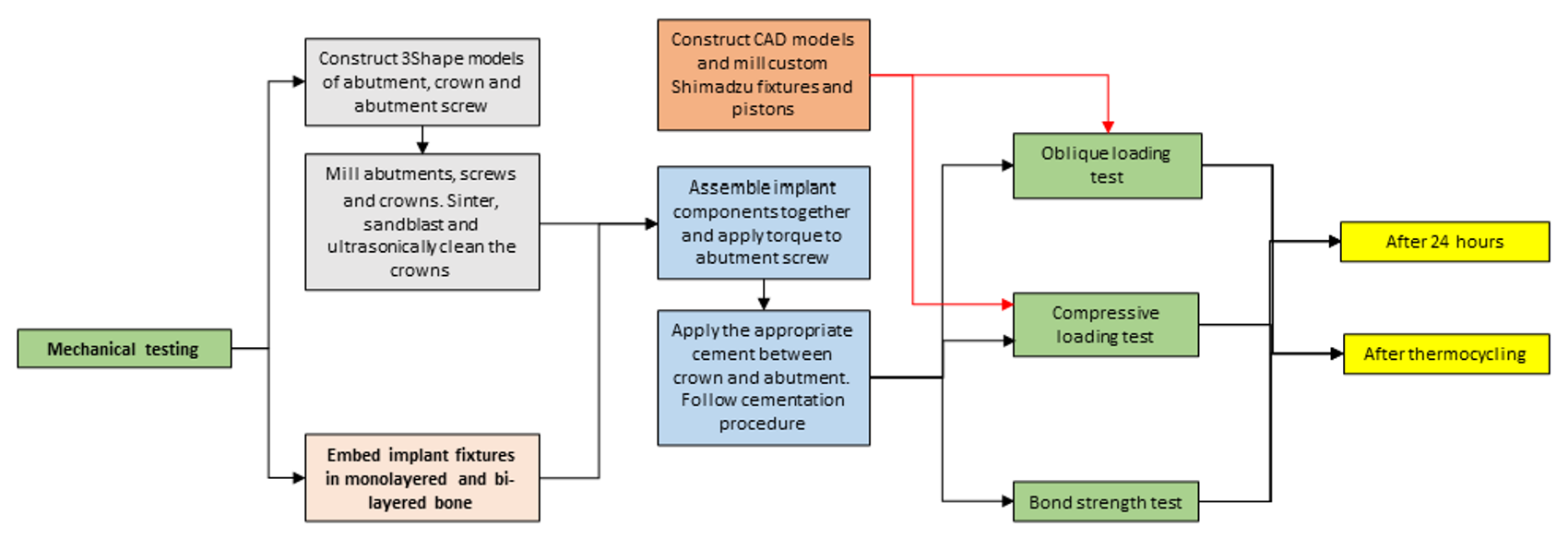
  • To construct an artificial bi-layered bone holder to mimic the natural structure of the jawbone.
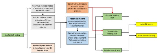
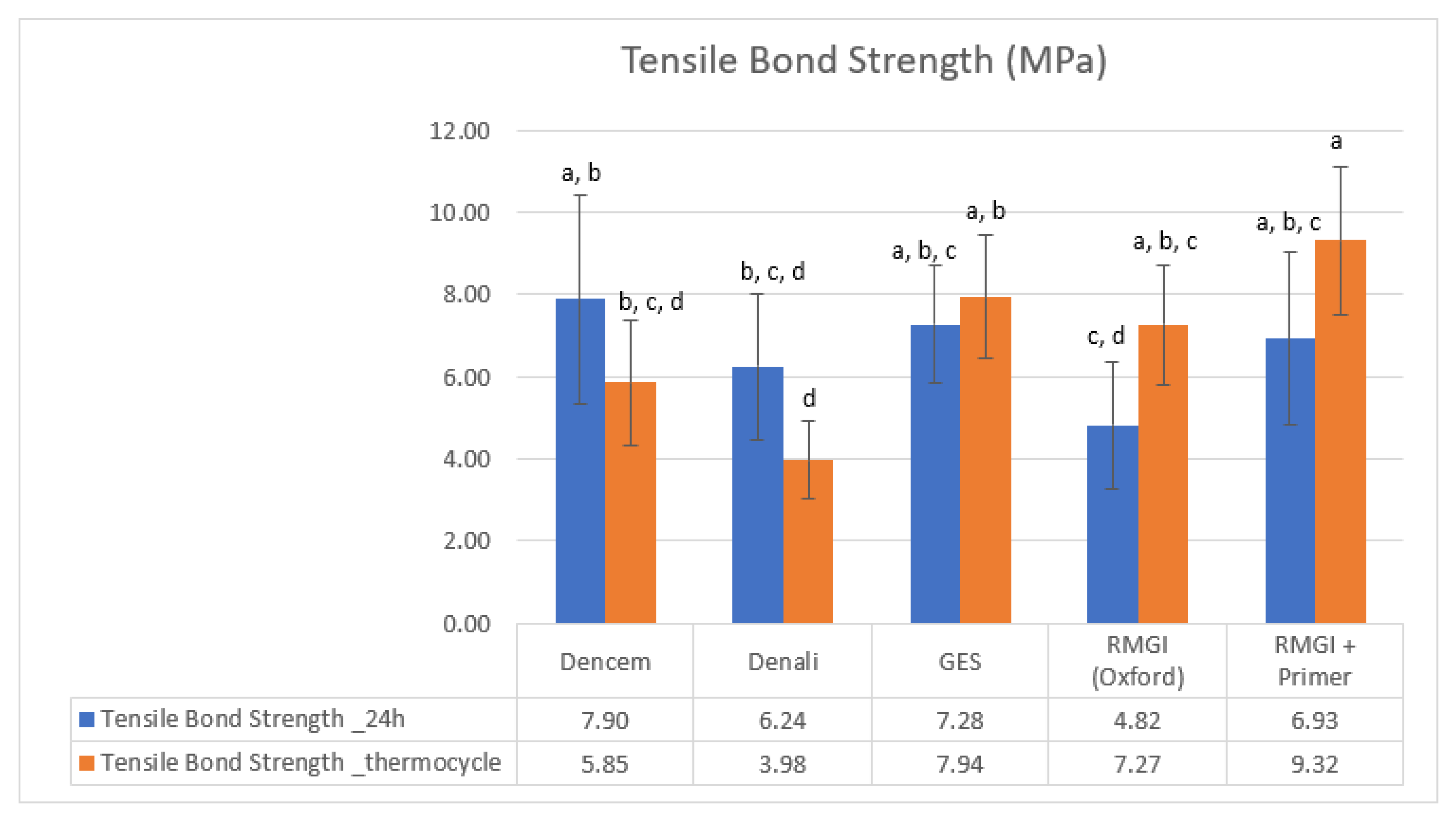
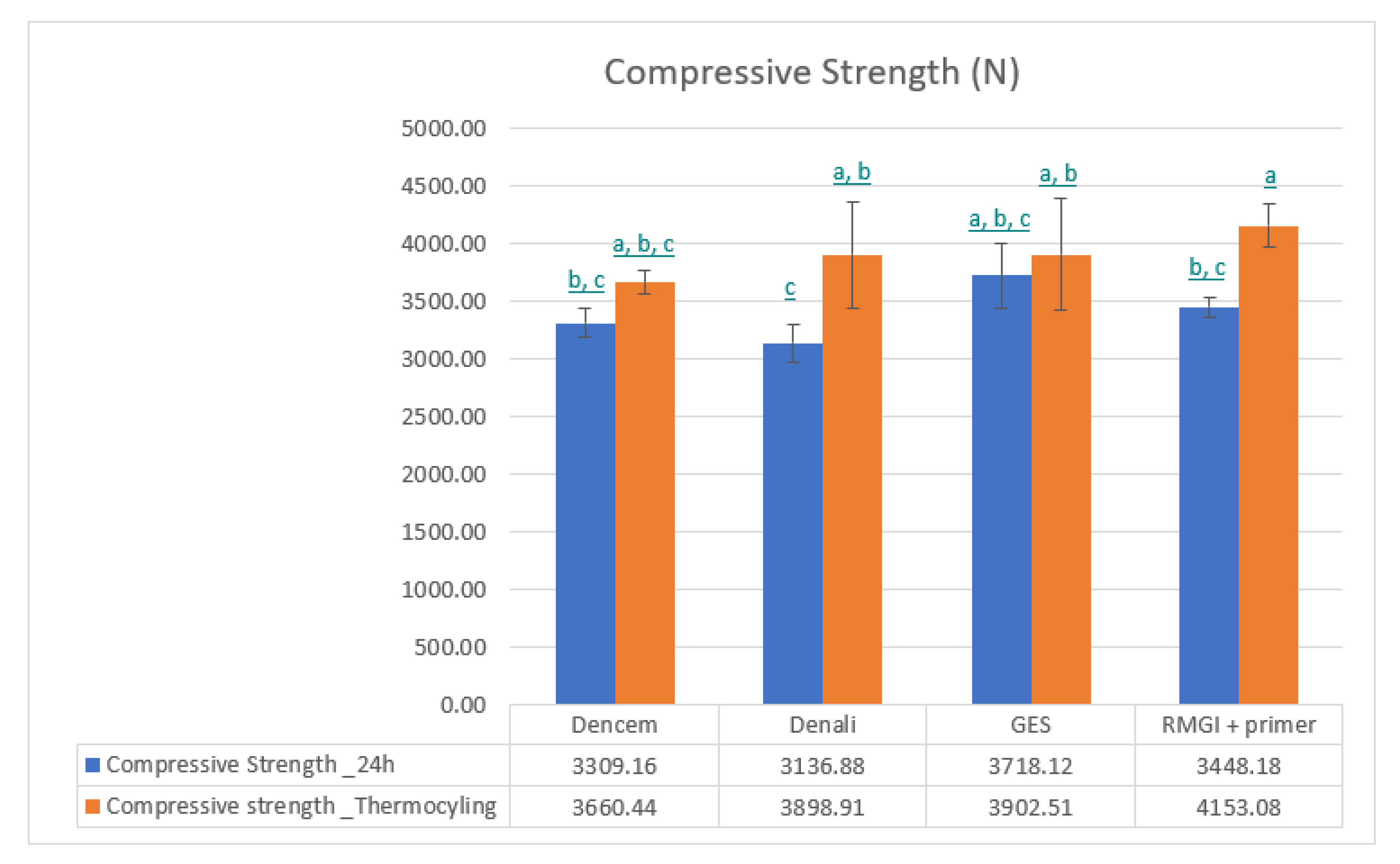
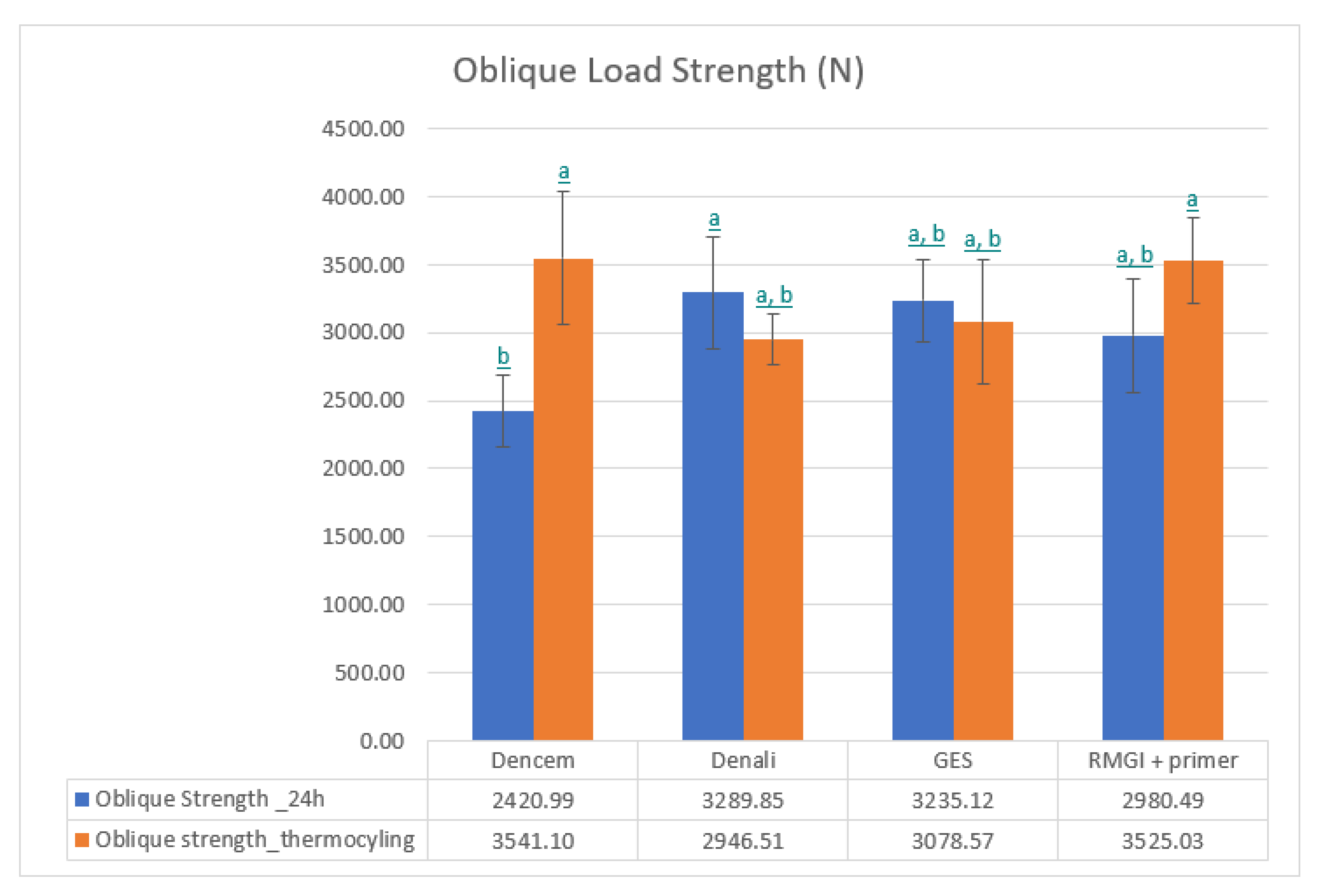
  • To perform a tensile bond strength test, a compressive load test, and an oblique load test on cement-retained crowns in titanium dental implant assemblies with different cement materials, before and after thermocycling.

2. Materials and Methods

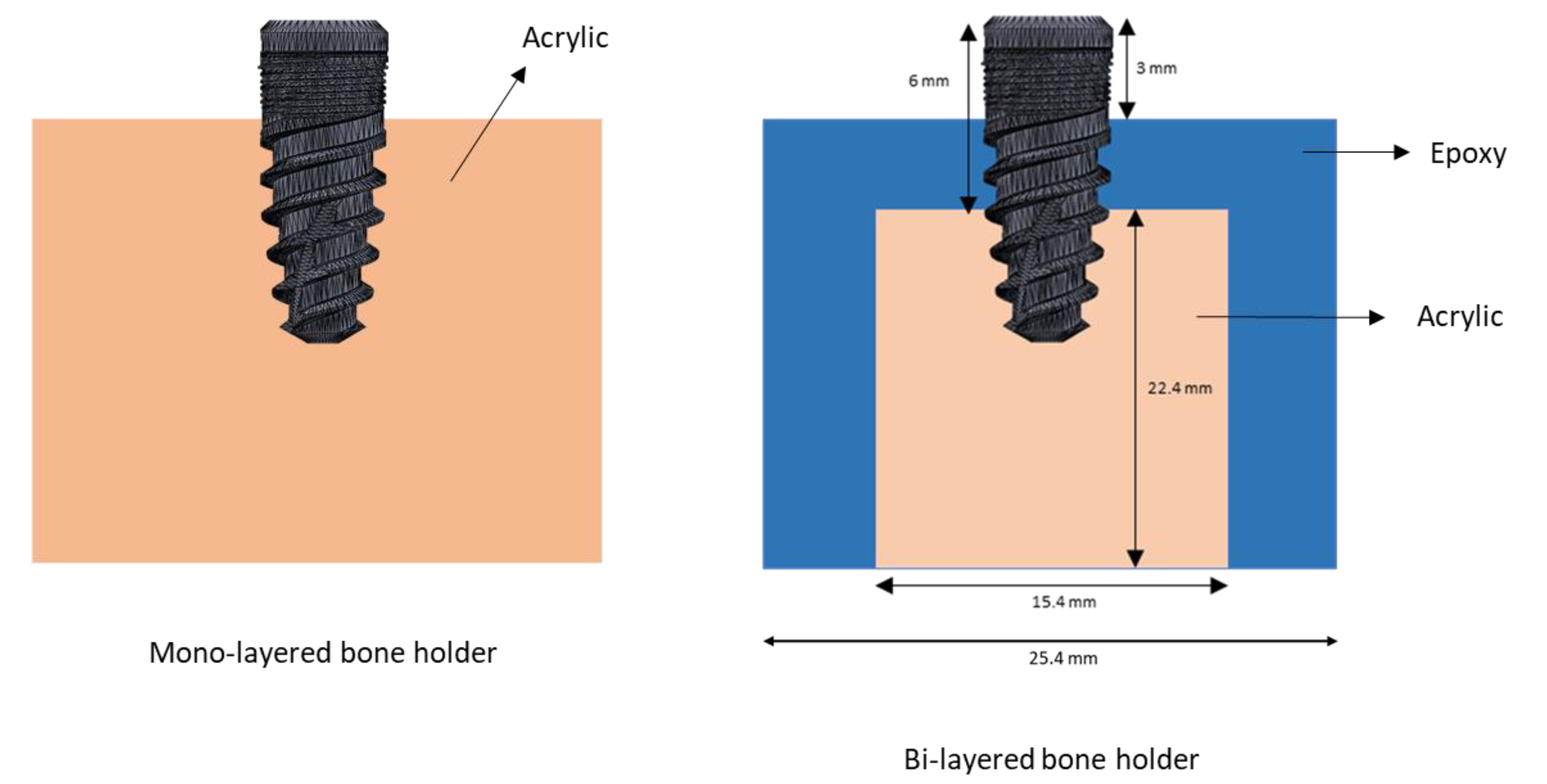
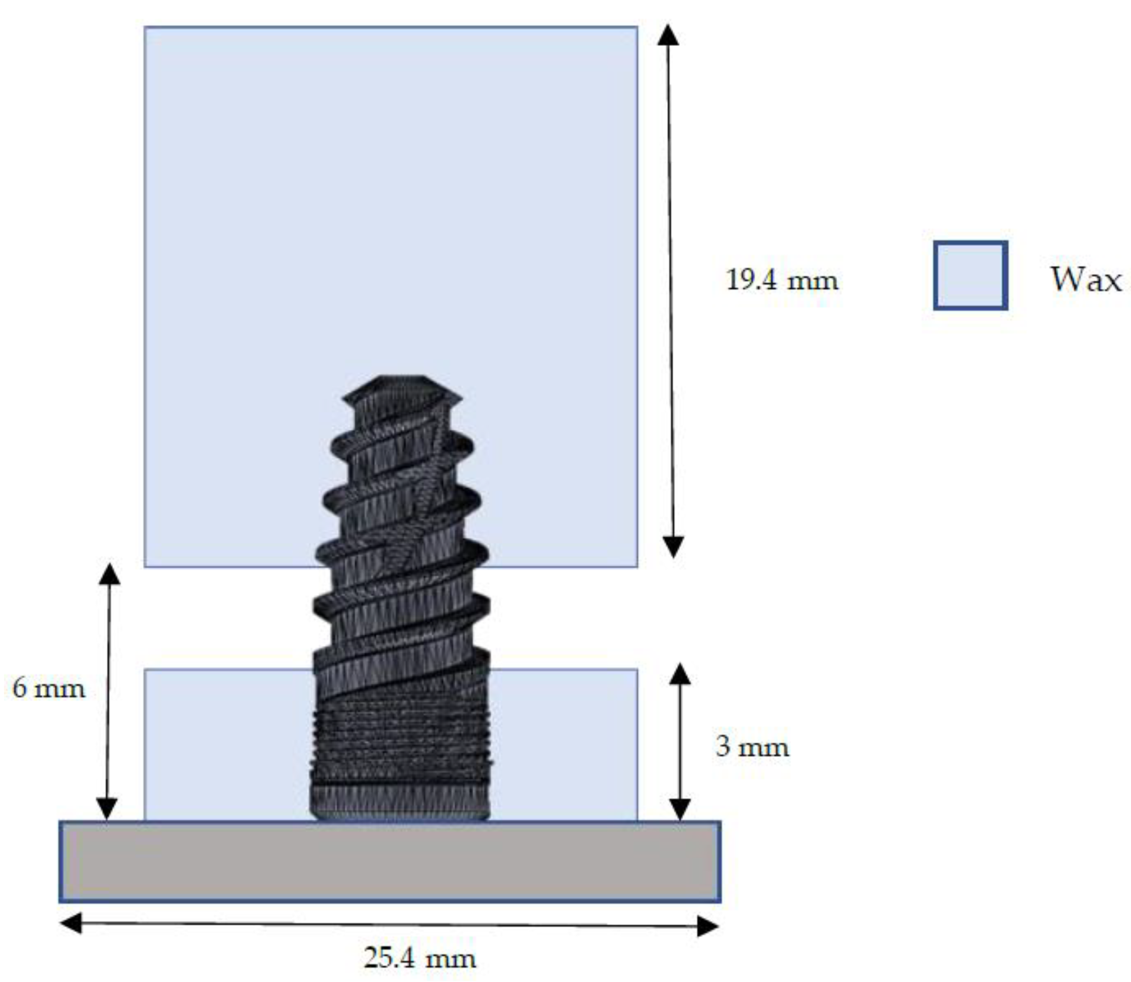
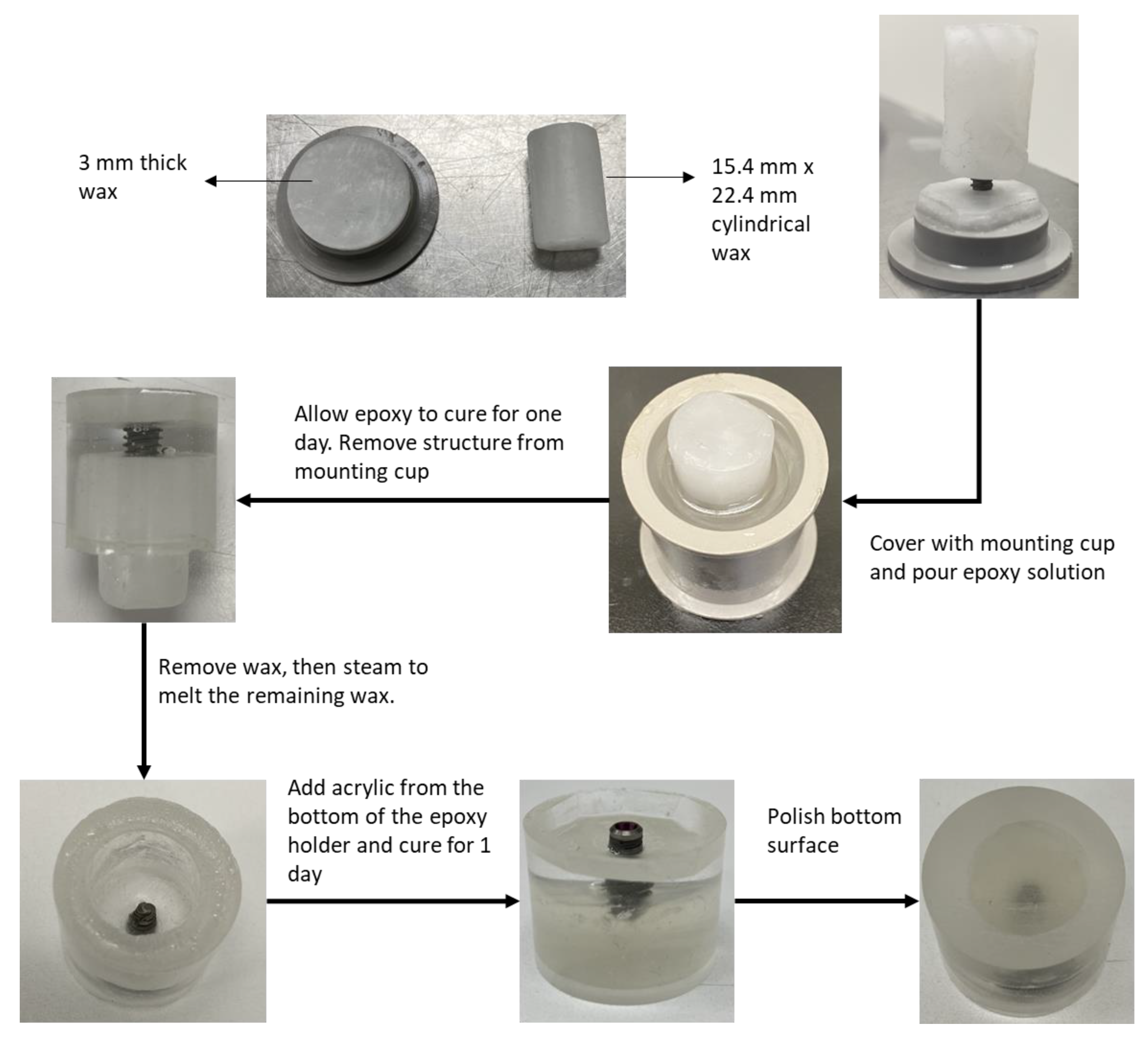
2.1. Mimicking the Bone Holder

Most in-vitro studies of dental implants involve assemblies with monolayered bone holders, typically made of acrylic. But the jawbone is a bi-layered structure, with dense cortical bone as the outer layer and soft trabecular bone comprising the inner layer. Hence, monolayered holders do not represent the natural structure of the jawbone. In the present study, an attempt was made to construct bi-layered epoxy-acrylic holders that will more closely match the structure of the jawbone. The implant assembly was constructed based on the International Organization for Standardization (ISO) 14801 standard such that there was a 3 mm gap between the implant nominal bone level and the artificial bone holder crest [33]. Figure 1 shows a schematic of the commonly used acrylic holder and the bi-layered holder we prepared for this study.
144 implants (HahnTM Tapered Implant System, Ø4.3 mm × 10 mm, Glidewell Laboratories, Irvine, CA, USA) were obtained. These implants were segregated based on the tests that were to be performed, as shown below:
  • Tensile bond strength test (after 24 h): 10 implants/cement × 4 cements = 40 implants
  • Tensile bond strength test (after thermocycling): 10 implants/cement × 4 cements = 40 implants
  • Compressive load test (after 24 h): 4 implants/cement × 4 cements = 16 implants
  • Compressive load test (after thermocycling): 4 implants/cement × 4 cements = 16 implants
  • Oblique load test (after 24 h): 4 implants/cement × 4 cements = 16 implants
  • Oblique load test (after thermocycling): 4 implants/cement × 4 cements = 16 implants
For constructing the bi-layered holder, the strategy was to embed the coronal section of the implant in epoxy and the rest of the section with wax. Then, the wax would be removed and replaced with acrylic. The dimensions of the holder were based on a previous simulation study [34]. The regions of the implant body to be covered in wax are shown in Figure 2. First, a 3 mm-thick cylindrical wax was constructed and stuck to the bottom of a mounting cup. The implant body was then embedded upside down in the center of the wax, so that it is completely submerged within the wax 3 mm from the nominal bone level. Another longer cylindrical wax structure was prepared (15.4 mm × 22.4 mm) and used to embed the bottom of the implant based on the dimensions shown in Figure 2. The structure was covered with a mounting cup.
The epoxy solution was prepared by mixing the epoxy resin with hardener (EpoxySet, Allied High Tech Products, Rancho Dominguez, CA, USA) in a 100:13 weight ratio. For a 25.4 mm diameter mounting cup, 12.87 g of resin was mixed with 1.67 g of hardener. The solution was then stirred slowly for about 3–4 min until it became clear. The solution was then poured to fill the hollow areas of the mounting cap with the implant-wax assembly. It was allowed to cure for about 24 h. The wax was then scraped off, and the implant-epoxy assembly was steam-cleaned to remove any remaining wax. The acrylic powder was mixed with an acrylic liquid (QuickSet, Allied High Tech Products, CA, USA) in a 2:1 volume ratio. After stirring the solution for about 1 min, it was poured into the cavity at the bottom of the epoxy holder and allowed to cure for 24 h. The bottom surface of the bi-layered holder was then polished to make it flat. Figure 3 shows the step-by-step procedure for constructing the bi-layered bone holder.
However, not all implants were embedded into the bi-layered holder. The 32 implants segregated for compressive load tests were embedded in monolayer polyurethane holders since the epoxy-acrylic bi-layered holders had less compressive strength and disintegrated before implant failure under compressive loading. To prepare the monolayered holders, a 3 mm thick wax was placed on the bottom of the mounting cup, and the implant body was embedded upside down such that 3 mm from the nominal bone level of the implant body was submerged within the wax. The inner walls of the cylindrical part of the mounting cup were coated with a lubricating agent for easy removal of the polyurethane. The polyurethane solution was prepared by thoroughly mixing the Polyurock base with a catalyst (Cendres+Métaux, Biel/Bienne, Switzerland). Because the intermediate steps used to prepare the bi-layered holder were unnecessary, the cylindrical part of the cup was replaced, and the cavity was completely filled with the polyurethane solution. The solution was then allowed to cure for about 30 min, following which the implant-holder assembly was removed from the cup and the wax was cleaned off.

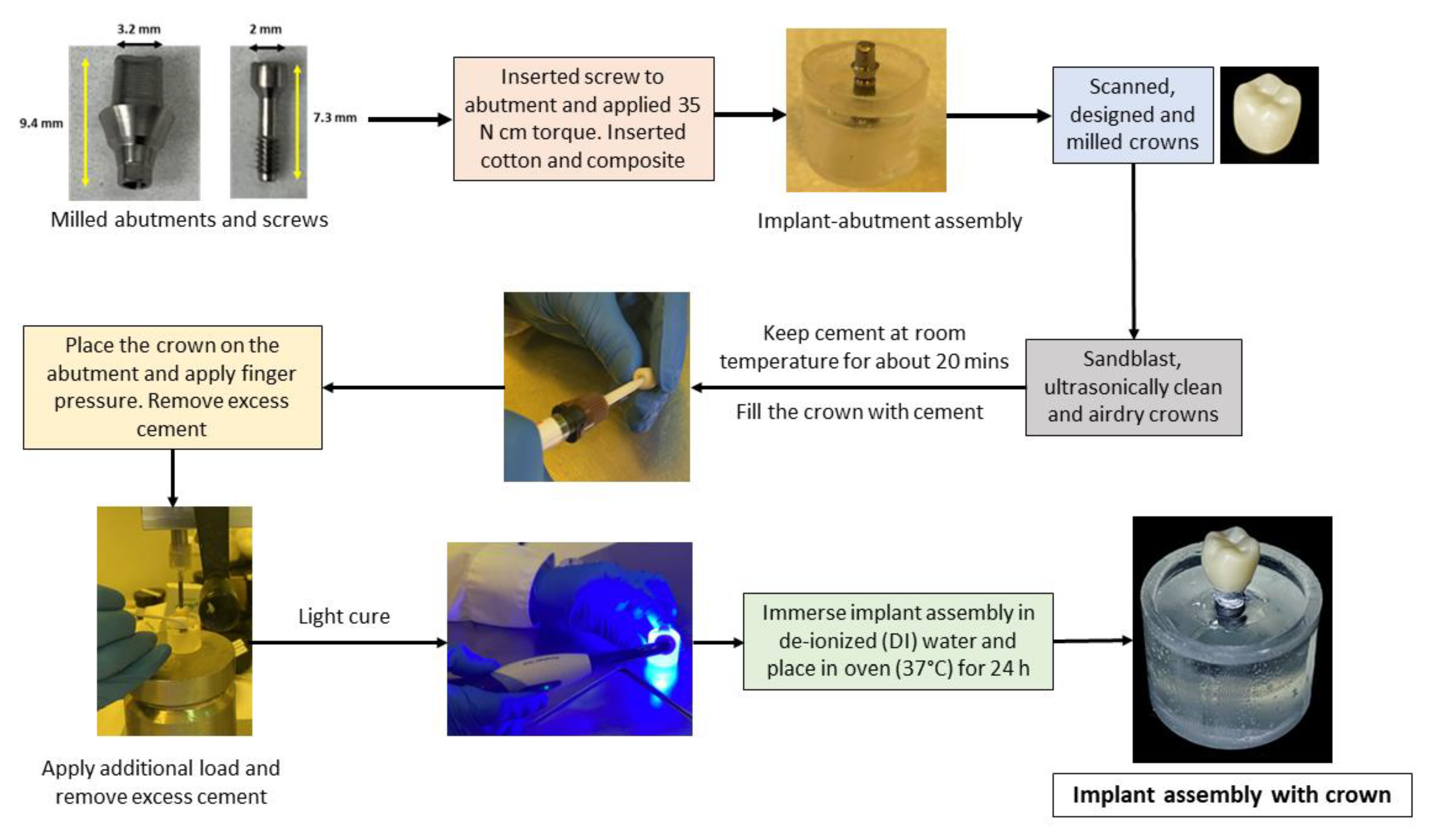
2.2. Cementation Procedure

Three SARC cements, namely, Dencem (Dentex, Changchun, China), Denali (Glidewell Laboratories, CA, USA), and Glidewell Experimental SARC Cement (GES, Glidewell Laboratories, CA, USA) cement, were used in this study. The RMGI (Glidewell Laboratories, CA, USA) was the fourth cement used. The corresponding custom titanium abutments and abutment screws were constructed in modeling software (3Shape, Copenhagen, Denmark) and milled. The abutment was fixed to the implant body using the abutment screw, and a 35 N cm torque was applied to the abutment screw based on the manufacturer’s recommendations. The cavity within the abutment was filled with cotton and a composite. In 3Shape, a special type of crown was created that was used specifically for tensile bond strength tests. For all other tests, a 1 mm-thick crown was modeled based on the structure of the mandibular first molar. These crowns were then milled out of zirconia milling blocks (BruxZir Shaded 16 PLUS A1, 20 mm blocks for bond strength test crowns, and 12 mm blocks for the first-molar crowns, Glidewell Laboratories, CA, USA) and sintered based on the instructions for use (IFU). The internal surface of the crowns was then sandblasted using 50 µm Al2O3 under 60 psi for 10 s, ultrasonically cleaned in de-ionized (DI) water for 5 min, and then air-dried. The four cements and the primer for RMGI cement (ZirconPrime M, S&C Polymer, Elmshorn, Germany) were kept at room temperature for 20 min. For each type of cement, about three-fourths of the crown was filled with cement, and the crown was placed on the abutment. Finger pressure was applied on the top of the crown, and excess cement was removed. A load cell was used to apply additional load to the crown, and any remaining excess cement was removed. The cement was light-cured based on the manufacturer’s instructions, followed by immersing the implant assembly in de-ionized (DI) water and placing it in the oven (37 °C) for 24 h. In the case of RMGI cement, a primer was applied to the abutment surface and the interior of the crown to allow proper bonding of the cement to these adjacent surfaces. Figure 4 shows the workflow of the cementation procedure.

2.3. Mechanical Tests

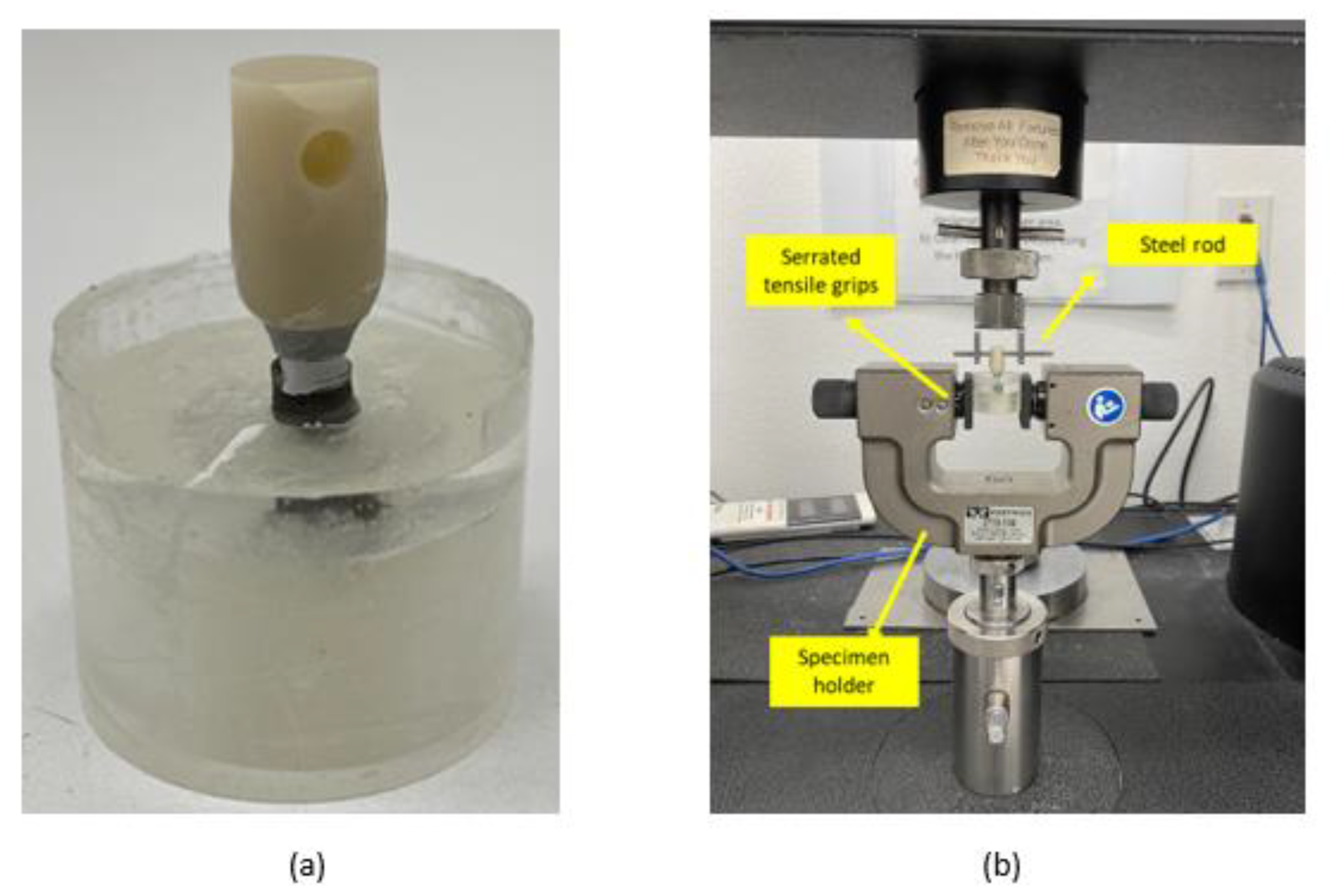
2.3.1. Tensile Bond Strength Test

The tensile bond strength test for the cements was performed based on the ISO/TS 11,405 standard [35]. As specified earlier, a different type of crown was modeled and cemented on the implant assemblies with the bi-layered holder for this test. In the 24-h test, the bond strength of the cements was tested 24 h after the cementation of the crown. Ten crowns and ten implant assemblies were used for each of the four cement types for the 24-h test. The test was performed on a Universal Testing Machine (UTM, Instron, Norwood, MA, USA), as shown in Figure 5. The bone holder was stabilized by using serrated grips, and a steel rod was inserted into the cemented crown. An axial force was applied in the upward direction on the rod until the crown was pulled apart from the rest of the assembly. The maximum force at which the crown debonded indicated the tensile bond strength of the cement with the abutment and zirconia crown.
The thermocycling procedure is an established method to simulate the artificial aging of materials. It is used for evaluating the effects of thermal stresses on the bond strength of dental materials. Hence, another set of ten samples per cement (40 samples total) was set up for thermocycling. The samples were immersed in cold water (5 °C), followed by hot water (55 °C), at regular intervals of time for 9 days, which is roughly equivalent to 10,000 cycles [36]. The dwell time was 30 s, with 5 s in the air. The tensile bond strength test was performed after 9 days in the same way as explained for the 24-h test.

2.3.2. Compressive Load Test

The compressive load test was performed to evaluate the overall compressive strength of the assembly with different cement materials. A custom fixture (or sample holder) with screws and a metal piston was modeled in computer-aided design (CAD) software (SOLIDWORKS, GoEngineer, San Diego, CA, USA) and milled out of stainless steel. In this test, the implant assemblies with a monolayered polyurock holder and the regular crown were used. For the 24-h test, four samples per cement (sixteen samples in total) were used. The test was set up in another universal testing machine (UTM, Shimadzu, Kyoto, Japan) that can go up to much higher loads (up to 10 kN) than the Instron UTM (up to 2 kN). It was necessary to use a UTM that can withstand high loads because it was anticipated that the compressive strength of the assembly components would be much higher than the cement bond strength. The metal fixture and piston, and the test setup, are shown in Figure 6. An axial load was applied at the center of the occlusal surface of the crown. Failure indicated the minimum load at which abutment screw loosening occurred.
Another set of four samples per cement (sixteen samples in total) was set up for thermocycling in the same way as explained under the tensile bond strength test. The samples were then tested for compressive strength after 9 days (or 10,000 cycles) of thermocycling.

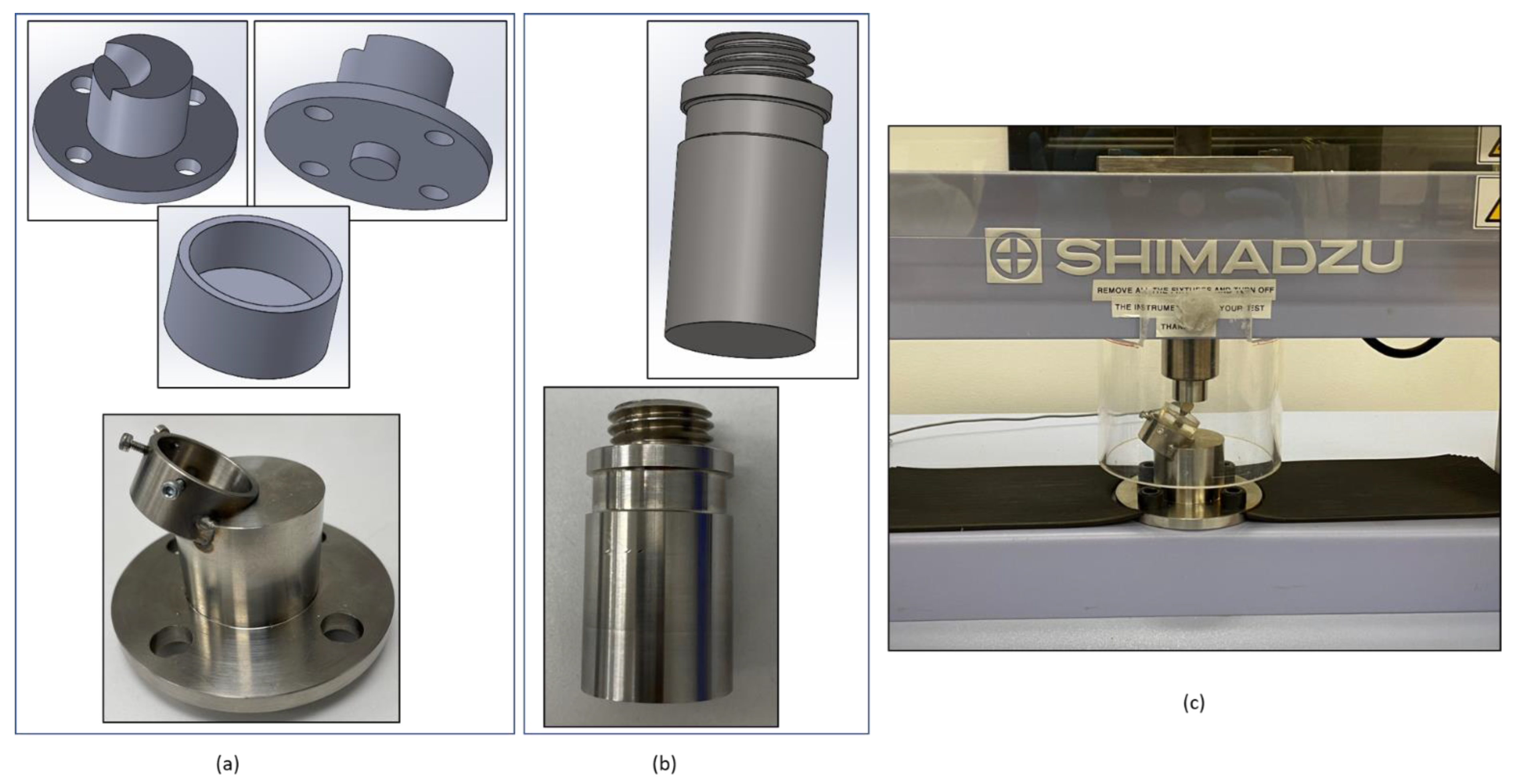
2.3.3. Oblique Load Test

The idea behind this test was to evaluate the strength of the assembly with different cement materials when subjected to an angular load. Dental prostheses may not always experience a vertical load from the opposing tooth. In mandibular molars, the buccal cusps are more functional than the lingual cusps, which means that the buccal cusp experiences more load compared to the lingual cusp. In this test, a simulated bite force was applied on the buccal cusp at an angle of 30 degrees from the implant axis (ISO 14801) [33]. A custom 30-degrees fixture (or sample holder) with screws and a flat metal piston was modeled in SOLIDWORKS and milled out of stainless steel. In this test, the implant assemblies with the bi-layered holder and the regular crown were used. For the 24-h test, four samples per cement (sixteen samples in total) were used. The metal fixture and piston, and the test setup, are shown in Figure 7. The Shimadzu 10 kN UTM was used for this test. Failure indicated the minimum load at which abutment screw loosening occurred. Another set of four samples per cement (sixteen samples in total) was set up for thermocycling in the same way as explained under the tensile bond strength test. The samples were then tested for oblique load strength after 9 days (or 10,000 cycles) of thermocycling.

2.4. Statistical Analysis

An analysis of variance (ANOVA) with a significance level of 0.05 was used to compare the results for each mechanical test. A Tukey pairwise comparison test (with 95% confidence) will be performed to evaluate any significant differences among the groups.

3. Results

3.1. Mechanical Tests

3.1.1. Tensile Bond Strength Test

The tensile bond strength test data for the cements after 24 h and after thermocycling for 9 days are shown in Figure 8 (n = 10, p < 0.05). To illustrate the importance of a primer, the tensile bond strength of RMGI cement was measured with and without the use of a primer. The SARC cements did not require a primer since their composition already contained the bonding agent. Before thermocycling, Dencem and GES cements had the highest mean tensile bond strengths. After thermocycling, however, RMGI with primer and GES cements had the highest mean tensile bond strengths. An ANOVA statistical test revealed a significant difference in the mean tensile bond strengths of the cements for both the 24-h test and the thermocycling test (p < 0.05).

3.1.2. Compressive Load Test

The compressive load test data for the implant assemblies with different cement materials after 24 h and after thermocycling are shown in Figure 9 (n = 4, p < 0.05). An ANOVA statistical test revealed that there was a significant difference among the mean compressive strengths of the implant assemblies with different cements for both the 24-h test and the test conducted after thermocycling. At 24 h, assemblies with GES cement and RMGI cement with primer had the highest load at failure, indicating the highest compressive strength. After thermocycling, though the loads at failure for assemblies with GES cement and Denali are comparable, the mean loads at failure for GES cement and RMGI cement with primer were the highest. In some cases, failure occurred at the crown before the screw, which was not the case during the testing after 24 h.

3.1.3. Oblique Load Test

The oblique load test data for the implant assemblies with different cement materials after 24 h and after thermocycling are shown in Figure 10 (n = 4, p < 0.05). This test was performed to mimic the natural biting mechanism according to the ISO 14,801 standard. An ANOVA statistical test revealed a significant difference between the mean strengths under oblique load of the implant assemblies with different cements for both the 24-h test and the thermocycling test. In the 24-h test, implant assemblies with Denali and GES cements had the highest load at failure. But in the test after thermocycling, Dencem and RMGI with primer had the highest load at failure.

4. Discussion

The procedure for constructing a bi-layered bone holder for testing dental implants has been a subject of interest for researchers. In this study, such a holder was successfully developed. Epoxy and acrylic were used to represent the cortical and cancellous bones, respectively. However, in the future, additional tests will be performed to replace these two polymers with other materials that will be compatible with the procedure and that will more closely represent the bone’s material properties. The in-vitro mechanical tests for tensile bond strength of the cements revealed that the GES cement and the RMGI cement with primer had better performance compared to the others tested. In addition, these two cements were the only ones whose bond strength increased after being exposed to thermocycling. In previous studies, it has been established that the exposure of dental cements to thermal cycling, in general, affects their thermal and mechanical properties [37,38,39,40,41]. Different cements react differently to temperature changes, depending on their composition. The exact effects of thermocycling on the microstructural changes and behavior of the cements considered in the present study are yet to be established. However, in general, thermocycling of dental components is performed to simulate the harsh conditions of the oral cavity that the samples may be exposed to in the clinical case. One would expect the bond strength to decrease after thermocycling. However, the results for GES cement and RMGI cement with primer indicate that water and its fluctuating temperature could have a beneficial effect on the composition of the cements by increasing their bonding to the substrate. Further research is required to justify this assumption.
Although the tensile bond strength tests were specifically for the cements, the compressive and oblique loading tests were performed to evaluate the strength of the overall assembly when different cement materials are used. The compressive load tests indicated that the implant assemblies with GES cement and RMGI cement with primer exhibited the highest maximum force at failure. The oblique test results, however, show different trends from the results obtained for the 24-h test and thermocycling. In the 24-h test, implant assemblies with Denali and GES cements had the highest strength under oblique load. However, after thermocycling, implant assemblies with Dencem and RMGI + Primer have the highest strength under oblique load. Since the test after thermocycling of the samples more closely represents the clinical case, these results should be given more priority, and it can be concluded that Dencem and RMGI + Primer are more desirable if angulated bite force is considered. Nevertheless, if all the mechanical tests performed in this study are cumulatively considered, RMGI + primer and GES cements have the most desirable performance so far. In addition, there was a significant difference among the means of different cements for each of the three tests. These results will help dentists and clinicians identify the appropriate cement for specific dental applications involving cement-retained zirconia crowns and titanium implant-abutment assemblies.

5. Conclusions

In the present study, three self-adhesive resin cements (SARC) and a resin-modified glass ionomer (RMGI) cement were evaluated for their performance in dental implant systems. The study showed that Glidewell Experimental SARC (GES) cement and Resin-modified Glass Ionomer (RMGI) cement with primer had better performances with zirconia crowns and Ti6Al4V implants compared to the other cements tested. In addition, a novel technique for constructing an artificial, bi-layered bone holder was successfully developed to mimic the natural structure of the jawbone.

Author Contributions

Conceptualization, M.S. and S.S.; data curation, M.S.; formal analysis, M.S. and H.P.; methodology, M.S. and H.P.; project administration, S.S.; resources, M.S. and S.S.; software, M.S. and H.P.; supervision, M.S. and S.S.; visualization, M.S.; writing—original draft, M.S.; writing—review & editing, M.S. and S.S. All authors have read and agreed to the published version of the manuscript.

Funding

This research received no external funding.

Institutional Review Board Statement

Not applicable.

Informed Consent Statement

Not applicable.

Data Availability Statement

The data are not publicly available due to data confidentiality resulting from the requirements of the accreditation.

Acknowledgments

The authors acknowledge Glidewell Laboratories for providing the necessary equipment and software for testing and data analysis.

Conflicts of Interest

The authors declare no conflict of interest.

References

  1. Sailer, I.; Sailer, T.; Stawarczyk, B.; Jung, R.E.; Hämmerle, C.H.F. In vitro study of the influence of the type of connection on the fracture load of zirconia abutments with internal and external implant-abutment connections. Int. J. Oral Maxillofac. Implants 2009, 245, 850–858. [Google Scholar]
  2. Griggs, J.A. Dental Implants. Dent. Clin. N. Am. 2017, 614, 857–871. [Google Scholar] [CrossRef] [PubMed]
  3. Alghamdi, H.S.; Jansen, J.A. The development and future of dental implants. Dent. Mater. J. 2020, 392, 167–172. [Google Scholar] [CrossRef] [PubMed] [Green Version]
  4. Kalluri, L.; Seale, B.; Satpathy, M.; Esquivel-Upshaw, J.F.; Duan, Y. Three-Dimensional Finite Element Analysis of the Veneer—Framework Thickness in an All-Ceramic Implant Supported Fixed Partial Denture. Ceramics 2021, 42, 199–207. [Google Scholar] [CrossRef]
  5. Leo, S.; Tan, M.; Yee, S.; Lee, F.; Tan, K. Rotational Load Fatigue Performance of Titanium vs Titanium-Zirconium Implant-Abutment Connections. Int. J. Oral Maxillofac. Implants 2022, 374, 740–747. [Google Scholar] [CrossRef]
  6. Parzham, V.; Judge, R.; Bailey, D. A Five-Year Retrospective Assay of Implant Treatments and Complications in Private Practice: Restorative Treatment Profiles of Long-Span, Implant-Supported Fixed and Removable Dental Prostheses. Int. J. Prosthodont. 2018, 313, 211–222. [Google Scholar] [CrossRef] [Green Version]
  7. Hamed, M.T.; Abdullah Mously, H.; Khalid Alamoudi, S.; Hossam Hashem, A.B.; Hussein Naguib, G. A Systematic Review of Screw versus Cement-Retained Fixed Implant Supported Reconstructions. Clin. Cosmet. Investig. Dent. 2020, 12, 9–16. [Google Scholar] [CrossRef] [Green Version]
  8. Hebel, K.S.; Gajjar, R.C. Cement-retained versus screw-retained implant restorations: Achieving optimal occlusion and esthetics in implant dentistry. J. Prosthet. Dent. 1997, 771, 28–35. [Google Scholar] [CrossRef]
  9. Karl, M.; Graef, F.; Taylor, T.D.; Heckmann, S.M. In vitro effect of load cycling on metal-ceramic cement- and screw-retained implant restorations. J. Prosthet. Dent. 2007, 973, 137–140. [Google Scholar] [CrossRef]
  10. Lee, J.H.; Jang, H.Y.; Lee, S.Y. Finite Element Analysis of Dental Implants with Zirconia Crown Restorations: Conventional Cement-Retained vs. Cementless Screw-Retained. Materials 2021, 14, 2666. [Google Scholar] [CrossRef]
  11. Michalakis, K.X.; Hirayama, H.; Garefis, P.D. Cement-retained versus screw-retained implant restorations: A critical review. Int. J. Oral Maxillofac. Implants 2003, 185, 719–728. [Google Scholar]
  12. Agarwal, S.; Ashok, V.; Maiti, S.; Agarwal, V. Dentists’ Preference toward Fixed Versus Removable Implant Prosthesis on Edentulous Jaws to Improve Quality of Life. J. Long Term Eff. Med. Implants 2023, 331, 83–89. [Google Scholar] [CrossRef] [PubMed]
  13. Wittneben, J.G.; Millen, C.; Brägger, U. Clinical Performance of Screw- Versus Cement-Retained Fixed Implant-Supported Reconstructions—A Systematic Review. Int. J. Oral Maxillofac. Implants 2014, 29, 84–98. [Google Scholar] [CrossRef] [PubMed] [Green Version]
  14. Kern, J.; Kern, T.; Wolfart, S.; Heussen, N. A systematic review and meta-analysis of removable and fixed implant-supported prostheses in edentulous jaws: Post-loading implant loss. Clin. Oral Implants Res. 2016, 272, 174–195. [Google Scholar] [CrossRef] [Green Version]
  15. Gundogdu, M.; Aladag, L.I. Effect of adhesive resin cements on bond strength of ceramic core materials to dentin. Niger. J. Clin. Pract. 2018, 213, 367–374. [Google Scholar] [CrossRef]
  16. Johnson, G.H.; Lepe, X.; Patterson, A.; Schäfer, O. Simplified cementation of lithium disilicate crowns: Retention with various adhesive resin cement combinations. J. Prosthet. Dent. 2018, 1195, 826–832. [Google Scholar] [CrossRef]
  17. Dressano, D.; Salvador, M.V.; Oliveira, M.T.; Marchi, G.M.; Fronza, B.M.; Hadis, M.; Palin, W.M.; Lima, A.F. Chemistry of novel and contemporary resin-based dental adhesives. J. Mech. Behav. Biomed. Mater. 2020, 110, 103875. [Google Scholar] [CrossRef]
  18. Carville, R.; Quinn, F. The selection of adhesive systems for resin-based luting agents. J. Ir. Dent. Assoc. 2008, 545, 218–222. [Google Scholar]
  19. Wilson, A.D. Glass-ionomer cement origins, development and future. Clin. Mater. 1991, 74, 275–282. [Google Scholar] [CrossRef]
  20. Ansari, S.; Moshaverinia, M.; Roohpour, N.; Chee, W.W.L.; Schricker, S.R.; Moshaverinia, A. Properties of a proline-containing glass ionomer dental cement. J. Prosthet. Dent. 2013, 1105, 408–413. [Google Scholar] [CrossRef]
  21. Bahsi, E.; Sagmak, S.; Dayi, B.; Cellik, O.; Akkus, Z. The evaluation of microleakage and fluoride release of different types of glass ionomer cements. Niger. J. Clin. Pract. 2019, 227, 961. [Google Scholar] [CrossRef] [PubMed]
  22. Berg, J.H.; Croll, T.P. Glass ionomer restorative cement systems: An update. Pediatr. Dent. 2015, 372, 116–124. [Google Scholar]
  23. Smith, D.C. Dental cements. Curr. Opin. Dent. 1991, 12, 228–234. [Google Scholar]
  24. Prasanth, B.K.; Prakasam, S.; Bharadwaj, P.; Loganathan, S. A comparative evaluation of compressive strength of Portland cement with zinc oxide eugenol and Polymer-reinforced cement: An in vitro analysis. Indian J. Dent. Res. 2014, 251, 73. [Google Scholar] [CrossRef] [PubMed]
  25. Bra¨nnstro¨m, M.; Nyborg, H. Pulp reaction to a temporary zinc oxide/eugenol cement. J. Prosthet. Dent. 1976, 352, 185–191. [Google Scholar] [CrossRef]
  26. Powers, J.M. Self-adhesive Resin Cements: Characteristics, Properties, and Manipulation. AEGIS Dental Network. Funct. Esthet. Restor. Dent. 2008, 1, 34–40. [Google Scholar]
  27. Weiser, F.; Behr, M. Self-Adhesive Resin Cements: A Clinical Review. J. Prosthodont. 2015, 242, 100–108. [Google Scholar] [CrossRef]
  28. Makkar, S.; Malhotra, N. Self-adhesive resin cements: A new perspective in luting technology. Dent. Update 2013, 409, 758–768. [Google Scholar] [CrossRef]
  29. Marghalani, H.Y. Sorption and solubility characteristics of self-adhesive resin cements. Dent. Mater. 2012, 2810, e187–e198. [Google Scholar] [CrossRef]
  30. Sidhu, S.K. Glass-ionomer cement restorative materials: A sticky subject? Aust. Dent. J. 2011, 56 (Suppl. S1), 23–30. [Google Scholar] [CrossRef]
  31. Walia, T.; Brigi, C.; Ziadkhani, M.M.; Khayat, A.A.; Tabibzadeh, Z. Retention Force of Glass Ionomer Based Luting Cements with Posterior Primary Zirconium Crowns—A Comparative in Vitro Study. J. Clin. Pediatr. Dent. 2021, 454, 259–264. [Google Scholar] [CrossRef] [PubMed]
  32. Satpathy, M.; Duan, Y.; Betts, L.; Priddy, M.; Griggs, J.A. Effect of Bone Remodeling on Dental Implant Fatigue Limit Predicted Using 3D Finite Element Analysis. J. Dent. Oral Epidemiol. 2022, 2. [Google Scholar] [CrossRef]
  33. ISO 14801; Dentistry—Implants—Dynamic Loading Test for Endosseous Dental Implants. ISO: London, UK, 2016.
  34. Satpathy, M.; Jose, R.M.; Duan, Y.; Griggs, J.A. Effects of abutment screw preload and preload simulation techniques on dental implant lifetime. JADA Found. Sci. 2022, 1, 100010. [Google Scholar] [CrossRef] [PubMed]
  35. ISO/TS 11405; Dentistry—Testing of Adhesion to Tooth Structure. ISO: London, UK, 2015.
  36. ISO/TR 11405; Dental Materials—Guidance on Testing on Adhesion to Tooth Structure. ISO: London, UK, 1994.
  37. Saghiri, M.A.; Asatourian, A.; Garcia-Godoy, F.; Gutmann, J.L.; Sheibani, N. The Impact of Thermocycling Process on the Dislodgement Force of Different Endodontic Cements. BioMed Res. Int. 2013, 2013, 317185. [Google Scholar] [CrossRef] [PubMed]
  38. Luthy, H.; Loeffel, O.; Hammerle, C. Effect of thermocycling on bond strength of luting cements to zirconia ceramic. Dent. Mater. 2006, 222, 195–200. [Google Scholar] [CrossRef]
  39. Bitter, K.; Meyer-Lueckel, H.; Priehn, K.; Kanjuparambil, J.P.; Neumann, K.; Kielbassa, A.M. Effects of luting agent and thermocycling on bond strengths to root canal dentine. Int. Endod. J. 2006, 3910, 809–818. [Google Scholar] [CrossRef]
  40. Miyazaki, M.; Sato, M.; Onose, H.; Moore, B.K. Influence of thermal cycling on dentin bond strength of two-step bonding systems. Am. J. Dent. 1998, 113, 118–122. [Google Scholar]
  41. Helvatjoglu-Antoniades, M.; Koliniotou-Kubia, E.; Dionyssopoulos, P. The effect of thermal cycling on the bovine dentine shear bond strength of current adhesive systems. J. Oral Rehabil. 2004, 319, 911–917. [Google Scholar] [CrossRef]
Figure 1. Mono- and bi-layered artificial bone holders.
Ceramics 06 00039 g001
Figure 2. Schematic of the implant body placement in the mounting cup. The regions left out were covered by epoxy. After the epoxy was cured, the wax was removed and replaced with acrylic.
Ceramics 06 00039 g002
Figure 3. Step-by-step procedure for preparing the bi-layered holder and embedding the implant body in the holder.
Ceramics 06 00039 g003
Figure 4. Abutment placement and cementation procedure on the implant assembly.
Ceramics 06 00039 g004
Figure 5. (a) Implant assembly used for tensile bond strength tests of the cements. (b) Tensile bond strength test setup.
Ceramics 06 00039 g005
Figure 6. (a) Custom Shimadzu fixture (left: CAD model; right: milled part). (b) Custom metal piston (left: CAD model; right: milled part). (c) Compressive load test setup.
Ceramics 06 00039 g006
Figure 7. (a) Custom Shimadzu 30-degree fixture (top: CAD model; bottom: milled part). (b) Custom flat metal piston (top: CAD model; bottom: milled part). (c) Oblique load test setup.
Ceramics 06 00039 g007
Figure 8. Tensile bond strength of the cements (blue) after 24 h of the samples placed in DI water at 37 °C and (orange) after thermocycling the samples for 9 days (p < 0.05). The bars that do not share a letter have a significant difference among their respective means, as per the Tukey pairwise comparison test (95% confidence).
Ceramics 06 00039 g008
Figure 9. Compressive load test data of the implants with different cements (blue) after 24 h of the samples placed in DI water at 37 °C and (orange) after thermocycling the samples for 9 days (p < 0.05). The bars that do not share a letter have a significant difference among their respective means, as per the Tukey pairwise comparison test (95% confidence).
Ceramics 06 00039 g009
Figure 10. Oblique load test data of the implants with different cements (blue) after 24 h of the samples placed in DI water at 37 °C and (orange) after thermocycling the samples for 9 days (p < 0.05). The bars that do not share a letter have a significant difference among their respective means, as per the Tukey pairwise comparison test (95% confidence).
Ceramics 06 00039 g010
Disclaimer/Publisher’s Note: The statements, opinions and data contained in all publications are solely those of the individual author(s) and contributor(s) and not of MDPI and/or the editor(s). MDPI and/or the editor(s) disclaim responsibility for any injury to people or property resulting from any ideas, methods, instructions or products referred to in the content.

