
First Observation of Protomicelles in the System with a Non-Colloidal Surfactant


1
Frumkin Institute of Physical Chemistry and Electrochemistry, Russian Academy of Sciences, 119071 Moscow, Russia
2
Mendeleev Center, Saint Petersburg State University, 199034 Saint Petersburg, Russia
*
Author to whom correspondence should be addressed.
Colloids Interfaces 2023, 7(2), 32; https://doi.org/10.3390/colloids7020032
Submission received: 13 March 2023
/
Revised: 10 April 2023
/
Accepted: 11 April 2023
/
Published: 13 April 2023
(This article belongs to the Special Issue A Themed Issue in Honor of Prof. Boris Noskov)
Abstract
:A spectrophotometric study of the system heptanol—Nile red (NR)—water was carried out, where, for the first time for such studies, a non-colloidal surfactant that does not form micelles was taken as a surfactant. The dependence of the solubility of NR on the concentration of heptanol in an aqueous solution was studied. The experiments were carried out at a given chemical potential of NR, which was provided by an excess of the solid phase of NR. The existence of a solubilization effect has been theoretically and experimentally established: An increase in the solubility of NR with an increase in the concentration of heptanol in solution. It was found that heptanol protomicelles with a solubilization core as an NR molecule are formed in such a system, so that in the absence of micelles, the protomicelles take on the entire solubilization load. From the experimental data, the concentration of protomicelle formation was calculated, which can also be taken as the concentration of NR monomerization in an aqueous solution, since the formation of protomicelles prevents the dye aggregation. Based on the results obtained, the following generalizations were made: (1) non-colloidal surfactants, although they do not give micelles, are capable of forming protomicelles; and (2) non-colloidal surfactants can serve as a practical means of dye monomerization.
1. Introduction
Due to the hydrophobic effect, the molecules of dyes, when placed in an aqueous medium, stick together into dimers and aggregates of higher orders, which leads to the loss of the chromophore properties of the dyes. It was discovered that surfactants destroy these aggregates and bring dyes to a monomeric state [1,2]. Most studies were carried out on phthalocyanines [3,4,5,6,7,8,9,10,11,12,13,14,15,16,17]. The authors also started with these dyes [14]. At that time, the common and standard explanation for the effect of dye monomerization was the solubilization of their molecules in surfactant micelles. This implies that only colloidal surfactants (surfactants capable of micellization) have the ability to monomerize the dye. However, it turned out that there is another mechanism of monomerization by adsorption of surfactant molecules or ions on the surface of a dye molecule with the formation of micelle-like particles with a solubilisate. Such particles were called protomicelles [14].
The concept of protomicelle is closely related to the concept of nano-adsorbent, which, despite the trivial sound, is a term of a new meaning in physical chemistry [18]. An ordinary adsorbent, even a highly crushed one, is always a phase, on the surface of which adsorption occurs, and the entire system is heterogeneous. The dye molecule, as a nano-adsorbent particle, is a component of the solution, i.e., homogeneous (single-phase) medium. As with any other component of the solution, the concept of chemical potential is applicable to the nano-adsorbent, and the act of adsorption itself can be described using the law of mass action [17]. In this case, adsorption is understood in the usual sense, i.e., as a fixation (at least in one direction) on the surface of somebody. In our case, the nano-adsorbent particles are dye molecules, and the surfactant performs the role of the adsorbate. The surface area of one dye molecule is small, and as it is filled with a monolayer of surfactant molecules, a protomicelle is formed. Outwardly, it practically does not differ from a micelle of the same surfactant (a micelle, as is known, is a closed surfactant monolayer [19]), which absorbed (by solubilization) one dye molecule. The formation of protomicelles does not require a critical micelle concentration (CMC) and begins immediately when the dye molecules enter the solution. It should be noted that the term proto-micelle occurs in polymer science [20,21,22,23] to designate a young and still nonequilibrium micelle (the opposite term is mature micelle), since polymer micelles require a long time to form. To avoid confusion, we may refer to our micelles as adsorption protomicelles. It is in this sense that the term protomicelle below must be understood.
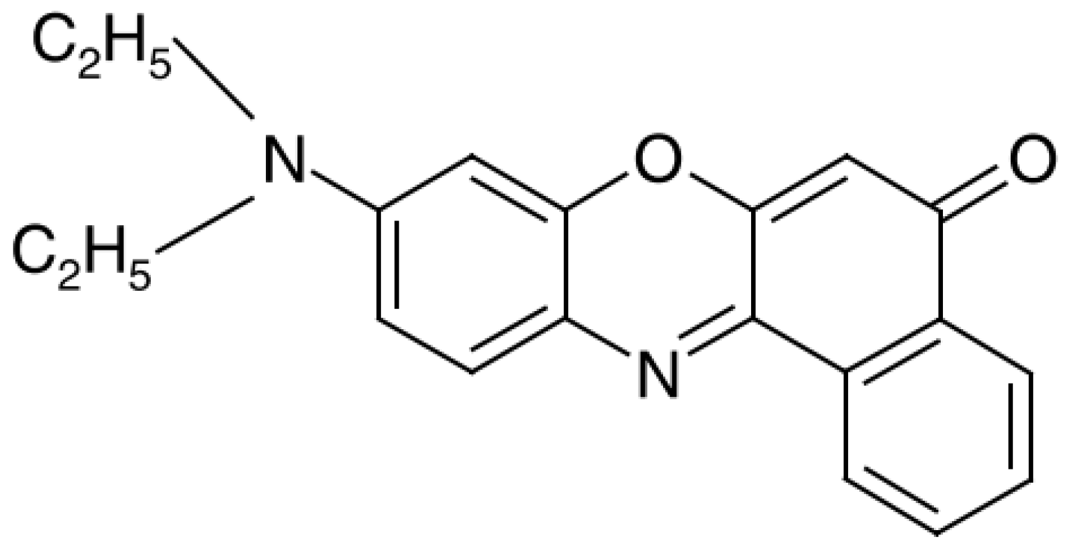
In addition to phthalocyanines, one of the popular hydrophobic dyes for such studies was Nile red (NR) [24,25,26,27,28,29,30,31,32], which is also used in this work. Its molecule also has a flat structure (Figure 1) and the ability to stick together into dimers and other molecular aggregates. Due to the hydrophobic effect, NR is poorly soluble in water (about 2 μM according to our data [30]). From here, it also follows the slowness of dissolution of NR in water at ordinary temperatures. Naturally, the dissolution mechanism consists of the transition of individual dye molecules from a solid state to a liquid state, so that at first the solution includes only NR monomers. As their concentration increases, the formation of dimers begins, then more complex aggregates, so that the final equilibrium state of the solution implies complete aggregative equilibrium. To accelerate the onset of equilibrium, it is necessary to increase the area of contact of the surface NR with water, i.e., take the dye in a large mass and finely dispersed state and mix intensively. With all these tricks, it takes several days to achieve equilibrium (at least four days according to Ref. [30]). In the presence of surfactants, these processes are accelerated, and most importantly, the molecular aggregates of the dye are destroyed, which eliminates the antichromophoric effects associated with them.
In addition to phthalocyanines and NR, other macrocyclic compounds whose molecules are prone to aggregation, can also serve as a core for the formation of protomicelles in aqueous solutions of surfactants. These compounds include chlorins with chromophoric properties. The effect of tetradecyltriphenylphosphonium bromide on the disaggregation of dicationic chlorine derivatives [33] manifests itself already at a concentration of 0.17 of CMC and can be explained by the formation of protomicelles. So far, colloidal surfactants have been used in experiments with combined solutions of dyes and surfactants. Aqueous solutions of NR have already been studied in combination with classical ionic surfactants, tetradecyltrimethylammonium bromide [30], and sodium dodecyl sulfate [32]. In Ref. [34], nonanoic acid (NA) was taken as a surfactant, which exhibits a number of transition properties. First, this is the transition from ionic to non-ionic surfactants: NA is a weak electrolyte and in the adsorbed state it is mainly in an undissociated form (at the water-air interface, the number of neutral molecules in the adsorption layer is 272 times higher than the number of adsorbed ions NA [35]). Second, this is the transition from colloidal to noncolloidal surfactants, although NA formally belongs to the first category and forms micelles in water at a concentration of less than 1 mM, according to the figures in Ref. [35]. According to our data, the CMC of NA in solution with NR is about 0.86 mM [34]).
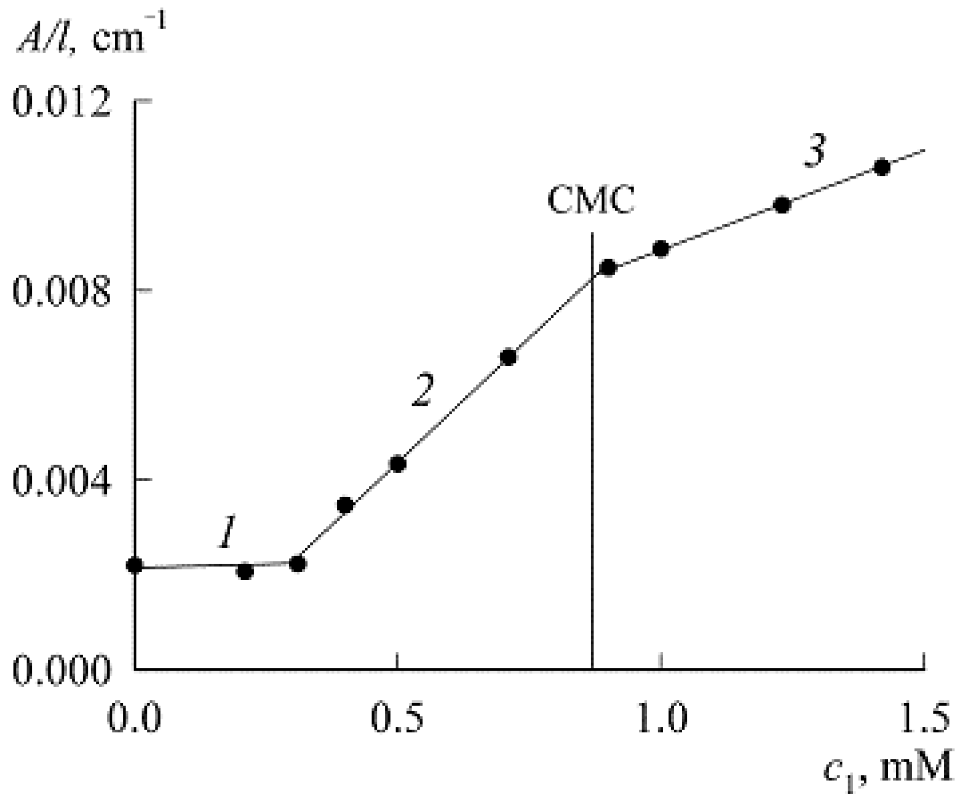
Figure 2 illustrates the results of a photospectrometric study of the NA–NR–water system, where NA acted as a solubilizer and NR as a solubilisate [34]. Region 2 refers to protomicelles, and region 3 to NA micelles with solubilized NR molecules. The slope of these areas characterizes the activity of the process, and we see that it is greater in the case of protomicelles. The calculation performed showed that NA micelles are only partially filled with solubilisate [34], which, by its location, is found to be greater in protomicelles (possibly because micelles are spherical, while protomicelles are disk-shaped). It can be said that the work [34] was the first study in which protomicelles turned out to be more important than micelles.
The next step, implemented in this work, is the use of a non-colloidal surfactant and the study of such a dye–surfactant–water system, where there are no micelles and cannot be. As such a non-colloidal surfactant, we took heptyl alcohol (1-heptanol) and offer the reader the results of a study of the heptanol–NR–water system (accordingly, in the notation we will consider heptanol as the first, and NR as the second component of the system). The purpose of this work is to study the effect of heptanol on the solubility of NR in water. Since solubility is the equilibrium concentration of a substance in contact with a solution of a pure (for example, solid) phase of the same substance, we can say that similar experiments with NR were carried out at the constant chemical potential of NR. In this work, for the first time, the protomicelles of heptanol were observed as a particular example of non-colloidal surfactants. The latter are often called hydrotropes. They are not able to form micelles but are seen in the formation of fluctuating dynamic structures in aqueous solutions [36,37,38].
2. Materials and Methods
2.1. Reagents
Experiments were carried out with the following reagents: NR (9-diethylamino-5-benzo[a]phenoxazinone, Figure 1) is a preparation from “Acros Organics” with a content of the main substance of 99.0%. 1-heptanol (C7H15OH) had the qualification “chemically pure”, the preparation of the company “OOO ChromeLab”, Russia, with the content of the main substance of 99.9%. Both preparations were used without additional purification. Water was taken in the form of a tridistillate with a specific electrical conductivity not higher than 4 × 10⁻⁴ Sm/m at 25 °C.
2.2. Methodology
The experimental technique was spectrophotometry, the measurement of the optical density A of the solution depending on the light wavelength λ and observation of the characteristic wavelengths for NR with a change in the concentration of heptanol. The relationship between the optical density (extinction) A and the concentration of the absorbing substance c is given by the Lambert–Beer law.
where ε is the extinction coefficient and l is the path length of the light beam. It can be seen from Equation (1) that in order to determine the concentration from the optical density, it is necessary to know the extinction coefficient. However, if it is unknown, then, from the direct proportionality between A and c in Equation (1), it follows that the optical density itself can serve as a certain measure of concentration in the absence of factors that violate this proportionality.
It is known [24,26,28] that the absorption bands characteristic of NR monomers and dimers are in the wavelength range λ = 450–700 nm. For NR monomers in pure water, the optical absorption maximum corresponds to the wavelengths λmax = 591 nm [24], 593 nm [28,30], and 600 nm [27], respectively. Previously, we found that NR monomers in its aqueous saturated solutions correspond to the maximum A at nm, and dimers at λmax = 543 nm [30]. In the presence of colloidal surfactants sodium dodecyl sulfate and tetradecyltrimethylammonium bromide, the bands characteristic of NR monomers and dimers are also in the range λ = 450–700 nm, although the positions of their maxima undergo changes. Therefore, to measure the spectra of the studied solutions, the wavelength range of 400–900 nm was chosen, which provides a margin for observing the characteristic NR wavelengths with a change in the heptanol concentration. Since the spectra of heptanol in this range do not overlap with the NR spectra, the spectra of the studied solutions with the dye were recorded relative to water and not to reference solutions of surfactants having a similar composition but without dye, as is sometimes done when studying solubilization using colloidal surfactants [39] To increase the sensitivity of the method, we used a cuvette with a longer optical path length (l = 5.007 cm) than we did earlier (1 and 2 cm) [30].
An important detail of the technique is the preparation of aqueous systems with NR and heptanol. First, heptanol solutions were prepared in water with concentrations (c1) of 8.5 and 10.3 mM (according to the reference book [40], these surfactant values are within the solubility range of heptanol (14–17 mM at 20 °C). By diluting the initial system with a concentration of 8.5 mM with water, heptanol solutions were obtained in the concentration range of 0.44–8.5 mM.
In each experiment, a weighed portion of NR powder of the same weight was poured with a heptanol solution (with volume of 25 mL) of a given concentration. The content of the NR sample in this volume was equivalent to a concentration (c2) of 30 µM, which is much higher than the solubility of the dye in pure water (about 2 µM [30]). The resulting systems were stirred at room temperature with a magnetic stirrer (at 400 rpm) for 3–5 h, followed by settling in a closed state for several days. Periodically, a liquid sample was taken over the NR precipitate and the absorption spectrum was recorded. In the absence of changes in the spectra, the onset of an equilibrium state of a saturated NR solution was noted. It was achieved in almost five days. All measurements were carried out at a room temperature (20 °C).
The subsequent analysis of the spectra included their decomposition. There are special computer programs for this purpose and we used the PeakFit mathematical package Version 4 for Win 32, AISN Software Inc., Mapleton, OR, USA.
3. Results and Discussion
As already noted, in our experiments studying the dependence of the solubility of NR on the concentration of heptanol, the chemical potential of NR at a given temperature is constant. For such conditions, the thermodynamic theory [18] leads to the expression:
where is the NR concentration in our case, is the NR concentration in the absence of heptanol, u is the work of detachment of one heptanol molecule from the NR molecule surface, n is the number of adsorbed heptanol molecules per NR molecule (protomicelle aggregation number), and kT has the usual meaning. Equation (2) shows that the NR concentration increases with the number of adsorbed heptanol molecules, which, whatever the adsorption equation, always increases with the heptanol concentration in solution. For small , this increase is linear:
where is Henry’s constant. In the limit of small n, the work u becomes a constant, and the dependence on in the leading term of the expansion of the exponent also becomes linear:
where Thus, Equation (2), and Equation (4) following from it, predicts an increase in the concentration of NR in solution with the addition of heptanol.
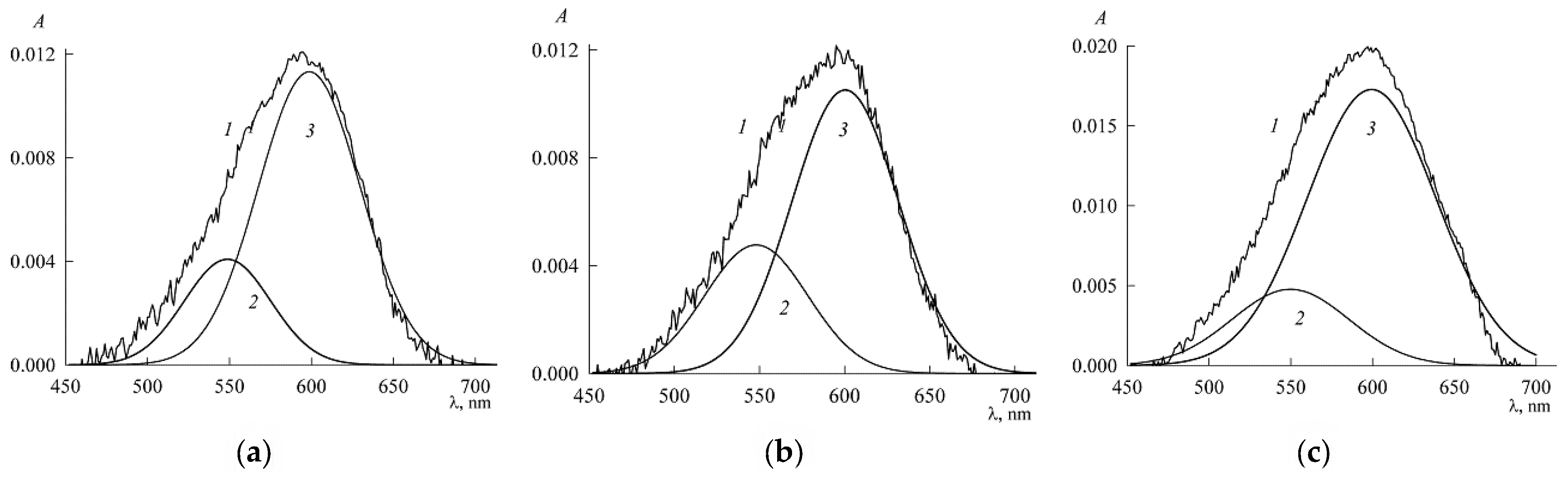
The predictions of the above equations are confirmed by experiment. Figure 3 shows the electronic absorption spectra of NR in aqueous solutions (saturated with respect to NR) with different concentrations of heptanol. For clarity, they are presented as three groups of spectra: a, b, and c. Region “a” corresponds to heptanol concentration c1 in the range 0–2.1 mM, region “b” corresponds to the c1 values 3 and 7 mM, and region “c” corresponds to c1 in the range 7.8–10.3 mM.
Figure 3 shows that the height of the optical density peaks A at the wavelength value of 593 nm, which is characteristic of NR monomers, gradually increases with an increase in the concentration of heptanol in the system. In this case, the shape of the absorption band also changes slightly. For the spectrum in pure water (curve 1 in Figure 3), a shoulder is visible on the left side, which indicates the presence of NR dimers as well. This can be demonstrated more clearly by decomposing the spectrum. The decomposition result of spectrum 1 is shown in Figure 4, where two peaks are clearly visible, corresponding to monomers (4, nm) and dimers (3, nm), and a small flattened peak (2, nm) possibly corresponding to other NR molecular aggregates.
When the NR spectra are decomposed in the presence of heptanol (curves 2–4 in Figure 3a), two main peaks are observed at λmax = 600 nm (3) and λmax = 550 nm (2). They also correspond to monomers [27] and NR dimers. The shift of the maximum of the monomer component (curve 2 in Figure 5) of NR compared to the maximum in an aqueous medium (curve 4 in Figure 4) apparently reflects the effect of heptanol on the microenvironment of dye molecules. Comparison of curve 3 in Figure 4 and curve 2 in Figure 5 shows that in the range of c1 0–2.1 mM, with an increase in the concentration of heptanol, the proportion of dimers in comparison with monomers in the dye–surfactant–water system decreases. In the other spectra of Figure 3b,c, the presence of dimers is practically not felt, and no decomposition was done for them, considering that the presence of dimers in them can be neglected. Thus, we can conclude that although heptanol does not form micelles, it has the ability to monomerize NR.
Among other features of the spectra in Figure 3, it can be noted that the position of maximum A has received a bathochromic shift and is located at a wavelength of 599 nm (curve 9 in Figure 3). At c1 > 7 mM, the left side of the band narrows, while the right side shows a shoulder characteristic of the protonated form of NR [25].
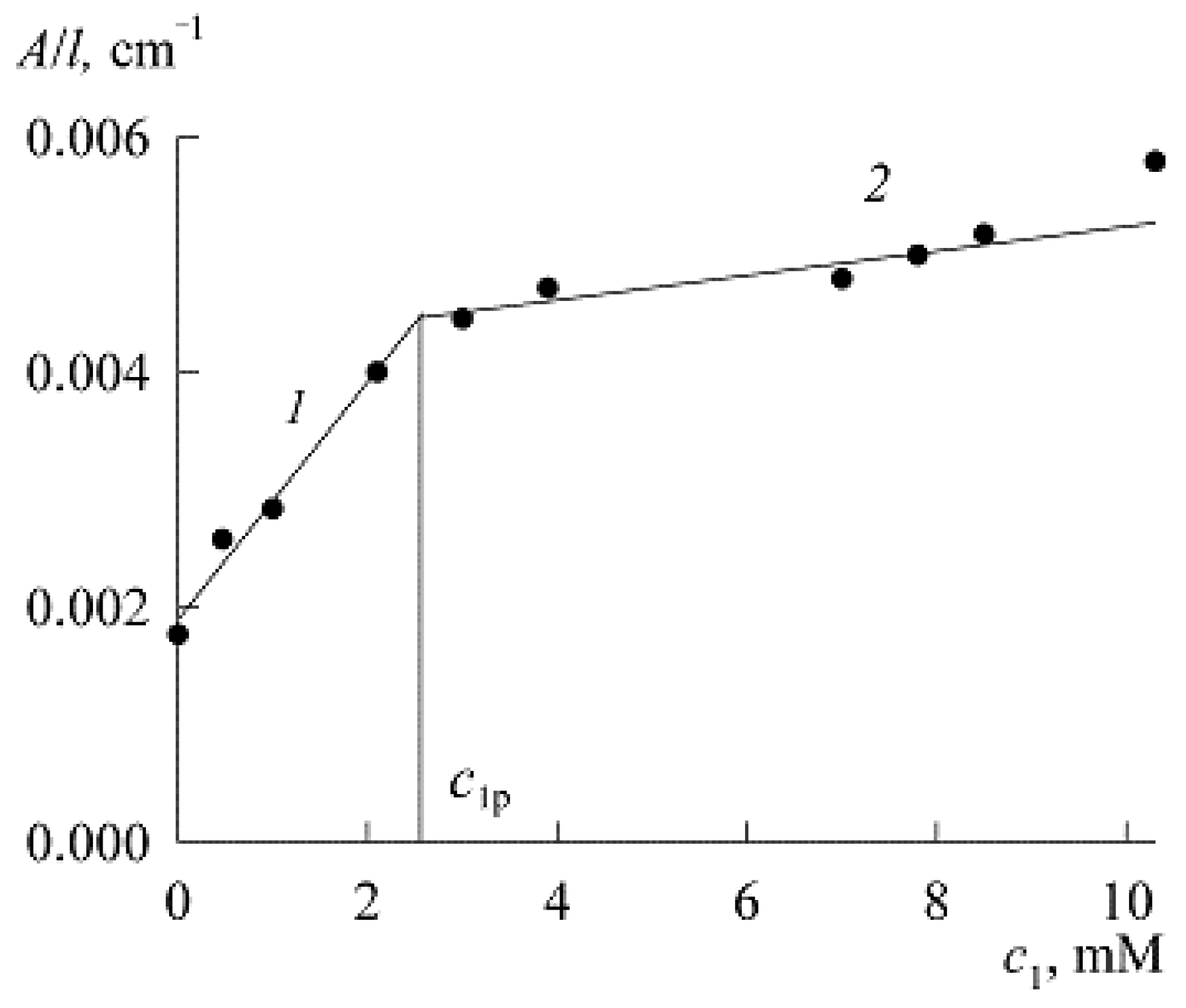
The maximum peak points for NR monomers in Figure 3, Figure 4 and Figure 5 are shown in Figure 6 as a dependence of the reduced (to the length l = 1 cm) optical density on the concentration of heptanol. Curve A/l vs. c1 can be divided into two sections. Section 1 with a stronger dependence corresponds to the process of protomicelle formation (filling of the adsorption layer with heptanol molecules). Section 2 corresponds to practically formed protomicelles. Figure 6 is similar to Figure 2 for the system with NA, where the process of protomicelle formation is interrupted by micellization. There are many micelles and the NR solubilization mechanism already passes through the micelles. In Figure 6, micellization is absent and protomicelles take over all the work of NR solubilization. The existence of two sections of the curve is due to the fact that when the protomicelle has already been formed, the introduction of new heptanol molecules into the adsorption layer requires incomparably more work than in the previous section (there are no more empty places, and in order to introduce another molecule, you need to push all the others). An increase in work also means an increase in the thermodynamic force of the process, which is the gradient or, more simply, the difference in the chemical potential of heptanol in solution and in the adsorption layer. This difference increases with the introduction of new portions of heptanol into the solution, and since the chemical potential depends on the concentration logarithmically, the latter requires a significant increase, which leads to a drop in the slope in Section 2 of Figure 6. The course of the second section itself depends on the adsorption isotherm, which is unknown and can be quite complicated, but in our case, it breaks off simply due to the limited solubility of heptanol itself.
From Figure 6, one can determine the value of the heptanol concentration (let us denote it as ) corresponding to the formation of protomicelles (when there are no more empty vacancies in the adsorption layer) as the break point. To do this, we approximate both the sections by straight lines described by mathematical equations (c1 is expressed in mM):
where R2 is the coefficient of determination. The joint solution of Equations (5) and (6) determines the coordinate of the break point as 2.57 mM. This is the value of , which is an important protomicelle parameter.
4. Concluding Remarks
Unlike surfactant micelles, surfactant protomicelles are formed by adsorption on nano-adsorbent particles (in our case, NR molecules), and therefore are never empty. Protomicelles are similar not just to micelles, but to micelles with solubilisate. For the latter, the phenomenon of solubilization is specific: not every micelle can absorb any nano-object. However, any body is capable of absorbing something on itself. Adsorption is a more universal phenomenon than solubilization, and therefore, it can be said that protomicelles are more universal objects than micelles. In the case of aqueous systems, the formation of both of them is associated with hydrophobic interactions, which, in comparison with conventional adsorption forces, have their own specifics. When surfactant molecules or ions are adsorbed on a hydrophobic surface or adhere to each other with their hydrophobic parts, they do not experience attraction, but are pushed out by water from the aquatic environment. The fact is that the structure of water is destroyed by the presence of hydrophobic particles. Since this is energetically unfavorable (requires work), water tends to get rid of such presence. Consequently, hydrophobic interactions are determined by the structure of water and manifest themselves both in adsorption and in micelle formation.
In the broad sense of the word, solubilization is not the penetration of a solubilizate into a micelle, but any increase in the solubility of a substance by the mere presence of another substance (solubilizer). Historically, solubilization has been associated with surfactant micelles and colloidal surfactants. However, now we see that the role of a surfactant as a solubilizer can be realized not only through micelles, but also through protomicelles in the absence of micelles, and any surfactant, including a non-colloidal one (in this work, it was heptanol) can serve as a solubilizer. This is the first conclusion that follows from this work, where a joint solution of a non-colloidal surfactant and a dye was studied for the first time and the formation of protomicelles in the absence of micelles was observed.
The formation of surfactant protomicelles in an aqueous solution is closely related to dye monomerization [17]. Indeed, if there are hydrophobic regions on the surface of the dye molecules (the contact of which with water is energetically unfavorable), then it is these regions that the dye molecules stick to each other, forming dimers and other molecular aggregates. Coating these areas with adsorbed surfactant molecules makes it impossible for the dye molecules to stick together. In this work, this has been experimentally demonstrated using heptanol as an example, and we can conclude that non-colloidal surfactants, no worse than colloidal surfactants, can serve as a means of dye monomerization. This conclusion is of great practical importance, because the choice of non-colloidal surfactants is much wider than colloidal ones.
Author Contributions
Conceptualization, A.I.R. and T.G.M.; methodology, T.G.M. and E.V.P.; validation, A.I.R. and T.G.M.; formal analysis, T.G.M. and E.V.P.; investigation, A.I.R.; resources, T.G.M. and E.V.P.; data curation, T.G.M. and E.V.P.; writing—original draft preparation, A.I.R.; writing—review and editing, A.I.R. and T.G.M.; supervision, A.I.R.; project administration, A.I.R. All authors have read and agreed to the published version of the manuscript.
Funding
This work was supported by the Ministry of Science and Higher Education of the Russian Federation on the topic “Physical and chemical problems of creating effective nano- and supramolecular systems” (the registration number 122011300052-1).
Data Availability Statement
Data is contained within the article.
Conflicts of Interest
The authors declare no conflict of interest.
References
- Li, X.Y.; He, X.; Ng, A.C.H.; Wu, C.; Ng, D.K.P. Influence of surfactants on the aggregation behavior of water-soluble dendritic phthalocyanines. Macromolecules 2000, 33, 2119–2123. [Google Scholar] [CrossRef]
- Chen, Z.J.; Li, X.Y.; Ngai, T.; Wu, C.; Ng, D.K.P. Monomerization of cationic phthalocyanine in AOT reversed micelles. Langmuir 2001, 17, 7957–7959. [Google Scholar] [CrossRef]
- Gol’dshleger, N.F.; Kalashnikova, I.P.; Baulin, V.E.; Tsivadze, A.Y. Octa- and tetra-(benzo-15-crown-5) phthalocyanines in surfactant-containing solutions. Prot. Met. Phys. Chem. Surf. 2011, 47, 471–477. [Google Scholar] [CrossRef]
- Gol’dshleger, N.F.; Chernyak, A.V.; Kalashnikova, I.P.; Baulin, V.E.; Tsivadze, A.Y. Magnesium octa(benzo-15-crown-5)phthalocyaninate in the sodium dodecyl sulfate solutions: A study using electron and 1H NMR spectroscopy. Russ. J. Gen. Chem. 2012, 82, 927–935. [Google Scholar] [CrossRef]
- Gol’dshleger, N.F.; Lobach, A.S.; Gak, V.Y.u.; Kalashnikova, I.P.; Baulin, V.E.; Tsivadze, A.Y. Magnesium octa[(4′-benzo-15-crown-5)oxy]phthalocyaninate in water micellar solutions of sodium deoxycholate. Prot. Met. Phys. Chem. Surf. 2014, 50, 599–607. [Google Scholar] [CrossRef]
- Goldshleger, N.F.; Chernyak, A.V.; Lobach, A.S.; Kalashnikova, I.P.; Baulin, V.E.; Tsivadze, A.Y. Monomerization of crown-containing phthalocyanines in microheterogeneous organized systems. Prot. Met. Phys. Chem. Surf. 2015, 51, 212–220. [Google Scholar] [CrossRef]
- Goldshleger, N.F.; Gak, V.Y.u.; Kalashnikova, I.P.; Baulin, V.E.; Ivanchikhina, A.V.; Smirnov, V.A.; Shiryaev, A.A.; Tsivadze, A.Y. Solubilization and photochemical stability of octa[(4′-benzo-15-crown-5)oxy]-phthalocyanines in aqueous micellar solutions. Prot. Met. Phys. Chem. Surf. 2018, 54, 1092–1101. [Google Scholar] [CrossRef]
- Goldshleger, N.F.; Kalashnikova, I.P.; Gorbunova, Y.G.; Martynov, A.G.; Baulin, V.E.; Tsivadze, A.Y. Solubilization of crown-substituted magnesium phthalocyaninates in solutions of salts of bile acids. Prot. Met. Phys. Chem. Surf. 2018, 54, 33–42. [Google Scholar] [CrossRef]
- Goldshleger, N.F.; Gak, V.Y.; Lapshina, M.A.; Baulin, V.E.; Shiryaev, A.A.; Tsivadze, A.Y. Supramolecular organization of crown- and phosphoryl-containing magnesium and zinc phthalocyaninates in solutions of synthetic and natural surfactants. Russ. Chem. Bull. 2018, 67, 2205–2211. [Google Scholar] [CrossRef]
- Movchan, T.G.; Averin, A.A.; Baulin, D.V.; Plotnikova, E.V.; Baulin, V.E.; Tsivadze, A.Y. Solubilization of magnesium octa[(4′-benzo-15-crown-5)oxy] phthalocyaninate in aqueous micellar solutions of hexadecyltriphenylphosphonium bromide. Colloid J. 2018, 80, 501–512. [Google Scholar] [CrossRef]
- Movchan, T.G.; Chernyad’ev, A.Y.; Plotnikova, E.V.; Averin, A.A.; Tsivadze, A.Y.; Baulin, V.E. Disaggregation of magnesium octa[(4′-benzo-15-crown-5)oxy] phthalocyaninate in aqueous micellar solutions of alkyltriphenylphosphonium bromide. Colloid J. 2018, 80, 667–675. [Google Scholar] [CrossRef]
- Movchan, T.G.; Chernyad’ev, A.Y.; Plotnikova, E.V.; Tsivadze, A.Y.; Baulin, V.E. Disaggregation of zinc tetra(4-carboxyphenoxy) phthalocyaninate in aqueous solutions of dodecyl-, tetradecyl-, and hexadecyltrimethylammonium bromides at ph 6–9. Colloid J. 2019, 81, 711–719. [Google Scholar] [CrossRef]
- Movchan, T.G.; Chernyad’ev, A.Y.; Plotnikova, E.V.; Tsivadze, A.Y.; Baulin, V.E. The processes zinc phosphoryl-substituted phthalocyaninate dissolution in water in the presence of tetradecyltrimethylammonium bromide within a ph range of 4–9. Colloid J. 2020, 82, 16–26. [Google Scholar] [CrossRef]
- Rusanov, A.I.; Movchan, T.G.; Plotnikova, E.V. A new type of micelles and concentration of monomerization for phthalocyanines in aqueous surfactant solutions. Dokl. Phys. Chem. 2020, 495, 181–185. [Google Scholar] [CrossRef]
- Movchan, T.G.; Rusanov, A.I.; Plotnikova, E.V. Thermodynamic study of solubilization of crown-substituted magnesium phthalocyaninate in aqueous solutions of sodium dodecyl sulfate. Colloid J. 2021, 83, 97–106. [Google Scholar] [CrossRef]
- Movchan, T.G.; Rusanov, A.I.; Plotnikova, E.V. Protomicelles of sodium dodecyl sulfate in a strongly diluted aqueous solution of crown-substituted magnesium phthalocyaninate. Colloid J. 2021, 83, 356–364. [Google Scholar] [CrossRef]
- Rusanov, A.I. On the theory of phthalocyanine monomerization in aqueous surfactant solutions. Colloid J. 2021, 83, 236–244. [Google Scholar] [CrossRef]
- Rusanov, A.I. Nano-adsorbents and protomicelles in colloid science. Colloids Surf. A 2021, 629, 127453. [Google Scholar] [CrossRef]
- Rusanov, A.I. Micellization in Surfactant Solutions; Harwood Academic Publishers: Reading, UK, 1996; pp. 1–326. ISBN 90-5702-297-4. [Google Scholar]
- Zhu, P.W.; Napper, D.H. Interfacial coil-to-globule transitions: The effects of molecular weight. Colloids Surf. A 1996, 113, 145–153. [Google Scholar] [CrossRef]
- El-Ejmi, A.A.S.; Huglin, M.B. Behaviour of poly(n,n-dimethylacrylamide-co-2-methoxyethylacrylate) in non-aqueous solution and LCST behaviour in water. Eur. Polym. J. 1997, 33, 1281–1284. [Google Scholar] [CrossRef]
- Qiao, B.; Littrell, K.C.; Ellis, R.J. Liquid worm-like and proto-micelles: Water solubilization in amphiphile–oil solutions. Phys. Chem. Chem. Phys. 2018, 20, 12908. [Google Scholar] [CrossRef]
- Wei, P.; Cornel, E.J.; Du, J. Breaking the corona symmetry of vesicles. Macromolecules 2021, 54, 7603–7611. [Google Scholar] [CrossRef]
- Greenspan, P.; Fowler, S.D. Spectrofluorometric studies of the lipid probe, nile red. J. Lipid Res. 1985, 26, 781–789. [Google Scholar] [CrossRef]
- Samsonova, L.G.; Selivanov, N.I.; Kopylova, T.N. Spectral properties of nile red in solutions and thin films. Opt. Spectrosc. 2014, 116, 72–76. [Google Scholar] [CrossRef]
- Ray, A.; Das, S.; Chattopadhyay, N. Aggregation of nile red in water: Prevention through encapsulation in β-cyclodextrin. ACS Omega 2019, 4, 15–24. [Google Scholar] [CrossRef] [Green Version]
- Swain, J.; Mishra, J.; Ghosh, G.; Mohr, A.K. Quantification of micropolarity of aggregation and salt-induced gelation of sodium deoxycholate (NaDC) using nile red fluorescence. Photochem. Photobiol. Sci. 2019, 18, 2773–2781. [Google Scholar] [CrossRef]
- Kurniasih, I.N.; Liang, H.; Mohr, P.C.; Khot, G.; Rabe, J.P.; Mohr, A. Nile red dye in aqueous surfactant and micellar solution. Langmuir 2015, 31, 2639–2648. [Google Scholar] [CrossRef]
- Datta, A.; Mandal, D.; Pal, S.K.; Bhattacharyya, K. Intramolecular charge transfer processes in confined systems. Nile red in reverse micelles. J. Phys. Chem. B 1997, 101, 10221–10225. [Google Scholar] [CrossRef]
- Movchan, T.G.; Rusanov, A.I.; Plotnikova, E.V. Solubilization of nile red in aqueous solutions oftetradecyltrimethylammonium bromide. Russ. J. Gen. Chem. 2022, 92, 2023–2032. [Google Scholar] [CrossRef]
- Krishna, M.M.G. Excited-state kinetics of the hydrophobic probe Nile red in membranes and micelles. J. Phys. Chem. A 1999, 103, 3589–3595. [Google Scholar] [CrossRef]
- Rusanov, A.I.; Movchan, T.G.; Plotnikova, E.V. Solubilization of Nile red in micelles and protomicelles of sodium dodecyl sulfate. Molecules 2022, 27, 7667. [Google Scholar] [CrossRef] [PubMed]
- Gradova, M.A.; Movchan, T.G.; Khudyaeva, I.S.; Chernyad’ev, A.Y.; Plotnikova, E.V.; Lobanov, A.V.; Belykh, D.V. Synthesis of the novel cationic chlorin derivatives with a phytol fragment on the periphery of the macrocycle and their aggregation state in aqueous surfactant solutions. Macroheterocycles 2020, 13, 23–32. [Google Scholar] [CrossRef]
- Rusanov, A.I.; Movchan, T.G.; Plotnikova, E.V. New concepts of colloid science: Nano-adsorbent and protomicelle. Dokl. Phys. Chem. 2023, in press. [Google Scholar]
- Badban, S.; Hyde, A.S.; Phan, C.M. Hydrophilicity of nonanoic acid and its conjugate base at the air/water interface. ACS Omega 2017, 2, 5565–5573. [Google Scholar] [CrossRef]
- Subramanian, D.; Boughter, C.T.; Klauda, J.B.; Hammoudac, B.; Anisimov, M.A. Mesoscale inhomogeneities in aqueous solutions of small amphiphilic molecules. Faraday Discuss. 2013, 167, 217. [Google Scholar] [CrossRef]
- Kunz, W.; Holmberg, K.; Zemb, T. Hydrotropes. Curr. Opin. Colloid Interface Sci. 2016, 22, 99–107. [Google Scholar] [CrossRef]
- Buchecker, T.; Krickl, S.; Winkler, R.; Grillo, I.; Bauduin, P.; Touraud, D.; Pfitzner, A.; Kunz, W. The impact of the structuring of hydrotropes in water on the mesoscale solubilization of a third hydrophobic component. Phys. Chem. Chem. Phys. 2017, 19, 1806–1816. [Google Scholar] [CrossRef] [Green Version]
- Zadymova, N.N.; Tsikurina, N.N.; Poteshnova, M.V. Solubilization of perfluorodealin in aqueous solutions of dodecaethoxylated nonylphenol. Colloid J. 2003, 65, 314–318. [Google Scholar] [CrossRef]
- Van Os, N.M.; Haak, J.R.; Rupert, L.A.M. Physico-Chemical Properties of Selected Anionic, Cationic and Nonionic Surfactants; Elsevier: Amsterdam, The Netherlands, 1993; pp. 1–608. [Google Scholar]
Figure 1.
The structural formula of Nile red.

Figure 2.
Dependence of the reduced (to the length l = 1 cm) optical density NR A/l on the concentration of NA according to the spectral data of the NA–NR–water system.
Figure 2.
Dependence of the reduced (to the length l = 1 cm) optical density NR A/l on the concentration of NA according to the spectral data of the NA–NR–water system.

Figure 3.
Electronic absorption spectra of saturated NR solutions at heptanol concentrations c1, mM: (a) 0 (1), 0.47 (2), 1 (3), 2.1 (4); (b) 3 (5), 7 (6), and (c) 7.8 (7), 8.5 (8), 10.3 (9); L = 5.007 cm.
Figure 3.
Electronic absorption spectra of saturated NR solutions at heptanol concentrations c1, mM: (a) 0 (1), 0.47 (2), 1 (3), 2.1 (4); (b) 3 (5), 7 (6), and (c) 7.8 (7), 8.5 (8), 10.3 (9); L = 5.007 cm.

Figure 4.
Decomposition of the NR absorption spectrum in water (curve 1) into components 2, 3, and 4.
Figure 4.
Decomposition of the NR absorption spectrum in water (curve 1) into components 2, 3, and 4.

Figure 5.
Decomposition of the absorption spectrum of NR (1) into components 2 and 3 at the concentration of heptanol in water c1, mM: 0.47 (a), 1 (b), and 2.1 (c).
Figure 5.
Decomposition of the absorption spectrum of NR (1) into components 2 and 3 at the concentration of heptanol in water c1, mM: 0.47 (a), 1 (b), and 2.1 (c).

Figure 6.
Dependence of the reduced (to the length l = 1 cm) optical density A/l of aqueous saturated solutions of NR on the concentration of heptanol c1.
Figure 6.
Dependence of the reduced (to the length l = 1 cm) optical density A/l of aqueous saturated solutions of NR on the concentration of heptanol c1.

Disclaimer/Publisher’s Note: The statements, opinions and data contained in all publications are solely those of the individual author(s) and contributor(s) and not of MDPI and/or the editor(s). MDPI and/or the editor(s) disclaim responsibility for any injury to people or property resulting from any ideas, methods, instructions or products referred to in the content. |
© 2023 by the authors. Licensee MDPI, Basel, Switzerland. This article is an open access article distributed under the terms and conditions of the Creative Commons Attribution (CC BY) license (https://creativecommons.org/licenses/by/4.0/).
Share and Cite
MDPI and ACS Style
Rusanov, A.I.; Movchan, T.G.; Plotnikova, E.V. First Observation of Protomicelles in the System with a Non-Colloidal Surfactant. Colloids Interfaces 2023, 7, 32. https://doi.org/10.3390/colloids7020032
AMA Style
Rusanov AI, Movchan TG, Plotnikova EV. First Observation of Protomicelles in the System with a Non-Colloidal Surfactant. Colloids and Interfaces. 2023; 7(2):32. https://doi.org/10.3390/colloids7020032
Chicago/Turabian StyleRusanov, Anatoly I., Tamara G. Movchan, and Elena V. Plotnikova. 2023. "First Observation of Protomicelles in the System with a Non-Colloidal Surfactant" Colloids and Interfaces 7, no. 2: 32. https://doi.org/10.3390/colloids7020032