Case Report

Tinnitus, Aural Fullness, and Hearing Loss in a Patient with Acoustic Neuroma and Pituitary Macroadenoma

1
Department of Clinical Sciences and Community Health, University of Milan, 20122 Milan, Italy
2
Audiology Unit, Department of Specialist Surgical Sciences, Fondazione IRCCS Ca’ Granda Ospedale Maggiore Policlinico, 20122 Milan, Italy
3
Otolaryngology Unit, Department of Specialist Surgical Sciences, Fondazione IRCCS Ca’ Granda Ospedale Maggiore Policlinico, 20122 Milan, Italy
*
Author to whom correspondence should be addressed.
J. Otorhinolaryngol. Hear. Balance Med. 2023, 4(1), 2; https://doi.org/10.3390/ohbm4010002
Submission received: 6 March 2023 / Revised: 19 March 2023 / Accepted: 23 March 2023 / Published: 27 March 2023

Abstract

:
We report the case of a 51-year-old woman with multiple otologic and vestibular symptoms. She presented with two different types of tinnitus in her right ear, vertigo, and fluctuating aural symptoms in the left ear. She also complained of disequilibrium; chronic headache; hyperhidrosis; amenorrhea; insomnia; broadened hands and feet; and widened, thickened, and stubby fingers. The patient underwent careful collection of medical history, otomiscroscopy, pure tone audiometry, tympanometry, reflex threshold measurements, vestibular assessments, blood tests, magnetic resonance imaging (MRI), and cone beam computed tomography (CBTC) of the head. The audiogram showed: (1) a mild low-to-mid frequency conductive hearing loss, and a sharply sloping sensorineural hearing loss above 4000 Hz in the right ear; (2) a mild low-frequency sensorineural hearing loss in the left ear. MRI with 3D FLAIR sequences detected an acoustic neuroma (7.4 mm × 5.2 mm) in the middle-third of the right internal auditory canal, a pituitary macroadenoma (13 mm × 10 mm × 10 mm) and left saccular hydrops. The CBCT scan documented an outbreak of otosclerosis (3 mm) around the fissula ante fenestram in the right ear. Therefore, acoustic neuroma (right ear), growth hormone-secreting macroadenoma of the pituitary gland, Menière’s disease (left ear), and otosclerosis (right ear) were diagnosed/strongly suspected. A watch-and-wait strategy was adopted for acoustic neuroma and otosclerosis, while transsphenoidal surgery was successfully performed to remove the pituitary macroadenoma. This case report confirms that multiple otologic disorders can occur simultaneously in the same patient, requiring prompt audiological and imaging evaluations.

The COVID-19 (coronavirus disease 19) pandemic led to both delayed diagnosis of ENT (Ear, and Throat) diseases and a high prevalence of balance disorders [1,2].
We discuss the complex case of a patient who presented with multiple otologic and vestibular symptoms.
A 51-year-old woman had experienced progressive hearing loss associated with continuous tinnitus (described as “buzzing”) in her right ear over the years.
She also reported 6 episodes of vertigo in the past 5 years, each lasting about 1–2 h, associated with fluctuating aural symptoms, including tinnitus, hearing loss, and fullness in the left ear.
Furthermore, she recently started complaining of a second tinnitus (described as “high-pitched whistling”) in her right ear; disequilibrium; chronic headache; hyperhidrosis (“My palms are always sweaty”); amenorrhea; insomnia; enlargement of the extremities (broadened hands and feet); and widened, thickened, and stubby fingers (“My wedding ring won’t fit anymore”).
Therefore, she presented to our third-level referral audiologic center for a complete audiological and vestibular evaluation.
Her medical history revealed no allergies, use of drugs, episodes of recurrent otitis media, head trauma, or recent infectious disease. She had no family history of hearing loss.
The patient underwent otomiscroscopy, pure tone audiometry, tympanometry, reflex threshold measurements, and vestibular assessments (bedside examination and video head impulse test [v-HIT]).
Otomicroscopy was bilaterally normal, with the exception of a reddish hue arising from the promontory behind an intact tympanic membrane in the right ear.
The audiogram showed:
(1)
a mild conductive hearing loss limited to low and middle frequencies, and a sharply sloping sensorineural hearing loss above 4000 Hz in the right ear (Figure 1);
(2)
a mild low-frequency sensorineural hearing loss with a characteristic rising curve in the left ear (Figure 2).
The acoustic reflexes were bilaterally absent. The tympanogram presented a shallow peak (type As) in the right ear, while it was normal (type A) in the left ear.
Vestibular examination revealed a left beating spontaneous nystagmus (grade I) and normal v-HIT gains.
In the suspicion of left endolymphatic hydrops with irritative nystagmus, the following medical therapy was prescribed: Amiloride + Hydrochlorothiazide (5 mg + 50 mg, 1 tablet/die), Betahistine (24 mg, 2 tablets/die), and citicoline (500 mg, 2 tablets/day) for 2 weeks.
Magnetic resonance imaging (MRI) with 3-dimensional (3D) fluid-attenuated inversion recovery (FLAIR) sequence and high-resolution cone beam computed tomography (CBCT) of the head were also requested.
After 2 weeks, pure tone audiometry was repeated, showing complete hearing recovery at low and middle frequencies in the left ear (Figure 3). Speech audiometry showed 100% understanding at 60 dB in the right ear and 100% understanding at 30 dB in the left ear.
MRI with 3D FLAIR sequences (with images taken at 10 min and 4 h after intravenous gadolinium injection) detected:
(a)
Acoustic neuroma (7.4 mm × 5.2 mm) in the middle-third of the right internal auditory canal (Figure 4a);
(b)
Pituitary macroadenoma (13 mm × 10 mm × 10 mm) (Figure 4b);
(c)
Left saccular hydrops (Figure 4c).
The CBCT scan documented:
(d)
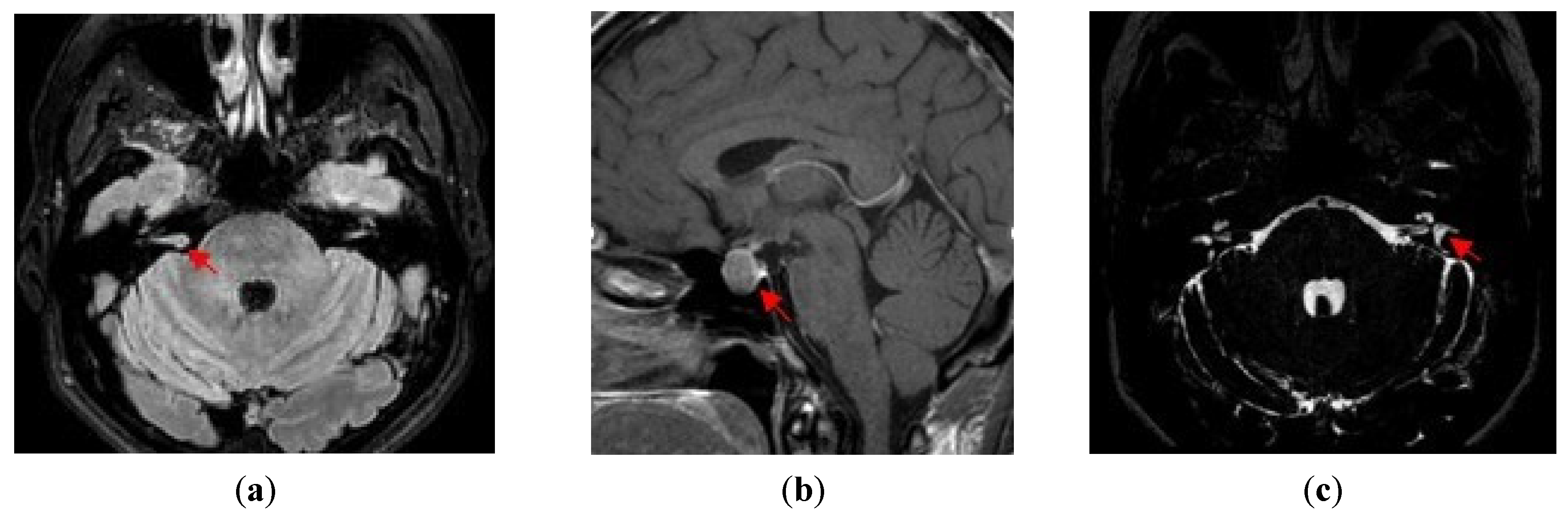
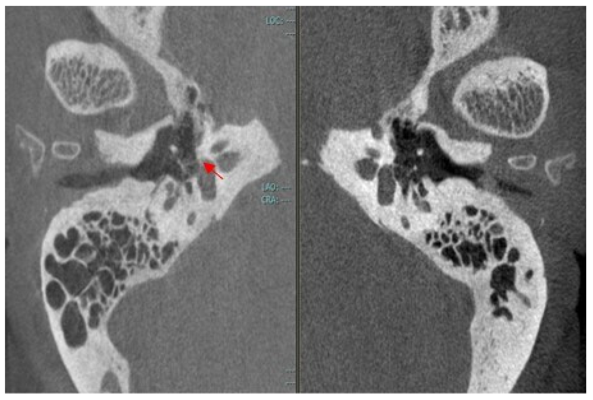
Outbreak of otosclerosis (3 mm) around the fissula ante fenestram in the right ear (Figure 5).
Subsequently, blood tests for autoimmune (anti-dsDNA, anti-RNP, anti-Sm, anti-SSA and SSB, anti-Scl-70, anti-Jo-1, and anti-CCP), cardiovascular (fibrinogen, D-dimer, activated partial thromboplastin time [aPTT], prothrombin time [PT], and homocysteine), and endocrine disorders (adrenocortisol [ACTH] and cortisol, growth hormone [GH], and insulin-like growth factor 1 [IGF-1], prolactin, thyroid-stimulating hormone [TSH] and thyroid hormone, luteinizing hormone (LH), and testosterone or estrogen, follicle-stimulating hormone [FSH]) were also requested.
Interestingly, a high value of IGF1 was found (>354.00 ng/mL [48.00–209.00]), while autoimmune and cardiovascular screenings were negative.
The following pathologies were diagnosed/strongly suspected:
(a)
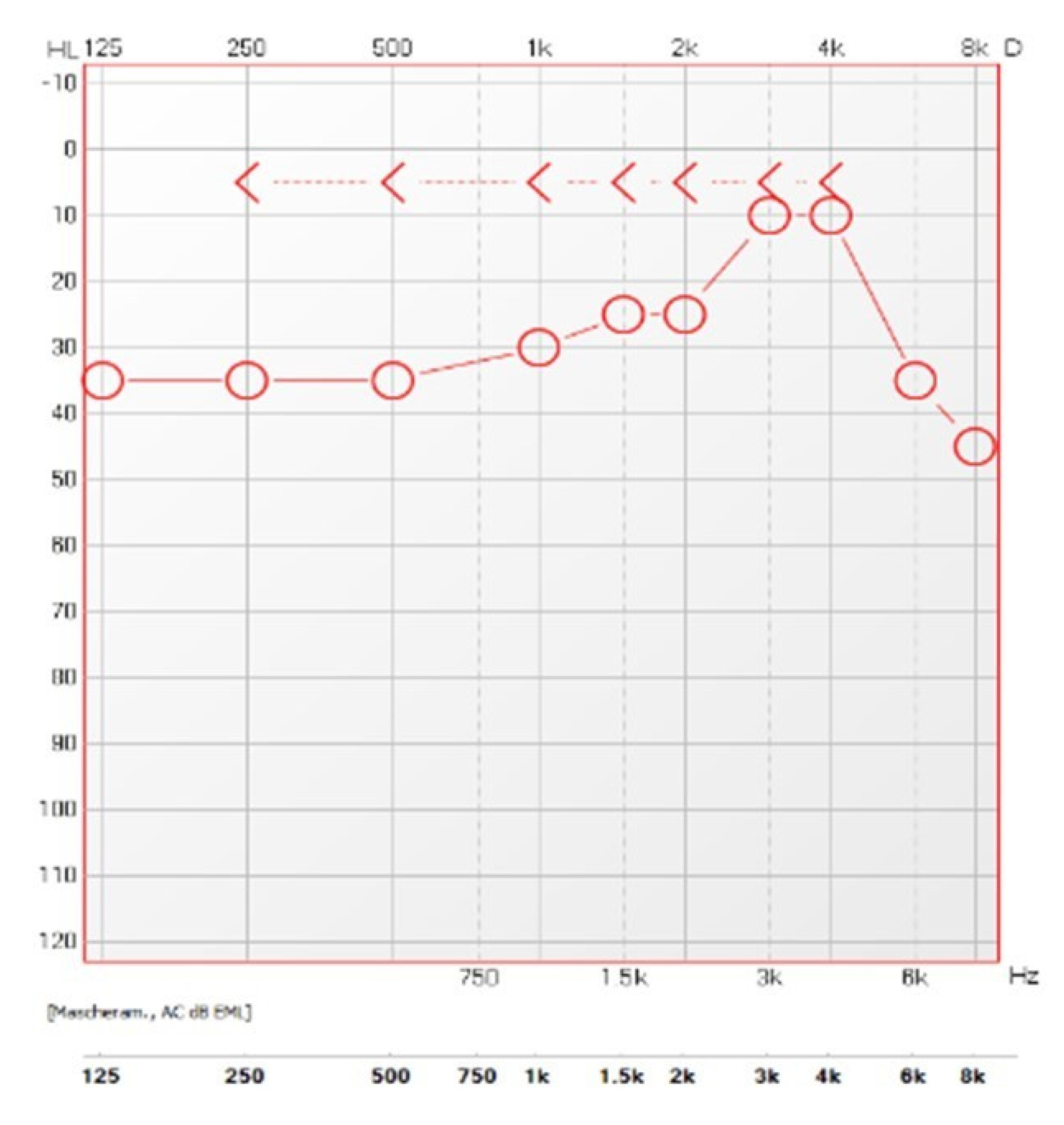
Acoustic neuroma (right ear): sharply sloping sensorineural hearing loss, high-pitched tinnitus, and MRI findings;
(b)
Growth hormone-secreting macroadenoma of the pituitary gland: chronic headache, hyperhidrosis, amenorrhea, insomnia, broadened hands and feet, widened and stubby fingers, and MRI findings;
(c)
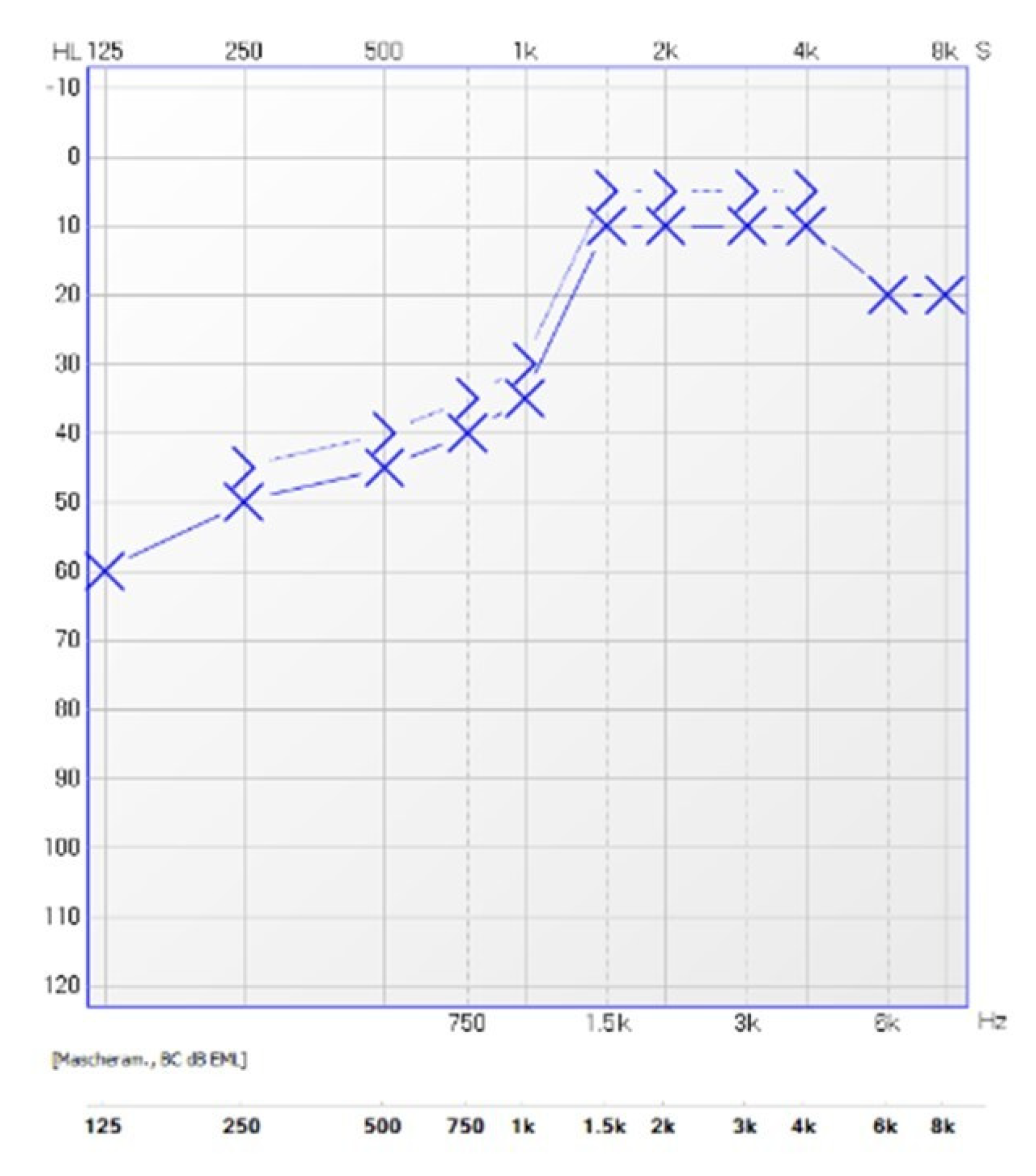
Menière’s disease (left ear): low-frequency sensorineural hearing loss, spontaneous episodes of vertigo (each lasting between 20 min and 12 h), fluctuating aural symptoms (hearing, tinnitus and fullness) in the affected ear, and saccular hydrops detected by 3D FLAIR MRI evaluation;
(d)
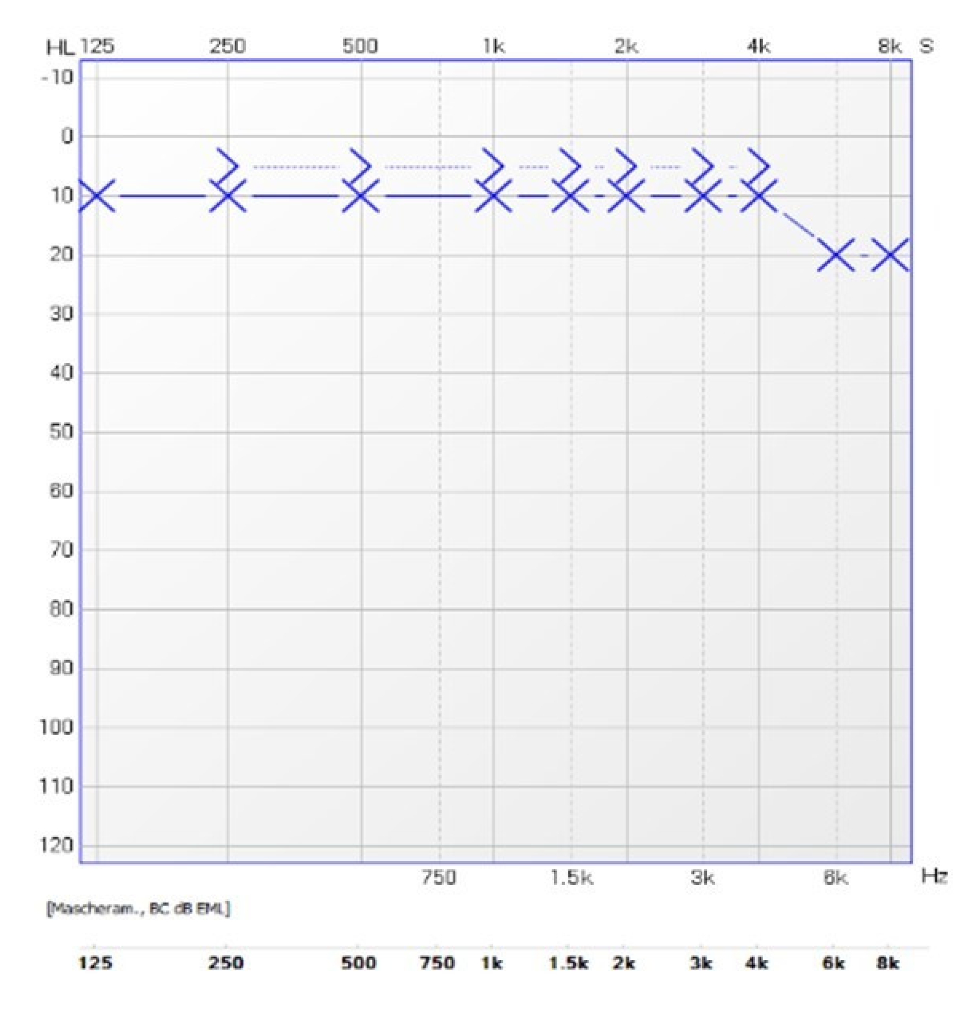
Otosclerosis (right ear): Schwartze’s sign, low-frequency conductive hearing loss, type As tympanogram, absent acoustic reflexes, and outbreak of otosclerosis detected by CBCT scan.
The patient’s symptoms were multiple and insidious, underlying complex pathologies that deserve some important considerations.
Tinnitus is defined as the perception of sound in the absence of an external auditory stimulus and can be attributable to hearing loss, somatosensory system dysfunction, or auditory cortex dysfunction [3]. Tinnitus is a complex symptom that requires detailed investigation to determine its origin, although the most common cause is hearing loss. However, hearing loss does not always lead to tinnitus, and cochlear damage can elicit a tinnitus signal that remains unperceived [3].
Many conditions and risk factors have been described to be associated with tinnitus, such as temporomandibular disorders, ototoxic medications, Menière’s disease, otitis media, Eustachian tube dysfunction, metabolic disorders, acoustic neuroma, loud noise, or psychiatric disorders [3,4,5].
Treatment strategies depend on the underlying causes of tinnitus [3].
Acoustic neuroma, also known as vestibular schwannoma, is a benign tumor that evolves from the Schwann cell sheath and tends to occupy the cerebellopontine angle. It is often unilateral and presents with unilateral high-frequency sensorineural hearing loss and tinnitus [6]. In contrast, bilateral acoustic neuromas are the hallmark feature of neurofibromatosis type 2 (NF2) [6].
Treatment options for acoustic neuroma include watchful waiting, microsurgical resection, and stereotactic radiation [6,7]. It has been suggested that active surveillance may be a valid choice for small and slow-growing tumors or elderly patients with poor medical conditions, while microsurgical resection should be performed in the case of large and rapid-growing tumors or hearing deterioration [7]. Stereotactic radiation was proposed as an alternative to microsurgery for selected patients, particularly elderly individuals with slow-growing tumor and mild hearing loss [7].
GH-secreting adenomas are the second most common type of secreting pituitary adenomas and usually present as macroadenomas, leading to acromegaly in adults and gigantism in children before fusion of bony epiphysis [8].
The main clinical features of acromegaly include enlarged extremities (hands and feet), macroglossia, marked facial lines (widened nose, forehead bulges, and prognathism), bitemporal hemianopia, joint pain due to hypertrophic arthropathy, wrist pain due to carpal tunnel syndrome, snoring and sleep disorders related to sleep apnea, headache, visual disturbances, hyperhidrosis, deepening of the voice, skin thickening, menstrual irregularity and other possible neurologic, cardiovascular, respiratory, and metabolic and neoplastic complications [8,9].
The mean age of diagnosis Is 40 for men and 45 for women [9].
Transsphenoidal surgery is the first-line treatment, except for patients with high surgical risk or invasive and unresectable tumors. Post-operative complications include panhypopituitarism or central diabetes insipidus [8,9].
Menière disease is characterized by excessive accumulation of endolymph and is considered idiopathic by definition. Conversely, Menière syndrome is caused by a specific condition, such as trauma, infections, and endocrine or autoimmune disorders [10].
The diagnostic criteria of Menière’s disease were defined by the Barany society as follows [11]:
(1)
Definite Menière’s disease:
  • Two or more spontaneous episodes of vertigo, each lasting 20 min to 12 h.
  • Audiometrically documented low-to-medium frequency sensorineural hearing loss in one ear, defining the affected ear on at least one occasion before, during or after one of the episodes of vertigo.
  • Fluctuating aural symptoms (hearing, tinnitus, or fullness) in the affected ear.
  • Not better accounted for by another vestibular diagnosis.
(2)
Probable Menière’s disease:
  • Two or more episodes of vertigo or dizziness, each lasting 20 min to 24 h.
  • Fluctuating aural symptoms (hearing, tinnitus, or fullness) in the affected ear.
  • Not better accounted for by another vestibular diagnosis.
The management of Menière’s diseases is focused on relieving acute attacks of vertigo and preventing recurrent attacks. When medical treatment fails, intratympanic gentamicin therapy or endolymphatic sac decompression surgery may be considered [12].
Menière’s disease should be differentiated from vestibular migraine, although several studies have shown a remarkable overlap between these two conditions [13].
According to The International Classification of Headache Disorders (third edition), the diagnostic criteria for vestibular migraine are as follows [14]:
  • At least five episodes fulfilling criteria C and D.
  • A current or past history of Migraine without aura or Migraine with aura.
  • Vestibular symptoms of moderate or severe intensity, lasting between 5 min and 72 h.
  • At least half of episodes are associated with at least one of the following three migrainous features:
    • headache with at least two of the following four characteristics:
      (a)
      unilateral location
      (b)
      pulsating quality
      (c)
      moderate or severe intensity
      (d)
      aggravation by routine physical activity
    • photophobia and phonophobia
    • visual aura
  • Not better accounted for by another ICHD-3 diagnosis or by another vestibular disorder.
Migraines are more common in patients with Menière’s disease than in healthy controls. Fluctuating hearing loss, tinnitus, and aural fullness may occur in vestibular migraine, and migraine headaches, photophobia, and migraine auras are common during Menière attacks [13,14].
Moreover, some patients with vestibular migraine have endolymphatic hydrops on MRI evaluation [15].
However, when the criteria for Menière’s disease are met and hearing loss is documented by audiometry, Menière’s disease should be diagnosed, even if migraine symptoms occur during the vestibular attacks [14].
Otosclerosis is a progressive disease of abnormal bone remodeling that affects the otic capsule of the temporal bone and leads to conductive hearing loss due to the fixation of the stapes. Extensive cochlear progression can result in mixed hearing loss in all frequencies [16].
Otosclerosis is considered as a multifactorial disease, caused by both genetic (an autosomal dominant mode of inheritance with reduced penetrance is found in some cases) and environmental factors (pregnancy, menopause, trauma, major operations, fluoride, and measles have been reported as possible factors that may cause or aggravate the disease) [16,17].
Approximately 10% of patients with otosclerosis have a characteristic otoscopic finding, called the “Schwartze’s sign”. The Schwartze sign, also known as “Flemingo’s flush sign” and “Rising sun sign”, is a characteristically reddish discoloration of the promontory induced by the increased blood flow due to the otosclerotic lesion [18].
High resolution computed tomography (HRCT) is considered the gold standard imaging modality in the diagnosis of otosclerosis. It has high diagnostic sensitivity and specificity, allowing the identification of smaller and initial outbreaks of otosclerosis [16,17]. However, the definitive diagnosis of otosclerosis is made by intraoperative inspection and confirmation of stapes foot plate fixation.
The treatment of choice is stapedotomy or stapedectomy, which may be indicated in the presence of conductive hearing loss with an air-bone gap of at least 20 dB and a speech discrimination score of 60% or greater [17]. In stapedectomy, the stapes footplate and the crura are removed and replaced with a prosthesis, while in stapedotomy, a small hole is made in the in the center of the fixed stapes footplate for the prosthesis without any removal of the structures [17].
In our clinical case, a watch-and-wait strategy was adopted for otosclerosis and acoustic neuroma, advising the patient to repeat the MRI of the head and pure tone audiometry after 6 months.
Transsphenoidal surgery was successfully performed to remove the pituitary macroadenoma.
The procedure was well tolerated, and the immediate and late postoperative course was uneventful. In particular, no postoperative complications, such as postoperative hyponatremia, were observed, and the patient experienced complete hormonal remission (normal IGF-1 levels and suppressed GH during oral glucose tolerance test) within 2 months.
It is important to highlight that a pituitary macroadenoma may induce endolymphatic hydrops; however, in our case the fluctuating aural symptoms affected only one ear and did not disappear after removing the pituitary macroadenoma. Indeed, the patient presented again to our third-level referral audiologic center complaining of two new episodes of vertigo: each episode occurred 2 and 3 months after the removal of the pituitary macroadenoma, lasted about 2 h, and was associated with fluctuating tinnitus, fullness and hearing loss in the left ear. Moreover, pure-tone audiometry performed during the attacks of vertigo confirmed the same low-frequency hearing loss detected before surgery (Figure 2). Each episode was well controlled by medical therapy.
To the best of our knowledge, this is the first study describing the simultaneous presence of acoustic neuroma, pituitary macroadenoma, Menière’s disease, and otosclerosis in the same patient.
Although the concurrent presence of multiple primary brain tumors with different histological characteristics is very rare [19,20], Carlson et al. reported that 1 in every 319 patients with vestibular schwannoma was also diagnosed with a pituitary adenoma [21]. These data suggest a possible common environmental and/or genetic predisposition that may favor the development of the two different tumors.
An association between otosclerosis and Menière’s disease was proposed in the literature, but the causal relationship remains controversial [22]. Some studies also hypothesized a possible correlation between prolactinoma and Menière’s disease, emphasizing the key role played by stress [23].
Decision making for solitary vestibular schwannoma and contralateral Menière’s disease is very complex, and a conservative management should be preferred whenever possible [24].
This case study confirms that multiple otologic disorders can occur simultaneously in a single patient, causing a multitude of symptoms.
Taking a careful medical history and performing accurate audiological and imaging evaluations are essential for a correct diagnosis.

Author Contributions

Conceptualization, M.A. and D.Z.; methodology, L.P.; validation, M.A., L.P. and D.Z.; formal analysis, M.A.; investigation, M.A.; resources, M.A.; data curation, M.A.; writing—original draft preparation, M.A.; writing—review and editing, L.P.; visualization, D.Z.; supervision, L.P. All authors have read and agreed to the published version of the manuscript.

Funding

This research received no external funding.

Institutional Review Board Statement

This retrospective study was conducted in accordance with the Declaration of Helsinki, and approved by the Institutional Review Board of Fondazione IRCCS Ca’ Granda Ospedale Maggiore Policlinico (468_2018bis).

Informed Consent Statement

Written informed consent has been obtained from the patient to publish this paper.

Data Availability Statement

Not applicable.

Conflicts of Interest

The authors declare no conflict of interest.

References

  1. Campagnoli, M.; Cerasuolo, M.; Renna, M.; Dell’Era, V.; Valletti, P.A.; Garzaro, M. ENT Referral From Emergency Department During COVID-19: A Single-Center Experience. Ear Nose Throat J. 2023, 102, NP95–NP98. [Google Scholar] [CrossRef] [PubMed]
  2. Aldè, M.; Barozzi, S.; Berardino, F.D.; Zuccotti, G.; Gonsonni, D.; Ambrosetti, U.; Socci, M.; Bertoli, S.; Battezzati, A.; Foppiani, A.; et al. Prevalence of symptoms in 1512 COVID-19 patients: Have dizziness and vertigo been underestimated thus far? Int. Emerg. Med. 2022, 17, 1343–1353. [Google Scholar] [CrossRef] [PubMed]
  3. Han, B.I.; Lee, H.W.; Ryu, S.; Kim, J.S. Tinnitus Update. J. Clin. Neurol. 2021, 17, 1–10. [Google Scholar] [CrossRef] [PubMed]
  4. Aldè, M.; Didier, H.A.; Giannì, A.B.; Sessa, F.; Borromeo, G.; Didier, A.H.; Barozzi, S.; Zanetti, D.; Di Berardino, F. Prevalence of New-Onset Otological Symptoms in Patients with Temporomandibular Disorders. J. Otorhinolaryngol. Hear. Balance Med. 2022, 3, 3. [Google Scholar] [CrossRef]
  5. Aldè, M.; Di Berardino, F.; Ambrosetti, U.; Barozzi, S.; Piatti, G.; Zanetti, D.; Pignataro, L.; Cantarella, G. Audiological and vestibular symptoms following SARS-CoV-2 infection and COVID-19 vaccination in children aged 5–11 years. Am. J. Otolaryngol. 2023, 44, 103669. [Google Scholar] [CrossRef]
  6. Greene, J.; Al-Dhahir, M.A. Acoustic, Neuroma; StatPearls Publishing: Treasure Island, FL, USA, 2022; [Updated 4 June 2022]. Available online: https://www.ncbi.nlm.nih.gov/books/NBK470177/ (accessed on 1 March 2023).
  7. Albera, R.; Luca, D.; Andrea, A.; Ettore, C.; Alfonso, S.; Ralli, M. What factors influence treatment decision making in acoustic neuroma? Our experience on 103 cases. Int. Tinnitus J. 2020, 24, 21–25. [Google Scholar] [CrossRef]
  8. Mehta, G.U.; Lonser, R.R. Management of hormone-secreting pituitary adenomas. Neuro Oncol. 2017, 19, 762–773. [Google Scholar] [CrossRef] [PubMed] [Green Version]
  9. Adigun, O.O.; Nguyen, M.; Fox, T.J.; Anastasopoulou, C. Acromegaly; StatPearls Publishing: Treasure Island, FL, USA, 2022. [Google Scholar]
  10. Paparella, M.M. Pathogenesis of Meniere’s disease and Meniere’s syndrome. Acta Otolaryngol. 1984, 406, 10–25. [Google Scholar] [CrossRef] [PubMed]
  11. Lopez-Escamez, J.A.; Carey, J.; Chung, W.H.; Goebel, J.A.; Magnusson, M.; Mandalà, M.; Newman-Toker, D.E.; Strupp, M.; Suzuki, M.; Trabalzini, F.; et al. Diagnostic criteria for Menière’s disease. J. Vestib. Res. 2015, 25, 1–7. [Google Scholar] [CrossRef] [PubMed] [Green Version]
  12. Nakashima, T.; Pyykkö, I.; Arroll, M.A.; Casselbrant, M.L.; Foster, C.A.; Manzoor, N.F.; Megerian, C.A.; Naganawa, S.; Young, Y.H. Meniere’s Disease. Nat. Views Dis. Prim. 2016, 2, 16028. [Google Scholar] [CrossRef] [PubMed]
  13. Tabet, P.; Saliba, I. Meniere’s Disease and Vestibular Migraine: Updates and Review of the Literature. J. Clin. Med. Res. 2017, 9, 733–744. [Google Scholar] [CrossRef] [PubMed] [Green Version]
  14. Arnold, M. Headache Classification Committee of the International Headache Society (IHS) The International Classification of Headache Disorders. Cephalalgia 2018, 38, 1–211. [Google Scholar] [CrossRef]
  15. Gürkov, R.; Kantner, C.; Strupp, M.; Flatz, W.; Krause, E.; Ertl-Wagner, B. Endolymphatic hydrops in patients with vestibular migraine and auditory symptoms. Eur. Arch. Otorhinolaryngol. 2014, 271, 2661–2667. [Google Scholar] [CrossRef] [PubMed]
  16. Zafar, N.; Jamal, Z.; Khan, M.A.B. Otosclerosis; StatPearls Publishing: Treasure Island, FL, USA, 2022; [Updated 25 July 2022]. Available online: https://www.ncbi.nlm.nih.gov/books/NBK560671/ (accessed on 1 March 2023).
  17. Batson, L.; Rizzolo, D. Otosclerosis: An update on diagnosis and treatment. JAAPA 2017, 30, 17–22. [Google Scholar] [CrossRef] [PubMed]
  18. Koenen, L.; Gupta, G. Schwartze Sign; StatPearls Publishing: Treasure Island, FL, USA, 2022; [Updated 5 September 2022]. Available online: https://www.ncbi.nlm.nih.gov/books/NBK532921/ (accessed on 1 March 2023).
  19. Amit, A.; Achawal, S.; Dorward, N. Pituitary macro adenoma and vestibular schwannoma: A case report of dual intracranial pathologies. Br. J. Neurosurg. 2008, 22, 695–696. [Google Scholar] [CrossRef] [PubMed]
  20. Niu, Y.; Ma, L.; Mao, Q.; Wu, L.; Chen, J. Pituitary adenoma and vestibular schwannoma: Case report and review of the literature. J. Postgrad. Med. 2010, 56, 281–283. [Google Scholar] [CrossRef] [PubMed]
  21. Carlson, M.L.; Patel, N.S.; Glasgow, A.E.; Habermann, E.B.; Grossardt, B.R.; Link, M.J. Vestibular schwannoma and pituitary adenoma in the same patient: Coincidence or novel clinical association? J. Neuro Ooncol. 2016, 128, 101–108. [Google Scholar] [CrossRef] [PubMed]
  22. Klockars, T.; Kentala, E. Case report: Meniere’s disease and otosclerosis—Different outcomes of the same disease? Auris Nasus Larynx 2007, 34, 101–104. [Google Scholar] [CrossRef] [PubMed]
  23. Horner, K.C.; Guieu, R.; Magnan, J.; Chays, A.; Cazals, Y. Prolactinoma in some Ménière’s patients—Is stress involved? Neuropsychopharmacology 2002, 26, 135–138. [Google Scholar] [CrossRef] [PubMed] [Green Version]
  24. Dispenza, F.; De Stefano, A.; Flanagan, S.; Romano, G.; Sanna, M. Decision making for solitary vestibular schwannoma and contralateral Meniere’s disease. Audiol. Neurootol. 2008, 13, 53–57. [Google Scholar] [CrossRef] [PubMed]
Figure 1. Right ear audiogram.
Ohbm 04 00002 g001
Figure 2. Left ear audiogram.
Ohbm 04 00002 g002
Figure 3. Left ear audiogram after medical therapy.
Ohbm 04 00002 g003
Figure 4. MRI of the head showing: (a) Right acoustic neuroma; (b) Pituitary macroadenoma; (c) Left saccular hydrops.
Ohbm 04 00002 g004
Figure 5. CBCT of the head showing right outbreak of otosclerosis.
Ohbm 04 00002 g005
Disclaimer/Publisher’s Note: The statements, opinions and data contained in all publications are solely those of the individual author(s) and contributor(s) and not of MDPI and/or the editor(s). MDPI and/or the editor(s) disclaim responsibility for any injury to people or property resulting from any ideas, methods, instructions or products referred to in the content.

