Proceeding Paper

Biomarkers in Patients with Hand-Arm Vibration Injury Entailing Raynaud’s Phenomenon and Cold Sensitivity, Compared to Referents †

1
Division of Occupational and Environmental Medicine, Department of Laboratory Medicine, Lund University, 221 00 Lund, Sweden
2
Division of Sustainable Health and Medicine, Department of Public Health and Clinical Medicine, Umeå University, 901 87 Umeå, Sweden
3
Department of Translational Medicine—Hand Surgery, Lund University, 205 02 Malmoe, Sweden
*
Author to whom correspondence should be addressed.
Presented at the 15th International Conference on Hand-Arm Vibration, Nancy, France, 6–9 June 2023.
Proceedings 2023, 86(1), 27; https://doi.org/10.3390/proceedings2023086027
Published: 14 April 2023
(This article belongs to the Proceedings of The 15th International Conference on Hand-Arm Vibration)

Abstract

:
The clinical evaluation of patients with neurosensory injury is based on quantitative sensory testing. Such tests require the patient’s cooperation, which may sometimes hinder a correct diagnosis. Objective findings, e.g., with biomarkers, would therefore be valuable. We evaluated serum biomarkers of vascular and neural injury in 92 patients with vibration injuries and in 64 referents. Thrombomodulin (TM), a biomarker for endothelial damage, was elevated in HAVS patients with Raynaud’s phenomenon (RP) compared to those without, as it also was in comparison to the referents. In the patients without RP, those with increased cold sensitivity showed somewhat higher—but not significantly different—values of TM (p = 0.4) than those without increased cold sensitivity, indicating an endothelial dysfunction or injury.

1. Introduction

The pathophysiology of hand-arm vibration syndrome (HAVS) is not fully clear. In most cases neurosensory symptoms proceed vascular ones [1]. Data suggest there is an individual susceptibility to HAVS, possibly due to contributions from coexisting medical conditions, for e.g., diabetes neuropathy [2]. Different pathophysiological mechanisms such as localized structural injuries affecting peripheral nerves and blood vessels and systemic inflammatory processes may exist [3,4,5,6,7]. Elevated plasma levels of thrombomodulin (TM), a marker for endothelial damage, are higher in vibration-exposed than in non-exposed workers [8,9]. Glial fibrillary acidic protein (GFAP), a proposed marker for axonal damage [10], has been detected in the nerve biopsies of type 2 diabetes subjects and controls [11], and elevated serum levels of GFAP correlate to decreased nerve action potentials [12]. In addition to symptoms of numbness, having tingling or not, having an impaired perception of touch, vibration, and temperature, and being diagnosed with Raynaud’s phenomenon (RP), the patients often describe increased cold sensitivity [13]—a phenomenon that can mechanistically be induced by a vascular or neural pathway following vibration exposure [14]. Individuals with nerve injury may recover from increased cold sensitivity, whereas patients with HAVS may not [15].
Biomarkers for neural and vascular injuries may shed light on mechanisms as well as be used as a complemental objective method for diagnosis, since clinical assessment of the neurosensory component of HAVS mostly relies on quantitative sensory tests requiring cooperation of the evaluated patients.
Our aims were to investigate serum levels of biomarkers for vascular (TM) and for nerve (GFAP) injury in patients with neurosensory HAVS, with or without RP, compared to referents, as well as to study the association of increased cold sensitivity with the levels of TM and GFAP in HAVS patients without RP.

2. Materials and Methods

2.1. Study Group

Ninety-two patients diagnosed with neurosensory manifestations of HAVS and/or RP were recruited from our department of occupational and environmental medicine, together with 64 referents, who were without a vibration injury.

2.2. Questionnaires

Data on descriptive characteristics and symptoms were collected via a questionnaire (Table 1). Also, questions about job tasks and vibration exposure was assessed, not presented here.

2.3. Perception of Touch

Perception of touch was tested with a Semmes Weinstein Monofilament. If participants were unable to detect stimulation with a filament, No. 3.61, corresponding to 0.4 g of force, the perception of touch was considered impaired.

2.4. Biomarkers

Blood samples were collected in the morning. Serum was removed and stored at −80 °C until analysis. TM and GFAP were measured by using commercially available ELISA assays (TM by ELISA kit from BioVendor (Brno, Czech Republic); GFAP from Proteintech, (Manchester, UK)) according to instructions provided by the manufacturers. Samples were diluted in a ratio of 1:2 in sample diluent, and after sample preparation the optical density was recorded in a microplate reader at a wavelength of 450 nm. Duplicate readings were made. The detection limits (LOD) were 0.625 ng/mL for TM and 31 ng/mL for GFAP.

2.5. Statistics

The Kruskal–Wallis test was used to compare median values between the group of patients with and without RP and referents. Mann–Whitney U tests were used for post hoc analyses, and between the subgroups of patients with and without increased cold sensitivity. p-values below 0.05 were considered statistically significant. The results have not been adjusted for confounding effects.

3. Results

3.1. Biomarkers in Patients with and without RP and Referents

The Kruskal–Wallis test showed a statistically significant difference in TM levels between groups. Post hoc analyses showed a significant increase in the serum levels of TM in patients with RP compared to patients without RP, as well in patients with RP compared to the referents (Table 2). TM levels were highest in patients with RP and lowest in the referents.

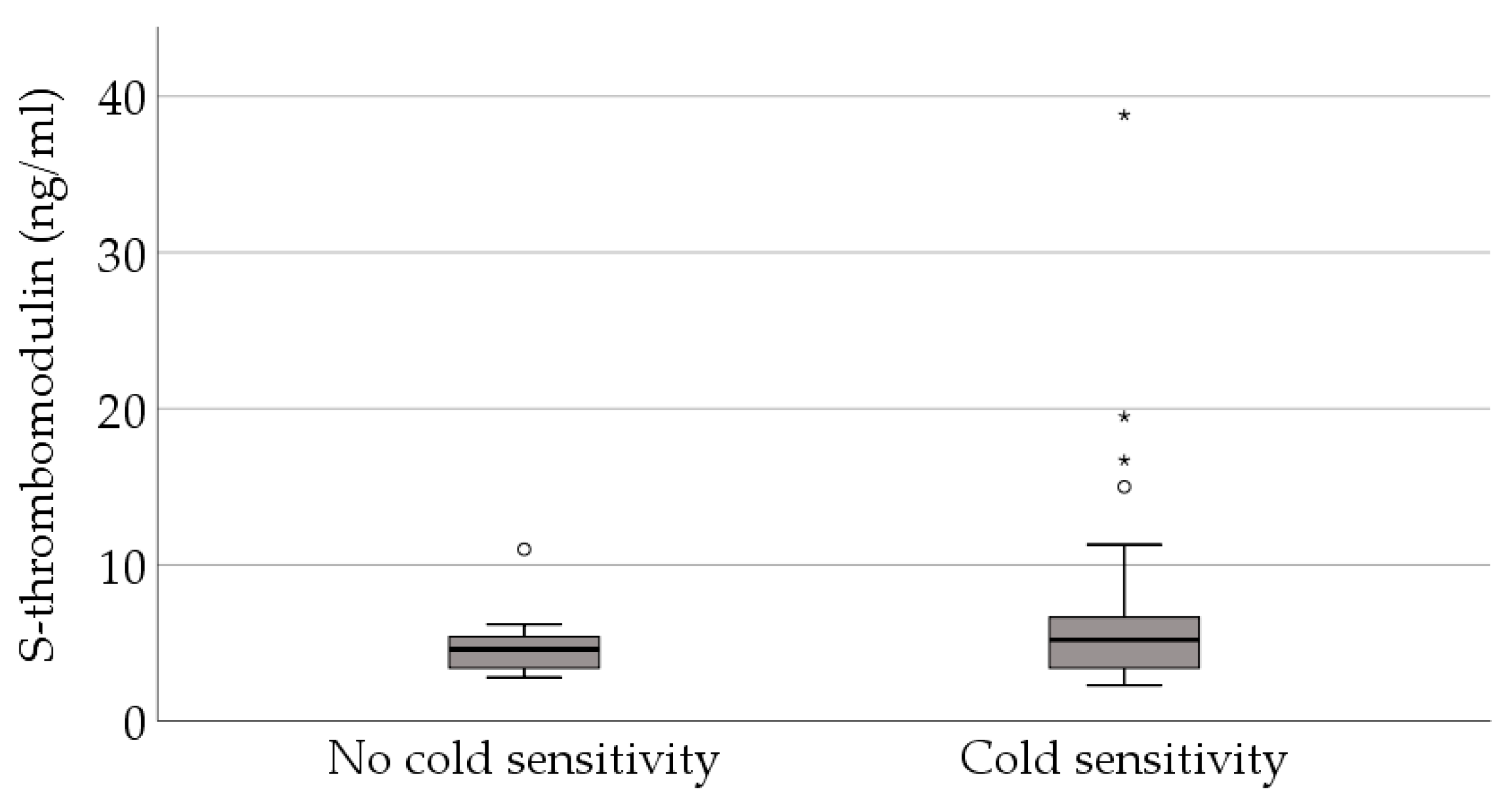
3.2. Biomarkers in Patients without RP and with and without Increased Cold Sensitivity

Patients without RP, but reporting increased cold sensitivity, showed a somewhat higher but not significant increase in the serum levels of TM (median 5.2; range 2.3–39) compared to patients not reporting this symptom (4.6; 2.8–11); p = 0.4, Figure 1. For GFAP, there were no significant differences between any of the groups.

4. Discussion

Patients with HAVS neurosensory injury and with RP had higher levels of TM than vibration-injured patients without RP. Patients with HAVS neurosensory injury and with RP also had higher levels of TM than the referents. There was no statistically significant difference between HAVS patients without RP and the referents. Increased levels of TM have been shown in previous studies on vibration-injured patients [8,9]. In one of the studies, however, there was no significant difference between patients with RP and those without RP [9]. This is in contrast to our findings that rather indicate that the RP symptoms is required to detect a significant change in the biomarker TM in HAVS patients. Patients with HAVS neurosensory injury without RP, reporting increased cold sensitivity, revealed higher levels of TM (although not significant) compared to those not reporting this symptom. Since TM is an endothelial marker, this may indicate that increased cold sensitivity originates from an endothelial dysfunction or injury which possibly could progress to RP.
The levels of GFAP did not differ significantly between the groups in this study, possibly because the neuropathy of the patients did not originate from any nerve injury involving GFAP or possibly because the samples were collected at a time point not reflecting the event for which the GFAP was released into the serum.

Author Contributions

Conceptualization, E.T. and C.N.; methodology, M.K.; formal analysis, E.T., C.N. and M.K.; data curation, E.T. and C.N. writing—original draft preparation, E.T., C.N. and M.K.; writing—review and editing, E.T., C.N., M.K., L.B.D., A.A., J.R. and T.N.; supervision, T.N. and L.B.D.; funding acquisition, C.N. All authors have read and agreed to the published version of the manuscript.

Funding

The study was funded by AFA Insurance (Grant number 170124).

Institutional Review Board Statement

The study was conducted in accordance with the Declaration of Helsinki and approved by the Regional Ethics Board in Lund. Sweden (No. 2018/15).

Informed Consent Statement

Participants gave informed consent to participate in the study before taking part.

Data Availability Statement

The datasets presented in this article are not readily available because public access to data is restricted to Swedish Authorities (Public Access to Information and Secrecy Act), but data can be available for researchers after a special review that includes approval of the research project by both an Ethics Committee and the Authorities’ Data Safety Committees.

Acknowledgments

The authors would like to thank Elizabeth Huynh for skilful analyses of biomarkers and Ulla Andersson, Else Åkerberg Krook, Eva Assarsson, and Anna Larsson for the collection of the blood samples and handling of the data. We would also like to thank all participants in the patient, as well as the referent group.

Conflicts of Interest

The authors declare no conflict of interest.

References

  1. Nilsson, T.; Wahlstrom, J.; Burstrom, L. Hand-arm vibration and the risk of vascular and neurological diseases-A systematic review and meta-analysis. PLoS ONE 2017, 12, e0180795. [Google Scholar] [CrossRef] [PubMed]
  2. Dahlin, L.B.; Sanden, H.; Dahlin, E.; Zimmerman, M.; Thomsen, N.; Bjorkman, A. Low myelinated nerve-fiber density may lead to symptoms associated with nerve entrapment in vibration-induced neuropathy. J. Occup. Med. Toxicol. 2014, 9, 7. [Google Scholar] [CrossRef] [PubMed]
  3. Stoyneva, Z.; Lyapina, M.; Tzvetkov, D.; Vodenicharov, E. Current pathophysiological views on vibration-induced Raynaud’s phenomenon. Cardiovasc. Res. 2003, 57, 615. [Google Scholar] [CrossRef] [PubMed]
  4. Kiedrowski, M.; Waugh, S.; Miller, R.; Johnson, C.; Krajnak, K. The effects of repetitive vibration on sensorineural function: Biomarkers of sensorineural injury in an animal model of metabolic syndrome. Brain Res. 2015, 1627, 216–224. [Google Scholar] [CrossRef] [PubMed]
  5. Krajnak, K.; Waugh, S. Systemic Effects of Segmental Vibration in an Animal Model of Hand-Arm Vibration Syndrome. J. Occup. Environ. Med. 2018, 60, 886–895. [Google Scholar] [CrossRef] [PubMed]
  6. Takeuchi, T.; Futatsuka, M.; Imanishi, H.; Yamada, S. Pathological changes observed in the finger biopsy of patients with vibration-induced white finger. Scand. J. Work Environ. Health 1986, 12, 280–283. [Google Scholar] [CrossRef] [PubMed]
  7. Govindaraju, S.R.; Curry, B.D.; Bain, J.L.; Riley, D.A. Comparison of continuous and intermittent vibration effects on rat-tail artery and nerve. Muscle Nerve 2006, 34, 197–204. [Google Scholar] [CrossRef] [PubMed]
  8. Kao, D.S.; Yan, J.G.; Zhang, L.L.; Kaplan, R.E.; Riley, D.A.; Matloub, H.S. Serological tests for diagnosis and staging of hand-arm vibration syndrome (HAVS). Hand 2008, 3, 129–134. [Google Scholar] [CrossRef] [PubMed]
  9. Kanazuka, M.; Shigekiyo, T.; Toibana, N.; Saito, S. Increase in plasma thrombomodulin level in patients with vibration syndrome. Thromb. Res. 1996, 82, 51–56. [Google Scholar] [CrossRef] [PubMed]
  10. Rossor, A.M.; Reilly, M.M. Blood biomarkers of peripheral neuropathy. Acta Neurol. Scand. 2022, 146, 325–331. [Google Scholar] [CrossRef] [PubMed]
  11. Ising, E.; Åhrman, E.; Thomsen, N.O.B.; Eriksson, K.F.; Malmström, J.; Dahlin, L.B. Quantitative proteomic analysis of human peripheral nerves from subjects with type 2 diabetes. Diabet. Med. J. Br. Diabet. Assoc. 2021, 38, e14658. [Google Scholar] [CrossRef] [PubMed]
  12. Notturno, F.; Capasso, M.; DeLauretis, A.; Carpo, M.; Uncini, A. Glial fibrillary acidic protein as a marker of axonal damage in chronic neuropathies. Muscle Nerve 2009, 40, 50–54. [Google Scholar] [CrossRef] [PubMed]
  13. Tekavec, E.; Löfqvist, L.; Larsson, A.; Fisk, K.; Riddar, J.; Nilsson, T.; Nordander, C. Adverse health manifestations in the hands of vibration exposed carpenters—A cross sectional study. J. Occup. Med. Toxicol. 2021, 16, 16. [Google Scholar] [CrossRef] [PubMed]
  14. Cooke, R.A.; Lawson, I.J. Cold intolerance and hand-arm vibration syndrome. Occup. Med. 2022, 72, 152–153. [Google Scholar] [CrossRef] [PubMed]
  15. Carlsson, I.K.; Dahlin, L.B. Self-reported cold sensitivity in patients with traumatic hand injuries or hand-arm vibration syndrome an eight year follow up. BMC Musculoskelet. Disord. 2014, 15, 83. [Google Scholar] [CrossRef] [PubMed]
Figure 1. S-thrombomodulin in relation to increased cold sensitivity among HAVS patients without Raynaud’s phenomenon. p = 0.4 (Mann–Whitney U test). Far outliers marked with a star and outliers marked with a circle.
Proceedings 86 00027 g001
Table 1. Descriptive characteristics of 92 patients with hand-arm vibration syndrome and 64 referents.
Patients with Raynaud’s
Phenomenon (n = 45)
Patients without Raynaud’s
Phenomenon (n = 47)
Referents
(n = 64)
Age [years; median (range)]45 (24–64)45 (21–64)43 (26–62)
Females [n (%)]1 (2)5 (11)9 (14)
Ongoing nicotine use [n (%)]23 (51)17 (36)25 (40)
Previous frost bites in hands [n (%)]6 (15) a4 (9) b 3 (5) c
Raynaud’s phenomenon [n (%)]45 (100)0 (0)6 (10)
Cold sensitivity [n (%)]44 (98)36 (77)10 (16)
Impaired perception of touch [n (%)]29 (64)16 (34)10 (16)
a missing data from 4 patients; b missing data from 3 patients; c missing data from 4 referents.
Table 2. Biomarkers in patients with and without Raynaud’s phenomenon and referents.
Patients with Raynaud’s
Phenomenon (n = 45)
Median (Range)
Patients without Raynaud’s
Phenomenon (n = 47)
Median (Range)
Referents
(n = 64)
Median (Range)
p-Value
S-TM (ng/mL)6.1 (2.7–30) a, b5.2 (2.3–39) a4.3 (<LOD–40) b0.003
S-GFAP (pg/mL)LOD c (LOD–3070)LOD (LOD–2528)LOD (LOD–3376)0.28
a p = 0.02; b p ≤ 0.001, c LOD = below lowest detection level. p-values from Kruskal–Wallis test. Post hoc analyses with Mann–Whitney U test.
Disclaimer/Publisher’s Note: The statements, opinions and data contained in all publications are solely those of the individual author(s) and contributor(s) and not of MDPI and/or the editor(s). MDPI and/or the editor(s) disclaim responsibility for any injury to people or property resulting from any ideas, methods, instructions or products referred to in the content.

