Case Report

Cerebral Cryptococcosis Associated with CD4+ T-lymphocytopenia in Non-HIV Patients after SARS-CoV-2 Infection: Case Series in a Specialized Institute in Lima, Peru

by
Juana M. Huamani-Córdova
1,
Miguel Hueda-Zavaleta
2,3,
Victor Vargas-Bellina
1,
Lourdes Simbron-Ribbeck
1,
Katty del Rosario Chong-Chinchay
1,
Juan Carlos Gómez de la Torre
4 and
Vicente A. Benítes-Zapata
5,*
1
Instituto Nacional de Ciencias Neurológicas, Lima 15003, Peru
2
Facultad de Ciencias de la Salud, Universidad Privada de Tacna, Tacna 23003, Peru
3
Hospital III Daniel Alcides Carrión—Essalud, Tacna 23000, Peru
4
Roe Clinical Laboratory, Lima 15076, Peru
5
Unidad de Investigación para la Generación y Síntesis de Evidencias en Salud, Universidad San Ignacio de Loyola, Lima 15024, Peru
*
Author to whom correspondence should be addressed.
Trop. Med. Infect. Dis. 2023, 8(3), 182; https://doi.org/10.3390/tropicalmed8030182
Submission received: 27 January 2023 / Revised: 20 February 2023 / Accepted: 3 March 2023 / Published: 22 March 2023

Abstract

:
Cases of cryptococcosis have been reported in patients with COVID-19. The majority are in patients with severe symptoms or who received immunosuppressants. However, there is still no clear association between COVID-19 and cryptococcosis. We report eight cases of cerebral cryptococcosis associated with CD4+ T lymphocytopenia in non-HIV patients after SARS-CoV-2 infection. The median age was 57 years and 5/8 were male. In addition, 2/8 of patients had diabetes, and 8/8 had a history of mild COVID-19, with a median of 75 days before diagnosis of cerebral cryptococcosis. All patients denied having received prior immunosuppressive therapy. The most frequent symptoms were confusion (8/8), headache (7/8), vomiting (6/8), and nausea (6/8) All patients were diagnosed by isolating Cryptococcus in cerebrospinal fluid. The median CD4+ and CD8+ T lymphocytes were 247 and 173.5, respectively. Other causes of immunosuppression, such as HIV or HTLV infection, were excluded in all patients. Finally, three patients died, and one presented long-term visual and auditory sequelae. The CD4+/CD8+ T lymphocyte count normalized during follow-up in those patients who survived. We hypothesize that CD4+ T lymphocytopenia in the patients in this case series could increase the risk of cryptococcosis after SARS-CoV-2 infection.

1. Introduction

More than three years have passed since the emergence of Coronavirus disease 2019 (COVID-19) caused by the severe acute respiratory syndrome coronavirus 2 (SARS-CoV-2) in the city of Wuhan at the end of December 2019. Since then, COVID-19 has spread rapidly worldwide, still threatening global health [1]. Likewise, multiple fungal co-infections such as Aspergillus, Candida, and Mucormycosis have been observed, mainly in patients with critical COVID-19 who required invasive mechanical ventilation or with a lengthy hospital stay [2,3,4]. Patients with severe COVID-19 may present with a pro-inflammatory state with the release of cytokines such as Interleukin (IL)-1, IL-2, IL-6, and tumor necrosis factor (TNF). It has also been described that they may present lymphocytopenia and reduced levels of T cells in peripheral blood, which is a factor associated with a greater risk of severity and mortality from COVID-19 [5]. CD4+ T-cell lymphocytopenia has been reported in some patients with COVID-19, mainly in those with severe symptoms [6,7]; however, its duration is unknown, and it could be a risk factor for opportunistic infections.
Cerebral cryptococcosis is an opportunistic infection that mainly affects patients with Human Immunodeficiency Virus (HIV) infection with CD4+ T-lymphocyte counts lower than 100 cells/µL, and also in transplant recipients, hematological malignancies, idiopathic CD4 lymphocytopenia, and users of prolonged corticosteroid therapy or other immunosuppressants [8]. Its presentation in immunocompetent hosts is infrequent. Although some cases associated with COVID-19 have been reported, these patients generally suffered severe disease, received corticosteroids, and were admitted to the intensive care unit (ICU) [9,10]. We report eight cases of cerebral cryptococcosis in patients without HIV infection, treated at the National Institute of Neurological Sciences (INCN) in Lima, Peru. All patients were diagnosed with COVID-19 by clinical picture and detection of IgM/IgG serum antibodies against COVID-19 and/or polymerase chain reaction (PCR) for SARS-CoV-2 from nasopharyngeal swabs. We included all patients diagnosed with cerebral cryptococcosis by isolating Cryptococcus. in cerebrospinal fluid culture and diagnosis or antecedent of confirmed COVID-19 (by RT-PCR of SARS-CoV-2 in those patients with suspicion of COVID-19 with a disease time of less than 7–10 days, or by detecting serum IgM/IgG antibodies against SARS-CoV-2 when disease time was greater than 7–10 days). The INCN Research Ethics Committee approved the study and all survivors gave informed consent.

2. Results

2.1. Demographic Characteristics

The median age was 57 years, and 5/8 were male. The most frequent comorbidity was diabetes, in 2/8. One patient (case 1) was diagnosed with type II naked lymphocyte syndrome after discharge, and 5/8 had no comorbidity, 8/8 of patients had a history of mild COVID-19, with a median time of 75 days (IQR: 30–180) before diagnosis of cerebral cryptococcosis. Finally, 7/8 had positive serum antibodies against SARS-CoV-2 and 2/8 were RT-PCR SARS-CoV-2 positive at the time of diagnosis of cryptococcosis (Table 1).

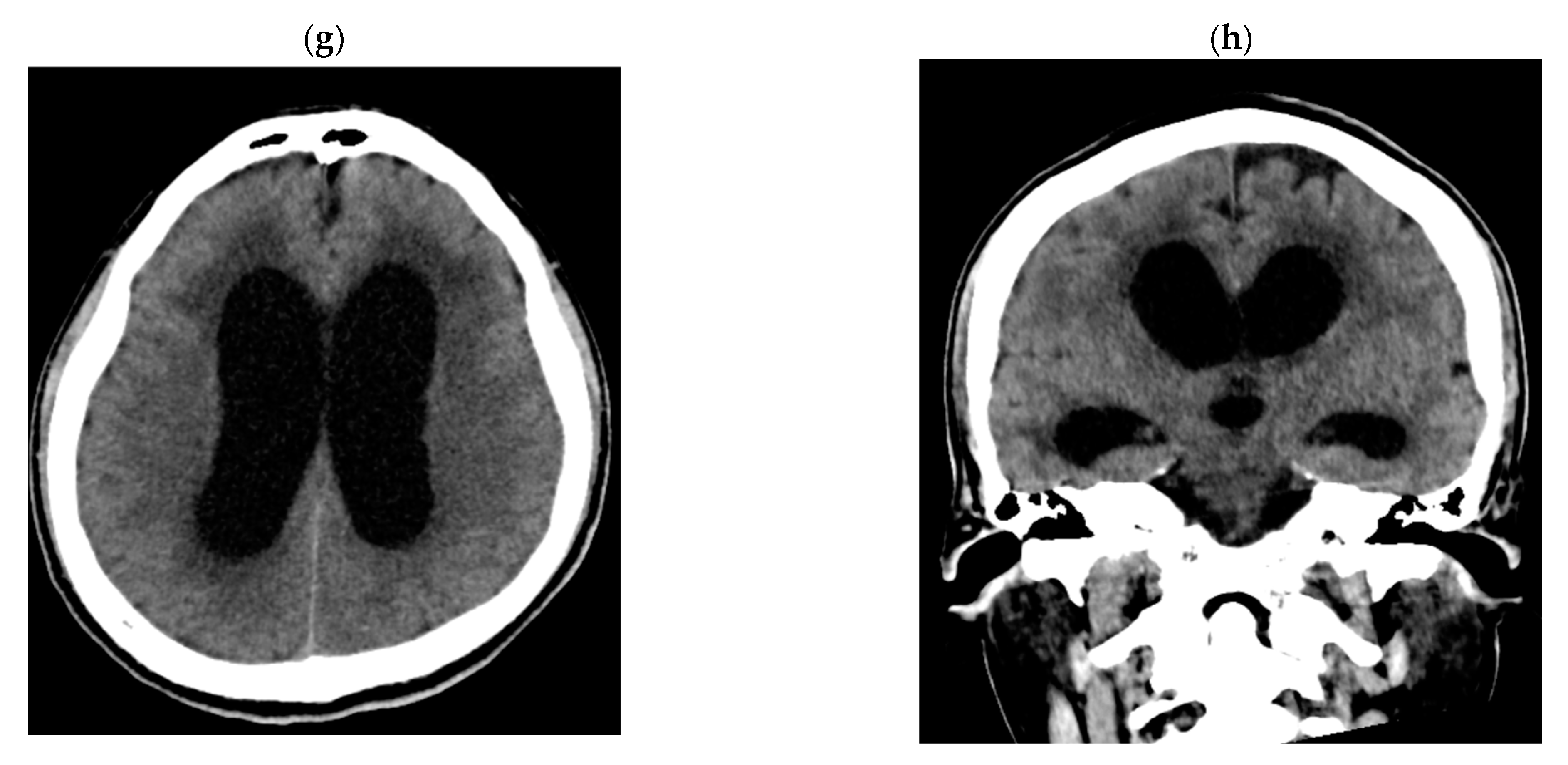
2.2. Detailed Case Descriptions

Case 1: 24-year-old female patient without comorbidities, without vaccination against COVID-19. She has a history of mild COVID-19 60 days before admission. She was admitted with a 30-day illness characterized by headache, nausea, vomiting, dysarthria, and confusion, with an 11-point Glasgow Coma Scale (GCS), nuchal stiffness, and 4/5 left hemiparesis. The CSF showed predominantly mononuclear pleocytosis, glucose 43 mg/dL (reference range: 40–76), protein: 54 mg/dL (reference range: 15–45), India ink positive, Cryptococcus latex antigen positive (1:1024), and isolation of Cryptococcus neoformans in Cerebrospinal Fluid (CSF) at 02 days of incubation. The blood CD4+ T lymphocyte count was 107/mm3 and CD8+ T lymphocytes were 253/mm3. Brain Magnetic Resonance Imaging (MRI) revealed cryptococomas and pseudocysts in the basal ganglia and subcortical white matter with basal leptomeningitis (Figure 1a,b). She received treatment with amphotericin B deoxycholate and fluconazole, presenting good evolution and discontinuation of therapy after five months of treatment. At three months, the CD4+ T lymphocyte count was 303/mm3 and CD8+ T lymphocytes were 1145/mm3 (Figure 2). Finally, a genomic sequencing study revealed “naked lymphocyte syndrome” during follow-up.
Case 2: 57-year-old female patient without comorbidities, without vaccination against COVID-19. She has a history of mild COVID-19 30 days before admission. She was admitted with a 17-day illness characterized by headache, nausea, vomiting, low-grade fever, confusion, and visual hallucinations. On examination, she presented a GCS of 13 points, no neck stiffness, and no focalization. The CSF showed predominantly mononuclear pleocytosis, glucose: 62 mg/dL, proteins: 39 mg/dL, negative India ink, positive Cryptococcus latex antigen (1:128), and isolation of Cryptococcus neoformans in CSF at 05 days of incubation. The CD4+ T lymphocyte count was 267/mm3 and CD8+ T lymphocytes were 143/mm3. Brain MRI was normal. She received treatment with amphotericin B deoxycholate and fluconazole, with good evolution and discontinuation of therapy after eight months of treatment. At three months, the CD4+ T lymphocyte count was 616/mm3 and CD8+ T lymphocytes were 613/mm3 (Figure 2).
Case 3: 58-year-old male patient, controlled diabetic, without vaccination against COVID-19. He has a history of mild COVID-19 05 months before admission. He was admitted with a disease period of 04 months characterized by headache, photophobia, nausea, vomiting, and confusion. On examination, he presents with a 14-point GCS, no nuchal stiffness, and no focusing. The CSF showed predominantly mononuclear pleocytosis, glucose: 32 mg/dL, proteins: 155 mg/dL, negative latex antigen for Cryptococcus, and isolation of Cryptococcus neoformans in CSF at 03 days of incubation. The CD4+ T lymphocyte count was 290/mm3 and CD8+ T lymphocytes were 161/mm3. Brain MRI was described as normal. He received treatment with amphotericin B deoxycholate and fluconazole, with good evolution and discontinuation of therapy after five months of treatment.
Case 4: 68-year-old male patient without comorbidities, without vaccination against COVID-19. He was admitted with a 14-day sick time characterized by headache, dizziness, diarrhea, confusion, on examination, mild quadriparesis, and no nuchal rigidity. He denies respiratory symptoms before admission but presents IgM and IgG (+) against SARS-CoV-2 and as RT-PCR SARS-CoV-2 positive in a nasopharyngeal swab. The CSF showed predominantly mononuclear pleocytosis, glucose: 21 mg/dL, proteins: 155, and isolation of Cryptococcus neoformans in CSF at 08 days of incubation. The CD4+ T lymphocyte count was 89/mm3 and CD8+ T lymphocytes were 20/mm3. Brain MRI was described as normal. He received treatment with amphotericin B deoxycholate and fluconazole, with good evolution and discontinuation of therapy after three months of treatment. At three months, the CD4+ T lymphocyte count was 976/mm3 and CD8+ T lymphocytes were 943/mm3 (Figure 2).
Case 5: 23-year-old male patient without comorbidities, without vaccination against COVID-19. He has a history of mild COVID-19 3 months before admission. He was admitted with a 22-day illness characterized by headache, nausea, vomiting, decreased visual acuity, and confusion. On examination, he presented a 14-point GCS, nuchal stiffness, bilateral VI ophthalmopareses, left homonymous hemianopsia, and 4/5 quadriparesis. The CSF showed predominantly mononuclear pleocytosis, glucose: 36 mg/dL, proteins: 35 mg/dL, India ink positive, Cryptococcus latex antigen positive (1:1024), and isolation of Cryptococcus neoformans in CSF at 04 days. The CD4+ T lymphocyte count was 584/mm3, and CD8+ T lymphocytes were 284/mm3, and the brain magnetic resonance evidenced symmetrical enlargement of the fissures and cisterns, with signs of basal arachnoiditis (Figure 1c,d). The patient developed obstructive hydrocephalus that required a ventriculoperitoneal shunt. He received treatment with amphotericin B deoxycholate and fluconazole, with good evolution and discontinuation of therapy after three months of treatment. At three months, the CD4+ T lymphocyte count was 642/µL and CD8+ T lymphocytes were 422/µL (Figure 2).
Case 6: 57-year-old female patient, without comorbidities, without vaccination against COVID-19. She has a history of mild COVID-19 40 days before admission. She is admitted with a 30-day sick time characterized by headache, nausea, vomiting, confusion, and visual hallucinations. On examination, she presented an 11-point GCS, nuchal rigidity, and quadriparesis 4/5. The CSF showed predominantly polymorphonuclear pleocytosis, glucose: 4 mg/dL, positive India ink, positive Cryptococcus latex antigen, and isolation of Cryptococcus neoformans in CSF at 05 days of incubation. The CD4+ T lymphocyte count was 228/µL and CD8+ T lymphocytes were 218/µL. Brain magnetic resonance showed subacute ischemic foci involving the body of the caudate nucleus, a posterior arm of the internal capsule, and the right cerebral peduncle. He received treatment with amphotericin B deoxycholate and fluconazole, which evolved unfavorably, presenting bacteremia associated with a central catheter due to Klebsiella pneumoniae, dying four weeks after admission.
Case 7: 66-year-old male patient, hypertensive, without vaccination against COVID-19. He was admitted with a 7-day sick time characterized by headache, confusion, visual hallucinations, and diaphoresis. On examination, he presented a GCS of 09 points, and right hemiplegia, without neck stiffness. He denies a history of COVID-19, but upon admission, he presents IgM and IgG (+) against SARS-CoV-2. The CSF showed predominantly mononuclear pleocytosis, glucose: 83 mg/dL, protein: 41 mg/dL, Chinese ink negative, Cryptococcus latex antigen negative, and isolation of Cryptococcus neoformans in CSF at 05 days of incubation. The CD4+ T lymphocyte count was 644/µL and CD8+ T lymphocytes were 186/µL. Brain tomography revealed a left parieto-occipital temporal ischemic infarction (Figure 1e,f). He received treatment with amphotericin B deoxycholate and fluconazole, presented unfavorable evolution, and presented with respiratory failure and multiple organ failure, dying seven days after admission.
Case 8: 51-year-old male patient, diabetic, without vaccination against COVID-19. He has a history of mild COVID-19 1 year before admission. He was admitted with a disease time of 335 days, characterized by unsteady gait, lateralization, headache, nausea, vomiting, progressive cognitive decline, and tonic-clonic seizures. On examination, he presented a GCS of 13 points, palsy of the right VI cranial nerve, and no nuchal rigidity. The CSF showed normal cellularity, glucose: 20 mg/dL, proteins: 64 mg/dL, positive Cryptococcus latex antigen (1:1200), and isolation of Cryptococcus neoformans in CSF at 02 days of incubation. The CD4+ T lymphocyte count was 46/µL, CD8+ T lymphocytes were 24/µL, and the brain tomography showed hydrocephalus with transependymal edema (Figure 1g,h). He received treatment with amphotericin B deoxycholate and fluconazole, presented unfavorable evolution with increased hydrocephalus, requiring external ventricular shunt, and later hospital-acquired pneumonia, dying eight weeks after admission.
A detailed description of the individual cases is shown in Table 2.

3. Discussion

There is not yet a clear association between COVID-19 and cryptococcosis. One study determined that the incidence of cryptococcosis after hospitalization for COVID-19 was 0.022%, and in the Latino population, 0.029% [10]. However, its incidence in patients with COVID-19 who do not require hospitalization is unknown. In this series of cases, most patients had a history of mild COVID-19, were managed on an outpatient basis, and did not receive immunosuppressants.
It has been observed that the majority of patients with cryptococcosis after COVID-19 were male, were of advanced age (>60 years), and had a higher prevalence of comorbidities such as HIV infection, diabetes mellitus, and chronic kidney disease [10,11]. Likewise, it has been described that most of these patients received corticosteroids or tocilizumab [9]. In our series of cases, we observed that most patients were male, and two had a history of diabetes, but none reported having received immunosuppressive treatment before admission.
Various studies have reported that COVID-19 can cause lymphocytopenia and reduced CD4+ and CD8+ T lymphocytes (without affecting the CD4:CD8 ratio), mainly in those with severe symptoms [5,6,7]. This lymphocytopenia is secondary to the migration of peripheral T lymphocytes to organs infected by SARS-CoV-2, mainly the lungs [6]. In addition to T-cell redistribution and sequestration, it has been postulated that T-lymphocytes undergo overactivation and depletion caused by SARS-CoV-2 antigens, ultimately leading to apoptosis and pyroptosis of these T-lymphocytes [6].
Hosts with impaired T lymphocytes are more vulnerable to cryptococcosis. In the event of Cryptococcus infection, CD4+/CD8+ T lymphocytes produce IFN-γ that promotes the fungicidal activity of macrophages, which is a necessary response to control the disease. In this case series, we hypothesized that this CD4+ T lymphocytopenia-associated SARS-CoV-2 infection could increase the risk of cerebral cryptococcosis [12]. Likewise, these values were normalized in patients who obtained control of CD4+/CD8+ T lymphocytes after discharge, suggesting the transient effect produced by SARS-CoV-2 infection (Figure 2).
Contrary to what was reported by Chastain et al., who observed that most episodes of cryptococcosis occurred within ten days after the diagnosis of COVID-19, in our study, the median time was 75 days. In addition, the symptoms presented and the mortality rate seem similar to cryptococcosis not associated with COVID-19 [11]. However, those patients with cryptococcosis have higher mortality than those hospitalized for COVID-19 but without cryptococcosis [10].
We must bear in mind that all the cases in this cohort and the majority of cases reported worldwide occurred in patients who were not immunized against COVID-19. Immunization against SARS-CoV-2 may have a protective role against these coinfections. Unlike other regions, Peru has many vaccines available and has one of the best vaccination coverages against COVID-19 in the Americas [13,14]. However, there is still high resistance to the COVID-19 vaccine [15], which is a constant threat.
The main limitation of our study is that the confirmed diagnosis of COVID-19 was performed by detecting serum IgM/IgG antibodies against SARS-CoV-2 and/or RT-PCR of SARS-CoV-2 in those patients with suspicion of COVID-19 according to disease time. Due to this, most of the patients in this cohort were diagnosed by serology, which is not exempt from false positives. In addition, due to the retrospective nature of this study, it was not possible to adequately control for missing data and evaluate some variables such as nutritional status or history of exposure to Cryptococcus, smoking, and alcohol consumption, among others.

4. Conclusions

The relevance of this series of cases lies in the rarity of the presentation of cerebral cryptococcosis after infection with SARS-CoV-2, mainly in patients without severe symptoms and without a history of having received immunosuppressive therapy such as corticosteroids or tocilizumab. Likewise, we observed that most patients had CD4+ T lymphocytopenia, which could increase the risk of cryptococcosis in patients after SARS-CoV-2 infection. Therefore, cerebral cryptococcosis should be a differential diagnosis in patients with COVID-19, regardless of its severity, who present confusion, headache, or other neurological manifestations.

Author Contributions

Conceptualization, J.M.H.-C., K.d.R.C.-C. and J.C.G.d.l.T.P.; Methodology, M.H.-Z., J.M.H.-C. and J.C.G.d.l.T.P.; Software, M.H.-Z.; Validation, V.V.-B. and L.S.-R.; Formal analysis, M.H.-Z. and J.C.G.d.l.T.P.; Investigation, J.M.H.-C. and V.V.-B.; Resources, J.M.H.-C., K.d.R.C.-C. and V.V.-B.; Data curation, V.V.-B., L.S.-R. and K.d.R.C.-C.; Writing—original draft preparation, M.H.-Z., J.M.H.-C. and J.C.G.d.l.T.P.; Writing—review and editing, M.H.-Z. and V.A.B.-Z.; Visualization, M.H.-Z.; Supervision, J.C.G.d.l.T.P. and V.A.B.-Z.; Project administration, J.C.G.d.l.T.P.; Funding acquisition, M.H.-Z. and V.A.B.-Z. All authors have read and agreed to the published version of the manuscript.

Funding

This research was funded by the private university “Universidad San Ignacio de Loyola”.

Institutional Review Board Statement

The study was conducted in accordance with the Declaration of Helsinki and approved by the Institutional Research Ethics Committee of the “Instituto Nacional de Ciencias Neurológicas” (protocol number 026-2022-CIEI-INCN, approved 20 December 2022).

Informed Consent Statement

The INCN Research Ethics Committee approved the study, and all survivors gave written informed consent to publish this paper.

Data Availability Statement

The data analyzed in this manuscript, as well as its definitions, can be downloaded at the DOI: 10.17632/f9g742pkff.1.

Acknowledgments

We thank all the healthcare personnel in the “Instituto Nacional de Ciencias Neurológicas” who have actively worked since the beginning of the pandemic period and participated in the biosafety controls of the institution as a way to avoid the transmission.

Conflicts of Interest

The authors declare no conflict of interest.

References

  1. Umakanthan, S.; Sahu, P.; Ranade, A.V.; Bukelo, M.M.; Rao, J.S.; Abrahao-Machado, L.F.; Dahal, S.; Kumar, H.; Kv, D. Origin, transmission, diagnosis and management of coronavirus disease 2019 (COVID-19). Postgrad. Med. J. 2020, 96, 753–758. [Google Scholar] [PubMed]
  2. Pemán, J.; Ruiz-Gaitán, A.; García-Vidal, C.; Salavert, M.; Ramírez, P.; Puchades, F.; García-Hita, M.; Alastruey-Izquierdo, A.; Quindós, G. Fungal co-infection in COVID-19 patients: Should we be concerned? Rev. Iberoam. Micol. 2020, 37, 41–46. [Google Scholar] [CrossRef] [PubMed]
  3. Bartoletti, M.; Pascale, R.; Cricca, M.; Rinaldi, M.; Maccaro, A.; Bussini, L.; Fornaro, G.; Tonetti, T.; Pizzilli, G.; Francalanci, E.; et al. Epidemiology of invasive pulmonary aspergillosis among COVID-19 intubated patients: A prospective study. Clin. Infect. Dis. 2021, 73, e3606–e3614. [Google Scholar] [CrossRef] [PubMed]
  4. Narayanan, S.; Chua, J.V.; Baddley, J.W. COVID-19 associated Mucormycosis (CAM): Risk factors and mechanisms of disease. Clin. Infect. Dis. 2022, 74, 1279–1283. [Google Scholar] [CrossRef] [PubMed]
  5. Toori, K.U.; Qureshi, M.A.; Chaudhry, A. Lymphopenia: A useful predictor of COVID-19 disease severity and mortality. Pak. J. Med. Sci. 2021, 37, 1984–1988. [Google Scholar] [CrossRef] [PubMed]
  6. Zhang, S.; Asquith, B.; Szydlo, R.; Tregoning, J.S.; Pollock, K.M. Peripheral T cell lymphopenia in COVID-19: Potential mechanisms and impact. Immunother. Adv. 2021, 1, ltab015. Available online: https://www.ncbi.nlm.nih.gov/pmc/articles/PMC9364037/ (accessed on 14 October 2022). [CrossRef] [PubMed]
  7. Tay, M.Z.; Poh, C.M.; Rénia, L.; MacAry, P.A.; Ng, L.F.P. The trinity of COVID-19: Immunity, inflammation and intervention. Nat. Rev. Immunol. 2020, 20, 363–374. [Google Scholar] [CrossRef] [PubMed]
  8. Ngan, N.T.T.; Flower, B.; Day, J.N. Treatment of Cryptococcal Meningitis: How Have We Got Here and Where are We Going? Drugs 2022, 82, 1237–1249. [Google Scholar] [CrossRef] [PubMed]
  9. Regalla, D.; VanNatta, M.; Alam, M.; Malek, A.E. COVID-19-associated Cryptococcus infection (CACI): A review of literature and clinical pearls. Infection 2022, 50, 1007–1012. [Google Scholar] [CrossRef] [PubMed]
  10. Chastain, D.B.; Kung, V.M.; Golpayegany, S.; Jackson, B.T.; Franco-Paredes, C.; Vargas Barahona, L.; Thompson, G.R., III; Henao-Martínez, A.F. Cryptococcosis among hospitalised patients with COVID-19: A multicentre research network study. Mycoses 2022, 65, 815–823. [Google Scholar] [CrossRef] [PubMed]
  11. Gushiken, A.C.; Saharia, K.K.; Baddley, J.W. Cryptococcosis. Infect. Dis. Clin. N. Am. 2021, 35, 493–514. [Google Scholar] [CrossRef] [PubMed]
  12. Chen, Y.; Shi, Z.W.; Strickland, A.B.; Shi, M. Cryptococcus neoformans Infection in the Central Nervous System: The Battle between Host and Pathogen. J. Fungi 2022, 8, 1069. [Google Scholar] [CrossRef] [PubMed]
  13. Umakanthan, S.; Bukelo, M.M.; Gajula, S.S. The Commonwealth Caribbean COVID-19: Regions Resilient Pathway During Pandemic. Front. Public Health 2022, 10, 844333. Available online: https://www.ncbi.nlm.nih.gov/pmc/articles/PMC9160791/ (accessed on 13 February 2023). [CrossRef] [PubMed]
  14. REUNIS: Repositorio Único Nacional de Información en Salud—Ministerio de Salud. Available online: https://www.minsa.gob.pe/reunis/data/vacunas-covid19.asp (accessed on 12 February 2023).
  15. Umakanthan, S.; Patil, S.; Subramaniam, N.; Sharma, R. COVID-19 vaccine hesitancy and resistance in India explored through a population-based longitudinal survey. Vaccines 2021, 9, 1064. [Google Scholar] [CrossRef] [PubMed]
Figure 1. Images of case 1: (a) FLAIR and (b) T1 with gadolinium show nodular hyperintensities in the basal ganglia, with irregular annular enhancement. Case 5 images: T1-weighted images (c) show irregular leptomeningeal enhancement with (d) underlying parenchymal edema observed on FLAIR. Case 7 images: (e,f) Cerebral tomography showing subacute left parietal and occipital infarction, characterized by cortical and subcortical hypodensity. Case 8 images: (g,h) Tomography showing acute hydrocephalus with transependymal edema.
Tropicalmed 08 00182 g001aTropicalmed 08 00182 g001b
Figure 2. Kinetics of CD4 lymphocyte count from the time of diagnosis of cerebral cryptococcosis, up to 300 days of follow-up.
Tropicalmed 08 00182 g002
Table 1. Demographic characteristics and history, clinical characteristics, and auxiliary examinations of the study population.
VariableAll Patients (n = 8)
Demographic characteristics and background
Age, years *57 (37.5–62)
Sex
-
Male (%)
5 (62.50)
-
Female (%)
3 (37.50)
Comorbidities 3 (37.5)
-
Diabetes (%)
2 (25.00)
-
Arterial hypertension (%)
1 (12.50)
-
No comorbidities (%)
5 (62.50)
COVID-19 diagnosis
-
Positive serums antibodies (%)
-
RT-PCR SARS-CoV-2 positive (%)

7 (87.5)
2 (25.00)
-
Days since history of COVID-19 *
75 (30–180)
COVID-19 mild8 (100.0)
Hospitalization time (days) *39 (27.5–55.5)
Clinical characteristics on admission
-
Illness time (days) *
26 (15.5–80)
-
Headache (%)
7 (87.50)
-
Nausea (%)
6 (75.00)
-
Vomiting (%)
6 (75.00)
-
Fever (%)
1 (12.50)
-
Confusion (%)
8 (100.0)
-
Hallucinations (%)
4 (50.00)
-
Focal motor deficit (%)
5 (62.50)
-
Seizures (%)
1 (12.50)
-
Heart rate per minute *
88.5 (81–90)
-
Rate respiratory per minute *
18 (18–20)
-
Systolic blood pressure (mmHg) *
105 (95–125)
-
Diastolic blood pressure (mmHg) *
60 (60–80)
-
Mean arterial pressure (mmHg) *
75 (71.66–95)
-
Temperature (°C) *
36.55 (36–36.65)
-
Oxygen saturation (%) *
97 (95.5–98)
-
Glasgow coma scale *
14 (13–14)
  • Laboratory characteristics
-
Leucocytes (cells/mm3) *
8935 (6830–14,000)
-
Lymphocytes (cells/mm3) *
1230 (815–1,770)
-
Platelets (cells/mm3) *
254,000 (209,000–295,000)
-
Hemoglobin (g/dL) *
13 (12.4–14.4)
-
Glucose (mg/dL) *
98 (86–107)
-
Creatinine (mg/dL) *
0.48 (0.43–1.06)
-
ALT (U/L) *
38 (29–74)
-
AST (U/L) *
29.5 (23–38)
-
CD4 lymphocyte count (cells/mm3) *
-
CD8 lymphocyte count (cells/mm3) *
-
Ratio CD4/CD8 *
247.5 (98–437)
173.5 (83.5–235.5)
1.89 (1.42–2.75)
-
ELISA VIH 4th generation positive (%)
0 (0.00)
-
ELISA HTLV positive (%)
0 (0.00)
-
Interstitial infiltrates in chest tomography (%)
3 (37.50)
* Median and interquartile range; RT-PCR: real-time polymerase chain reaction; SARS-CoV-2: Severe Acute Respiratory Syndrome Coronavirus 2; COVID-19: Coronavirus disease 2019; TOG: aspartate aminotransferase; GPT: alanine aminotransferase, ELISA: Enzyme-Linked ImmunoSorbent Assay; HIV: human immunodeficiency virus; HTLV: human T-lymphotropic virus.
Table 2. Characteristics of patients with cerebral cryptococcosis after SARS-CoV-2 infection.
CaseAgeSexCOVID SeverityTime from COVIDIllness TimeHea-dacheSei-zuresAlutina-tionsCerebrospinal Fluid (CSF)Ag. Latex (+)Ly CD4Ly CD8Dead
CelMonProtGlucChinese Ink (+)Culture (+)
124FMild6030YesNotNot641005443YesYes1024107253Not
257FMild3017YesNotYes4891003962NotYes128267143Not
358MMild180120YesNotNot13210015532NotYes-290161Not
468MMild-14YesNotYes1919429821NotYes-8920Not
523MMild9022YesNotNot201003536YesYes1024584284Not
657FMild4030YesNotYes70-4YesYes16228218Yes
766MMild-7YesNotYes201004183NotYes-644186Yes
851MMild365365NotYesNot31006420NotYes10244624Yes
CSF: cerebrospinal fluid, Cel: cellularity in CSF, Mon: percentage of mononuclear cells in CSF; prot: proteins in CSF (mg/dL), gluc: glucose in CSF (mg/dL), Ly: lymphocytes (cel/mL); M: male; F: female.
Disclaimer/Publisher’s Note: The statements, opinions and data contained in all publications are solely those of the individual author(s) and contributor(s) and not of MDPI and/or the editor(s). MDPI and/or the editor(s) disclaim responsibility for any injury to people or property resulting from any ideas, methods, instructions or products referred to in the content.

