
Usability Assessments for Augmented Reality Head-Mounted Displays in Open Surgery and Interventional Procedures: A Systematic Review
 , and
, and
Abstract
:1. Introduction
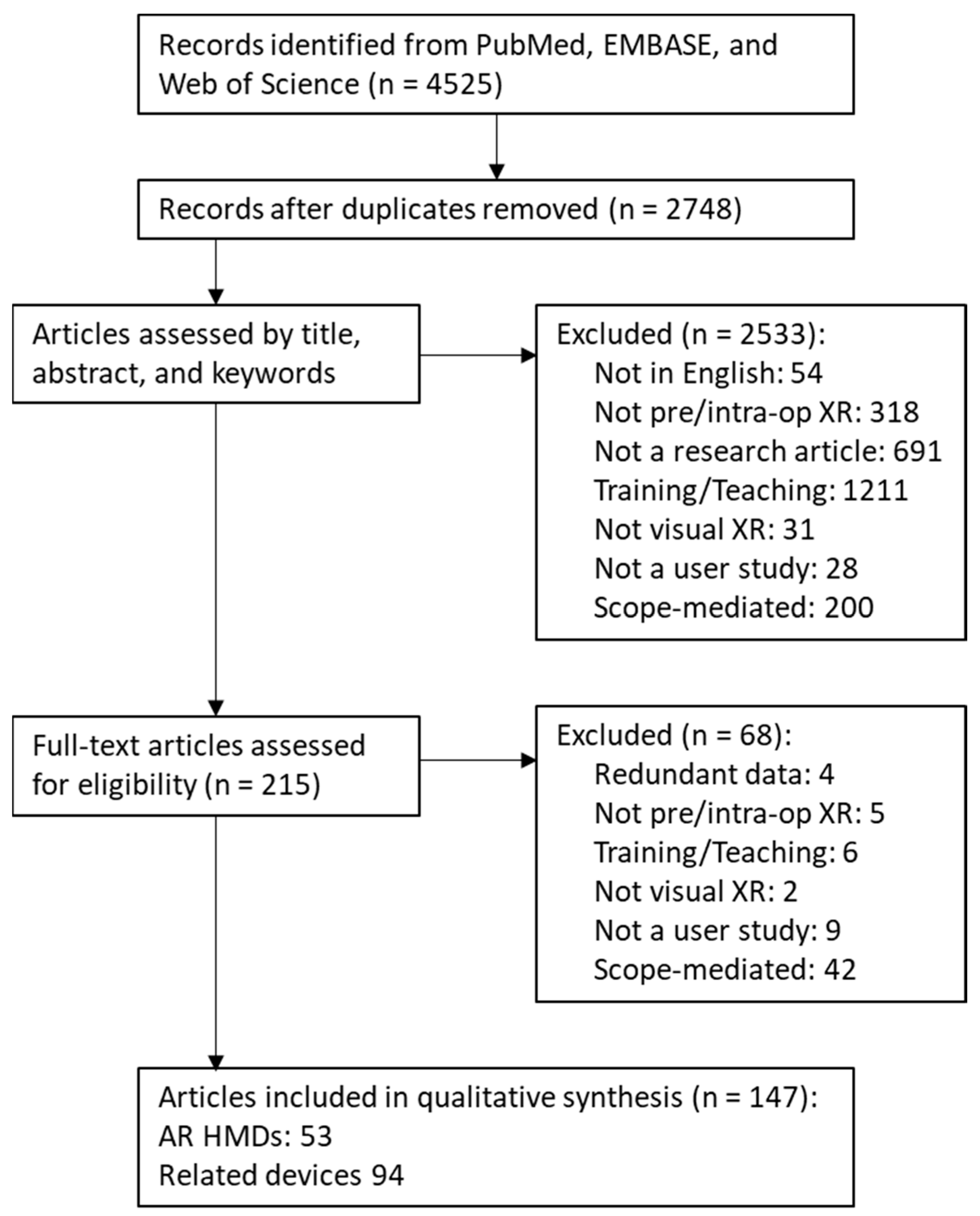
2. Materials and Methods
3. Results
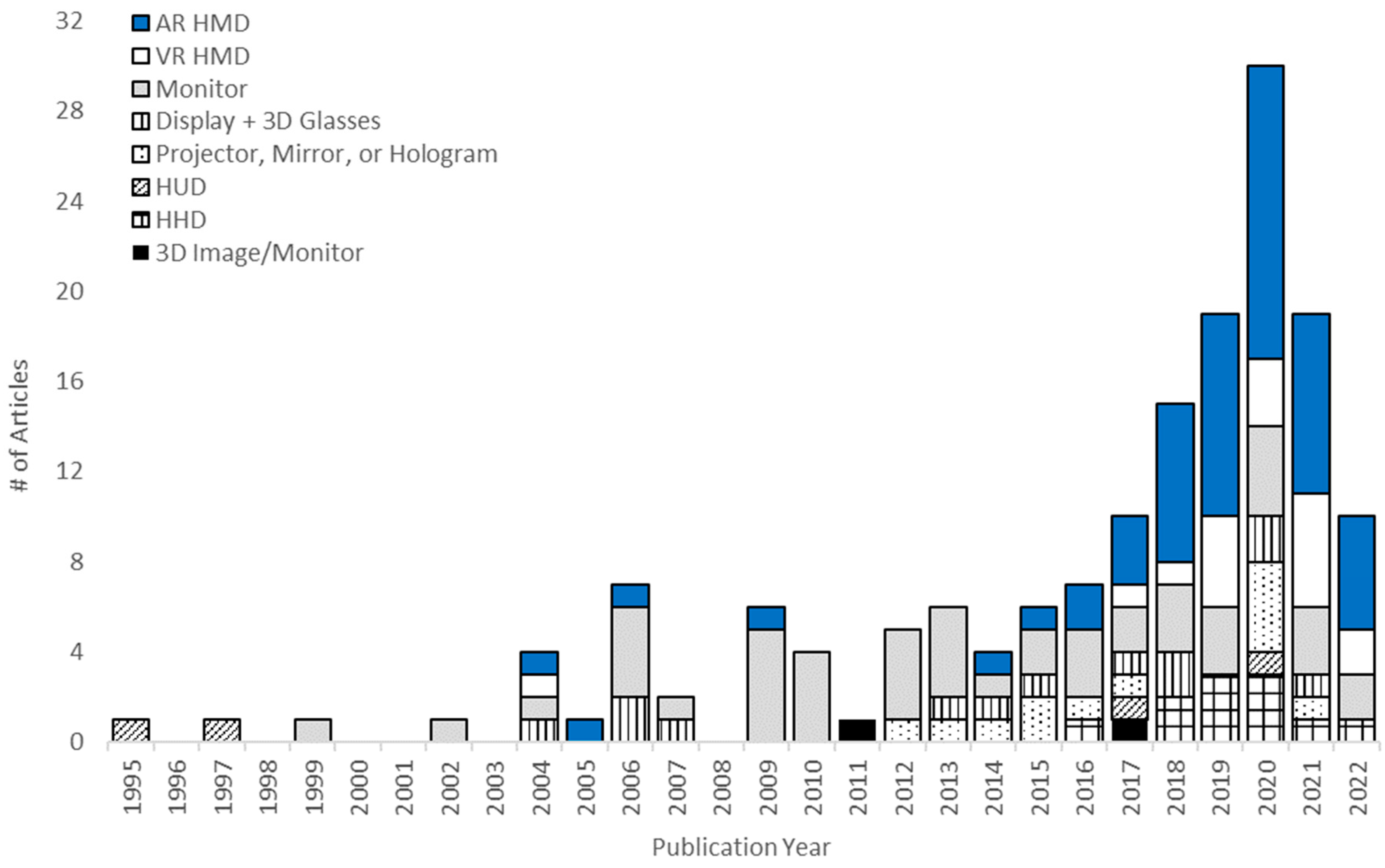
3.1. Device Types
3.2. Surgical Applications
3.3. User Demographics
3.4. Usability Assessments
- Task Performance (84)—Assessments of motor or visuomotor task success.
- User Experience (80)—Interviews, surveys, or other user-reported feedback about the usability and effectiveness of visualization type or hardware.
- Completion Times (55)—Duration of setup or task performance.
- Cognition (27)—Assessments of mental and attentional demands or changes in decision making.
- Visual Effects (22)—Objective assessments of visualization quality or accuracy, relative effectiveness among visual augmentations or rendering options, visual perception, or adverse physiological events related to visual perception.
- Efficiency (17)—Quantification of intraoperative imaging use, material use, or tool path length.
- Physical loads (1)—Quantification of muscle activity or body movement.
- 8.
- System Performance (39)—Measurements of visualization hardware and software accuracy and speed.
- 9.
- Validity/Reliability (12)—Comparison of simulators to real situations, or comparisons within and across users or observers.
4. Discussion
4.1. Usability Assessments for XR+ Devices
4.1.1. Task Performance
4.1.2. XR+ User Experience
4.1.3. Completion Times
4.1.4. Cognition
4.1.5. Visual Effects
4.1.6. Efficiency
4.1.7. Physical Loads
4.2. Additional Assessments and Reporting Related to Usability
4.2.1. User Demographics
4.2.2. System Performance
4.2.3. Validity and Reliability
4.3. Limitations
5. Conclusions
Supplementary Materials
Author Contributions
Funding
Institutional Review Board Statement
Informed Consent Statement
Data Availability Statement
Conflicts of Interest
Note
References
- Dey, A.; Billinghurst, M.; Lindeman, R.W.; Swan, J.E. A systematic review of 10 years of augmented reality usability studies: 2005 to 2014. Front. Robot. AI 2018, 5, 37. [Google Scholar] [CrossRef]
- Linte, C.A.; Davenport, K.P.; Cleary, K.; Peters, C.; Vosburgh, K.G.; Navab, N.; Jannin, P.; Peters, T.M.; Holmes III, D.R.; Robb, R.A. On mixed reality environments for minimally invasive therapy guidance: Systems architecture, successes and challenges in their implementation from laboratory to clinic. Comput. Med. Imaging Graph. 2013, 37, 83–97. [Google Scholar] [CrossRef] [PubMed]
- Friets, E.M.; Strohbehn, J.W.; Hatch, J.F.; Roberts, D.W. A frameless stereotaxic operating microscope for neurosurgery. IEEE Trans. Biomed. Eng. 1989, 36, 608–617. [Google Scholar] [CrossRef] [PubMed]
- Kelly, P.J.; Alker, G.J., Jr.; Goerss, S. Computer-assisted stereotactic laser microsurgery for the treatment of intracranial neoplasms. Neurosurgery 1982, 10, 324–331. [Google Scholar] [CrossRef] [PubMed]
- Kress, B.; Starner, T. A review of head-mounted displays (HMD) technologies and applications for consumer electronics. In Proceedings of the Conference on SPIE Defense, Security, and Sensing, Baltimore, MD, USA, 29 April–3 May 2013; p. 87200A. [Google Scholar]
- Qian, L.; Barthel, A.; Johnson, A.; Osgood, G.; Kazanzides, P.; Navab, N.; Fuerst, B. Comparison of optical see-through head-mounted displays for surgical interventions with object-anchored 2D-display. Int. J. Comput. Assist. Radiol. Surg. 2017, 12, 901–910. [Google Scholar] [CrossRef] [PubMed]
- Brun, H.; Bugge, R.A.B.; Suther, L.K.R.; Birkeland, S.; Kumar, R.; Pelanis, E.; Elle, O.J. Mixed reality holograms for heart surgery planning: First user experience in congenital heart disease. Eur. Heart J. Cardiovasc. Imaging 2018, 20, 883–888. [Google Scholar] [CrossRef]
- Si, W.X.; Liao, X.; Qian, Y.; Wang, Q. Mixed reality guided radiofrequency needle placement: A pilot study. IEEE Access 2018, 6, 31493–31502. [Google Scholar] [CrossRef]
- Deib, G.; Johnson, A.; Unberath, M.; Yu, K.; Andress, S.; Qian, L.; Osgood, G.; Navab, N.; Hui, F.; Gailloud, P. Image guided percutaneous spine procedures using an optical see-through head mounted display: Proof of concept and rationale. J. Neurointerv. Surg. 2018, 10, 1187–1191. [Google Scholar] [CrossRef]
- Incekara, F.; Smits, M.; Dirven, C.; Vincent, A. Clinical feasibility of a wearable mixed-reality device in neurosurgery. World Neurosurg. 2018, 118, e422–e427. [Google Scholar] [CrossRef]
- Koesveld, J.; Tetteroo, G.; Graaf, E. Use of head-mounted display in transanal endoscopic microsurgery. Surg. Endosc. 2003, 17, 943–946. [Google Scholar] [CrossRef]
- Qian, L.; Deguet, A.; Kazanzides, P. ARssist: Augmented reality on a head-mounted display for the first assistant in robotic surgery. Healthc. Technol. Lett. 2018, 5, 194–200. [Google Scholar] [CrossRef] [PubMed]
- Liebert, C.A.; Zayed, M.A.; Aalami, O.; Tran, J.; Lau, J.N. Novel use of Google Glass for procedural wireless vital sign monitoring. Surg. Innov. 2016, 23, 366–373. [Google Scholar] [CrossRef] [PubMed]
- Liu, D.; Jenkins, S.A.; Sanderson, P.M.; Fabian, P.; Russell, W.J. Monitoring with head-mounted displays in general anesthesia: A clinical evaluation in the operating room. Anesth. Analg. 2010, 110, 1032–1038. [Google Scholar] [CrossRef] [PubMed]
- Kumar, S.; Singhal, P.; Krovi, V.N. Computer-vision-based decision support in surgical robotics. IEEE Des. Test 2015, 32, 89–97. [Google Scholar] [CrossRef]
- Sakata, S.; Watson, M.O.; Grove, P.M.; Stevenson, A.R. The conflicting evidence of three-dimensional displays in laparoscopy: A review of systems old and new. Ann. Surg. 2016, 263, 234–239. [Google Scholar] [CrossRef]
- Bernhardt, S.; Nicolau, S.A.; Soler, L.; Doignon, C. The status of augmented reality in laparoscopic surgery as of 2016. Med. Image Anal. 2017, 37, 66–90. [Google Scholar] [CrossRef]
- Luo, X.; Mori, K.; Peters, T.M. Advanced Endoscopic Navigation: Surgical Big Data, Methodology, and Applications. Annu. Rev. Biomed. Eng. 2018, 20, 221–251. [Google Scholar] [CrossRef]
- Condino, S.; Carbone, M.; Piazza, R.; Ferrari, M.; Ferrari, V. Perceptual limits of optical see-through visors for augmented reality guidance of manual tasks. IEEE Trans. Biomed. Eng. 2019, 67, 411–419. [Google Scholar] [CrossRef]
- Beams, R.; Brown, E.; Cheng, W.C.; Joyner, J.S.; Kim, A.S.; Kontson, K.; Amiras, D.; Baeuerle, T.; Greenleaf, W.; Grossmann, R.J.; et al. Evaluation Challenges for the Application of Extended Reality Devices in Medicine. J. Digit. Imaging 2022, 35, 1409–1418. [Google Scholar] [CrossRef]
- Shuhaiber, J.H. Augmented reality in surgery. Arch. Surg. 2004, 139, 170–174. [Google Scholar] [CrossRef]
- Yoon, J.W.; Chen, R.E.; Kim, E.J.; Akinduro, O.O.; Kerezoudis, P.; Han, P.K.; Si, P.; Freeman, W.D.; Diaz, R.J.; Komotar, R.J.; et al. Augmented reality for the surgeon: Systematic review. Int. J. Med. Robot. Comput. Assist. Surg. 2018, 14, 13. [Google Scholar] [CrossRef] [PubMed]
- Meola, A.; Cutolo, F.; Carbone, M.; Cagnazzo, F.; Ferrari, M.; Ferrari, V. Augmented reality in neurosurgery: A systematic review. Neurosurg. Rev. 2017, 40, 537–548. [Google Scholar] [CrossRef] [PubMed]
- Neubauer, A.; Wolfsberger, S. Virtual endoscopy in neurosurgery: A review. Neurosurgery 2013, 72 (Suppl. 1), A97–A106. [Google Scholar] [CrossRef] [PubMed]
- de Ribaupierre, S.; Eagleson, S. Editorial: Challenges for the usability of AR and VR for clinical neurosurgical procedures. Healthc. Technol. Lett. 2017, 4, 151. [Google Scholar] [CrossRef]
- Guha, D.; Alotaibi, N.M.; Nguyen, N.; Gupta, S.; McFaul, C.; Yang, V.X. Augmented reality in neurosurgery: A review of current concepts and emerging applications. Can. J. Neurol. Sci. 2017, 44, 235–245. [Google Scholar] [CrossRef]
- Jud, L.; Fotouhi, J.; Andronic, O.; Aichmair, A.; Osgood, G.; Navab, N.; Farshad, M. Applicability of augmented reality in orthopedic surgery—A systematic review. BMC Musculoskelet. Disord. 2020, 21, 103. [Google Scholar] [CrossRef]
- Kim, Y.; Kim, H.; Kim, Y.O. Virtual reality and augmented reality in plastic surgery: A review. Arch. Plast. Surg. 2017, 44, 179. [Google Scholar] [CrossRef]
- Qian, L.; Wu, J.Y.; DiMaio, S.P.; Navab, N.; Kazanzides, P. A Review of Augmented Reality in Robotic-Assisted Surgery. IEEE Trans. Med. Robot. Bionics 2019, 2, 1. [Google Scholar] [CrossRef]
- Birlo, M.; Edwards, P.E.; Clarkson, M.; Stoyanov, D. Utility of optical see-through head mounted displays in augmented reality-assisted surgery: A systematic review. Med. Image Anal. 2022, 77, 102361. [Google Scholar] [CrossRef]
- Dünser, A.; Grasset, R.; Billinghurst, M. A survey of evaluation techniques used in augmented reality studies. In Proceedings of the International Conference on Computer Graphics and Interactive Techniques, Los Angeles, CA, USA, 11–15 August 2008. [Google Scholar]
- Cutolo, F.; Carli, S.; Parchi, P.D.; Canalini, L.; Ferrari, M.; Lisanti, M.; Ferrari, V. AR interaction paradigm for closed reduction of long-bone fractures via external fixation. In Proceedings of the 22nd ACM Conference on Virtual Reality Software and Technology, Munich, Germany, 2–4 November 2016; Spencer, S.N., Ed.; Assoc Computing Machinery: New York, NY, USA, 2016; pp. 305–306. [Google Scholar]
- El-Hariri, H.; Pandey, P.; Hodgson, A.J.; Garbi, R. Augmented reality visualisation for orthopaedic surgical guidance with pre- and intra-operative multimodal image data fusion. Healthc. Technol. Lett. 2018, 5, 189–193. [Google Scholar] [CrossRef]
- Fotouhi, J.; Alexander, C.P.; Unberath, M.; Taylor, G.; Lee, S.C.; Fuerst, B.; Johnson, A.; Osgood, G.; Taylor, R.H.; Khanuja, H.; et al. Plan in 2-D, execute in 3-D: An augmented reality solution for cup placement in total hip arthroplasty. J. Med. Imaging 2018, 5, 021205. [Google Scholar] [CrossRef] [PubMed]
- Molina, C.A.; Theodore, N.; Ahmed, A.K.; Westbroek, E.M.; Mirovsky, Y.; Harel, R.; Khan, M.; Witham, T.; Sciubba, D.M. Augmented reality-assisted pedicle screw insertion: A cadaveric proof-of-concept study. J. Neurosurg. Spine 2019, 31, 139–146. [Google Scholar] [CrossRef] [PubMed]
- Wang, H.X.; Wang, F.; Leong, A.P.Y.; Xu, L.; Chen, X.; Wang, Q. Precision insertion of percutaneous sacroiliac screws using a novel augmented reality-based navigation system: A pilot study. Int. Orthop. 2016, 40, 1941–1947. [Google Scholar] [CrossRef]
- Cutolo, F.; Parchi, P.D.; Ferrari, V. Video see through AR head-mounted display for medical procedures. In Proceedings of the 2014 IEEE International Symposium on Mixed and Augmented Reality, Munich, Germany, 10–12 September 2014; Julier, S., Lindeman, R.W., Sandor, C., Eds.; IEEE: New York, NY, USA, 2014; pp. 393–396. [Google Scholar]
- Traub, J.; Stefan, P.; Heining, S.M.; Sielhorst, T.; Riquarts, C.; Euler, E.; Navab, N. Stereoscopic augmented reality navigation for trauma surgery: Cadaver experiment and usability study. Int. J. Comput. Assist. Radiol. Surg. 2006, 1, 30–32. [Google Scholar]
- Nguyen, N.Q.; Cardinell, J.; Ramjist, J.M.; Androutsos, D.; Yang, V.X. Augmented Reality and Human Factors Regarding the Neurosurgical Operating Room Workflow. In Proceedings of the Conference on Optical Architectures for Displays and Sensing in Augmented, Virtual, and Mixed Reality, Virtual, 28–31 March 2020; Kress, B.C., Peroz, C., Eds.; Spie-Int Soc Optical Engineering: Bellingham, WA, USA, 2020. [Google Scholar]
- Urakov, T.M. Augmented Reality-assisted Pedicle Instrumentation: Versatility Across Major Instrumentation Sets. Spine 2020, 45, E1622–E1626. [Google Scholar] [CrossRef] [PubMed]
- Viehofer, A.F.; Wirth, S.H.; Zimmermann, S.M.; Jaberg, L.; Dennler, C.; Fürnstahl, P.; Farshad, M. Augmented reality guided osteotomy in hallux Valgus correction. BMC Musculoskelet. Disord. 2020, 21, 1–6. [Google Scholar] [CrossRef]
- Bichlmeier, C.; Heining, S.M.; Feuerstein, M.; Navab, N. The virtual mirror: A new interaction paradigm for augmented reality environments. IEEE Trans. Med. Imaging 2009, 28, 1498–1510. [Google Scholar] [CrossRef]
- Gu, W.; Martin-Gomez, A.; Cho, S.M.; Osgood, G.; Bracke, B.; Josewski, C.; Knopf, J.; Unberath, M. The impact of visualization paradigms on the detectability of spatial misalignment in mixed reality surgical guidance. Int. J. Comput. Assist. Radiol. Surg. 2022, 17, 921–927. [Google Scholar] [CrossRef]
- Harel, R.; Anekstein, Y.; Raichel, M.; Molina, C.A.; Ruiz-Cardozo, M.A.; Orrú, E.; Khan, M.; Mirovsky, Y.; Smorgick, Y. The XVS System During Open Spinal Fixation Procedures in Patients Requiring Pedicle Screw Placement in the Lumbosacral Spine. World Neurosurg. 2022, 164, e1226–e1232. [Google Scholar] [CrossRef]
- Yanni, D.S.; Ozgur, B.M.; Louis, R.G.; Shekhtman, Y.; Iyer, R.R.; Boddapati, V.; Iyer, A.; Patel, P.D.; Jani, R.; Cummock, M.; et al. Real-time navigation guidance with intraoperative CT imaging for pedicle screw placement using an augmented reality head-mounted display: A proof-of-concept study. Neurosurg. Focus 2021, 51, E11. [Google Scholar] [CrossRef]
- Saylany, A.; Spadola, M.; Blue, R.; Sharma, N.; Ozturk, A.K.; Yoon, J.W. The Use of a Novel Heads-Up Display (HUD) to View Intra-Operative X-rays during a One-Level Cervical Arthroplasty. World Neurosurg. 2020, 138, 369–373. [Google Scholar] [CrossRef] [PubMed]
- Yoon, J.W.; Chen, R.E.; Han, P.K.; Si, P.; Freeman, W.D.; Pirris, S.M. Technical feasibility and safety of an intraoperative head-up display device during spine instrumentation. Int. J. Med. Robot. Comput. Assist. Surg. 2017, 13, e1770. [Google Scholar] [CrossRef] [PubMed]
- Alexander, C.; Loeb, A.E.; Fotouhi, J.; Navab, N.; Armand, M.; Khanuja, H.S. Augmented Reality for Acetabular Component Placement in Direct Anterior Total Hip Arthroplasty. J. Arthroplast. 2020, 35, 1636–1641.e3. [Google Scholar] [CrossRef]
- Bong, J.H.; Kim, H.; Park, S. Development of a surgical navigation system for corrective osteotomy based on augmented reality. Int. J. Precis. Eng. Manuf. 2017, 18, 1057–1062. [Google Scholar] [CrossRef]
- Fischer, M.; Fuerst, B.; Lee, S.C.; Fotouhi, J.; Habert, S.; Weidert, S.; Euler, E.; Osgood, G.; Navab, N. Preclinical usability study of multiple augmented reality concepts for K-wire placement. Int. J. Comput. Assist. Radiol. Surg. 2016, 11, 1007–1014. [Google Scholar] [CrossRef]
- Ma, L.F.; Zhao, Z.; Chen, F.; Zhang, B.; Fu, L.; Liao, H. Augmented reality surgical navigation with ultrasound-assisted registration for pedicle screw placement: A pilot study. Int. J. Comput. Assist. Radiol. Surg. 2017, 12, 2205–2215. [Google Scholar] [CrossRef]
- Ogawa, H.; Hasegawa, S.; Tsukada, S.; Matsubara, M. A pilot study of augmented reality technology applied to the acetabular cup placement during total hip arthroplasty. J. Arthroplast. 2018, 33, 1833–1837. [Google Scholar] [CrossRef]
- Ponce, B.A.; Brabston, E.W.; Zu, S.; Watson, S.L.; Baker, D.; Winn, D.; Guthrie, B.L.; Shenai, M.B. Telemedicine with mobile devices and augmented reality for early postoperative care. In Proceedings of the Annual International Conference of the IEEE Engineering in Medicine and Biology Society, Orlando, FL, USA, 16–20 August 2016; pp. 4411–4414. [Google Scholar]
- Tsukada, S.; Ogawa, H.; Nishino, M.; Kurosaka, K.; Hirasawa, N. Augmented reality-based navigation system applied to tibial bone resection in total knee arthroplasty. J. Exp. Orthop. 2019, 6, 44. [Google Scholar] [CrossRef]
- Wang, X.; Habert, S.; Zu Berge, C.S.; Fallavollita, P.; Navab, N. Inverse visualization concept for RGB-D augmented C-arms. Comput. Biol. Med. 2016, 77, 135–147. [Google Scholar] [CrossRef]
- Chen, X.; Naik, H.; Wang, L.; Navab, N.; Fallavollita, P. Video-guided calibration of an augmented reality mobile C-arm. Int. J. Comput. Assist. Radiol. Surg. 2014, 9, 987–996. [Google Scholar] [CrossRef]
- Erat, O.; Pauly, O.; Weidert, S.; Thaller, P.; Euler, E.; Mutschler, W.; Navab, N.; Fallavollita, P. How a surgeon becomes Superman by visualization of intelligently fused multi-modalities. In Proceedings of the Medical Imaging 2013: Image-Guided Procedures, Robotic Interventions, and Modeling, Orlando, FL, USA, 9–14 February 2013; Holmes, D.R., Yaniv, Z.R., Eds.; SPIE-Int Soc Optical Engineering: Bellingham, WA, USA, 2013. [Google Scholar]
- Gavaghan, K.; Oliveira-Santos, T.; Peterhans, M.; Reyes, M.; Kim, H.; Anderegg, S.; Weber, S. Evaluation of a portable image overlay projector for the visualisation of surgical navigation data: Phantom studies. Int. J. Comput. Assist. Radiol. Surg. 2012, 7, 547–556. [Google Scholar] [CrossRef] [PubMed]
- Heining, S.M.; Wiesner, S.; Euler, E.; Navab, N. Pedicle screw placement under video-augmented flouroscopic control: First clinical application in a cadaver study. Int. J. Comput. Assist. Radiol. Surg. 2006, 1, 189–190. [Google Scholar]
- Juhnke, B.; Berron, M.; Philip, A.; Williams, J.; Holub, J.; Winer, E. Comparing the Microsoft (R) Kinect (TM) to a traditional mouse for adjusting the viewed tissue densities of three-dimensional anatomical structures. In Proceedings of the Medical Imaging 2013: Image Perception, Observer Performance, and Technology Assessment, Lake Buena Vista, FL, USA, 28 March 2013; Abbey, C.K., MelloThoms, C.R., Eds.; SPIE-Int Soc Optical Engineering: Bellingham, WA, USA, 2013. [Google Scholar]
- Kendoff, D.; Citak, M.; Gardner, M.J.; Stübig, T.; Krettek, C.; Hüfner, T. Intraoperative 3D Imaging: Value and Consequences in 248 Cases. J. Trauma Inj. Infect. Crit. Care 2009, 66, 232–238. [Google Scholar] [CrossRef] [PubMed]
- Londei, R.; Esposito, M.; Diotte, B.; Weidert, S.; Euler, E.; Thaller, P.; Navab, N.; Fallavollita, P. Intra-operative augmented reality in distal locking. Int. J. Comput. Assist. Radiol. Surg. 2015, 10, 1395–1403. [Google Scholar] [CrossRef] [PubMed]
- Marschollek, M.; Teistler, M.; Bott, O.J.; Stuermer, K.M.; Pretschner, D.P.; Dresing, K. Pre-operative dynamic interactive exploration of complex articular fractures using a novel 3D navigation tool. Methods Inf. Med. 2006, 45, 384–388. [Google Scholar]
- Pahuta, M.A.; Schemitsch, E.H.; Backstein, D.; Papp, S.; Gofton, W. Virtual fracture carving improves understanding of a complex fracture: A randomized controlled study. J. Bone Jt. Surg. Am. Vol. 2012, 94, e182.1–e182.7. [Google Scholar] [CrossRef]
- Testi, D.; Lattanzi, R.; Benvegnù, M.; Petrone, M.; Zannoni, C.; Viceconti, M.; Toni, A. Efficacy of stereoscopic visualization and six degrees of freedom interaction in preoperative planning of total hip replacement. Med. Inform. Internet Med. 2006, 31, 205–218. [Google Scholar] [CrossRef]
- Vaghela, K.R.; Lee, J.; Akhtar, K. Performance on a virtual reality DHS simulator correlates with performance in the operating theatre. Surg. Technol. Int. 2018, 33, sti33/1040. [Google Scholar]
- Alsofy, S.Z.; Nakamura, M.; Ewelt, C.; Kafchitsas, K.; Lewitz, M.; Schipmann, S.; Molina, E.S.; Santacroce, A.; Stroop, R. Retrospective Comparison of Minimally Invasive and Open Monosegmental Lumbar Fusion, and Impact of Virtual Reality on Surgical Planning and Strategy. J. Neurol. Surg. Part A Cent. Eur. Neurosurg. 2021, 82, 399–409. [Google Scholar]
- Moreta-Martinez, R.; Pose-Díez-de-la-Lastra, A.; Calvo-Haro, J.A.; Mediavilla-Santos, L.; Pérez-Mañanes, R.; Pascau, J. Combining Augmented Reality and 3D Printing to Improve Surgical Workflows in Orthopedic Oncology: Smartphone Application and Clinical Evaluation. Sensors 2021, 21, 1370. [Google Scholar] [CrossRef]
- Pandey, P.U.; Guy, P.; Lefaivre, K.A.; Hodgson, A.J. What are the optimal targeting visualizations for performing surgical navigation of iliosacral screws? A user study. Trauma Surg. 2021, 143, 677–690. [Google Scholar] [CrossRef] [PubMed]
- Cartucho, J.; Shapira, D.; Ashrafian, H.; Giannarou, S. Multimodal mixed reality visualisation for intraoperative surgical guidance. Int. J. Comput. Assist. Radiol. Surg. 2020, 15, 819–826. [Google Scholar] [CrossRef] [PubMed]
- Cutolo, F.; Meola, A.; Carbone, M.; Sinceri, S.; Cagnazzo, F.; Denaro, E.; Esposito, N.; Ferrari, M.; Ferrari, V. A new head-mounted display-based augmented reality system in neurosurgical oncology: A study on phantom. Comput. Assist. Surg. 2017, 22, 39–53. [Google Scholar] [CrossRef] [PubMed]
- Kubben, P.; Sinlae, R.N. Feasibility of using a low-cost head-mounted augmented reality device in the operating room. Surg. Neurol. Int. 2019, 10, 26. [Google Scholar] [CrossRef]
- Fick, T.; van Doormaal, J.A.M.; Hoving, E.W.; Regli, L.; van Doormaal, T.P.C. Holographic patient tracking after bed movement for augmented reality neuronavigation using a head-mounted display. Acta Neurochir. 2021, 163, 879–884. [Google Scholar] [CrossRef] [PubMed]
- Benmahdjoub, M.; Niessen, W.J.; Wolvius, E.B.; Walsum, T.V. Multimodal markers for technology-independent integration of augmented reality devices and surgical navigation systems. Virtual Real. 2022, 26, 1637–1650. [Google Scholar] [CrossRef]
- Condino, S.; Montemurro, N.; Cattari, N.; D’Amato, R.; Thomale, U.; Ferrari, V.; Cutolo, F. Evaluation of a Wearable AR Platform for Guiding Complex Craniotomies in Neurosurgery. Ann. Biomed. Eng. 2021, 49, 2590–2605. [Google Scholar] [CrossRef]
- Thabit, A.; Benmahdjoub, M.; Van Veelen, M.L.C.; Niessen, W.J.; Wolvius, E.B.; van Walsum, T. Augmented reality navigation for minimally invasive craniosynostosis surgery: A phantom study. Int. J. Comput. Assist. Radiol. Surg. 2022, 17, 1453–1460. [Google Scholar] [CrossRef]
- Coelho, G.; Rabelo, N.N.; Vieira, E.; Mendes, K.; Zagatto, G.; de Oliveira, R.S.; Raposo-Amaral, C.E.; Yoshida, M.; de Souza, M.R.; Fagundes, C.F.; et al. Augmented reality and physical hybrid model simulation for preoperative planning of metopic craniosynostosis surgery. Neurosurg. Focus 2020, 48, E19. [Google Scholar] [CrossRef]
- Gerard, I.J.; Kersten-Oertel, M.; Drouin, S.; Hall, J.A.; Petrecca, K.; De Nigris, D.; Di Giovanni, D.A.; Arbel, T.; Collins, D.L. Combining intraoperative ultrasound brain shift correction and augmented reality visualizations: A pilot study of eight cases. J. Med. Imaging 2018, 5, 021210. [Google Scholar] [CrossRef]
- Léger, É.; Reyes, J.; Drouin, S.; Collins, D.L.; Popa, T.; Kersten-Oertel, M. Gesture-based registration correction using a mobile augmented reality image-guided neurosurgery system. Healthc. Technol. Lett. 2018, 5, 137–142. [Google Scholar] [CrossRef] [PubMed]
- Léger, É.; Reyes, J.; Drouin, S.; Popa, T.; Hall, J.A.; Collins, D.L.; Kersten-Oertel, M. MARIN: An open-source mobile augmented reality interactive neuronavigation system. Int. J. Comput. Assist. Radiol. Surg. 2020, 15, 1013–1021. [Google Scholar] [CrossRef] [PubMed]
- Acker, G.; Schlinkmann, N.; Piper, S.K.; Onken, J.; Vajkoczy, P.; Picht, T. Stereoscopic versus monoscopic viewing of aneurysms: Experience of a single institution with a novel stereoscopic viewing system. World Neurosurg. 2018, 119, E491–E501. [Google Scholar] [CrossRef] [PubMed]
- Azar, F.S.; Perrin, N.; Khamene, A.; Vogt, S.; Sauer, F. User performance analysis of different image-based navigation systems for needle placement procedures. In Proceedings of the Medical Imaging 2004: Visualization, Image-Guided Procedures, and Display, San Diego, CA, USA, 15–17 February 2004; Galloway, R.L., Ed.; SPIE-Int Soc Optical Engineering: Bellingham, WA, USA, 2004; pp. 110–121. [Google Scholar]
- Gökyar, A.; Bahadır, S.; Çokluk, C. Evaluation of projection-based augmented reality technique in cerebral catheter procedures. Ann. Clin. Anal. Med. 2020, 11, 630–633. [Google Scholar]
- Kersten-Oertel, M.; Chen, S.J.-S.; Collins, D.L. An Evaluation of Depth Enhancing Perceptual Cues for Vascular Volume Visualization in Neurosurgery. IEEE Trans. Vis. Comput. Graph. 2014, 20, 391–403. [Google Scholar] [CrossRef]
- Rolls, A.E.; Riga, C.V.; Bicknell, C.D.; Stoyanov, D.V.; Shah, C.V.; Van Herzeele, I.; Hamady, M.; Cheshire, N.J. A pilot study of video-motion analysis in endovascular surgery: Development of real-time discriminatory skill metrics. Eur. J. Vasc. Endovasc. Surg. 2013, 45, 509–515. [Google Scholar] [CrossRef]
- Stadie, A.T.; Kockro, R.A. Mono-stereo-autostereo: The evolution of 3-dimensional neurosurgical planning. Neurosurgery 2013, 72, A63–A77. [Google Scholar] [CrossRef]
- Tabrizi, L.B.; Mahvash, M. Augmented reality-guided neurosurgery: Accuracy and intraoperative application of an image projection technique. J. Neurosurg. 2015, 123, 206–211. [Google Scholar] [CrossRef]
- Tai, A.X.; Riga, C.V.; Bicknell, C.D.; Stoyanov, D.V.; Shah, C.V.; Van Herzeele, I.; Hamady, M.; Cheshire, N.J. The Benefits of Limited Orbitotomy on the Supraorbital Approach: An Anatomic and Morphometric Study in Virtual Reality. Neurosurgery 2020, 18, 542–550. [Google Scholar] [CrossRef]
- Willaert, W.; Aggarwal, R.; Bicknell, C.; Hamady, M.; Darzi, A.; Vermassen, F.; Cheshire, N.; European Virtual Reality Endovascular Research Team. Patient-specific simulation in carotid artery stenting. J. Vasc. Surg. 2010, 52, 1700–1705. [Google Scholar] [CrossRef]
- Willaert, W.I.M.; Aggarwal, R.; Daruwalla, F.; Van Herzeele, I.; Darzi, A.W.; Vermassen, F.E.; Cheshire, N.J.; European Virtual Reality Endovascular Research Team EVEResT. Simulated procedure rehearsal is more effective than a preoperative generic warm-up for endovascular procedures. Ann. Surg. 2012, 255, 1184–1189. [Google Scholar] [CrossRef] [PubMed]
- Willaert, W.I.M.; Aggarwal, R.; Van Herzeele, I.; Plessers, M.; Stroobant, N.; Nestel, D.; Cheshire, N.; Vermassen, F. Role of patient-specific virtual reality rehearsal in carotid artery stenting. Br. J. Surg. 2012, 99, 1304–1313. [Google Scholar] [CrossRef]
- Zawy Alsofy, S.; Sakellaropoulou, I.; Stroop, R. Evaluation of Surgical Approaches for Tumor Resection in the Deep Infratentorial Region and Impact of Virtual Reality Technique for the Surgical Planning and Strategy. J. Craniofac. Surg. 2020, 31, 1865–1869. [Google Scholar] [CrossRef] [PubMed]
- Koike, T.; Kin, T.; Tanaka, Y.; Uchikawa, H.; Shiode, T.; Saito, N. Development of Innovative Neurosurgical Operation Support Method Using Mixed-Reality Computer Graphics. World Neurosurg. X 2021, 11, 100102. [Google Scholar] [CrossRef] [PubMed]
- Huang, L.; Collins, S.; Kobayashi, L.; Merck, D.; Sgouros, T. Shared visualizations and guided procedure simulation in augmented reality with Microsoft HoloLens. In Proceedings of the Medical Imaging 2019: Image-Guided Procedures, Robotic Interventions, and Modeling, San Diego, CA, USA, 16–21 February 2019; Fei, B., Linte, C.A., Eds.; SPIE-Int Soc Optical Engineering: Bellingham, WA, USA, 2019. [Google Scholar]
- Perkins, S.L.; Lin, M.A.; Srinivasan, S.; Wheeler, A.J.; Hargreaves, B.A.; Daniel, B.L. A mixed-reality system for breast surgical planning. In Proceedings of the 2017 IEEE International Symposium on Mixed and Augmented Reality (ISMAR-Adjunct), Nantes, France, 9–13 October 2017; IEEE: Nantes, France, 2017; pp. 269–274. [Google Scholar]
- Amini, S.; Kersten-Oertel, M. Augmented reality mastectomy surgical planning prototype using the HoloLens template for healthcare technology letters. Healthc. Technol. Lett. 2019, 6, 261–265. [Google Scholar] [CrossRef]
- Galati, R.; Simone, M.; Barile, G.; De Luca, R.; Cartanese, C.; Grassi, G. Experimental Setup Employed in the Operating Room Based on Virtual and Mixed Reality: Analysis of Pros and Cons in Open Abdomen Surgery. J. Health Eng. 2020, 2020, 8851964. [Google Scholar] [CrossRef]
- Werkgartner, G.; Lemmerer, M.; Hauser, H.; Sorantin, E.; Beichel, R.; Reitinger, B.; Bornik, A.; Leberl, F.; Mischinger, H.J. Augmented-reality-based liver-surgical planning system. Eur. Surg. 2004, 36, 270–274. [Google Scholar] [CrossRef]
- Wacker, F.K.; Vogt, S.; Khamene, A.; Sauer, F.; Wendt, M.; Duerk, J.L.; Lewin, J.S.; Wolf, K.J. MR image-guided needle biopsies with a combination of augmented reality and MRI: A pilot study in phantoms and animals. In Proceedings of the CARS 2005: Computer Assisted Radiology and Surgery, Berlin, Germany, 22–25 June 2005; Elsevier Science Bv: Amsterdam, The Netherlands, 2005; pp. 424–428. [Google Scholar]
- Heinrich, F.; Schwenderling, L.; Joeres, F.; Lawonn, K.; Hansen, C. Comparison of Augmented Reality Display Techniques to Support Medical Needle Insertion. IEEE Trans. Vis. Comput. Graph. 2020, 26, 3568–3575. [Google Scholar] [CrossRef]
- Cattari, N.; Condino, S.; Cutolo, F.; Ferrari, M.; Ferrari, V. In Situ Visualization for 3D Ultrasound-Guided Interventions with Augmented Reality Headset. Bioengineering 2021, 8, 131. [Google Scholar] [CrossRef]
- Cardin, M.A.; Wang, J.X.; Lobaugh, N.J.; Guimont, I.; Plewes, D.B. A quantitative evaluation of human coordination interfaces for computer assisted surgery. Comput. Aided Surg. 2007, 12, 71–81. [Google Scholar] [CrossRef]
- El-Khameesy, N.; El-Wishy, H. Evaluating the impact of virtual reality on liver surgical planning procedures. In Modeling and Simulation in Engineering, Economics, and Management; Engemann, K.J., GilLafuente, A.M., Merigo, J.M., Eds.; Springer: Berlin, Germany, 2012; pp. 210–218. [Google Scholar]
- Freschi, C.; Troia, E.; Ferrari, V.; Megali, G.; Pietrabissa, A.; Mosca, F. Ultrasound guided robotic biopsy using augmented reality and human-robot cooperative control. In Proceedings of the 2009 Annual International Conference of the IEEE Engineering in Medicine and Biology Society, Minneapolis, MN, USA, 3–6 September 2009; IEEE: New York, NY, USA, 2009; Volume 1–20, pp. 5110–5113. [Google Scholar]
- Pfeiffer, M.; Kenngott, H.; Preukschas, A.; Huber, M.; Bettscheider, L.; Müller-Stich, B.; Speidel, S. IMHOTEP: Virtual reality framework for surgical applications. Int. J. Comput. Assist. Radiol. Surg. 2018, 13, 741–748. [Google Scholar] [CrossRef] [PubMed]
- Reitinger, B.; Bornik, A.; Beichel, R.; Schmalstieg, D. Liver surgery planning using virtual reality. IEEE Comput. Graph. Appl. 2006, 26, 36–47. [Google Scholar] [CrossRef] [PubMed]
- Tian, F.; Wu, J.X.; Rong, W.Q.; Wang, L.M.; Wu, F.; Yu, W.B.; An, S.L.; Liu, F.Q.; Feng, L.; Bi, C.; et al. Three-dimensional morphometric analysis for hepatectomy of centrally located hepatocellular carcinoma: A pilot study. World J. Gastroenterol. 2015, 21, 4607–4619. [Google Scholar] [CrossRef] [PubMed]
- Vos, E.L.; Koning, A.H.; Obdeijn, I.M.; van Verschuer, V.M.; Verhoef, C.; van der Spek, P.J.; Menke-Pluijmers, M.B.; Koppert, L.B. Preoperative prediction of cosmetic results in breast conserving surgery. J. Surg. Oncol. 2014, 111, 178–184. [Google Scholar] [CrossRef] [PubMed]
- Wellens, L.M.; Meulstee, J.; van de Ven, C.P.; Van Scheltinga, C.T.; Littooij, A.S.; van den Heuvel-Eibrink, M.M.; Fiocco, M.; Rios, A.C.; Maal, T.; Wijnen, M.H. Comparison of 3-Dimensional and Augmented Reality Kidney Models with Conventional Imaging Data in the Preoperative Assessment of Children with Wilms Tumors. JAMA Netw. Open 2019, 2, e192633. [Google Scholar] [CrossRef]
- Asgar-Deen, D.; Carriere, J.; Wiebe, E.; Peiris, L.; Duha, A.; Tavakoli, M. Augmented Reality Guided Needle Biopsy of Soft Tissue: A Pilot Study. Front. Robot. AI 2020, 7, 72. [Google Scholar] [CrossRef]
- Davrieux, C.F.; Carriere, J.; Wiebe, E.; Peiris, L.; Duha, A.; Tavakoli, M. Mixed reality navigation system for ultrasound-guided percutaneous punctures: A pre-clinical evaluation. Surg. Endosc. 2020, 34, 226–230. [Google Scholar] [CrossRef]
- Gao, Y.; Zhao, Y.; Xie, L.; Zheng, G. A Projector-Based Augmented Reality Navigation System for Computer-Assisted Surgery. Sensors 2021, 21, 2931. [Google Scholar] [CrossRef]
- Huettl, F.; Saalfeld, P.; Hansen, C.; Preim, B.; Poplawski, A.; Kneist, W.; Lang, H.; Huber, T. Virtual reality and 3D printing improve preoperative visualization of 3D liver reconstructions-results from a preclinical comparison of presentation modalities and user’s preference. Ann. Transl. Med. 2021, 9, 1074. [Google Scholar] [CrossRef]
- Kumar, R.P.; Pelanis, E.; Bugge, R.; Brun, H.; Palomar, R.; Aghayan, D.L.; Fretland, Å.A.; Edwin, B.; Elle, O.J. Use of mixed reality for surgery planning: Assessment and development workflow. J. Biomed. Inform. 2020, 112, 100077. [Google Scholar] [CrossRef]
- Southworth, M.K.; Silva, J.N.A.; Blume, W.M.; Van Hare, G.F.; Dalal, A.S.; Silva, J.R. Performance Evaluation of Mixed Reality Display for Guidance during Transcatheter Cardiac Mapping and Ablation. IEEE J. Transl. Eng. Health Med. 2020, 8, 1–10. [Google Scholar] [CrossRef] [PubMed]
- Ye, W.; Zhang, X.; Li, T.; Luo, C.; Yang, L. Mixed-reality hologram for diagnosis and surgical planning of double outlet of the right ventricle: A pilot study. Clin. Radiol. 2020, 76, 237.e1–237.e7. [Google Scholar] [CrossRef] [PubMed]
- Ballocca, F.; Meier, L.M.; Ladha, K.; Hiansen, J.Q.; Horlick, E.M.; Meineri, M. Validation of quantitative 3-dimensional transesophageal echocardiography mitral valve analysis using stereoscopic display. J. Cardiothorac. Vasc. Anesth. 2018, 33, 732–741. [Google Scholar] [CrossRef]
- Bruckheimer, E.; Rotschild, C.; Dagan, T.; Amir, G.; Kaufman, A.; Gelman, S.; Birk, E. Computer-generated real-time digital holography: First time use in clinical medical imaging. Eur. Hearth J. Cardiovasc. Imaging 2016, 17, 845–849. [Google Scholar] [CrossRef]
- Guiraudon, G.M.; Jones, D.L.; Bainbridge, D.; Linte, C.; Pace, D.; Moore, J.; Wedlake, C.; Lang, P.; Peters, T.M. Augmented reality image guidance during off-pump mitral valve replacement through the guiraudon universal cardiac introducer. Innov. Technol. Tech. Cardiothorac. Vasc. Surg. 2010, 5, 430–438. [Google Scholar] [CrossRef]
- Harake, D.; Gnanappa, G.K.; Alvarez, S.G.; Whittle, A.; Punithakumar, K.; Boechler, P.; Noga, M.; Khoo, N.S. Stereoscopic Display Is Superior to Conventional Display for Three-Dimensional Echocardiography of Congenital Heart Anatomy. J. Am. Soc. Echocardiogr. 2020, 33, 1297–1305. [Google Scholar] [CrossRef]
- Kim, B.; Loke, Y.H.; Mass, P.; Irwin, M.R.; Capeland, C.; Olivieri, L.; Krieger, A. A Novel Virtual Reality Medical Image Display System for Group Discussions of Congenital Heart Disease: Development and Usability Testing. JMIR Cardio 2020, 4, e20633. [Google Scholar] [CrossRef]
- Kozlowski, P.; Urheim, S.; Samset, E. Evaluation of a multi-view autostereoscopic real-time 3D ultrasound system for minimally invasive cardiac surgery guidance. In Proceedings of the 2017 IEEE 14th International Symposium on Biomedical Imaging, Melbourne, Australia, 18–21 April 2017; IEEE: New York, NY, USA, 2017; pp. 604–607. [Google Scholar]
- Lo, J.; Moore, J.; Wedlake, C.; Guiraudon, G.; Eagleson, R.; Peters, T. Surgeon-controlled visualization techniques for virtual reality-guided cardiac surgery. Stud. Health Technol. Inform. 2009, 142, 162–167. [Google Scholar]
- Napa, S.; Moore, M.; Bardyn, T. Advancing Cardiac Surgery Case Planning and Case Review Conferences Using Virtual Reality in Medical Libraries: Evaluation of the Usability of Two Virtual Reality Apps. JMIR Hum. Factors 2019, 6, e12008. [Google Scholar] [CrossRef]
- Seitel, M.; Maier-Hein, L.; Rietdorf, U.; Nikoloff, S.; Seitel, A.; Franz, A.; Kenngott, H.; Karck, M.; De Simone, R.; Wolf, I.; et al. Towards a mixed reality environment for preoperative planning of cardiac surgery. Stud. Health Technol. Inform. 2009, 142, 307–309. [Google Scholar]
- Pushparajah, K.; Chu, K.Y.K.; Deng, S.; Wheeler, G.; Gomez, A.; Kabir, S.; Schnabel, J.A.; Simpson, J.M. Virtual reality three-dimensional echocardiographic imaging for planning surgical atrioventricular valve repair. JTCVS Tech. 2021, 7, 269–277. [Google Scholar] [CrossRef] [PubMed]
- Kim, B.; Nguyen, P.; Loke, Y.H.; Cleveland, V.; Liu, X.; Mass, P.; Hibino, N.; Olivieri, L.; Krieger, A. Virtual Reality Cardiac Surgical Planning Software (CorFix) for Designing Patient-Specific Vascular Grafts: Development and Pilot Usability Study. JMIR Cardio 2022, 6, e35488. [Google Scholar] [CrossRef] [PubMed]
- Pellegrino, G.; Mangano, C.; Mangano, R.; Ferri, A.; Taraschi, V.; Marchetti, C. Augmented reality for dental implantology: A pilot clinical report of two cases. BMC Oral Health 2019, 19, 158. [Google Scholar] [CrossRef] [PubMed]
- Pepe, A.; Trotta, G.F.; Mohr-Ziak, P.; Gsaxner, C.; Wallner, J.; Bevilacqua, V.; Egger, J. A Marker-Less Registration Approach for Mixed Reality-Aided Maxillofacial Surgery: A Pilot Evaluation. J. Digit. Imaging 2019, 32, 1008–1018. [Google Scholar] [CrossRef] [PubMed]
- Katic, D.; Spengler, P.; Bodenstedt, S.; Castrillon-Oberndorfer, G.; Seeberger, R.; Hoffmann, J.; Dillmann, R.; Speidel, S. A system for context-aware intraoperative augmented reality in dental implant surgery. Int. J. Comput. Assist. Radiol. Surg. 2015, 10, 101–108. [Google Scholar] [CrossRef]
- Glas, H.H.; Kraeima, J.; van Ooijen, P.M.A.; Spijkervet, F.K.L.; Yu, L.; Witjes, M.J.H. Augmented Reality Visualization for Image-Guided Surgery: A Validation Study Using a Three-Dimensional Printed Phantom. J. Oral Maxillofac. Surg. 2021, 79, 1943.e1–1943.e10. [Google Scholar] [CrossRef]
- Kim, H.; Jeong, S.; Seo, J.; Park, I.; Ko, H.; Moon, S.Y. Augmented reality for botulinum toxin injection. Concurr. Comput. Pract. Exp. 2019, 32, e5526. [Google Scholar] [CrossRef]
- Al-Saud, L.M.; Mushtaq, F.; Mirghani, I.A.; Balkhoyor, A.; Keeling, A.; Manogue, M.; Mon-Williams, M.A. Drilling into the functional significance of stereopsis: The impact of stereoscopic information on surgical performance. Ophthalmic Physiol. Opt. 2017, 37, 498–506. [Google Scholar] [CrossRef]
- Bartella, A.K.; Kamal, M.; Scholl, I.; Schiffer, S.; Steegmann, J.; Ketelsen, D.; Hölzle, F.; Lethaus, B. Virtual reality in preoperative imaging in maxillofacial surgery: Implementation of “the next level”? Br. J. Oral Maxillofac. Surg. 2019, 57, 644–648. [Google Scholar] [CrossRef]
- Enislidis, G.; Wagner, A.; Ploder, O.; Ewers, R. Computed intraoperative navigation guidance—A preliminary report on a new technique. Br. J. Oral Maxillofac. Surg. 1997, 35, 271–274. [Google Scholar] [CrossRef]
- Stamm, T.; Meyer, U.; Meier, N.; Ehmer, U.; Joos, U. Public domain computer-aided surgery (CAS) in orthodontic and maxillofacial surgery. J. Orofac. Orthop. 2002, 63, 62–75. [Google Scholar] [CrossRef] [PubMed]
- Suenaga, H.; Tran, H.H.; Liao, H.; Masamune, K.; Dohi, T.; Hoshi, K.; Takato, T. Vision-based markerless registration using stereo vision and an augmented reality surgical navigation system: A pilot study. BMC Med. Imaging 2015, 15, 11. [Google Scholar] [CrossRef] [PubMed]
- Suenaga, H.; Hoang Tran, H.; Liao, H.; Masamune, K.; Dohi, T.; Hoshi, K.; Mori, Y.; Takato, T. Real-time in situ three-dimensional integral videography and surgical navigation using augmented reality: A pilot study. Int. J. Oral Sci. 2013, 5, 98–102. [Google Scholar] [CrossRef]
- Rojas-Munoz, E.; Cabrera, M.E.; Andersen, D.; Popescu, V.; Marley, S.; Mullis, B.; Zarzaur, B.; Wachs, J. Surgical Telementoring Without Encumbrance A Comparative Study of See-through Augmented Reality-based Approaches. Ann. Surg. 2019, 270, 384–389. [Google Scholar] [CrossRef]
- Qian, L.; Deguet, A.; Wang, Z.; Liu, Y.H.; Kazanzides, P. Augmented Reality Assisted Instrument Insertion and Tool Manipulation for the First Assistant in Robotic Surgery. In Proceedings of the 2019 International Conference on Robotics and Automation, Montreal, QC, Canada, 20–24 May 2019; Howard, A., Ed.; IEEE: New York, NY, USA, 2019; pp. 5173–5179. [Google Scholar]
- Moosburner, S.; Remde, C.; Tang, P.; Queisner, M.; Haep, N.; Pratschke, J.; Sauer, I.M. Real world usability analysis of two augmented reality headsets in visceral surgery. Artif. Organs 2018, 43, 694–698. [Google Scholar] [CrossRef] [PubMed]
- Arpaia, P.; De Benedetto, E.; De Paolis, L.; D’Errico, G.; Donato, N.; Duraccio, L. Performance and Usability Evaluation of an Extended Reality Platform to Monitor Patient’s Health during Surgical Procedures. Sensors 2022, 22, 3908. [Google Scholar] [CrossRef]
- Andersen, D.; Popescu, V.; Cabrera, M.E.; Shanghavi, A.; Mullis, B.; Marley, S.; Gomez, G.; Wachs, J.P. An augmented reality-based approach for surgical telementoring in austere environments. Mil. Med. 2017, 182, 310–315. [Google Scholar] [CrossRef]
- Boedecker, C.; Huettl, F.; Saalfeld, P.; Paschold, M.; Kneist, W.; Baumgart, J.; Preim, B.; Hansen, C.; Lang, H.; Huber, T. Using virtual 3D-models in surgical planning: Workflow of an immersive virtual reality application in liver surgery. Langenbeck’s Arch. Surg. 2021, 406, 911–915. [Google Scholar] [CrossRef]
- Hansen, C.; Wieferich, J.; Ritter, F.; Rieder, C.; Peitgen, H.O. Illustrative visualization of 3D planning models for augmented reality in liver surgery. Int. J. Comput. Assist. Radiol. Surg. 2010, 5, 133–141. [Google Scholar] [CrossRef]
- Sampogna, G.; Pugliese, R.; Elli, M.; Vanzulli, A.; Forgione, A. Routine clinical application of virtual reality in abdominal surgery. Minim. Invasive Ther. Allied Technol. 2017, 26, 135–143. [Google Scholar] [CrossRef]
- Vertemati, M.; Cassin, S.; Rizzetto, F.; Vanzulli, A.; Elli, M.; Sampogna, G.; Gallieni, M. A Virtual Reality Environment to Visualize Three-Dimensional Patient-Specific Models by a Mobile Head-Mounted Display. Surg. Innov. 2019, 26, 359–370. [Google Scholar] [CrossRef] [PubMed]
- Allgaier, M.; Chheang, V.; Saalfeld, P.; Apilla, V.; Huber, T.; Huettl, F.; Neyazi, B.; Sandalcioglu, I.E.; Hansen, C.; Preim, B.; et al. A comparison of input devices for precise interaction tasks in VR-based surgical planning and training. Comput. Biol. Med. 2022, 145, 105429. [Google Scholar] [CrossRef]
- Hombeck, J.; Meuschke, M.; Zyla, L.; Heuser, A.J.; Toader, J.; Popp, F.; Bruns, C.J.; Hansen, C.; Datta, R.R.; Lawonn, K. Evaluating Perceptional Tasks for Medicine: A Comparative User Study between a Virtual Reality and a Desktop Application. In Proceedings of the IEEE Conference on Virtual Reality and 3D User Interfaces (IEEE VR), Christchurch, New Zealand, 12–16 March 2022. [Google Scholar]
- Wu, X.; Zeng, N.; Hu, H.; Pan, M.; Jia, F.; Wen, S.; Tian, J.; Yang, J.; Fang, C. Preliminary Exploration on the Efficacy of Augmented Reality-Guided Hepatectomy for Hepatolithiasis. J. Am. Coll. Surg. 2022, 235, 677–688. [Google Scholar] [CrossRef] [PubMed]
- Sadri, S.; Kohen, S.A.; Elvezio, C.; Sun, S.H.; Grinshpoon, A.; Loeb, G.J.; Basu, N.; Feiner, S.K. Manipulating 3D Anatomic Models in Augmented Reality: Comparing a Hands-Free Approach and a Manual Approach. In Proceedings of the 2019 IEEE International Symposium on Mixed and Augmented Reality, Beijing, China, 10–18 October 2019; IEEE: New York, NY, USA, 2019; pp. 93–102. [Google Scholar]
- Zuo, Y.; Jiang, T.; Dou, J.; Yu, D.; Ndaro, Z.N.; Du, Y.; Li, Q.; Wang, S.; Huang, G. A Novel Evaluation Model for a Mixed-Reality Surgical Navigation System: Where Microsoft HoloLens Meets the Operating Room. Surg. Innov. 2020, 27, 193–202. [Google Scholar] [CrossRef] [PubMed]
- Bartlett, C.; LeRoy, N.; Schofield, D.; Ford, J.; Decker, S. Assessing the Feasibility of using Augmented Reality to Visualize Interventional Radiology Imagery. In Proceedings of the Ivapp: 15th International Joint Conference on Computer Vision, Imaging and Computer Graphics Theory and Applications, Valetta, Malta, 27–29 February 2020; Kerren, A., Hurter, C., Braz, J., Eds.; Scitepress: Setubal, Portugal; Volume 3, pp. 169–176. [Google Scholar]
- Desender, L.; Rancic, Z.; Aggarwal, R.; Duchateau, J.; Glenck, M.; Lachat, M.; Vermassen, F.; Van Herzeele, I.; EVEREST (European Virtual Reality Endovascular RESearch Team). Patient-specific Rehearsal Prior to EVAR: A Pilot Study. Eur. J. Vasc. Endovasc. Surg. 2013, 45, 639–647. [Google Scholar] [CrossRef]
- Furtado, H.; Stüdeli, T.; Sette, M.; Morita, T.; Trunk, P.; Freudenthal, A.; Samset, E.; Bergsland, J.; Geršak, B. Endoclamp Balloon Visualization and Automatic Placement System. Hearth Surg. Forum 2010, 13, E205–E211. [Google Scholar] [CrossRef]
- Guo, S.; Cai, X.; Gao, B. A tensor-mass method-based vascular model and its performance evaluation for interventional surgery virtual reality simulator. Int. J. Med. Robot. Comput. Assist. Surg. 2018, 14, e1946. [Google Scholar] [CrossRef]
- Wang, J.; Fallavollita, P.; Wang, L.; Kreiser, M.; Navab, N. Augmented reality during angiography: Integration of a virtual mirror for improved 2D/3D visualization. In Proceedings of the 2012 IEEE International Symposium on Mixed and Augmented Reality, Atlanta, GA, USA, 5–8 November 2012; IEEE: New York, NY, USA, 2012; pp. 257–264. [Google Scholar]
- Scherl, C.; Stratemeier, J.; Karle, C.; Rotter, N.; Hesser, J.; Huber, L.; Dias, A.; Hoffmann, O.; Riffel, P.; Schoenberg, S.O.; et al. Augmented reality with HoloLens in parotid surgery: How to assess and to improve accuracy. Eur. Arch. Oto-Rhino-Laryngol. 2020, 278, 2473–2483. [Google Scholar] [CrossRef]
- Scherl, C.; Stratemeier, J.; Rotter, N.; Hesser, J.; Schönberg, S.O.; Servais, J.J.; Männle, D.; Lammert, A. Augmented Reality with HoloLens (R) in Parotid Tumor Surgery: A Prospective Feasibility Study. Orl-J. Oto-Rhino-Laryngol. Head Neck Surg. 2021, 10, 439–448. [Google Scholar] [CrossRef]
- Gsaxner, C.; Pepe, A.; Li, J.; Ibrahimpasic, U.; Wallner, J.; Schmalstieg, D.; Egger, J. Augmented Reality for Head and Neck Carcinoma Imaging: Description and Feasibility of an Instant Calibration, Markerless Approach. Comput. Methods Programs Biomed. 2021, 200, 8. [Google Scholar] [CrossRef]
- Andersen, S.A.W.; Varadarajan, V.V.; Moberly, A.C.; Hittle, B.; Powell, K.A.; Wiet, G.J. Patient-specific Virtual Temporal Bone Simulation Based on Clinical Cone-beam Computed Tomography. Laryngoscope 2021, 131, 1855–1862. [Google Scholar] [CrossRef] [PubMed]
- Hans, P.; Grant, A.J.; Laitt, R.D.; Ramsden, R.T.; Kassner, A.; Jackson, A. Comparison of three-dimensional visualization techniques for depicting the scala vestibuli and scala tympani of the cochlea by using high-resolution MR imaging. Am. J. Neuroradiol. 1999, 20, 1197–1206. [Google Scholar] [PubMed]
- Timonen, T.; Iso-Mustajärvi, M.; Linder, P.; Lehtimäki, A.; Löppönen, H.; Elomaa, A.P.; Dietz, A. Virtual reality improves the accuracy of simulated preoperative planning in temporal bones: A feasibility and validation study. Eur. Arch. Oto-Rhino-Laryngol. 2020, 278, 2795–2806. [Google Scholar] [CrossRef] [PubMed]
- Ungar, O.J.; Handzel, O.; Haviv, L.; Dadia, S.; Cavel, O.; Fliss, D.M.; Oron, Y. Optimal Head Position Following Intratympanic Injections of Steroids, As Determined by Virtual Reality. Otolaryngol. Neck Surg. 2019, 161, 1012–1017. [Google Scholar] [CrossRef] [PubMed]
- Hendel, K.; Ortner, V.K.; Fuchs, C.S.; Eckhouse, V.; Haedersdal, M. Dermatologic Scar Assessment with Stereoscopic Imaging and Digital Three-Dimensional Models: A Validation Study. Lasers Surg. Med. 2021, 7, 1043–1049. [Google Scholar] [CrossRef]
- Jinnin, M.; Fukushima, S.; Masuguchi, S.; Tanaka, H.; Kawashita, Y.; Ishihara, T.; Ihn, H. Evaluation of usefulness of 3D views for clinical photography. Biosci. Trends 2011, 5, 211–216. [Google Scholar] [CrossRef]
- van den Berg, N.S.; Engelen, T.; Brouwer, O.R.; Mathéron, H.M.; Valdés-Olmos, R.A.; Nieweg, O.E.; van Leeuwen, F.W. A pilot study of SPECT/CT-based mixed-reality navigation towards the sentinel node in patients with melanoma or Merkel cell carcinoma of a lower extremity. Nucl. Med. Commun. 2016, 37, 812–817. [Google Scholar] [CrossRef]
- Rojas-Muñoz, E.; Lin, C.; Sanchez-Tamayo, N.; Cabrera, M.E.; Andersen, D.; Popescu, V.; Barragan, J.A.; Zarzaur, B.; Murphy, P.; Anderson, K.; et al. Evaluation of an augmented reality platform for austere surgical telementoring: A randomized controlled crossover study in cricothyroidotomies. Npj Digit. Med. 2020, 3, 75. [Google Scholar] [CrossRef]
- Rojas-Munoz, E.; Cabrera, M.E.; Lin, C.; Sánchez-Tamayo, N.; Andersen, D.; Popescu, V.; Anderson, K.; Zarzaur, B.; Mullis, B.; Wachs, J.P. Telementoring in Leg Fasciotomies via Mixed-Reality: Clinical Evaluation of the STAR Platform. Mil. Med. 2020, 185, 513–520. [Google Scholar] [CrossRef]
- Andersen, D.S.; Cabrera, M.E.; Rojas-Muñoz, E.J.; Popescu, V.S.; Gonzalez, G.T.; Mullis, B.; Marley, S.; Zarzaur, B.L.; Wachs, J.P. Augmented Reality Future Step Visualization for Robust Surgical Telementoring. Simul. Healthc. J. Soc. Simul. Healthc. 2019, 14, 59–66. [Google Scholar] [CrossRef]
- Block, F.E.; Yablok, D.O.; McDonald, J.S. Clinical-evaluation of the head-up display of anesthesia data—Preliminary communication. Int. J. Clin. Monit. Comput. 1995, 12, 21–24. [Google Scholar] [CrossRef] [PubMed]
- Jeon, Y.; Choi, S.; Kim, H. Evaluation of a simplified augmented reality device for ultrasound-guided vascular access in a vascular phantom. J. Clin. Anesth. 2014, 26, 485–489. [Google Scholar] [CrossRef] [PubMed]
- Novotny, J.; Miller, W.R.; Luks, F.I.; Merck, D.; Collins, S.; Laidlaw, D.H. Towards Placental Surface Vasculature Exploration in Virtual Reality. IEEE Comput. Graph. Appl. 2020, 40, 28–39. [Google Scholar] [CrossRef]
- Niitsu, H.; Hirabayashi, N.; Yoshimitsu, M.; Mimura, T.; Taomoto, J.; Sugiyama, Y.; Murakami, S.; Saeki, S.; Mukaida, H.; Takiyama, W. Using the Objective Structured Assessment of Technical Skills (OSATS) global rating scale to evaluate the skills of surgical trainees in the operating room. Surg. Today 2013, 43, 271–275. [Google Scholar] [CrossRef]
- Martin, J.; Regehr, G.; Reznick, R.; Macrae, H.; Murnaghan, J.; Hutchison, C.; Brown, M. Objective structured assessment of technical skill (OSATS) for surgical residents. Br. J. Surg. 1997, 84, 273–278. [Google Scholar] [PubMed]
- Brooke, J. SUS: A “quick and dirty’usability. Usability Eval. Ind. 1996, 189, 4–7. [Google Scholar]
- Sevdalis, N.; Davis, R.; Koutantji, M.; Undre, S.; Darzi, A.; Vincent, C.A. Reliability of a revised NOTECHS scale for use in surgical teams. Am. J. Surg. 2008, 196, 184–190. [Google Scholar] [CrossRef] [PubMed]
- Malec, J.F.; Torsher, L.C.; Dunn, W.F.; Wiegmann, D.A.; Arnold, J.J.; Brown, D.A.; Phatak, V. The mayo high performance teamwork scale: Reliability and validity for evaluating key crew resource management skills. Simul. Healthc. 2007, 2, 4–10. [Google Scholar] [CrossRef]
- Wiegmann, D.A.; ElBardissi, A.W.; Dearani, J.A.; Daly, R.C.; Sundt III, T.M. Disruptions in surgical flow and their relationship to surgical errors: An exploratory investigation. Surgery 2007, 142, 658–665. [Google Scholar] [CrossRef]
- Hart, S.G. NASA-task load index (NASA-TLX); 20 years later. Proc. Hum. Factors Ergon. Soc. Annu. Meet. 2006, 50, 904–908. [Google Scholar] [CrossRef]
- Hart, S.G.; Staveland, L.E. Development of NASA-TLX (Task Load Index): Results of empirical and theoretical research. In Advances in Psychology; North-Holland: Amsterdam, The Netherlands, 1988; Volume 52, pp. 139–183. [Google Scholar]
- McKendrick, R.D.; Cherry, E. A deeper look at the NASA TLX and where it falls short. Proc. Hum. Factors Ergon. Soc. Annu. Meet 2018, 62, 44–48. [Google Scholar] [CrossRef]
- Byers, J.C.; Bittner, A.C.; Hill, S.G. Traditional and raw task load index (TLX) correlations: Are paired comparisons necessary? In Advances in Industrial Ergonomics and Safety; Mital, A., Ed.; CRC Press: Cincinnati, OH, USA, 1989; pp. 481–485. [Google Scholar]
- Miyake, S. Factors influencing mental workload indexes. J. Univ. Occup. Environ. Health 1997, 19, 313–325. [Google Scholar] [CrossRef]
- Hughes-Hallett, A.; Mayer, E.K.; Marcus, H.J.; Pratt, P.; Mason, S.; Darzi, A.W.; Vale, J.A. Inattention blindness in surgery. Surg. Endosc. 2015, 29, 3184–3189. [Google Scholar] [CrossRef] [PubMed]
- Dixon, B.J.; Daly, M.J.; Chan, H.; Vescan, A.D.; Witterick, I.J.; Irish, J.C. Surgeons blinded by enhanced navigation: The effect of augmented reality on attention. Surg. Endosc. 2013, 27, 454–461. [Google Scholar] [CrossRef] [PubMed]
- Vandenberg, S.G.; Kuse, A.R. Mental Rotations, a Group Test of Three-Dimensional Spatial Visualization. Percept. Mot. Ski. 1978, 47, 599–604. [Google Scholar] [CrossRef] [PubMed]
- Dain, S.J. Clinical colour vision tests. Clin. Exp. Optom. 2004, 87, 276–293. [Google Scholar] [CrossRef]
- Tsurutani, K.; Naruse, K.; Oshima, K.; Uehara, S.; Sato, Y.; Inoguchi, K.; Otsuka, K.; Wakemoto, H.; Kurashige, M.; Sato, O.; et al. 65-2: Optical attachment to measure both eye-box/FOV characteristics for AR/VR eyewear displays. In Proceedings of the SID Symposium Digest of Technical Papers, Los Angeles, CA, USA, 21–26 May 2017; pp. 954–957. [Google Scholar]
- Draper, R.S.; Penczek, J.; Varshneya, R.; Boynton, P.A. 72-2: Standardizing fundamental criteria for near eye display optical measurements: Determining eye point position. In Proceedings of the SID Symposium Digest of Technical Papers, Los Angeles, CA, USA, 20–25 May 2018; pp. 961–964. [Google Scholar]
- Nichols, S.; Patel, H. Health and safety implications of virtual reality: A review of empirical evidence. Appl. Ergon. 2002, 33, 251–271. [Google Scholar] [CrossRef]
- LaViola, J.J., Jr. A discussion of cybersickness in virtual environments. Spec. Interest Group Comput. Hum. Interact. Bull. 2000, 32, 47–56. [Google Scholar] [CrossRef]
- DiZio, P.; Lackner, J.R. Circumventing Side Effects of Immersive Virtual Environments; HCI International: San Francisco, CA, USA, 1997; pp. 893–896. [Google Scholar]
- Sielhorst, T.; Bichlmeier, C.; Heining, S.M.; Navab, N. Depth perception–a major issue in medical AR: Evaluation study by twenty surgeons. In Proceedings of the International Conference on Medical Image Computing and Computer-Assisted Intervention, Copenhagen, Denmark, 1–6 October 2006; Springer: Berlin, Germany, 2006; pp. 364–372. [Google Scholar]
- Duane, A. Normal values of the accommodation at all ages. J. Am. Med. Assoc. 1912, 59, 1010–1013. [Google Scholar] [CrossRef]
- Padmanaban, N.; Konrad, R.; Stramer, T.; Cooper, E.A.; Wetzstein, G. Optimizing virtual reality for all users through gaze-contingent and adaptive focus displays. Proc. Natl. Acad. Sci. USA 2017, 114, 2183–2188. [Google Scholar] [CrossRef]
- Condino, S.; Turini, G.; Parchi, P.D.; Viglialoro, R.M.; Piolanti, N.; Gesi, M.; Ferrari, M.; Ferrari, V. How to Build a Patient-Specific Hybrid Simulator for Orthopaedic Open Surgery: Benefits and Limits of Mixed-Reality Using the Microsoft HoloLens. J. Health Eng. 2018, 2018, 5435097. [Google Scholar] [CrossRef] [PubMed]
- Lee, H.J.; Drag, L.L.; Bieliauskas, L.A.; Langenecker, S.A.; Graver, C.; O’Neill, J.; Greenfield, L. Results from the cognitive changes and retirement among senior surgeons self-report survey. J. Am. Coll. Surg. 2009, 209, 668–671.e2. [Google Scholar] [CrossRef] [PubMed]
- Garcia, A.; Baldwin, C.; Dworsky, M. Gender differences in simulator sickness in fixed-versus rotating-base driving simulator. Proc. Hum. Factors Ergon. Soc. Annu. Meet. 2010, 54, 1551–1555. [Google Scholar] [CrossRef]
- Jaeger, B.K.; Mourant, R.R. Comparison of simulator sickness using static and dynamic walking simulators. Proc. Hum. Factors Ergon. Soc. Annu. Meet. 2001, 45, 1896–1900. [Google Scholar] [CrossRef]
- Stanney, K.M.; Hale, K.S.; Nahmens, I.; Kennedy, R.S. What to expect from immersive virtual environment exposure: Influences of gender, body mass index, and past experience. Hum. Factors 2003, 45, 504–520. [Google Scholar] [CrossRef]
- Linn, M.C.; Petersen, A.C. Emergence and characterization of sex differences in spatial ability: A meta-analysis. Child Dev. 1985, 56, 1479–1498. [Google Scholar] [CrossRef]
- Voyer, D.; Saunders, K.A. Gender differences on the mental rotations test: A factor analysis. Acta Psychol. 2004, 117, 79–94. [Google Scholar] [CrossRef]
- Voyer, D.; Voyer, S.; Bryden, M.P. Magnitude of sex differences in spatial abilities: A meta-analysis and consideration of critical variables. Psychol. Bull. 1995, 117, 250. [Google Scholar] [CrossRef]
- McWilliams, W.; Hamilton, C.J.; Muncer, S.J. On Mental Rotation in Three Dimensions. Percept. Mot. Ski. 1997, 85, 297–298. [Google Scholar] [CrossRef]
- Larson, P.; Rizzo, A.A.; Buckwalter, J.G.; van Rooyen, A.; Kratz, K.; Neumann, U.; Kesselman, C.; Thiébaux, M.; van der Zaag, C. Gender Issues in the Use of Virtual Environments. CyberPsychol. Behav. 1999, 2, 113–123. [Google Scholar] [CrossRef]
- Parsons, T.D.; Larson, P.; Kratz, K.; Thiebaux, M.; Bluestein, B.; Buckwalter, J.G.; Rizzo, A.A. Sex differences in mental rotation and spatial rotation in a virtual environment. Neuropsychologia 2004, 42, 555–562. [Google Scholar] [CrossRef] [PubMed]
- Rodán, A.; Contreras, M.J.; Elosúa, M.R.; Gimeno, P. Experimental but Not Sex Differences of a Mental Rotation Training Program on Adolescents. Front. Psychol. 2016, 7, 1050. [Google Scholar] [CrossRef] [PubMed]
- Neubauer, A.C.; Bergner, S.; Schatz, M. Two- vs. three-dimensional presentation of mental rotation tasks: Sex differences and effects of training on performance and brain activation. Intelligence 2010, 38, 529–539. [Google Scholar] [CrossRef] [PubMed]
- Soler, L.; Nicolau, S.; Schmid, J.; Koehl, C.; Marescaux, J.; Pennec, X.; Ayache, N. Virtual reality and augmented reality in digestive surgery. In Proceedings of the Third IEEE and ACM International Symposium on Mixed and Augmented Reality, Arlington, VA, USA, 5 November 2004; pp. 278–279. [Google Scholar]
- Shekhar, R.; Dandekar, O.; Bhat, V.; Philip, M.; Lei, P.; Godinez, C.; Sutton, E.; George, I.; Kavic, S.; Mezrich, R.; et al. Live augmented reality: A new visualization method for laparoscopic surgery using continuous volumetric computed tomography. Surg. Endosc. 2010, 24, 1976–1985. [Google Scholar] [CrossRef] [PubMed]
- Nicolau, S.; Pennec, X.; Soler, L.; Buy, X.; Gangi, A.; Ayache, N.; Marescaux, J. An augmented reality system for liver thermal ablation: Design and evaluation on clinical cases. Med. Image Anal. 2009, 13, 494–506. [Google Scholar] [CrossRef]
- FDA. Applying Human Factors and Usability Engineering to Medical Devices; FDA: Silver Spring, MD, USA, 2016.


| Medical Specialty | Publications for AR HMDs and Other XR+ Devices |
|---|---|
| Orthopedic/ Spinal Surgery (40) | AR HMD (16): [6,9,32,33,34,35,36,37,38,39,40,41,42,43,44,45] Other (24): [46,47,48,49,50,51,52,53,54,55,56,57,58,59,60,61,62,63,64,65,66,67,68,69] |
| Neurosurgery/ Interventional Neuroradiology (26) | AR HMD (8): [10,70,71,72,73,74,75,76] Other (18): [53,77,78,79,80,81,82,83,84,85,86,87,88,89,90,91,92,93] |
| Surgical Oncology/Interventional Oncology (23) | AR HMD (9): [8,94,95,96,97,98,99,100,101] Other (14): [58,68,102,103,104,105,106,107,108,109,110,111,112,113] |
| Cardiac Surgery/Interventional Cardiology (16) | AR HMD (4): [7,114,115,116] Other (12): [105,117,118,119,120,121,122,123,124,125,126,127] |
| Oral and Maxillofacial Surgery (13) | AR HMD (4): [128,129,130,131] Other (9): [58,77,132,133,134,135,136,137,138] |
| General Surgery (12) | AR HMD (4): [139,140,141,142] Other (8): [143,144,145,146,147,148,149,150] |
| Endovascular Surgery (7) | AR HMD (3): [151,152,153] Other (4): [154,155,156,157] |
| Otolaryngology (7) | AR HMD (3): [158,159,160] Other (4): [161,162,163,164] |
| Dermatology/Plastic Surgery (3) | AR HMD: -- Other (3): [165,166,167] |
| Emergency Medicine/Trauma (3) | AR HMD (2): [168,169] Other (1): [170] |
| Anesthesiology (2) | AR HMD: -- Other (2): [171,172] |
| Obstetrics (1) | AR HMD: -- Other (1): [173] |
| Experience-Level Subset of Included Users | ||||
|---|---|---|---|---|
| Article Subset | Novice | Trainee | Expert | Not Reported |
| All XR+ Articles | 31 | 48 | 104 | 17 |
| Includes Number of Users | 30 | 48 | 88 | 8 |
| Includes Number of Female Users | 13 | 17 | 15 | 0 |
| Includes Age Statistics | 16 | 16 | 18 | 0 |
| All AR HMD Articles | 12 | 14 | 36 | 6 |
| Includes Number of Users | 11 | 14 | 30 | 2 |
| Includes Number of Female Users | 5 | 8 | 6 | 0 |
| Includes Age Statistics | 7 | 8 | 9 | 0 |
| Category | Assessments |
|---|---|
| Task Performance (84 XR+ Articles) | Error of position or orientation; Number of task completions; Success rate; Complication Rate; Performance rating; Feasibility; Objective Structured Assessment of Technical Skills (OSATS); Surgical Outcomes. |
| User Experience (80) | Feedback on effectiveness, usefulness, quality, ergonomics, or visual effects; User preferences for system tools or visualization technique; User interaction/engagement with the visualization; Tool, material, or procedure choices compared with standard tools; System Usability Scale (SUS); Non-technical skills (NOTECHS) for surgeons; Mayo High Performance Teamwork Scale (MHPTS). |
| Time Management (55) | Task completion time; Diagnosis/Planning time; Setup/Co-registration time. |
| System Performance (39) | Frame rate; System display lag; Co-registration error; Calibration error; Effects of lighting and glove color on hand gesture controls; Effects of user movement on visualization accuracy. |
| Visual Effects (22) | Effects of shading/color/transparency on depth perception; Identification of anatomical/surgical landmarks; Text readability; Contrast perception; Stereo Fly Test (SFT); Pseudo-Isochromatic Plate (PIP) color vision test; Color and greyscale perception under varying ambient lighting conditions; Geometric and rendering distortions; Visual discomfort. |
| Efficiency (17) | Tool path length/deviations; X-ray acquisitions; Contrast volume used; Cineloops used. |
| Cognition (27) | Attention shifts; Cognitive load (NASA-TLX, Surg-TLX); Task recall; Mental rotation test; Influence on diagnosis, surgical approach, or revisions. |
| Validity/Reliability (12) | Face validity; Content validity; Interrater variability for anatomical landmark detection; Intra- and inter-operator variability of measurements; Inter-observer variability of performance assessment; Statistical similarity of virtual and real landmarks. |
| Physical Load (1) | Muscle fatigue; Posture. |
Disclaimer/Publisher’s Note: The statements, opinions and data contained in all publications are solely those of the individual author(s) and contributor(s) and not of MDPI and/or the editor(s). MDPI and/or the editor(s) disclaim responsibility for any injury to people or property resulting from any ideas, methods, instructions or products referred to in the content. |
© 2023 by the authors. Licensee MDPI, Basel, Switzerland. This article is an open access article distributed under the terms and conditions of the Creative Commons Attribution (CC BY) license (https://creativecommons.org/licenses/by/4.0/).
Share and Cite
Brown, E.J.; Fujimoto, K.; Blumenkopf, B.; Kim, A.S.; Kontson, K.L.; Benz, H.L. Usability Assessments for Augmented Reality Head-Mounted Displays in Open Surgery and Interventional Procedures: A Systematic Review. Multimodal Technol. Interact. 2023, 7, 49. https://doi.org/10.3390/mti7050049
Brown EJ, Fujimoto K, Blumenkopf B, Kim AS, Kontson KL, Benz HL. Usability Assessments for Augmented Reality Head-Mounted Displays in Open Surgery and Interventional Procedures: A Systematic Review. Multimodal Technologies and Interaction. 2023; 7(5):49. https://doi.org/10.3390/mti7050049
Chicago/Turabian StyleBrown, Ellenor J., Kyoko Fujimoto, Bennett Blumenkopf, Andrea S. Kim, Kimberly L. Kontson, and Heather L. Benz. 2023. "Usability Assessments for Augmented Reality Head-Mounted Displays in Open Surgery and Interventional Procedures: A Systematic Review" Multimodal Technologies and Interaction 7, no. 5: 49. https://doi.org/10.3390/mti7050049