

A Machine for Ionizing Radiation Treatment of Bio-Deteriogens Infesting Artistic Objects
Italian National Agency for New Technologies, Energy and Sustainable Economic Development, Development of Particle Accelerators and Medical Applications, Physical Technologies for Safety and Health Division, Fusion and Technologies for Nuclear Safety Department (ENEA-FSN-TECFIS-APAM), c.r. Frascati, Via E. Fermi, 45, 00044 Frascati, Italy
Quantum Beam Sci. 2022, 6(4), 33; https://doi.org/10.3390/qubs6040033
Submission received: 8 August 2022
/
Revised: 13 December 2022
/
Accepted: 13 December 2022
/
Published: 16 December 2022
(This article belongs to the Special Issue Quantum Beam Science: Feature Papers 2022)
Abstract
:Precious cultural heritage has been inherited through past activities and maintained by the generations, and it includes artifacts and objects preserved in institutes or museum areas. As part of the study, the conservation of art objects and other cultural assets was carried out at the ENEA Frascati Research Center and attention was paid to the biodegradation aspect caused by microorganisms that cause the loss of information and artistic characteristics contained in the artifacts, for example, through covering them, the loss of color and the smearing of decorative or writing strokes. A non-chemical and non-toxic, completely ecological approach is used as an alternative bio-removal treatment to control the pathogens: these are the disinfection procedures that were applied using the REX machine. The beams of photons and electrons produced by this facility carried out anti-biodegradation activities for the control of deteriogens isolated from multi-material works. This communication concerns the REX machine, which is framed in the context of ENEA and in the panorama of activities carried out for the conservation of cultural heritage, presenting its application to case studies in which the developed technique was demonstrated as a non-invasive treatment for bio-degradation removal.
1. Introduction
The wealth of cultural heritage (CH) need to be preserved for humanity in the present day and the future of it. Emergency or rescue treatments are often required to mitigate the consequences of accidents or improper storage conditions, but many CH artifacts are naturally biodegradable: wood, paper, leather and fabrics are threatened by various biological agents, for example insects, fungi or bacteria.
Wooden objects are damaged by beetles or wood larvae, but they are also damaged by fungi and some xylophages, as well as by bacteria. Even paper stored at high levels of humidity is easily attacked by fungi, and this happens in libraries and archives that are not properly ventilated. Likewise, old skin, films and photos are quickly attacked by fungi in the presence of water. Cotton and other cellulose fibers are affected by insects and fungi. The microorganisms that easily attack the supports also create a microenvironment with its own ecological system, involving more than one species, which makes it difficult to evaluate the effectiveness of a particular decontamination technique.
In this context, ionizing radiation offers an effective and efficient solution, as it is able to carry out rapid and complete treatments that are, above all, non-toxic to the environment, for the operators and for the materials.
Radiation sterilization has been considered a mass decontamination technique for CH, since its wide application in the medical field and its adequacy is demonstrated by extensive applications, including in mass decontamination treatments. A thorough review of the side effects on most CH materials is presented in the IAEA publication [1].
The effects of ionizing radiation on CH materials have been studied mainly for cellulosic materials, especially for paper [2,3,4,5] and pigments [6,7]. Pest control irradiation produces no measurable effects on wood (most historic panel paintings have not been modified up to absorbed radiation doses of 10 kGy) [8,9,10]. Fewer studies have been published on leather or parchment [11,12], textiles [13] and photographic/film materials [14].
The experimental research studies were carried out at the ENEA Frascati Research Center (r.c.), which is one of the major national and international research centers dedicated to the study and development in the fields of nuclear fusion, laser sources and particle accelerators, which are applied in this sector.
There is a long tradition at the Frascati r.c. in the design of radiation sources: the activities of the Technical Unit for the Development of Radiation Applications and the Laboratory of Radiation Sources by electron and proton accelerators have concentrated on research and development for industrial applications such as the sterilization of foodstuffs, medical applications such as cancer therapy and radiochemistry and scientific applications such as the study of materials damage, etc. This set of these technologies has long been applied in many sectors, and the research group involved has innovated in: the environment, biomedicine, industry, telecommunications, microlithography, nanotechnology, photonics, aerospace sensors and security.
The REX source is developed in this context. The acronym stands for Removable Electron to X-ray linac. The machine was created in an industrial feasibility study guided by the evidence that the physical, chemical or biological characteristics of products and materials can be improved by controlled interaction with radiation.
REX accelerates electrons with an average energy of 5 MeV. This are the beams preferred for thin products treatments which are processed at high speeds. The structure is equipped with removable X-ray targets so that the irradiation processes can be performed with both types of ionizing radiation.
REX allows intrinsically and completely green operations: the machine does not use chemical products, it does not produce waste, it does not have systems to dispose of (this happens for sophisticated radioactive sources) and it works with electrical ignition from the mains. Furthermore, its applicability to safeguard the materials from which the objects of art are made was demonstrated with a rich series of tests and analyzes that were specifically conducted with an interest in the science of the materials and which produced an extensive scientific bibliography. The peculiarities of the plant, its applications and the research programs in which it is inserted are described in the next paragraphs.
2. The REX Facility
The entire design and construction of the REX machine was carried out at the ENEA Frascati r.c., and the technological demonstrator was developed under the research projects of nuclear technologies applied to the science and conservation of cultural heritage which were carried out first at the preparation stage, and then, they were promoted and financed as part of the activities of the Technological District for Cultural Heritage and Activities (DTC) of the Lazio region. This action is part of the strategic infrastructure of the Center of Excellence of the entire territorial system of the Regione Lazio, Italy, which includes research, innovation and technology transfer, together with training, with the aim of strengthening, relaunching and internationalizing the system itself, starting from the development of new skills which are applied to the conservation of cultural heritage.
The ambition, shared by the global community, towards the conservation of art and cultural heritage, and the care of an important part of it, has led ENEA to use the nuclear science and technology to characterize and preserve the artifacts. Another interesting aspect among the applications of the REX machine is the connection with the space and aerospace sector: in fact, it is included in a work program with the aim of validating it as an irradiation plant for systems and devices in the space field.
Our recent works are oriented around the search for a reliable method to quantify the materials or devices that have been subjected to processes with ionizing radiation and to the definition of safe protocols operating in the context of research and development projects aimed at the study, research and use of equipped and cutting-edge facilities, which have been funded in the national and European contexts (additionally, for its use in the field of radiation in the space environment and the damage caused to humans and flight equipment).
2.1. The REX machine
The REX is an irradiation facility consisting of subsystems that can deliver electrons or X-rays (see Table 1) depending on the selected use. The machine generates high-performance, highly customizable, high-quality pulsed beams involved in radiation processing research for the evaluation and characterization of the radiation-induced effects on materials for different applications in collaborative working with academic and industrial partners.
In addition to this exposed versatility, the added value of the REX machine is the technological difference between the gamma sources used for the materials irradiation tests: REX is not a radioactive source, instead, it is easier to handle thanks to the simple feature of being externally powered.
Furthermore, REX allows us to employ different electrons in the X-ray converters, thus, it is able to operate with photon beams of different characteristics in the energy range of the electrons accelerated by the linac.
The equipment has a compact design in which the main sub-systems are assembled.
The linear electron accelerator is presented in Figure 1, Table 2 and Figure 2, in which the design of the entire system and the view of the inside of the irradiation chamber are presented, with the particular view of the vacuum exit window of the electron beams monitored by the machine system.
The mode with electrons is operated directly, and the dosimetry is performed with the active machine systems consisting of a Faraday cup and a Rogowsky torus and also with passive systems using radiochromic films. The films used are of different types: Gafchromic EBT3 and HD-V2. These are devices conventionally used in specific applications of medical radiology and radiation therapy, as well as other domains of radiation physics. They differ from each other in their structural characteristics, such as the composition and dimensions of the active layer or substrate, and this leads to different sensitivity to radiation dose and energy. Exposure to ionizing radiation involves a polymerization process that makes the films darker. The intensity of the color is in accordance with the absorbed dose. Their analysis is carried out using the appropriate digitizer flatbed scanner. The digitized information is read using appropriate computational tools (commonly used image analysis software, such as ImageJ or MATLAB). Their dosimetric capacity ranges from 0.1 Gy to 1000 Gy.
Such systems are employed for checking the delivered dose, but above all, they are used for checking the homogeneity of the electron beams that propagate in the air or through measurements in water.
A characterization of the electron beams emitted by the linac was also performed using alanine-based dosimeters in the form of small cylinders containing gold nanoparticles, which have been extensively used to increase the sensitivity of the radiation dosimeters. These instruments allow us to make electron dose rate measurements up to 400 kGy/h (water equivalent), which can be obtained in the most robust irradiation conditions (central geometry, 10 cm from the linac window).
The REX operating mode that involves the use of X-rays is used for very thick products because the penetration capacity of the X-ray beams into the materials is higher than that of electrons, and it is even slightly greater than that of gamma rays (which in the past had been identified as a conventional source for industrial sterilization treatments, while the use of X-rays has been, to a certain extent, less common, given the lower rate of production), with significantly lower capital and operating costs and also with access and safety procedures that are much less complex, while it also allows for slower processing speeds.
In any case, it should be noted that the attenuation process depends on the energy of the photons, on the nature and density of the target matter and on the length of the travel path of the radiation through this matter. When gamma or X radiation interacts with the atoms of the matter that they are passing through, three processes mainly may occur, which are responsible for the attenuation of the beam. For low-energy radiation, the photo-electric process is very probable (by this process, the photon, gamma ray or X-ray collides with an inner shell electron, is completely absorbed, and as a consequence, the electron is ejected from the atom); for medium energy photons, the Compton effect is the most probable (here, a photon also collides with an electron, but there is only a partial energy loss, and the ray is deviated from its original trajectory, and through this process, gamma or X radiation is scattered and changes its energy and wavelength); only for energies higher that are than 1.02 MeV, the photons may interact with the target nuclei and become transformed into an electron and a positron. This process is called pair production.
In the REX machine, the X-rays emerging by the conversion target have a high energy level with a broad spectrum: the bremsstrahlung effect (meaning braking radiation). The continuous spectrum of photonic energies produced extends from about 30 keV up to the maximum amount of energy that is equal to the kinetic energy of the incident electrons. For example, with 5 MeV accelerated electron beams, the most probable photon energy value is around 300 keV, the average photon energy value is close to 1.0 MeV, while the maximum photon energy value is 5.0 MeV. The intensity of the X-rays increases with the energy of the electron, with the beam current and with the atomic number of the target material. Heavy metals, such as tungsten, tantalum, lead and bismuth, are practical target materials (they have high atomic numbers and high fusion temperatures).
The maximum X-ray yield can be obtained with an optimal target thickness of about 40% of the electron range in that material; the maximum CSDA (continuous slow down approximation) for 5 MeV electrons in tungsten is about 3.4 g/cm2, with a volumetric density of approximately 19.2 g/cm3, which equates to a thickness of 5.6 mm. Hence, given the radiation yield at this incident electron energy, the optimum thickness of tungsten is about 2.2 mm, and the efficiency for converting the power of the electron beam into the power of the X-rays emitted through the target in the direction of the incident electron beam increases in proportion to the energy of the electrons.
The X-ray irradiation setup of the REX machine employs a removable head with an interchangeable target that is housed upstream of a lead collimator with a conical aperture that is extended forward by 10 cm to cut the photon beam in the air.
Radiation monitoring is performed with an on-line system consisting of an ionization chamber (IC). The ICs are the most used equipment in radiotherapy dosimetry, and the plane-parallel ionization chambers are the recommended secondary standard systems for clinical reference dosimetry which are used daily in clinics. The REX machine is based on a linac, with the same characteristics of a clinical machine. The recommended correction factors for IC dosimetry and protocols are determined for conventional LINACs in IAEA TRS-381, IAEA TRS-398 and AAPM TG-51 (https://inis.iaea.org/collection/NCLCollectionStore/_Public/34/017/34017886.pdf?r=1, accessed on 12 December 2022).
The ionization chamber used is a commercial system that is equipped with a wide guard ring for perturbation-free measurements. It has a thin entrance window, allowing us to take measurements in solid-state phantoms up to the surface, and a protection cap makes the chamber waterproof to take measurements in water phantoms. The high spatial resolution is guaranteed by its small size, while the sensitive volume is hundredths of cm3.
As for the electrons, radiochromic films are also used with X-rays for the two-dimensional dose distribution assessment (both for reading the imparted dose and for studying the spatial and energetic distribution of the beam).
2.2. The REX Applications
The REX machine [15] meets the needs of the scientific community, operating in various fields, and it is related to both testing the resistance and verifying the improvement of the characteristics of materials or devices by green operations. The irradiation campaigns which, more recently, the REX machine has been used in have involved various and concerned innovative process development activities in collaboration with academic and industrial realities.
The most significant activity, within which its introduction was made, concerns the irradiation of materials, substrates and objects of historical and cultural interest for the care and conservation of artistic heritage. The REX machine was used in campaigns for the treatment of the biodegradation of wooden, stone and membranous artifacts that are both vegetable-based, such as cellulose, and animal-based, such as collagen (wood, paper, leather and parchment). The conservation problem that is to be addressed is delicate due to the importance of its application not only for studies related to material science but also, and above all, for the preservation of the historical heritage that the artifacts themselves belong to [16,17,18,19]. Micro- and macroorganisms’ must be attacked by definitively destroying the degrada-tion colonies that they have formed but the conditions of the assets must be completely safeguarded. This situation is critical for objects that have not been properly preserved (e.g., during earthquakes, floods, conflicts, and pollution, etc.) by the owners. The use of more traditional treatment methods are not always effective and sustainable (as they usually involve rather long intervention times and the use of non-green and harmful chemicals). A very effective, non-destructive and non-invasive method to remove biodeteriogens (bacteria, insects, molds and fungi) is the use of ionizing radiation at the REX plant [20,21,22,23]. In many countries [France and Brazil, etc.] the growing collaboration between irradiation facilities, research laboratories and cultural institutions, archives, museums and libraries has opened new perspectives for use of irradiation technologies.
The REX machine operates in the context of research programs that are developed in collaboration with academies, central institutes, superintendencies, and companies that carry out experimental campaigns for microbiology and science studies for the conservation of artifacts. The mycological analyzes accompanied by those of denaturation, and by spectrometric characterizations and colorimetric measurements, indicate the complete applicability of the care processes for the identified assets which are performed by the REX machine [24,25,26,27].
Additionally, this application has a direct involvement in a different sector, and specifically, in the characterization of extremophilic organisms which are studied for the identification of the limits of life on Earth and for the characterization of adaptation mechanisms to environmental extremes. Specifically, the studies concern the high level of adaptation of the microbial communities that inhabit the Antarctic valleys and that are highly susceptible to the potential impact of ongoing climate change. Electron and X-ray irradiation campaigns of the REX machine are involved in the experiments that designed to study microbic and fungal communities extracted during exploration campaigns in Antarctica with the interest of defining their molecular adaptations, and also, with the aim to define the possibility of persistence of life in a active form on other planets in space (the samples are subjected to different irradiation treatments with radiation doses of particles that are compatible with those experienced by putative organisms that colonized the primordial Martian surface).
Another example of this concerns the studies in the field of Earth sciences and geoenvironmental research that are being carried out. Both for the knowledge of some geological processes and for the industrial applications of the color change on gems, it is necessary to use the REX machine to induce the structural chemistry reaction with the activation of the centers of the minerals of different origins and different geological environments, which have been obtained during numerous sampling campaigns in different mines around the world.
This interest is followed, almost as a consequence, by the studies on the problems linked to the containment of radioactive emissions by the rocks and sediments constituting the specific geological environments, and in particular, the effects of radiation on the minerals characterizing the sites of national interest identified for the storage of radioactive waste.
3. Conclusions
In this work, the REX machine has been described in the scientific context of which it operates, providing information about the disinfection and disinfestation treatments for objects of historical and cultural interest. The facility, which delivers electron beams which are accelerated and are directly or converted into X-rays, allows us to apply irradiation procedures in order to cure biodegradation by inhibiting the growth of biological pests, both microorganisms and macroorganisms, in a completely non-toxic, and therefore, green way.
The motivation for the green operation is absolutely intrinsic since REX is a machine that does not use chemical products, that does not produce waste, that does not have systems to dispose of (as happens for sophisticated radioactive sources) and which works with electrical ignition from the mains. Furthermore, the demonstration of its applicability to safeguard the materials from which the objects of art are made of was presented with a rich series of tests and analyzss which have been specifically conducted with an interest in the science of materials and which produced an extensive scientific bibliography.
The use of the REX machine, its availability and the scalability of the system make it particularly interesting for applications in the field of cultural heritage conservation, and therefore, it has been approved by numerous stakeholders belonging to the public sector, such as central and specialized institutes in the private sector with small and medium-sized enterprises that operate there and that have an interest in technologies for CH.
Another important aspect of the research conducted with the REX machine is linked to the extensive multidisciplinarity of the studies which can be conducted and explored with its application.
Funding
This research received no external funding.
Data Availability Statement
Details of the reported data are held in archives with access provided to the requesting community.
Acknowledgments
The work summarized in this communication involved L. Picardi and F. Borgognoni, to whom the important work that has been conducted is attributed. In particular, F. Borgognoni also created the technical drawing in Figure 1.
Conflicts of Interest
The author declares no conflict of interest.
References
- International Atomic Energy Agency. Uses of Ionizing Radiation for Conservation of Tangible Cultural Heritage; Radiation Technology Series No. 6. STI/PUB/1747; IAEA: Vienna, Austria, 2017. [Google Scholar]
- Adamo, A.M.; Giovannotti, M.; Magaudda, G.; Plossi Zappalà, M.; Rocchetti, F.; Rossi, G. Effect of gamma rays on pure cellulose paper as a model for the study of a treatment of biological recovery of biodeteriorated books. Restaur. Int. J. Preserv. Libr. Arch. Mater. 1998, 19, 41–59. [Google Scholar] [CrossRef]
- Magaudda, G.G. The recovery of bio-deteriorat-ed books and archive documents through gamma radiation—Some considerations on the results achieved. J. Cult. Herit. 2004, 5, 113–118. [Google Scholar] [CrossRef]
- Moise, I.V.; Virgolici, M.; Negut, C.D.; Manea, M.; Alexandru, M.; Trandafir, L.; Zorila, F.L.; Talasman, C.M.; Manea, D.; Nisipeanu, S.; et al. Establishing the irradiation dose for paper decontamination. Radiat. Phys. Chem. 2012, 81, 1045–1050. [Google Scholar] [CrossRef]
- Moise, I.V.; Stanculescu, I.; Meltzer, V. Thermogravimetric and calorimetric study of cellulose paper at low dose irradiation. J. Therm. Anal. Calorim. 2014, 115, 1417–1425. [Google Scholar] [CrossRef]
- Manea, M.M.; Negut, C.D.; Stanculescu, I.R.; Ponta, C.C. Radiation effects on canvas oil painting: Spectroscopic observations. Radiat. Phys. Chem. 2012, 81, 1595–1599. [Google Scholar] [CrossRef]
- Manea, M.M.; Moise, I.V.; Virgolici, M.; Negut, C.D.; Barbu, O.H.; Cutrubinis, M.; Fugaru, V.; Stanculescu, I.R.; Ponta, C.C. Spectroscopic evalua-tion of painted layer structural changes induced by gamma radiation in experimental models. Radiat. Phys. Chem. 2012, 81, 160–167. [Google Scholar] [CrossRef]
- Ponta, C. Irradiation conservation of cultural heritage. Nucl. Phys. News 2008, 18, 22–24. [Google Scholar] [CrossRef]
- Ramiere, R. Protection de l’environnement culturel par les techniques nucléaires. In Proceedings of the Industrial Application of Radioisotopes and Radiation Technology, Grenoble, France, 28 September–2 October 1981; Proceedings Series STI/PUB/598. IAEA: Vienna, Austria, 1982. [Google Scholar]
- Severiano, L.C.; Lahr, F.A.; Bardi, M.A.; Santos, A.C.; Machado, L.D. Influence of gamma radiation on properties of common Brazilian wood species used in art-work. Prog. Nucl. Energy 2010, 52, 730–734. [Google Scholar] [CrossRef]
- Sendrea, C.; Badea, E.; Stanculescu, I.; Miu, L.; Iovu, H. Dose-dependent effects of gamma irradiation on collagen in vegetable tanned leather by mobile NMR spectroscopy. Leather Footwear J. 2015, 15, 139–150. [Google Scholar] [CrossRef]
- Nunes, I.; Mesquita, N.; Verde, S.C.; Trigo, M.J.; Ferreira, A.; Carolino, M.M.; Portugal, A.; Botelho, M.L. Gamma radiation effects on physical properties of parchment documents: Assessment of Dmax. Radiat. Phys. Chem. 2012, 81, 1943–1946. [Google Scholar] [CrossRef]
- Geba, M.; Lisa, G.; Ursescu, C.M.; Olaru, A.; Spiridon, I.; Leon, A.L.; Stanculescu, I. Gamma irradiation of protein-based textiles for historical col-lections decontamination. J. Therm. Anal. Calorim. 2014, 118, 977–985. [Google Scholar] [CrossRef]
- Mitran, A.; Ponta, C.C.; Danis, A. Traitement antimicrobien des films cinématographiques au moyen du rayonnement gamma. In Proceedings of the La conservation à l’ère du numérique—Actes des Quatrièmes Journées Internationales D’études de l’ARSAG, Paris, France, 27–30 May 2002; Groupe Liénart Press: Paris, France, 2002. [Google Scholar]
- Vadrucci, M.; Ferrari, P.; Borgognoni, F.; Campani, L. The REX irradiation facility and its applications. Nucl. Instrum. Methods Phys. Res. A 2019, 930, 126–131. [Google Scholar] [CrossRef]
- Vadrucci, M.; Cicero, C.; Mazzuca, C.; Mercuri, F.; Missori, M.; Orazi, N.; Severini, L.; Zammit, U. Effect of X-ray and artificial aging on parchment. Eur. Phys. J. Plus 2021, 136, 873. [Google Scholar] [CrossRef]
- Cicero, C. Nanoscale characterization of the fibrillar networking in collagen-based cultural heritage artefacts. Il Nuovo Cim. C 2021, 44, 1–10. [Google Scholar] [CrossRef]
- Vadrucci, M.; Cicero, C.; Parisse, P.; Casalis, L.; De Bellis, G. Surface evaluation of the effect of X-rays irradiation on parchment artefacts through AFM and SEM. Appl. Surf. Sci. 2020, 513, 145881. [Google Scholar] [CrossRef]
- Vadrucci, M.; de Bellis, G.; Mazzuca, C.; Mercuri, F.; Borgognoni, F.; Schifano, E.; Uccelletti, D.; Cicero, C. Effects of the Ionizing Radiation Disinfection Treatment on Historical Leather. Front. Mater. 2020, 7, 21. [Google Scholar] [CrossRef] [Green Version]
- Vadrucci, M.; De Bellis, G.; Mazzuca, C.; Mercuri, F.; Borgognoni, F.; Schifano, E.; Uccelletti, D.; Cicero, C. Caratterizzazione su Scala Nanoscopica Della Rete Fibrillare di Collagene di Beni Archivistici e Librari, 106° Congresso Nazionale; Società Italiana di Fisica: Bologna, Italy, 2020; ISBN 978-88-7438-123-4. [Google Scholar]
- Vadrucci, M.; Cicero, C.; Borgognoni, F. Rimozione del Biodegrado da Beni Culturali con Fasci di Elettroni, 106° Congresso Nazionale; Società Italiana di Fisica: Bologna, Italy, 2020; ISBN 978-88-7438-123-4. [Google Scholar]
- Cicero, C.; Vadrucci, M.; de Bellis, G. Caratterizzazione Degli Effetti Dell’irraggiamento con Raggi X sul Collagene di Manufatti artistici Mediante Tecniche di Microscopia Avanzata. Atti del I Convegno Annuale del Distretto Tecnologico per i Beni e le Attività Culturali della Regione Lazio; Centro di Eccellenza: Roma, Italy, 2019; ISBN 978-88-913-1971-5. [Google Scholar]
- Vadrucci, M.; Cicero, C.; de Bellis, G. Assessing the irradiation-driven restoration of biodeteriorated parchments through AFM and SEM. In Proceedings of the TECHNART 2019: The European Conference on the Use of Analytical Methods for Characterization of Works of Art, Bruges, Belgium, 7–10 May 2019; Available online: https://www.iperionch.eu/technart-2019-the-european-conference-on-the-use-of-analytical-methods-for-characterization-of-works-of-art/ (accessed on 12 December 2022).
- Vadrucci, M.; Cicero, C.; Parisse, P.; Casalis, L.; De Bellis, G. Nanoscale evaluation of the effect of X-rays irradiation on parchment artefacts through AFM and SEM. In Proceedings of the Nano Innovation Conference & Exhibition, Roma, Italy, 11–14 June 2019; Available online: www.nanoinnovation2019.eu (accessed on 12 December 2022).
- Vadrucci, M.; Cicero, C.; de Bellis, G.; Mercuri, F. Evaluation of the effects of the X-rays irradiation on collagen matrix: A new proposed disinfection method. In Proceedings of the Physics of Parchments Workshop, Namur, Belgio, 28–29 November 2019; Available online: https://www.pergamenum21.eu/sites/en/pergamenum/events/workshop/abstract-book-pdf (accessed on 12 December 2022).
- Vadrucci, M.; Borgognoni, F.; Cicero, C.; Perini, N.; Migliore, L.; Mercuri, F.; Orazi, N.; Rubechini, A. Parchment processing and analysis: Ionizing radiation treatment by the REX source and multidisciplinary approach characterization. Appl. Radiat. Isot. 2019, 149, 159–164. [Google Scholar] [CrossRef] [PubMed]
- Vadrucci, M.; Cicero, C.; Borgognoni, F.; Ceres, G.; Perini, N.; Migliore, L.; Mercuri, F.; Orazi, N.; Paoloni, S.; Rubechini, A. Parchment disinfection treatment by ionizing radiation. In Proceedings of the Metrology for Archaeology and Cultural Heritage (MetroArchaeo), Cassino, Italy, 22–24 October 2018; pp. 367–372. [Google Scholar] [CrossRef]
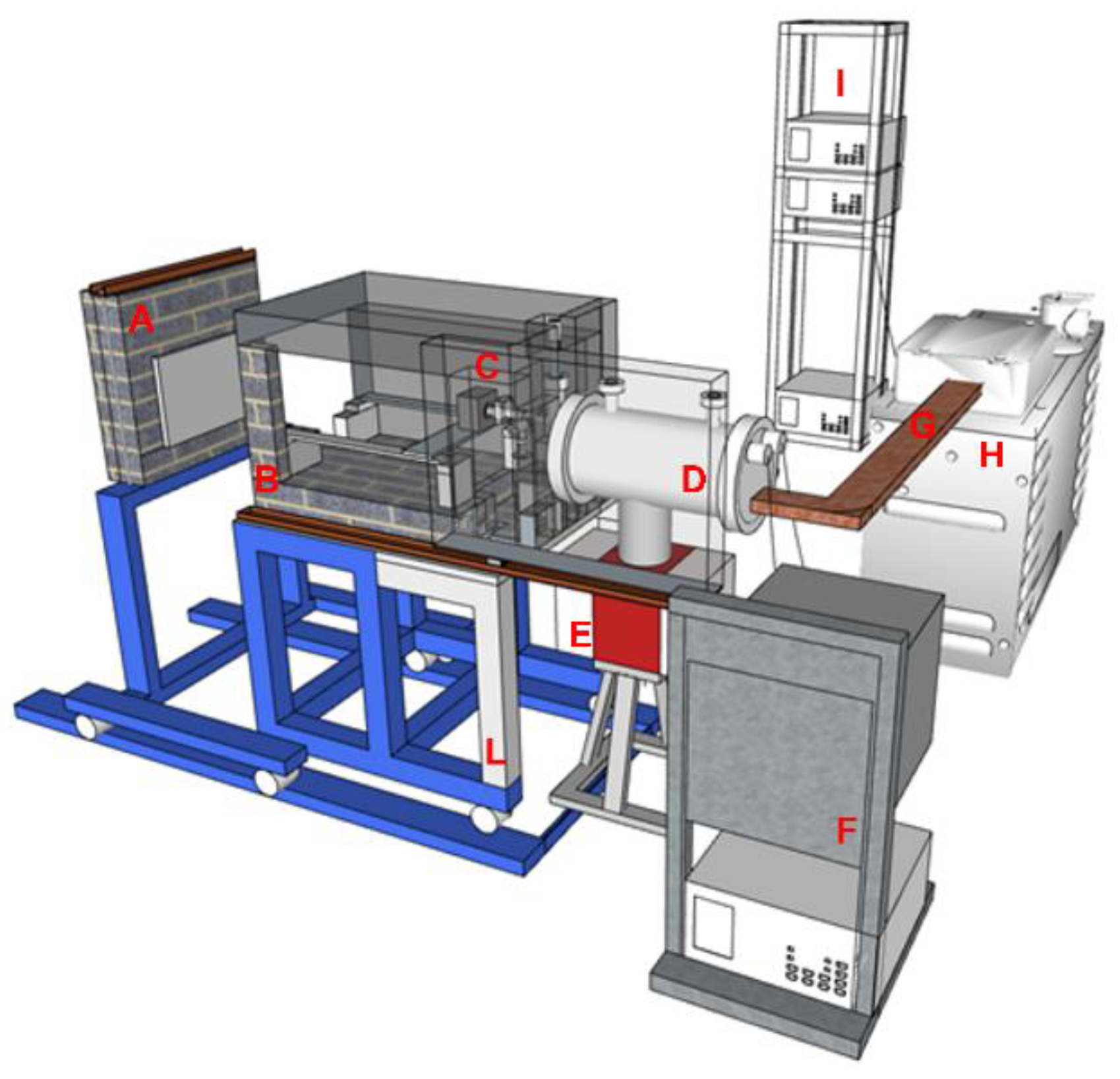
Figure 1.
Representation of the design scheme of the REX machine made entirely in the laboratories of the c.r. di Frascati of the entire REX irradiation facility. The drawing on the left shows the shielded irradiation chamber in which the disinfection treatment takes place, and we are able to insert the objects that are to be treated using a three-axis mover that is remotely piloted for complete 3D processes. The chamber can also accommodate cells that are specifically designed to operate in a controlled atmosphere and temperature. The measurements of the irradiation chamber (presented prototype) and of the support can be modified for the specific use of interest: this drawing refers to housings of a few tens of centimeters, but the method can also be applied in the raster scanning of large objects. The accelerator is housed in the cylindrical vacuum chamber that appears in the center of the image. It is directly connected to the power supply and microwave power generation services, and the elements of these are schematized.

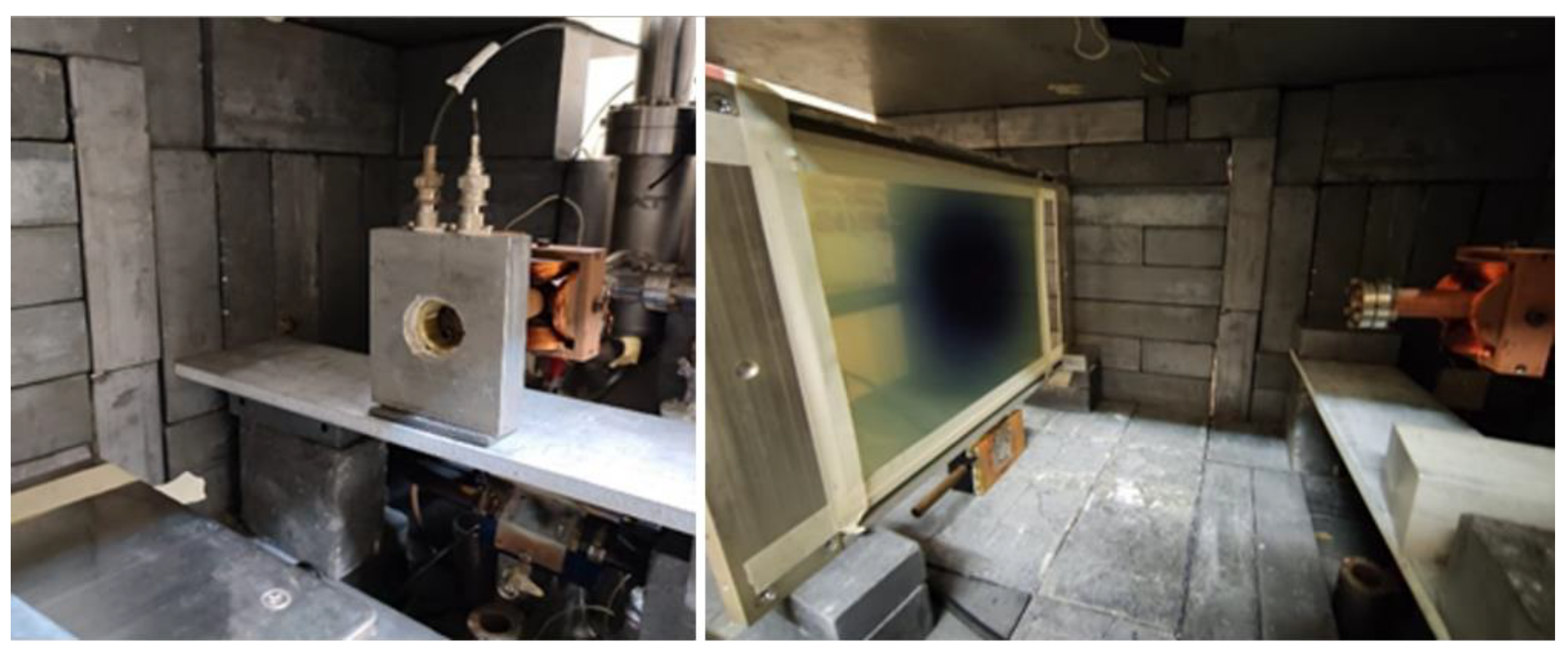
Figure 2.
View of the inside of the irradiation chamber, with the particular view of the exit window of the vacuum of the electron beams monitored by the machine system in the left image, while on the right, a photograph of a radiochromic film used for the impression is presented of the beam and the measurement of the light cone of the treatment process.


Table 1.
Technical specifications of the 5 MeV REX—linac.
| Item | Value |
|---|---|
| Power | 900 W |
| RF Frequency: | 2999 Hz |
| Pulse Duration (FWHM): | 3.5 μs |
| Electron Beam Energy: | up to 5.7 MeV |
| X-rays mean energy (from 4 mm W): | 1.25 MeV |
Table 2.
Summary of the plant specifications shown in Figure 1.
Table 2.
Summary of the plant specifications shown in Figure 1.
| A: wheeled front-closing door of the irradiation chamber of the REX facility used for radiation dumping during operations. |
| B: the irradiation chamber specially made for the operations of the REX machine, with a scalable size, equipped with a large, wheeled side door for accessing the objects, structures and devices necessary for the specific treatment. |
| C: output of the radiation beams produced by the REX machine (in particular: the output stretch of the electrons accelerated by the linac and emerging from the vacuum window and also the beta-gamma conversion system). |
| D: vacuum chamber containing the electron accelerator. |
| E: pumping system. |
| F: tower containing the power supplies necessary for the electron gun. |
| G: radiofrequency waveguide equipped with direct and reflected power measurement devices. |
| H: radio frequency generation system. |
| I: tower containing the power systems necessary for the radiofrequency formation line. |
| L: modifiable floor support structure for housing liquid phase materials which require treatments with radiation from above. |
Publisher’s Note: MDPI stays neutral with regard to jurisdictional claims in published maps and institutional affiliations. |
© 2022 by the author. Licensee MDPI, Basel, Switzerland. This article is an open access article distributed under the terms and conditions of the Creative Commons Attribution (CC BY) license (https://creativecommons.org/licenses/by/4.0/).
Share and Cite
MDPI and ACS Style
Vadrucci, M. A Machine for Ionizing Radiation Treatment of Bio-Deteriogens Infesting Artistic Objects. Quantum Beam Sci. 2022, 6, 33. https://doi.org/10.3390/qubs6040033
AMA Style
Vadrucci M. A Machine for Ionizing Radiation Treatment of Bio-Deteriogens Infesting Artistic Objects. Quantum Beam Science. 2022; 6(4):33. https://doi.org/10.3390/qubs6040033
Chicago/Turabian StyleVadrucci, Monia. 2022. "A Machine for Ionizing Radiation Treatment of Bio-Deteriogens Infesting Artistic Objects" Quantum Beam Science 6, no. 4: 33. https://doi.org/10.3390/qubs6040033