Article

Periodontal Maintenance Therapy: Efficacy of Oral Irrigator in the Home Oral Hygiene Protocol Associated with Microbiological Analysis with Phase Contrast Microscope

1
School of Medicine and Surgery, University of Milano-Bicocca, 20900 Monza, Italy
2
Independent Researcher, 20900 Monza, Italy
*
Author to whom correspondence should be addressed.
Inventions 2022, 7(4), 104; https://doi.org/10.3390/inventions7040104
Submission received: 19 October 2022 / Revised: 8 November 2022 / Accepted: 11 November 2022 / Published: 15 November 2022
(This article belongs to the Collection Feature Innovation Papers)

Abstract

:
Objective: The aim of this research was to assess how the application of an oral irrigator modifies the subgingival bacterial flora in patients undergoing periodontal maintenance therapy. We used a qualitative microbiological analysis with a phase contrast microscope that can differentiate a non-pathogenic (immobile) bacterial flora from a pathogenic (mobile). Methods: In this study, 60 patients with a diagnosis of moderate periodontitis were enrolled. They were treated with non-surgical laser-assisted causal therapy and after one month a re-evaluation was performed that established the stabilization of the periodontal pathology. They were instructed in the home oral hygiene protocol with sonic toothbrush, interdental brushes and oral irrigator, and were included in the periodontal maintenance therapy program with a phase contrast microscope control every 6 months. At T0 phase contrast–phase microscopic examinations, the existence of non-pathogenic bacterial flora was displayed in all patients. They were then randomly divided into two groups, A and B. After professional oral hygiene treatment, group B suspended the usage of oral irrigator. Patients of group A, after the professional oral hygiene treatment, were motivated to continue their oral hygiene protocol at home. After three months, the patients underwent microscopic analysis of the bacterial plaque. Results: At T1 in Group B, 90% of patients had undergone a pathogenic bacterial flora change. In group A, in which patients kept using the oral irrigator for the three months, 100% of them showed immobile plaque on phase contrast microscopic analysis. Conclusions: This research showed that oral irrigator in the practice of home hygiene protocols plays a role in the long-term maintenance of a non-pathogenic bacterial flora in periodontal patients.

1. Introduction

Maintaining a periodontal state of health over time is crucial in evaluating the success of dental treatments. Periodontal maintenance therapy (PMT) and home oral hygiene protocols are the keys to this success. We know from the literature and clinical experiences that, without meticulously organized and performed PMT, patients with a predisposition to periodontal disease are at high risk of reinfection and the progression of periodontal lesions [1]. A large number of failures after nonsurgical or surgical periodontal instrumentation are due to an incomplete cleaning of the root surfaces as well as the near-soft tissues. This is because of the existence of aggressive bacterial aggregations [2] belonging to red and orange Socransky complexes, such as Porphyromonas gingivalis, Treponema denticola, Bacteroides forsythus, Fusobacterium nucleatum and Peptostreptococcus micros. Several works in the literature showed that, with these bacteria, at a one-year follow up, even with frequent dentist appointments, the repopulation of the periodontal pockets is total. This is case whether the treatment is conducted with either with nonsurgical therapies or with regenerative surgeries [3]. Since the 1980s there has been a great deal of research on the use of lasers in periodontology. Various articles available in the literature show that decontamination by laser radiation is possible [4,5,6,7]. Lasers can be applied alone or in association with a photosensitizer. Photo-dynamic therapy (PDT) is the association of light with a chromophore and oxygenated tissues [8]. The aim of this technique is to bring singlet oxygen to all tissues affected by pathogenic bacteria. If the wavelengths used are absorbed in the chromophore used, a laser ray cannot break through very deep, and if the power density is too modest [usually PDT protocols use photobiomodulation (PBM) energy, in order to avoid any thermal damage], the decontaminating effects are not sufficient. Recent research proposed that a combination between high power settings and high frequencies of diode laser 980 nm (using really high peak power combined with a low average power, in order to reduce thermal effects) and hydrogen peroxide 10 volume 3%, or modified hydrogen peroxide 10 volumes 3% could be effective as a means of treatment [9]. This treatment protocol was named “photodynamic therapy without dye” [Oxygen high-level laser therapy (OHLLT)] in the protocol proposed by Gerard Rey in 1999 [10]. Several in vitro and in vivo studies have shown bactericidal activity of laser irradiation combined with hydrogen peroxide on numerous bacterial species [11,12,13,14,15,16]. Another important feature is the photobiomodulation properties of laser at the bone level: these promote the healing process in the post-surgical period and reduce the post-surgical pain [17,18], as well as orthodontic pain [19,20,21]. There are also other in vitro studies that have tested different materials for maintaining oral hygiene, such as MicroRepair (MicroR, a biomimetic hydroxyapatite) and pomegranate (PomeGr, a natural compound) [22], and Copper–Calcium–Hydroxide Solution [23].
The aim of this research was to assess how the application of the oral irrigator modifies the bacterial flora in patients affected by chronic periodontitis treated with OHLLT (diode laser in association with hydrogen peroxide) and following a strictly home care protocol. The diagnostic tool is the phase contrast microscope [24] which provides us with a qualitative data of the bacterial flora that integrates the standard periodontal parameters such as plaque index, bleeding index, loss of clinical attachment, etc., collected and monitored during the six-monthly recalls of professional hygiene.

2. Materials and Methods

2.1. Study Design and Patient Selection

A total of 60 patients were recruited for this study. These patients during 2016, 2017 and 2018 were diagnosed with generalized moderate periodontitis and were treated in a private dental clinic in Bergamo, Italy. The diagnosis was made with the classic periodontal clinical parameters (bleeding on probing, plaque index, sounding depth) and with qualitative microbiological analysis with a phase contrast microscope.
They were treated with laser-assisted non-surgical causal therapy. Following a periodontal re-evaluation (1 month after treatment) that determined the stabilization of the disease, the patients were placed in a periodontal maintenance therapy protocol. This consists of teaching patients a specific home hygiene protocol and performing a re-evaluation session every 6 months.

2.2. Inclusion Criteria

At the start of this study, all 60 patients had achieved stabilized periodontal disease and were undergoing periodontal maintenance therapy. They met the following inclusion criteria:
  • age from 35 to 65;
  • no systemic diseases;
  • clinical attachment loss of less than 5 mm in at least two non-near teeth;
  • no dental mobility;
  • bleeding index under 30%;
  • plaque index under 30%.
Below is a detailed analysis of the diagnostic-therapeutic path that led to the condition of stability of the pathology and the start of periodontal maintenance therapy (starting point of this study).

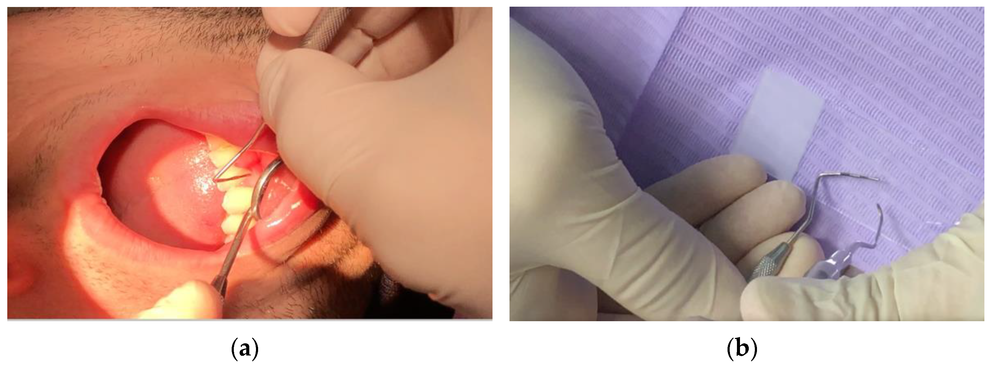
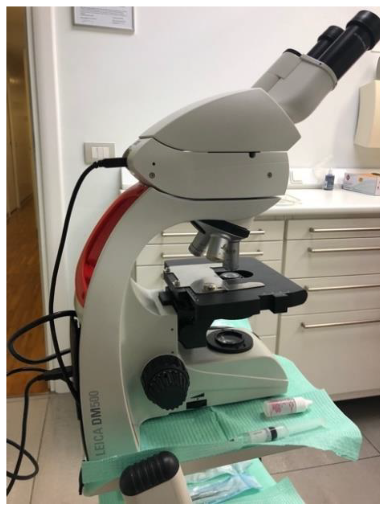
2.3. Phase Contrast Microscope: Qualitative Microbiological Analysis of Bacterial Plaque for Periodontal Diagnosis and Re-Evaluation

For periodontal diagnosis and each periodontal maintenance therapy (PMT) session the authors suggested an evaluation of subgingival plaque sample with analysis by phase contrast microscopy (DM500, Leica, Wetzlar, Germany). (Figure 1 and Figure 2)
The subgingival plaque is taken with a periodontal curette inserted in the gingival sulci in the interdental spaces between the molars and/or in the most represented periodontal pockets, where bacterial plaque is more likely to be present. The plaque is deposited on a slide and irrigated with a drop of physiological solution. A counterslide is then affixed to the slide. Finally, a drop of oil is placed on top of the slide to concentrate the light of the microscope.
A 40× objective is recommended for an eyepiece of 15 (600×).
Thanks to a camera integrated into the microscope, it is possible to view the bacterial plaque samples on the computer and take screenshots (Figure 3).
In the preparation, it is possible to visualize which bacteria are present, as well as the number and structures of epithelial cells and polymorphonuclear cells found there. It seems simple to distinguish between:
-
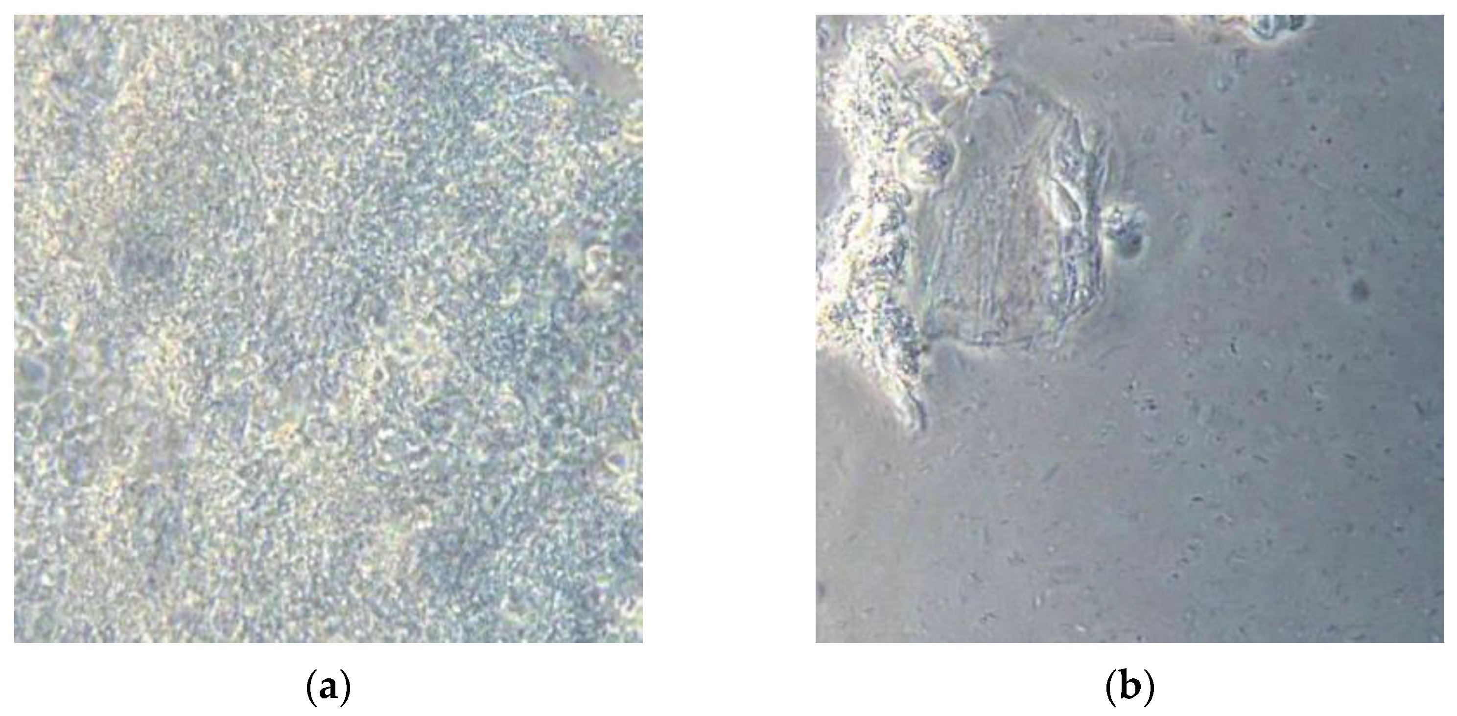
Non-pathogenic bacterial flora: immobile, similar to the aerial view of the mainland (Figure 3a);
-
Pathogenic bacterial flora: in which are visualized “Streams” or “basins” with mobile bacteria, mostly composed of spirochetes (including Treponema Denticola) and Trichomonas Tenax that flow in motion, as if dragged by the current. The most consistent similarity is that of the aerial visualization of the Norwegian fjords (Figure 3b).
After first diagnosis or periodontal re-evaluation during PMT session, because of the phase contrast microscopy results, it was decided the following protocol:
-
Non-pathogenic bacterial flora (static flora, Gram-positive bacteria): usual periodontal maintenance therapy with appointments every 6 months;
-
Pathogenic bacterial flora (spirochete, moving flora): immediate treatment with supra-gingival and sub-gingival ultrasonic instrumentation, air flow with bicarbonate powder, and a single-stage session of photodynamic therapy without dye (OHLLT) in the entire mouth. This protocol has also been applied without signs of inflammation.

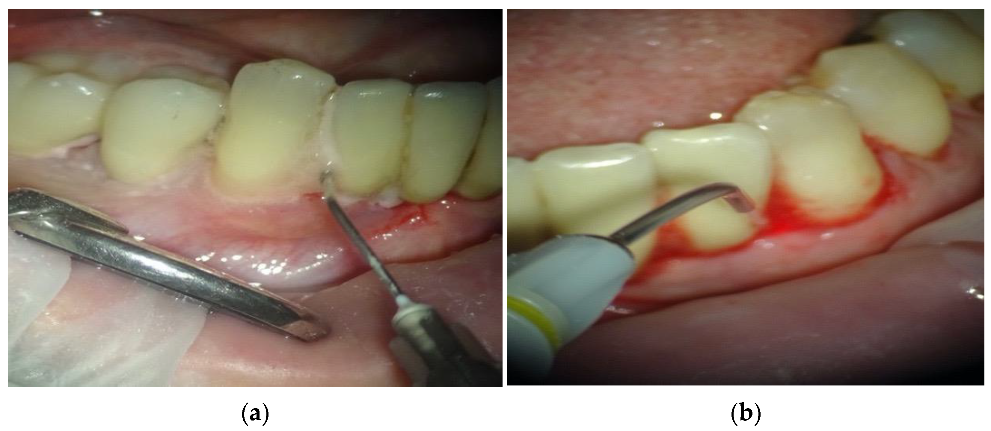
2.4. Laser-Assisted Non-Surgical Causal Therapy: OHLLT

The single-stage session of photodynamic therapy without dye consists of (Figure 4):
  • Irrigation of periodontal pockets with Sioxyl solution (derivative of hydrogen peroxide);
  • Aspiration of Sioxyl solution emerging from the gingival sulcus and leaving the remaining solution inside the pocket for 2 min;
  • Introduction of the HF Diode Laser 980 nm, Fiber 400 microns (Wiser Doctor Smile, Lambda, Vicenza, Italy) within the pocket and reaching the bottom;
  • Radiation of subgingival tissues with a movement back and forth using the dedicated program, 60 s for each side (2.5 W peak power, high frequency, 10 KHz, power average 0.5 W, Fluency 25,000/cm2).
At 1 month, a follow-up is conducted with the phase contrast microscope:
-
Non-pathogenic bacterial flora: Patients begin periodontal maintenance therapy with 6-month controls;
-
Pathogenic bacterial flora: It is recommended re-motivation to home oral hygiene, and a single-stage session of laser-assisted PDT without dye.

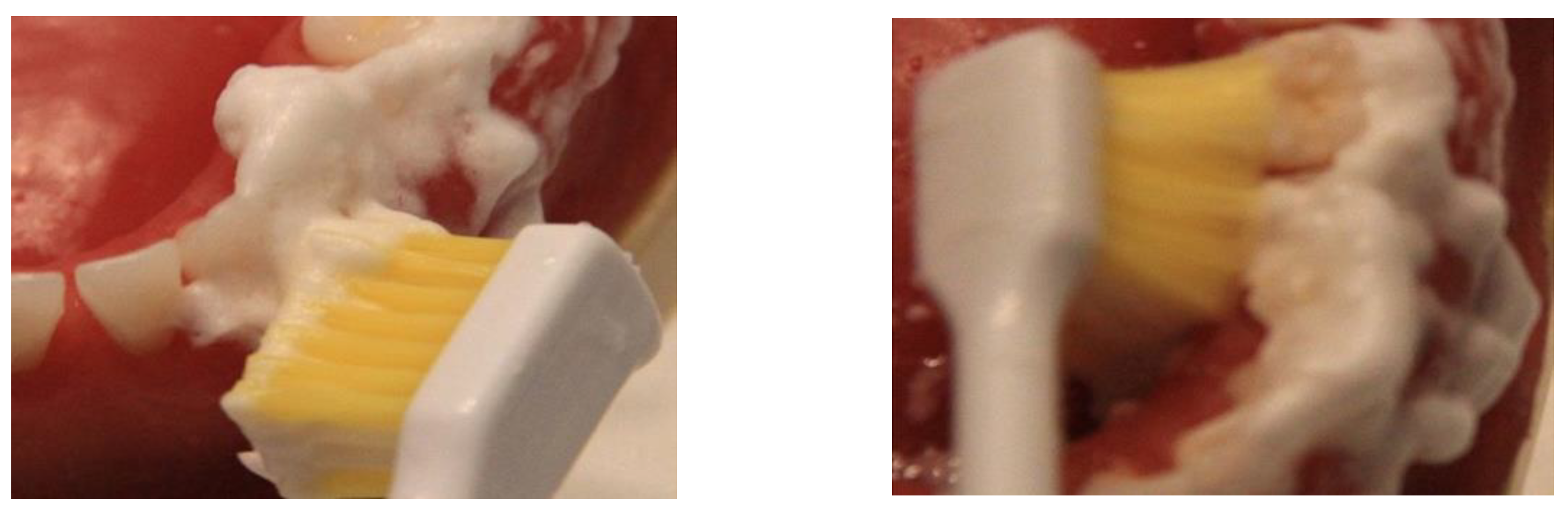
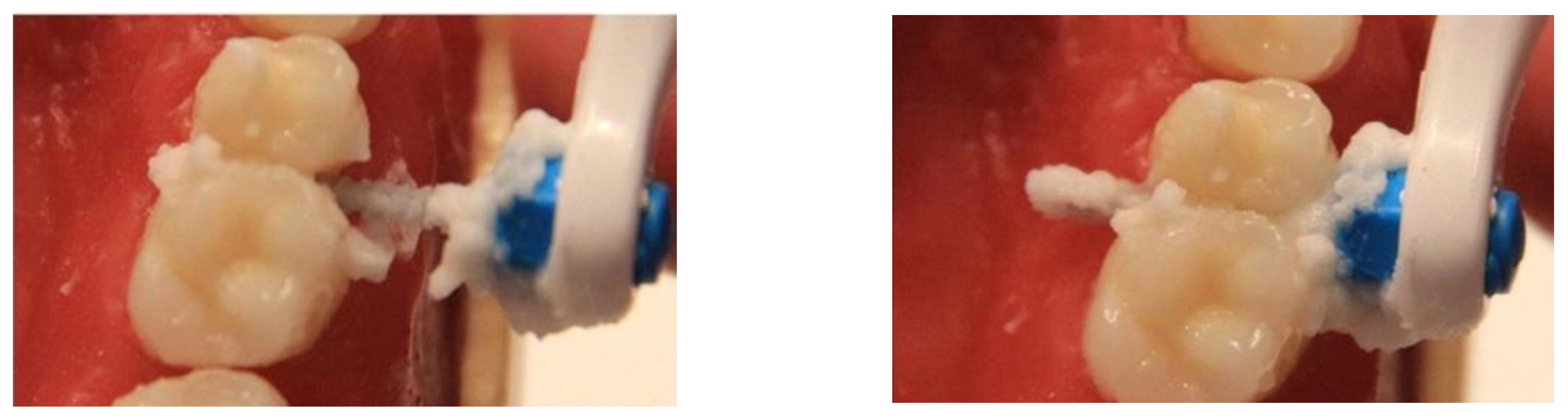
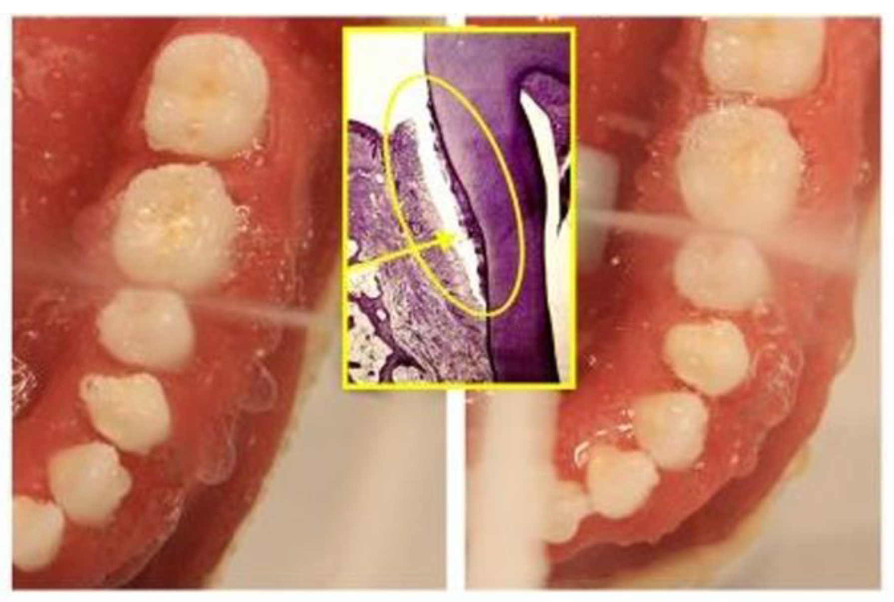
2.5. Modified Home Oral Hygiene Protocol: Sonic Toothbrush, Interdental Brushes, Oral Irrigator

To each patient, after the diagnosis of periodontal disease, before and after treatment, and during all periodontal maintenance therapy, the authors suggested the following maintenance home hygiene protocol: Sonic brush with vertical movement (Broxo OraBrush, Santé Parodonte, Geneva, Switzeland), interdental brushes, and oral irrigators (Broxo OraJets, Santé Parodonte, Geneva, Switzeland) at least two times every day (Figure 5, Figure 6 and Figure 7).

2.6. Methods for a Study Aimed at Demonstrating the Efficacy of the Oral Irrigator in Modifying the Subgingival Bacterial Flora

In summary, all 60 patients enrolled in this study received a diagnosis of moderate periodontitis, were treated with non-surgical laser-assisted causal therapy. After one month, a re-evaluation was performed that established the stabilization of the periodontal pathology (non-pathogenic bacterial plaque). Patients were instructed in the previously described home oral hygiene protocol and included in the periodontal maintenance therapy program with phase contrast microscope control every 6 months.
At time T0, all these patients were harvested from subgingival plaque with qualitative analysis under a phase contrast microscope. They were evaluated by the same operator, as explained in Section 2.1. Microscopic analysis showed non-pathogenic bacterial flora for each sample (T0). They all underwent a routine professional oral hygiene session.
All patients were then divided into two groups at random, A and B. After a professional oral hygiene session, group B was asked to suspend the use of oral irrigator and to continue the home hygiene protocol using only the sonic toothbrush and interdental brushes. Group A after the professional oral hygiene session was told to continue the complete protocol at home (sonic toothbrush, interdental brushes, oral irrigator).
Three months later, the patients once again underwent the bacterial plaque analysis (T1). At T0 and T1, the classic clinical periodontal indices on the whole mouth were also examined: bleeding on probing, plaque index, probing depth.

2.7. Statistical Analysis

The data were obtained and accessed from the Statistical Package for Social Sciences (SPSS, Chicago, IL, USA). Periodontal index data (BoP, P.I., P.D.) between patients in group A and group B at times T0 and T1 were compared with the chi-square statistical test. The threshold for statistical significance was set at p < 0.001.

3. Results

Patients belonging to Group B (Table 1), who did not use the oral irrigator for three months, following a protocol at home that included only the use of a sonic toothbrush with vertical movement and interdental brushes, saw change in pathogenic bacterial flora in 90% of cases. For instance, on examination of a pathogenic bacterial flora of a collection of plaque, 27 out of 30 patients had an active plaque in the phase contrast microscopic analysis. 100% of patients belonging to group A, who for the three months of the study kept using the standard protocol with sonic toothbrush, interdental brushes and oral irrigator, did not have mobile plaque on phase contrast microscopic evaluation. All 30 patients still had compatible flora after 3 months.
Patients at the time of recruitment, already using the complete home protocol, had a plaque index of less than 30% (Table 2). At the time of re-evaluation, group B showed a slight worsening of the plaque index. Only 7 out of 30 patients had a plaque index greater than 30%. Within group A, on the other hand, there was no significant worsening of the plaque index. In fact, the control group maintained a plaque index of below 30% in the totality of the sample.
Table 3 and Table 4 show the difference between periodontal and microbial parameters of group A and B at T0 and T1. Statistically significant differences were found between bleeding on probing and plaque index values between groups A and B at time T1.

4. Discussion

The phase contrast microscope uses two principles of geometry (wavelength and amplitude) to create an image of the illuminated cells [24,25]. Methodologically, the next aspects are important as they strongly influence the result of the analysis: sample contamination, sampling technique and sample preparation. The reproducibility of the above-mentioned techniques is high when a large number of parameters are kept constant. Sample analysis gives us some clinically relevant information [24,26]. Direct examination of the sub-gingival dental plaque under a phase contrast microscope allows you to view the bacterial morphotypes present in the sample and to immediately obtain information on microbial diversity even in the absence of inflammatory clinical signs. It also allows the evaluation of the microbial density and the presence of motile bacteria, associated or not with active periodontal lesions.
The effectiveness of phase contrast microscopy in order to provide a qualitative data of the bacterial flora that integrates the standard periodontal parameters such as plaque index, bleeding index, loss of clinical attachment, etc., was collected and monitored during the six-monthly recalls of professional hygiene. Quirynen [27] based the results of his research and his convictions precisely on the microbiological evaluation using a phase contrast microscope. Bollen and Quirynen [28] published a pilot study in 1996 to examine the long-term microbiological effects of a “full mouth” disinfection, controlled with phase contrast microscopy. Yeom [29] in 1997 used the phase contrast microscope to evaluate the clinical and microbiological effects of the subgingival deposition of bioabsorbable microcapsules, loaded at 10% of minocycline (MC) in 15 adult patients with periodontitis. Quirynen [30] used bacteriological tests with a phase contrast microscope to evaluate the bacterial plaque around the implant surfaces. In 2012, Acharya [31] compared the efficiency of three different motivational techniques at maintaining good oral hygiene during fixed appliance orthodontic treatment. Phase contrast microscopy together with the conventional plaque detection method and the demonstration of the horizontal brushing washing method have a lasting effect on the patient. This reduces the need for frequent strengthening sessions of plaque control programs compared to motivational tests and conventional plaque control measures.
The analysis of the literature led us to believe that the home hygiene protocols in patients with pathogenic bacterial flora and in those on periodontal support therapy can preferably include the use of a sonic electric toothbrush [32,33,34]. The shockwave generated by the movement of the bristles, combines with the oral irrigator [35,36,37] which supports the solution for damaging subgingival plaque potential by means of the disorganization of the salivary biofilm. This, which appears to be the cause of the spread in the gingival sulcus of pathogenic microorganisms, is carried by the salivary biofilm [2,3,38].
The present study showed that the use of a specific home oral hygiene protocol is essential in periodontal maintenance therapy. In fact, oral irrigators are important in order to maintain a non-pathogenic bacterial flora in subgingival plaque, as showed in the results of this research. Within 3 months, if patients in PMT avoid using oral irrigators, periodontal conditions deteriorate, an increase in pathogenic bacterial flora is detected with phase contrast microscopy and worst periodontal parameters emerge (PI, BOP, PD).
It is essential that the patient is always monitored and continuously remotivated to maintain excellent levels of oral hygiene, even if the proposed protocol can seem somewhat demanding both for the patient and for the dental hygienist. The scientific literature proves that a good feasible method to stop the onset and advancement of bacteria, avoid relapses and stabilize the results obtained in patients predisposed to periodontal disease is to keep bacteria below the pathological limits with an accurate home protocol [13,14].
The scientific assumptions that can lead to the development of a new home protocol for patients in periodontal maintenance therapy are based on the knowledge of bacterial plaque evaluation techniques through investigations using a phase contrast microscope. With this instrument, it is possible to perform a qualitative microbiological analysis and to classify two specific types of bacterial flora: non-pathogenic and pathogenic.

5. Conclusions

The present study shows how phase contrast microscopy can detect sub-gingival plaque modifications early due to poor home oral hygiene protocol application by patients, and how the use of the oral irrigator is decisive in the long-term maintenance of a non-pathogenic bacterial flora in periodontal patients. In fact, it is clear that, although it is possible to maintain an acceptable plaque index only with a sonic toothbrush and interdental brushes, oral irrigators have allowed patients to maintain non-pathogenic bacterial flora in the oral cavities of patients who have not stopped using it.

Author Contributions

Conceptualization, G.C.; methodology, G.C.; validation, P.C.; formal analysis, P.C.; investigation, P.E.; Data curation, P.C.; writing original draft preparation, G.C.; writing—review and editing, P.C.; visualization, A.A.B.; supervision, G.C.; project administration, G.C. All authors have read and agreed to the published version of the manuscript.

Funding

This research received no external funding.

Institutional Review Board Statement

The study was conducted according to the guidelines of the Ethics Committee of the School of Medicine and Surgery at the Milano-Bicocca University (protocol n. 11/17) and it was executed in conformity with the Declaration of Helsinki.

Informed Consent Statement

Informed consent was obtained from all subjects involved in the study.

Data Availability Statement

Not applicable.

Conflicts of Interest

The authors declare no conflict of interest.

References

  1. Manresa, C.; Sanz-Miralles, E.C.; Twigg, J.; Bravo, M. Supportive periodontal therapy (SPT) for maintaining the dentition in adults treated for periodontitis. Cochrane Database Syst. Rev. 2018, 1, CD009376. [Google Scholar] [CrossRef] [PubMed]
  2. Rhemrev, G.E.; Timmerman, M.F.; Veldkamp, I.; van Winkelhoff, A.J.; van der Velden, U. Immediate effect of instrumen-tation on the subgingival microflora in deep inflamed pockets under strict plaque control. J. Clin. Periodontol. 2006, 33, 42–48. [Google Scholar] [CrossRef] [PubMed]
  3. Heitz-Mayfield, L.; Tonetti, M.S.; Cortellini, P.; Lang, N.P. Microbial colonization patterns predict the outcomes of surgi-cal treatment of intrabony defects. J. Clin. Periodontol. 2006, 33, 62–68. [Google Scholar] [CrossRef] [PubMed]
  4. Gursoy, H.; Ozcakir-Tomruk, C.; Tanalp, J.; Yilmaz, S. Photodynamic therapy in dentistry: A literature review. Clin. Oral Investig. 2013, 17, 1113–1125. [Google Scholar] [CrossRef]
  5. Ando, Y.; Aoki, A.; Watanabe, H.; Ishikawa, I. Bactericidal Effect of Erbium YAG Laser on Periodontopathic Bacteria. Lasers Surg. Med. 1996, 19, 190–200. [Google Scholar] [CrossRef]
  6. Moritz, A.; Gutnetcht, N.; Schoop, U.; Gobarkhay, K.; Doerbudak, O.; Speer, W. Irradiation of infected root canals with a diode laser in vivo: Results of microbiological examinations lasers in surgery and medecine. Lasers Surg. Med. 1997, 21, 221–226. [Google Scholar] [CrossRef]
  7. Nammour, S.; El Mobadder, M.; Maalouf, E.; Namour, M.; Namour, A.; Rey, G.; Matamba, P.; Matys, J.; Zeinoun, T.; Grzech-Leśniak, K. Clinical Evaluation of Diode (980 nm) Laser-Assisted Nonsurgical Periodontal Pocket Therapy: A Randomized Comparative Clinical Trial and Bacteriological Study. Photobiomodul. Photomed. Laser Surg. 2021, 39, 10–22. [Google Scholar] [CrossRef] [PubMed]
  8. Azarpazhooh, A.; Shah, P.S.; Tenenbaum, H.C.; Berg, M.B.G. Photodynamic therapy for periodontitis: A systematic review and meta-analysis. J. Periodontol. 2010, 81, 4–14. [Google Scholar] [CrossRef]
  9. Caccianiga, G.; Rey, G.; Baldoni, M.; Paiusco, A. Clinical, radiographic and microbiological evaluation of high level laser therapy, a new photodynamic therapy protocol, in peri-implantitis treatment; a pilot experience. Biomed. Res. Int. 2016, 2016, 6321906. [Google Scholar] [CrossRef] [Green Version]
  10. Rey, G. L’apport du laser dans le traitement des poches paradontales. Implantodontie 2000, 38, 37–44. [Google Scholar]
  11. Caccianiga, G.; Baldoni, M.; Ghisalberti, C.A.; Paiusco, A. A Preliminary In Vitro Study on the Efficacy of High-Power Photodynamic Therapy (HLLT): Comparison between Pulsed Diode Lasers and Superpulsed Diode Lasers and Impact of Hydrogen Peroxide with Controlled Stabilization. Biomed. Res. Int. 2016, 2016, 1386158. [Google Scholar] [CrossRef]
  12. Caccianiga, G.; Cambini, A.; Donzelli, E.; Baldoni, M.; Rey, G.; Paiusco, A. Effects of laser biostimulation on the epithelial tissue for keratinized layer differentiation: An in vitro study. J. Biol. Regul. Homeost. Agents 2016, 30 (Suppl. 1), 99–105. [Google Scholar]
  13. Caccianiga, G.; Rey, G.; Baldoni, M.; Caccianiga, P.; Baldoni, A.; Ceraulo, S. Periodontal Decontamination Induced by Light and Not by Heat: Comparison between Oxygen High Level Laser Therapy (OHLLT) and LANAP. Appl. Sci. 2021, 11, 4629. [Google Scholar] [CrossRef]
  14. Caccianiga, G.; Rey, G.; Caccianiga, P.; Leonida, A.; Baldoni, M.; Baldoni, A.; Ceraulo, S. Rough Dental Implant Surfaces and Peri-Implantitis: Role of Phase-Contrast Microscopy, Laser Protocols, and Modified Home Oral Hygiene in Maintenance. A 10-Year Retrospective Study. Appl. Sci. 2021, 11, 4985. [Google Scholar] [CrossRef]
  15. Caccianiga, G.; Rey, G.; Caccianiga, P.; Leonida, A.; Baldoni, M.; Baldoni, A.; Ceraulo, S. Peri-Implantitis Management: Surgical versus Non-Surgical Approach Using Photodynamic Therapy Combined with Hydrogen Peroxide (OHLLT—Oxygen High Level Laser Therapy). A Retrospective Controlled Study. Appl. Sci. 2021, 11, 5073. [Google Scholar] [CrossRef]
  16. Caccianiga, G.; Rey, G.; Caccianiga, P.; Leonida, A.; Baldoni, M.; Baldoni, A.; Ceraulo, S. Laser Management of Peri-Implantitis: A Comparison between Photodynamic Therapy Combined with Hydrogen Peroxide (OHLLT) and OHLLT + Er:YAG Laser. A Retrospective Controlled Study. Appl. Sci. 2021, 11, 6771. [Google Scholar] [CrossRef]
  17. Caccianiga, G.; Rey, G.; Baldoni, M.; Caccianiga, P.; Porcaro, G.; Baldoni, A.; Ceraulo, S. Laser Decontamination and LED Photobiomodulation Promote Bone Regeneration and Wound Healing by Secondary Intention, in Alveolar Ridge Preservation-Clinical and Radiographic Evaluation: A Pilot Experience. Photobiomodul. Photomed. Laser Surg. 2022, 40, 343–354. [Google Scholar] [CrossRef]
  18. Caccianiga, G.; Ferri, L.; Baldoni, M.; Bader, A.A.; Caccianiga, P. Magnetic Mallet and Laser for a Minimally Invasive Implantology: A Full Arch Case Report. Appl. Sci. 2022, 12, 9995. [Google Scholar] [CrossRef]
  19. Caccianiga, G.; Caccianiga, P.; Baldoni, M.; Lo Giudice, A.; Perillo, L.; Moretti, N.; Ceraulo, S. Pain Reduction during Rapid Palatal Expansion Due to LED Photobiomodulation Irradiation: A Randomized Clinical Trial. Life 2022, 12, 37. [Google Scholar] [CrossRef]
  20. Lo Giudice, A.; Nucera, R.; Leonardi, R.; Paiusco, A.; Baldoni, M.; Caccianiga, G. A Comparative Assessment of the Efficiency of Orthodontic Treatment with and Without Photobiomodulation During Mandibular Decrowding in Young Subjects: A Single-Center, Single-Blind Randomized Controlled Trial. Photobiomodulation Photomed. Laser Surg. 2020, 38, 272–279. [Google Scholar] [CrossRef] [Green Version]
  21. Caccianiga, G.; Lo Giudice, A.; Paiusco, A.; Portelli, M.; Militi, A.; Baldoni, M.; Nucera, R. Maxillary Orthodontic Expansion Assisted by Unilateral Alveolar Corticotomy and Low-Level Laser Therapy: A Novel Approach for Correction of a Posterior Unilateral Cross-Bite in Adults. J. Lasers Med. Sci. 2019, 10, 225–229. [Google Scholar] [CrossRef]
  22. Odorici, A.; Colombari, B.; Bellini, P.; Meto, A.; Venturelli, I.; Blasi, E. Novel Options to Counteract Oral Biofilm Formation: In Vitro Evidence. Int. J. Environ. Res. Public Health 2022, 19, 8056. [Google Scholar] [CrossRef]
  23. Meto, A.; Colombari, B.; Castagnoli, A.; Sarti, M.; Denti, L.; Blasi, E. Efficacy of a Copper-Calcium-Hydroxide Solution in Reducing Microbial Plaque on Orthodontic Clear Aligners: A Case Report. Eur. J. Dent. 2019, 13, 478–484. [Google Scholar] [CrossRef] [Green Version]
  24. Callens, A. Darkfield or phase contrast microscopy. Usefulness in periodontology. Ned. Tijdschr. Tandheelkd. 1992, 99, 381–384. [Google Scholar]
  25. Rose, G.G. Phase-Contrast Microscopy in living cells. Microsc. Soc. 1964, 83, 97–114. [Google Scholar] [CrossRef]
  26. Leggott, P.J.; Anderson, A.W.; Punwani, I.; Sabet, T.; Murphy, R.; Crawford, J. Phase contrast microscopy of microbial aggregates in the gingival sulcus of Macaca mulatta. Subgingival plaque bacteria in macaca mulatta. J. Clin. Periodontol. 1983, 10, 412–421. [Google Scholar] [CrossRef]
  27. Quirynen, M.; Mongardini, C.; de Soete, M.; Pauwels, M.; Coucke, W.; van Eldere, J.; van Steenberghe, D. The role of chlorhexi-dine in the one-stage full-mouth disinfection treatment of patients with advanced adult periodontitis. Long-term clinical and microbiological observations. J. Clin. Periodontol. 2000, 27, 578–589. [Google Scholar] [CrossRef]
  28. Bollen, C.M.; Vandekerckhove, B.N.; Papaioannou, W.; Van Eldere, J.; Quirynen, M. Full-versus partial-mouth disinfection in the treatment of periodontal infections. A pilot study: Long-term microbiological observations. J. Clin. Periodontol. 1996, 23, 960–970. [Google Scholar] [CrossRef]
  29. Yeom, H.R.; Park, Y.J.; Lee, S.J.; Rhyu, I.C.; Chung, C.P.; Nisengard, R.J. Clinical and microbiological effects of minocycline-loaded microcapsules in adult periodontitis. J. Periodontol. 1997, 68, 1102–1109. [Google Scholar] [CrossRef]
  30. Quirynen, M.; Bollen, C.M.; Papaioannou, W.; Van Eldere, J.; van Steenberghe, D. The influence of titanium abutment surface roughness on plaque accumulation and gingivitis: Short-term observations. Int. J. Oral Maxillofac. Implant. 1996, 11, 169–178. [Google Scholar]
  31. Acharya, S.; Goyal, A.; Utreja, A.K.; Mohanty, U. Effect of three different motivational techniques on oral hygiene and gingi-val health of patients undergoing multibracketed orthodontics. Angle Orthod. 2011, 81, 884–888. [Google Scholar] [CrossRef] [PubMed] [Green Version]
  32. Gallie, A. Home use of interdental cleaning devices and toothbrushing and their role in disease prevention. Evid. Based Dent. 2019, 20, 103–104. [Google Scholar] [CrossRef] [PubMed]
  33. Goyal, C.R.; Lyle, D.M.; Qaqish, J.G.; Schuller, R. The addition of a water flosser to power tooth brushing: Effect on bleeding, gingivitis, and plaque. J. Clin. Dent. 2012, 23, 57–63. [Google Scholar]
  34. Tawakoli, P.N.; Sauer, B.; Becker, K.; Buchalla, W.; Attin, T. Interproximal biofilm removal by intervallic use of a sonic tooth brush compared to an oral irrigation system. BMC Oral Health 2015, 15, 91. [Google Scholar] [CrossRef] [Green Version]
  35. Eakle, W.S.; Ford, C.; Boyd, R.L. Depth of penetration in periodontal pockets with oral irrigation. J. Clin. Periodontol. 1986, 13, 39–44. [Google Scholar] [CrossRef]
  36. Cutler, C.W.; Stanford, T.W.; Abraham, C.; Cederberg, R.A.; Boardman, T.J.; Ross, C. Clinical benefits of oral irrigation for per-iodontitis are related to reduction of pro-inflammatory cytokine levels and plaque. J. Clin. Periodontol. 2000, 27, 134–143. [Google Scholar] [CrossRef]
  37. Costa, F.O.; Costa, A.A.; Cota, L.O.M. The use of interdental brushes or oral irrigators as adjuvants to conventional oral hygiene associated with recurrence of periodontitis in periodontal maintenance therapy: A 6-year prospective study. J. Periodontol. 2020, 91, 26–36. [Google Scholar] [CrossRef] [Green Version]
  38. Quirynen, M.; Teughels, W.; van Steenberghe, D. Impact of antiseptics on one-stage, full-mouth disinfection. J. Clin. Periodontol. 2006, 33, 49–52. [Google Scholar] [CrossRef]
Figure 1. (a) Removal of supra and subgingival plaque; (b) placement of the plaque on the slide.
Inventions 07 00104 g001
Figure 2. Contrast phase microscope in periodontal office.
Inventions 07 00104 g002
Figure 3. Subgingival plaque images observed under a phase contrast microscope at T1: (a) Non-pathogenic bacterial flora (patient belonging to Group A); (b) Pathogenic bacterial flora (patient belonging to Group B).
Inventions 07 00104 g003
Figure 4. Photodynamic therapy without dye (a) Periodontal pockets irrigation with sioxyl solution; (b) Radiation with diode laser.
Inventions 07 00104 g004
Figure 5. Sonic toothbrush in action.
Inventions 07 00104 g005
Figure 6. Interdental brush in action.
Inventions 07 00104 g006
Figure 7. Oral irrigator in action, in order to remove sub-gingival biofilm.
Inventions 07 00104 g007
Table 1. Patients with non-pathogenic flora at first (T0) and second (T1) evaluation.
Compatible FloraT0T1
Group A3030
Group B303
Table 2. Patients with plaque index less than 30% at first (T0) and second (T1) evaluation.
Plaque Index < 30%T0T1
Group A3030
Group B3023
Table 3. Periodontal and microbial parameters among the A group and B group before suppression of oral irrigator in group B. (T0).
Group A (n = 30)Group B (n = 30)p Value
Mean ± SdMean ± Sd
BoP0.14 ± 0.090.11 ± 0.10<0.018
P.I.0.23 ± 0.120.21 ± 0.110.006
P.D.2.24 ± 0.232.25 ± 0.210.267
BoP: bleeding on probing; P.I.: plaque index; and P.D.: periodontal depth. * p value < 0.001 is statistically significant.
Table 4. Periodontal and microbial parameters among the A group and B group 3 months after suppression of oral irrigator in group B. (T1).
Group A (n = 30)Group B (n = 30)p Value
Mean ± SdMean ± Sd
BoP0.12 ± 0.051.12 ± 0.21<0.0005 *
P.I.0.12 ± 0.150.93 ± 0.21<0.001 *
P.D.2.16 ± 0.182.41 ± 0.240.135
BoP: bleeding on probing; P.I.: plaque index; and P.D.: periodontal depth. * p value < 0.001 is statistically significant.
Publisher’s Note: MDPI stays neutral with regard to jurisdictional claims in published maps and institutional affiliations.

