Article

High-Temperature Stress Induces Autophagy in Rainbow Trout Skeletal Muscle

by
Alfredo Molina
1,2,3,
Phillip Dettleff
4,
Valentina Valenzuela-Muñoz
2,5,
Cristian Gallardo-Escarate
2,5 and
Juan Antonio Valdés
1,2,3,*
1
Laboratorio de Biotecnología Molecular, Facultad de Ciencias de la Vida, Universidad Andres Bello, Santiago 8370035, Chile
2
Interdisciplinary Center for Aquaculture Research (INCAR), Universidad de Concepción, Concepción 4030000, Chile
3
Centro de Investigación Marina Quintay (CIMARQ), Universidad Andres Bello, Quintay 2340000, Chile
4
Escuela de Medicina Veterinaria, Facultad de Agronomía e Ingeniería Forestal, Facultad de Ciencias Biológicas y Facultad de Medicina, Pontificia Universidad Católica de Chile, Santiago 7820436, Chile
5
Laboratory of Biotechnology and Aquatic Genomics, Department of Oceanography, University of Concepción, Concepción 4030000, Chile
*
Author to whom correspondence should be addressed.
Fishes 2023, 8(6), 303; https://doi.org/10.3390/fishes8060303
Submission received: 2 May 2023 / Revised: 28 May 2023 / Accepted: 2 June 2023 / Published: 6 June 2023
(This article belongs to the Special Issue Effect of Climate Change on Salmonid Fishes in Rivers)

Abstract

:
Ectothermic animals, such as teleosts, have increasingly been exposed to stressful high-temperature events due to global warming. Currently, the effects of thermal stress on skeletal muscle, a key tissue for fish growth, are unknown. This study examined the impact of high-temperature stress on the skeletal muscle transcriptome of rainbow trout (Oncorhynchus mykiss) in control (15 °C) and high-temperature (20 °C) conditions. Additionally, we examined the plasmatic levels of cortisol, glucose, and creatine kinase activity, and examined oxidative damage and autophagy activation in skeletal muscle. High-temperature stress induced significant increases in cortisol and glucose plasmatic levels. Nevertheless, no changes were observed in creatine kinase activity in plasma and skeletal muscle oxidation. Skeletal muscle RNA was isolated and sequenced using the HiSeq Illumina platform. A total of 383,796,290 reads were mapped onto the reference rainbow trout genome. The transcriptomic analysis showed that 293 genes were upregulated in the high-temperature group, mainly associated with autophagosome assembly, amino acid transport, and the glutamine metabolic process. On the other hand, 119 genes were downregulated in the high-temperature group, mainly associated with digestion, proteolysis, and the muscle contraction process. In addition, RT-qPCR of differentially expressed representative genes and Western blot analysis of LC3-II/LC3-I levels confirmed skeletal muscle autophagy induced by high temperature. This study sheds light on intriguing facets of the adaptive response of rainbow trout skeletal muscle to high-temperature stress and provides significant insights into the physiology of autophagy in teleosts.
Key Contribution: This study evaluates the effects of high-temperature stress on the skeletal muscle transcriptome of rainbow trout (O. mykiss) and contributes to a better understanding of the potential role of autophagy as a negative regulator of skeletal muscle atrophy.

Graphical Abstract

1. Introduction

Global warming refers to the ongoing rise in the average global temperature of the Earth [1]. This phenomenon is exerting an impact on animal populations across the globe, primarily through chronic temperature increases and a heightened incidence of heatwaves [2]. Increases in atmospheric temperatures are reflected in higher temperatures of marine and freshwater habitats, home to a great diversity of living organisms [3]. It is projected that, by the end of this century, the mean temperature of the ocean will have risen by 1–4 °C, which will exert negative effects on the physiology of aquatic organisms [3]. Additionally, changes in ocean temperatures will influence the intensity and frequency of phenomena on the Pacific coast known as El Niño–Southern Oscillation (ENSO) [4]. Given that fishes are ectothermic organisms, and their body temperature is equivalent to that of the surrounding water environment, teleosts are particularly vulnerable to the effects of global warming [5].
The physiological mechanisms that are essential for mounting an adequate response to temperature stress entail the activation of a well-coordinated network of neuroendocrine pathways, including the brain–sympathetic–chromaffin (BSC) axis and the hypothalamic–pituitary–interrenal (HPI) axis, which are responsible for the production of catecholamines and cortisol, respectively [6]. The secretion of these hormones plays a critical role in regulating energy metabolism and maintaining an organism’s homeostasis [7]. Specifically, the cortisol-mediated response in teleosts plays a fundamental role in facilitating energetic adaptation to temperature stress by enabling long-term glucose metabolism via gluconeogenesis and protein catabolism [8].
Skeletal muscle is considered to be one of the primary tissues impacted by the stress induced by rising temperatures [8]. Skeletal muscles make up approximately 60% of the body mass in teleosts, playing a crucial role in their locomotion, metabolism, and growth [9]. The growth of skeletal muscle is a multifaceted process that is coregulated by mechanisms associated with myoblast proliferation (hyperplasia), increase in muscular fiber size through protein synthesis (hypertrophy), and muscle protein degradation (atrophy) [10]. Although several studies have described the physiological and molecular effects of temperature stress in fish tissues, very few have focused on skeletal muscles [11]. In a recent study conducted on a marine teleost species, red cusk-eel (Genypterus chilensis), it was found that thermal stress leads to skeletal muscle oxidation and atrophy [12]. Additionally, RNA-Seq assays revealed that high-temperature stress induced the expression of various autophagy-associated genes in the liver of the species [13]. Autophagy is a fundamental cellular mechanism that plays an essential role in energetic catabolism and the lysosome-mediated degradation of cell components. It is a highly conserved process that allows the cell to recycle damaged or unwanted organelles and proteins [14]. In mammalian skeletal muscle, autophagy has been described as an essential mechanism for maintaining the structure and proper functioning of this tissue [15]. Skeletal muscle with an impaired autophagy process can be affected by myopathy disorder, which is related to excessive protein accumulation in the muscle cells [16]. However, very little is known about autophagy in teleost skeletal muscles and its regulation by environmental variables such as temperature [17]. In this study, we evaluated the effects of high-temperature stress on global gene expression in the skeletal muscle of rainbow trout to comprehend the impact of temperature stress on skeletal muscle and predict the influence of global warming on the adaptive capability of fish during their development. The transcriptomic information obtained here enabled us to identify potential genes and signaling pathways associated with the high-temperature stress response in rainbow trout.

2. Materials and Methods

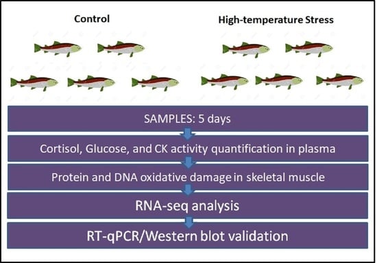
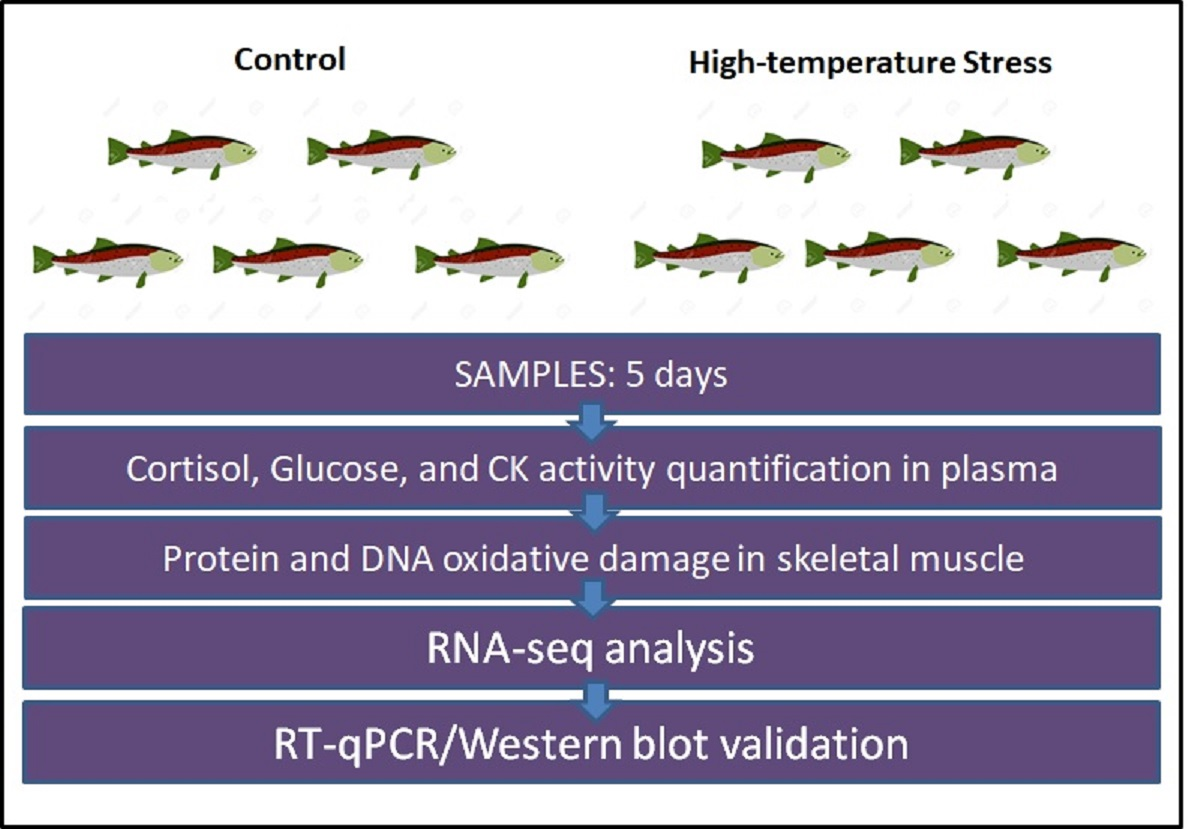
2.1. Experimental Thermal Stress Protocol

Juvenile rainbow trout (Oncorhynchus mykiss) (1 year old; 13.22 g ± 1.34; total n = 20) were obtained from Pisciculture Rio Blanco (V region, Chile). Fish were maintained under natural temperatures and light:dark photoperiod conditions (15 °C ± 1 °C and L:D 12:12), in aerated dechlorinated water, with water turnover of 0.5 L min−1 and fed daily with commercial pellets containing 45% protein, 22% lipids, 16% carbohydrates, and 17% other components. Fish were acclimatized for 1 week before the trial and exposed to a thermal stress protocol previously described [12]. Briefly, this protocol consists of increasing the temperature over 24 h at a rate of 1 °C in 5 h. During the remaining 4 days of the trial, the control temperature was maintained at 15 ± 1 °C (control group; n = 5) and the stress temperature at 20 ± 1 °C (stress group; n = 5). Biological replicates of the trials were included. On the fifth day, five individuals per group were sampled. For plasma isolation, blood samples were collected and centrifuged at 5000× g for 10 min and stored at −80 °C. Finally, sample individuals were euthanized by an overdose of anesthetic (benzocaine, 300 mg/L). Skeletal muscle was collected, frozen in liquid nitrogen, and maintained at −80 °C until further analysis.

2.2. Cortisol, Creatine Kinase Activity, and Glucose Quantification in Blood Plasma

The cortisol plasmatic concentration was quantified using the Cayman Cortisol ELISA Kit (Cayman Chemical, Ann Arbor, MI, USA; catalog number 500360). The plasmatic activity of creatine kinase (CK) was quantified using the Abcam Creatine Kinase Activity Assay Kit (Abcam, Cambridge, UK; catalog number 155901). The glucose plasmatic concentration was quantified using the Abcam Glucose Uptake Assay Kit (Abcam, Cambridge, UK; catalog number 136955). The use of these kits in rainbow trout has been previously verified [18].

2.3. DNA and Protein Oxidative Damage in Skeletal Muscle

DNA oxidative damage and protein carbonylation were determined using the commercially available kits OxiSelect Oxidative DNA Damage Quantification (catalog number STA-320) and OxiSelect Protein Carbonyl Spectrophotometric Assay (catalog number STA-310) (Cell Biolabs, San Diego, CA, USA), respectively, following the manufacturer’s instructions. For further details, see Rivas-Aravena et al. [18].

2.4. Skeletal Muscle RNA Extraction and Sequencing

Total RNA was extracted from the skeletal muscles of both the control and stress groups using the EZNA Total RNA Kit II (OMEGA Bio-Tek Inc., Norcross, GA, USA). The RNA concentration was measured using a Qubit 2.0 Fluorometer (Life Technology, Carlsbad, CA, USA), and RNA integrity was confirmed by capillary electrophoresis using a Fragment Analyzer Automated CE System (Advanced Analytical Technologies, Inc., Ankeny, IA, USA). The ratio of absorbance at 260 nm and 280 nm was used to assess the purity of RNA. Samples with RQN values of at least 9 were selected for further analysis. The construction of cDNA libraries was carried out with 1 μg of RNA using the TruSeq RNA Sample Preparation Kit v2 (Illumina, San Diego, CA, USA). Libraries were sequenced with the Hiseq technology (Illumina) at Macrogen (Seoul, Republic of Korea) using a paired-end technique (2 × 150 bp).

2.5. RNA-Seq and GO Analysis

The sequencing reads were processed to remove sequences of low quality (Q20) and those less than 30 bp in length. To detect differentially expressed genes (DEGs), the reads were mapped to the last version of rainbow trout (O. mykiss) reference genome by CLC Genomics Workbench 9.0 (Qiagen, Germantown, MD USA), using default parameters. Gene expression levels were estimated using the RPKM value (reads per kilobase per million mapped reads). Genes exhibiting a fold-change value greater than 2.0 and a false discovery rate (FDR) p-value less than 0.05 were considered as differentially expressed. The identification of DAVID GO and KEGG enrichment analysis of DEGs was previously described [19].

2.6. RNA-Seq Validation by Real-Time RT-qPCR

All qPCR assays followed MIQE guidelines [20]. Preserved skeletal muscles from each sampled fish were homogenized, and total RNA was extracted using TRIzol reagent (Invitrogen, Carlsbad, CA, USA). Isolated RNAs with A260/280 ratios between 1.9 and 2.0 were selected for further processing. Next, 1 μg of RNA from each sample was reverse transcribed into cDNA using the ImProm-II Reverse Transcription System (Promega, WI, USA). Real-time qPCR was performed using a Stratagene MX3000P qPCR system (Stratagene, La Jolla, CA, USA) following the procedure described by Rivas-Aravena et al. [18]. The list of primers used in this study is provided in Supplementary Table S1. The housekeeping genes used for normalization were β-actin (actβ) and 40S ribosomal protein S30 (fau). These genes were previously obtained by using the geNorm program, which obtained the normalization factor and subsequent relative expression levels [18].

2.7. LC3 Western Blot Analysis

To validate the effects of high-temperature stress on the induction of autophagy, we analyzed the levels of the microtubule-associated proteins 1A/1B light chain 3B (LC3) by Western blot. To extract skeletal muscle proteins, 0.1 g of tissue was homogenized in 1 mL of lysis buffer containing 50 mM Tris-HCl (pH 7.4), 150 mM NaCl, 1 mM EDTA, 1% NP-40, and a protease inhibitor cocktail (Calbiochem, Billerica, MA, USA). Pierce BCA Protein Assay Kit (Thermo Scientific, Rockford, IL, USA) was used for protein concentration measurement. Then, 50 g of proteins was resolved in SDS-PAGE and analyzed using the Western blot procedure described by Rivas-Aravena et al. [18]. Antibodies against LC3 (catalog number 12741; dilution 1:2000) and β-actin (catalog number 4967; dilution 1:5000) were obtained from Cell Signaling Technology (Danvers, MA, USA). After incubation for 1 h with HRP-conjugated secondary antibodies (dilution 1:2000), membranes were developed by enhanced chemiluminescence (Amersham Biosciences, Amersham, UK). The films were scanned and densitometric analysis was carried out with ImageJ [21].

2.8. Statistical Analysis

Based on the raw data, the mean and standard error of the mean (±SEM) were calculated for each indicator. Differences in means among the groups were assessed using one-way ANOVA, followed by Bonferroni’s post hoc test for multiple comparisons. All statistical analyses were performed using GraphPad Prism v.8.0 software (GraphPad Software Inc., La Jolla, CA, USA).

3. Results

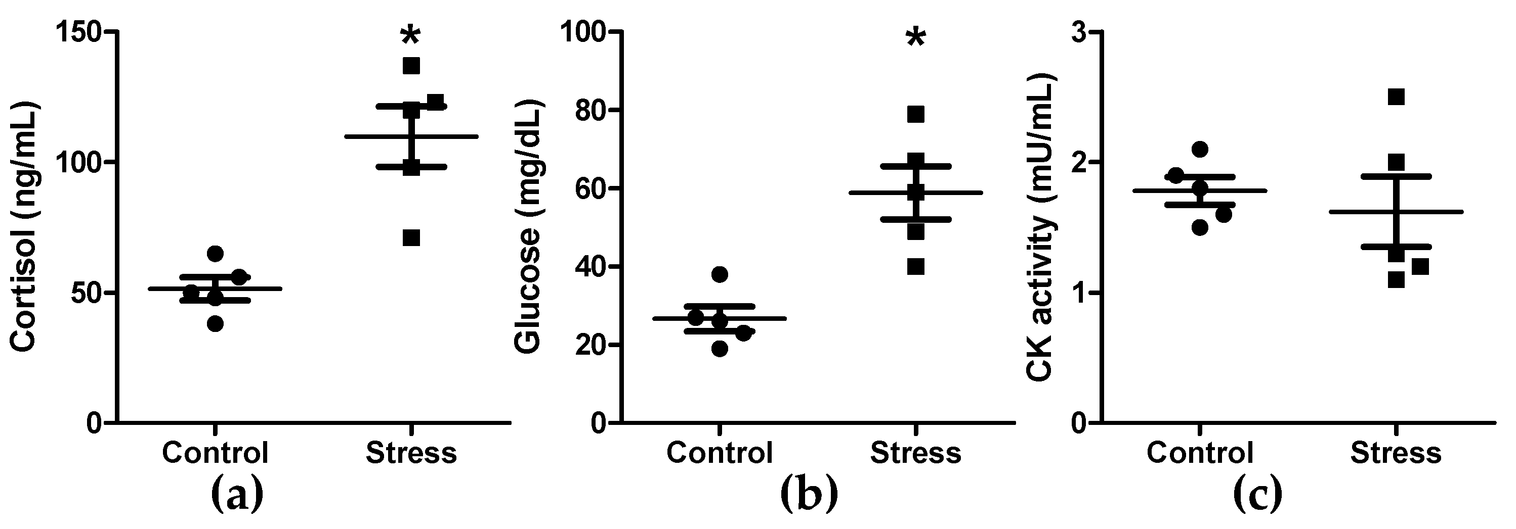
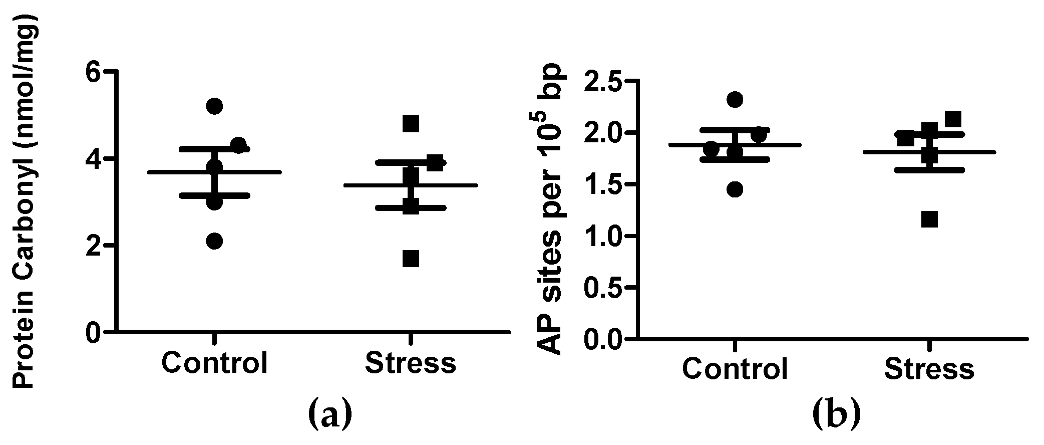
3.1. Cortisol, Glucose, and Creatine Kinase Activity Quantification in Plasma, and Oxidation in Skeletal Muscle Tissue

Blood plasma cortisol and glucose levels significantly increased after five days of exposure to high temperature in the stressed group (Figure 1a,b). No significant differences in plasma creatine kinase activity were observed between the control and stressed groups (Figure 1c).
To evaluate the skeletal muscle oxidation induced by high temperature, we measured protein and DNA oxidative damage. Thermal stress did not induce protein carbonylation and DNA oxidation as compared with the control group (Figure 2a,b).

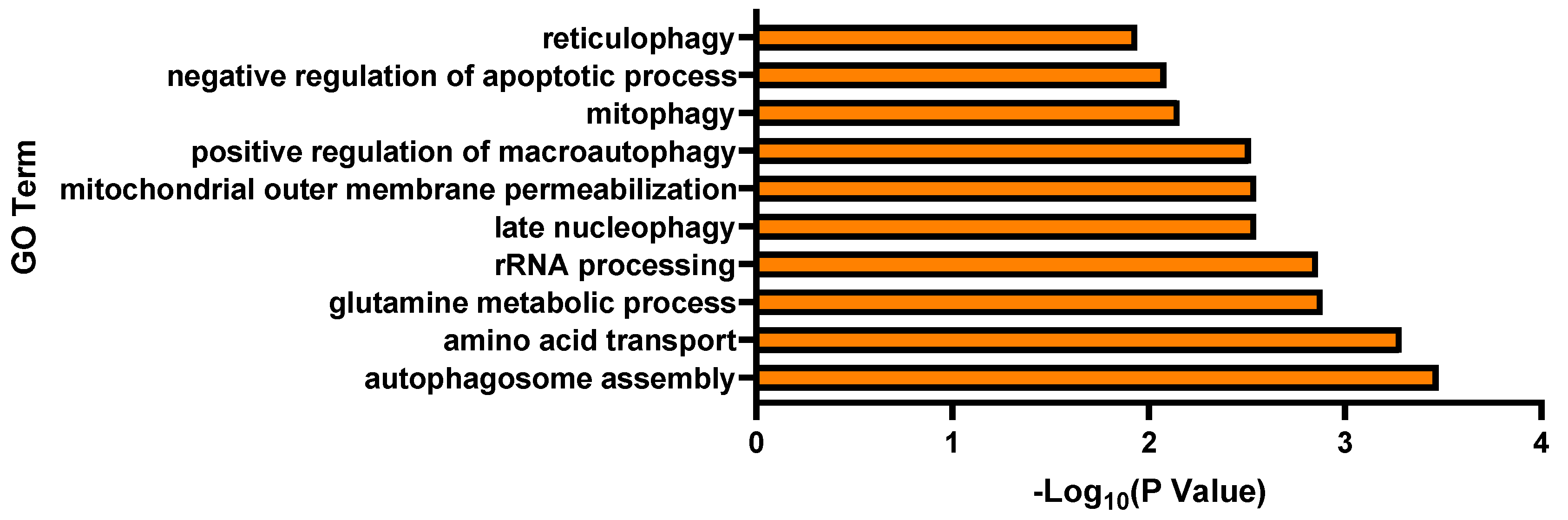
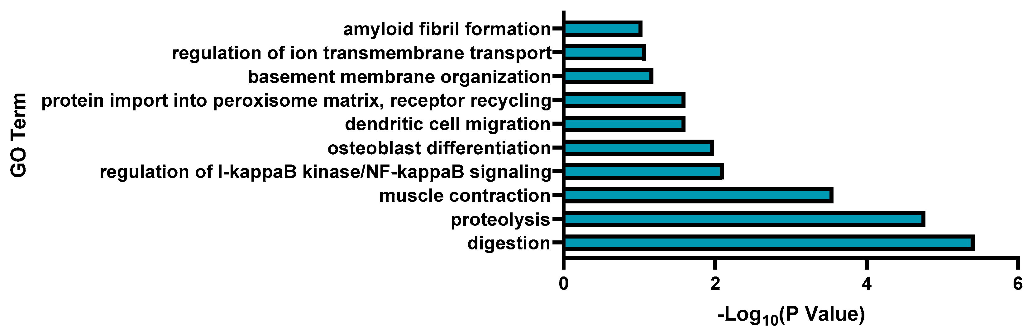
3.2. Transcriptomic Analysis and Pathway Enrichment Analysis

To analyze the effect of high-temperature stress on global gene expression, we performed RNA-Seq analysis. A total of 383,796,290 trimmed reads were mapped in the rainbow trout reference genome, covering 85.9% of its size. Principal component analysis (PCA) revealed a high similarity in the biological replicates (Supplementary Figure S1). Differential expression analysis revealed that 293 genes were upregulated and 119 genes were downregulated under high-temperature stress (Supplementary Table S2). To analyze the biological role of the DEGs, GO term enrichment analysis was performed with the DAVID database. The upregulated DEGs were enriched in biological processes (BPs), such as autophagosome assembly, amino acid transport, and glutamine metabolic process (Figure 3). Gene Ontology (GO) terms for upregulated genes were assigned to RNA binding and nucleoplasm for molecular function (MF) and cellular component (CC), respectively (Supplementary Tables S3 and S4). Among KEGG pathways, mitophagy—animal, autophagy—animal, and spinocerebellar ataxia were over-represented (Table 1).
The downregulated differentially expressed genes (DEGs) were enriched in biological processes (BPs) such as digestion, proteolysis, and muscle contraction (Figure 4). The Gene Ontology (GO) terms for downregulated genes were assigned to serine-type endopeptidase activity and cytosol for molecular function (MF) and cellular component (CC), respectively (Supplementary Tables S5 and S6). The KEGG pathways assigned to the differentially expressed genes (DEGs) included pancreatic secretion, protein digestion and absorption, and adrenergic signaling in cardiomyocytes (Table 2).

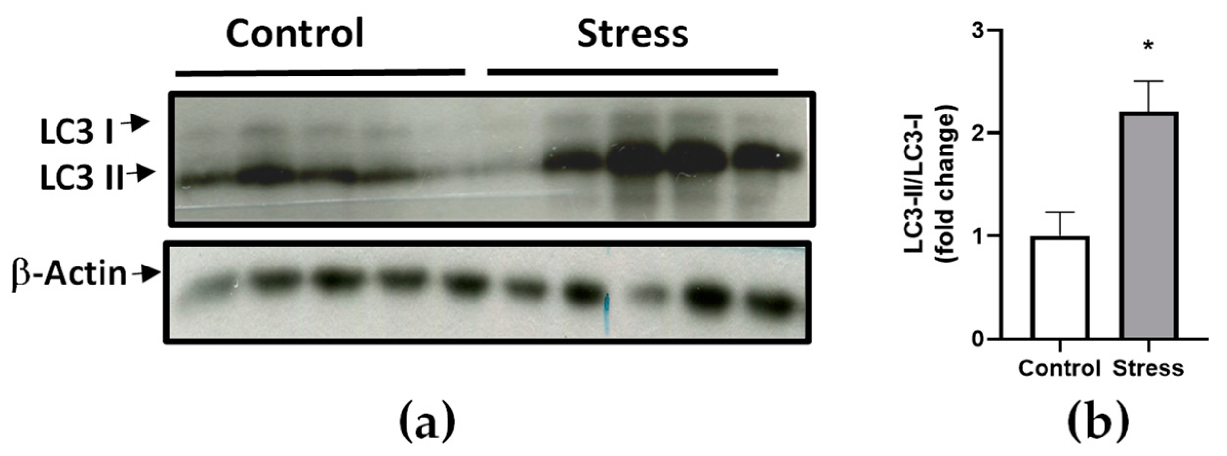
3.3. RNA-Seq Result Validation by Real-Time RT-qPCR and Western Blot

For real-time RT-qPCR validation, we selected four upregulated genes associated with autophagy (mapk10, bnip3, atg9a, and raf1) and four downregulated genes related to protein digestion (prss1, cela1, ctrb2, and prss2) (Supplementary Figure S2). The gene expression fold-changes measured by these two methods (RNA-Seq and RT-qPCR) were highly correlated, with a significant R2 value of 0.86 (p-value, 0.0001). Finally, to verify the presence of autophagy in skeletal muscle, we analyzed the LC3-II/LC3-I levels. We found that LC3-II protein was significantly upregulated in the stressed group (Figure 5a), measuring 2.1-fold that of control conditions (Figure 5b).

4. Discussion

In the present study, we investigated the effects of high-temperature stress on the global gene expression response in the skeletal muscle of rainbow trout (O. mykiss). The effect of temperature stress on fish physiology is an important research focus in the face of ongoing global warming and its potential impact on freshwater and marine aquaculture [22]. The ability of fish to cope with temperature variations is related to their phenotypic plasticity, that is, different phenotypes triggered by variable environmental conditions. In this sense, the acclimatization process mediated by transcriptional changes is essential for the implementation of an ad hoc metabolic, neuroendocrine, and immunological response to climate change [23]. In the present study, we found that the applied stress protocol resulted in a significant increase in the blood plasma levels of cortisol and glucose, reaching concentrations of approximately 110 ng/mL and 59 mg/dL, respectively. These results are consistent with those previously reported to date in multiple other teleost fishes [24,25,26,27,28]. For instance, studies conducted on adult rainbow trout (O. mykiss) have shown that sudden increases in temperature can induce a similar rise in glucose and blood plasma cortisol levels [26,27,28]. This contrasts with studies where the temperature was gradually increased [29]. In a recently published article, it was determined that a similar protocol of gradually increasing temperature induced an increment in cortisol and plasma glucose levels in the red cusk-eel (G. chilensis), a marine teleost [12]. Considering that cortisol has been described as a potent catabolic hormone, inducing muscle atrophy in vertebrates [19,30], we quantified plasma creatine kinase and oxidative damage in muscle tissue as indicators of skeletal muscle atrophy. Interestingly, our results showed that high-temperature stress did not elevate the levels of blood plasma creatine kinase and oxidative damage markers in skeletal muscle tissue. This observation contrasts with the findings of thermal stress studies on red cusk-eel (G. chilensis), which reported an increase in skeletal muscle oxidative damage and an upregulation in the expression of several genes associated with muscle atrophy [12]. Similarly, in the African sharptooth catfish (Clarias gariepinus), it was reported that the long-term exposure to high-temperature stress has a negative consequence on skeletal muscle growth performance and structural integrity [31]. In Atlantic salmon (Salmo salar), thermal stress has been shown to cause skeletal muscle discoloration and loss of skeletal muscle integrity [32], while in gilt-head seabream (Sparus aurata), exposure to high temperatures induces an inflammatory and oxidative response in red muscle [33]. To gain further insights into the molecular mechanisms underlying these responses, we conducted RNA-Seq analysis in the present study.
Differential expression analysis revealed that upregulated genes were mainly associated with autophagosome assembly and mitophagy. The obtained results were validated by RT-qPCR analysis of selected genes and LC3 I/II Western blot, confirming that temperature stress induces autophagy in rainbow trout skeletal muscle. Autophagy is a catabolic mechanism that is fundamental for physiological balance and responsible for the delivery of cytoplasmic components to the lysosomes for digestion [34]. Autophagy is triggered by various stimuli, including lack of nutrients, reactive oxygen species (ROS), endoplasmic reticulum stress, and the presence of microorganisms [35]. Although various reports in teleosts have investigated autophagy in processes such as reproduction [36,37,38], hepatic metabolism [13,39,40], and immune response to infections [41,42], there is limited evidence of the role of autophagy in skeletal muscle catabolism. Studies of fine flounder (Paralichthys adspersus) have shown that stress induced by high-density farming can upregulate the expression of genes involved in the autophagy process, which is believed to be a protective mechanism against apoptosis regulated by the ubiquitin–proteasome pathway [43]. Similar findings were reported in red cusk-eel (G. chilensis), where it was demonstrated that exposure to handling stress increased the expression of genes associated with autophagy and the ubiquitin–proteasome pathway [44]. In rainbow trout (O. mykiss), it was determined that infectious pancreatic necrosis virus (IPNV) infection induced a dynamic response between autophagy and the proteasomal pathways in skeletal muscle [45]. Furthermore, autophagy-mediated skeletal muscle atrophy has been found to be induced by intensive exercise in zebrafish (Danio rerio) [46]. Among the genes with differential expression validated by RT-qPCR are mapk10, bnip3, atg9a, and raf1. Mapk10, also known as Jun Kinase 3 (JNK3), encodes for a serine/threonine-protein kinase involved in various processes such as cell differentiation, apoptosis, and autophagy [47]. In fish, its expression has been related to stress due to hypoxia and salinity in Asian seabass (Lateolabrax maculatus) [48]. Bnip3, also known as BCL2/adenovirus E1B protein-interacting protein 3, regulates apoptosis, modulating the permeability of the outer mitochondrial membrane [49]. It has also been described that bnip3 is also a potent inducer of autophagy in many tissues [49]. Its overexpression in skeletal muscle induced by starvation was reported in the fine flounder (P. adspersus) [50]. Atg9a, also known as autophagy-related protein 9A, encodes for a lipid scramblase involved in autophagosomal membrane expansion, directly regulating autophagy [51]. Gene expression analysis showed that cadmium presence in water induced atg9a gene expression, positively modulating autophagy in the liver tissue of Chinese ink carp (Procypris merus) [40]. Raf1 encodes for the RAF proto-oncogene serine/threonine-protein kinase, which acts as a critical regulator of autophagy and the link between the membrane-associated Ras GTPases and the MAPK/ERK signaling pathway [52]. In Chinese rare minnows (Gobiocypris rarus), the chemical compound Carbamazepine increases its expression, inducing DNA damage and apoptosis in the liver [53]. To our knowledge, there are no previous reports linking the expression of these genes with high-temperature stress and autophagy in lower vertebrates.
Interestingly, temperature-induced autophagy is a phenomenon that has been reported in mammalian skeletal muscle models. In wild boar (Sus scrofa), it has been reported that short-term temperature stress results in apoptosis and autophagy in skeletal muscle [54]. Similarly, it has been reported that in rat (Rattus norvegicus) skeletal muscle, the autophagy pathway is activated in response to temperature stress, as a compensatory mechanism for muscle atrophy induced by tendon cutting [55]. In mice (Mus musculus), temperature stress treatment rescues denervation-induced mitophagy (autophagy in mitochondria) and the consequent skeletal muscle atrophy [56]. Further, in cell models of mammalian skeletal muscle (C2C12 myotubes), it was reported that acute heat exposure induced autophagy resulting in an elevation in AMPK, Beclin-1, and LC3 II levels, similar to our observations [57]. In recent years, evidence has emerged showing that autophagy is a fundamental mechanism of the cellular redox balance, acting in the molecular responses to reactive oxygen species (ROS) [58]. Reactive oxygen species (ROS) are produced during mitochondrial ATP production, which can cause mitochondrial damage, triggering further cell apoptosis [59]. To prevent apoptosis, dysfunctional mitochondria are eliminated through mitophagy, a selective process of autophagy that targets mitochondria [60]. Surprisingly, we did not detect oxidative damage in the muscle tissue of fish subjected to high-temperature stress. However, we did observe an over-representation of biological processes related to mitophagy and negative regulation of apoptosis, indicating that autophagy in teleost skeletal muscle is a protective mechanism against oxidative damage induced by temperature stress.

5. Conclusions

In the present study, we demonstrated that temperature stress induces autophagy in the skeletal muscle of O. mykiss. Considering the absence of protein carbonylation and DNA oxidation in skeletal muscle and the absence of skeletal muscle atrophy markers in plasma, we propose that autophagy acts as a negative regulator of damage in this tissue, inducing the mitophagy process and inhibiting cell apoptosis.

Supplementary Materials

The following supporting information can be downloaded at https://www.mdpi.com/article/10.3390/fishes8060303/s1. Figure S1: Principal component analyses (PCAs) of cDNA libraries; Figure S2: RT-qPCR validation of selected differentially expressed genes in the skeletal muscle rainbow trout in response to high-temperature stress. Table S1: Primer sequences used in qPCR analysis; Table S2: List of DEGs in control vs. stress groups; Table S3: Molecular function (MF) of differentially upregulated genes; Table S4: Cellular component (CC) of differentially upregulated genes; Table S5: Molecular function (MF) of differentially downregulated genes; Table S6: Cellular component (CC) of differentially downregulated genes.

Author Contributions

Conceptualization, A.M. and J.A.V.; methodology, P.D., V.V.-M. and J.A.V.; software, J.A.V.; validation, P.D.; formal analysis, C.G.-E. and J.A.V.; investigation, J.A.V.; resources, A.M. and J.A.V.; data curation, J.A.V.; writing—original draft preparation, J.A.V.; writing—review and editing, J.A.V.; visualization, A.M. and J.A.V.; supervision, J.A.V.; project administration, A.M. and J.A.V.; funding acquisition, J.A.V. All authors have read and agreed to the published version of the manuscript.

Funding

This research was funded by the Fondo Nacional de Desarrollo Científico y Tecnológico (FONDECYT) under grant numbers 1230794 (to Juan Antonio Valdés), 11230153 (to Phillip Dettleff), 11220307 (to Valentina Valenzuela-Muñoz), and 1210852 (to Cristian Gallardo-Escarate); Fondo de Financiamiento de Centros de Investigación en Áreas Prioritarias (FONDAP) INCAR 15110027 grant; and the Concurso de Apoyo a Centros de Excelencia en Investigación FONDAP 2022 1522A0004.

Institutional Review Board Statement

The study adhered to animal welfare procedures, and the protocol was approved by the bioethical committee of Universidad Andres Bello and the National Commission for Scientific and Technological Research of the Chilean government (protocol code 010-2023).

Informed Consent Statement

Not applicable.

Data Availability Statement

The raw read sequences obtained from sequencing were deposited in the Sequence Read Archive (SRA) under BioProject accession number PRJNA930332 (SRR23318096, SRR23318099). The datasets generated and analyzed during the current study are not publicly available owing to privacy or ethical restrictions but are available from the corresponding author upon reasonable request.

Conflicts of Interest

The authors declare no conflict of interest. The funders had no role in the design of the study; in the collection, analyses, or interpretation of data; in the writing of the manuscript; or in the decision to publish the results.

References

  1. Abbass, K.; Qasim, M.Z.; Song, H.; Murshed, M.; Mahmood, H.; Younis, I. A Review of the Global Climate Change Impacts, Adaptation, and Sustainable Mitigation Measures. Environ. Sci. Pollut. Res. 2022, 29, 42539–42559. [Google Scholar] [CrossRef] [PubMed]
  2. Chophel, Y. Global Warming and Climate Change (GWCC) Realities. In The Nature, Causes, Effects and Mitigation of Climate Change on the Environment; Harris, S.A., Ed.; IntechOpen: Rijeka, Croatia, 2022; ISBN 978-1-83968-611-5. [Google Scholar]
  3. Garcia-Soto, C.; Cheng, L.; Caesar, L.; Schmidtko, S.; Jewett, E.B.; Cheripka, A.; Rigor, I.; Caballero, A.; Chiba, S.; Báez, J.C.; et al. An Overview of Ocean Climate Change Indicators: Sea Surface Temperature, Ocean Heat Content, Ocean PH, Dissolved Oxygen Concentration, Arctic Sea Ice Extent, Thickness and Volume, Sea Level and Strength of the AMOC (Atlantic Meridional Overturning Circulation). Front. Mar. Sci. 2021, 8, 642372. [Google Scholar] [CrossRef]
  4. Wang, B.; Luo, X.; Yang, Y.-M.; Sun, W.; Cane, M.A.; Cai, W.; Yeh, S.-W.; Liu, J. Historical Change of El Niño Properties Sheds Light on Future Changes of Extreme El Niño. Proc. Natl. Acad. Sci. USA 2019, 116, 22512–22517. [Google Scholar] [CrossRef] [PubMed] [Green Version]
  5. Alfonso, S.; Gesto, M.; Sadoul, B. Temperature Increase and Its Effects on Fish Stress Physiology in the Context of Global Warming. J. Fish. Biol. 2021, 98, 1496–1508. [Google Scholar] [CrossRef] [PubMed]
  6. Pankhurst, N.W. The Endocrinology of Stress in Fish: An Environmental Perspective. General. Comp. Endocrinol. 2011, 170, 265–275. [Google Scholar] [CrossRef] [Green Version]
  7. Chen, S.; Yong, Y.; Ju, X. Effect of Heat Stress on Growth and Production Performance of Livestock and Poultry: Mechanism to Prevention. J. Therm. Biol. 2021, 99, 103019. [Google Scholar] [CrossRef]
  8. Faught, E.; Vijayan, M.M. Mechanisms of Cortisol Action in Fish Hepatocytes. Comp. Biochem. Physiol. Part B Biochem. Mol. Biol. 2016, 199, 136–145. [Google Scholar] [CrossRef]
  9. Manneken, J.D.; Dauer, M.V.P.; Currie, P.D. Dynamics of Muscle Growth and Regeneration: Lessons from the Teleost. Exp. Cell Res. 2022, 411, 112991. [Google Scholar] [CrossRef]
  10. Johnston, I.A.; Bower, N.I.; Macqueen, D.J. Growth and the Regulation of Myotomal Muscle Mass in Teleost Fish. J. Exp. Biol. 2011, 214, 1617–1628. [Google Scholar] [CrossRef] [Green Version]
  11. Sadoul, B.; Vijayan, M.M. Stress and Growth. In Fish Physiology; Elsevier: Amsterdam, The Netherlands, 2016; Volume 35, pp. 167–205. ISBN 978-0-12-802728-8. [Google Scholar]
  12. Dettleff, P.; Zuloaga, R.; Fuentes, M.; Gonzalez, P.; Aedo, J.; Estrada, J.M.; Molina, A.; Valdés, J.A. Physiological and Molecular Responses to Thermal Stress in Red Cusk-Eel (Genypterus chilensis) Juveniles Reveals Atrophy and Oxidative Damage in Skeletal Muscle. J. Therm. Biol. 2020, 94, 102750. [Google Scholar] [CrossRef]
  13. Dettleff, P.; Zuloaga, R.; Fuentes, M.; Gonzalez, P.; Aedo, J.; Estrada, J.M.; Molina, A.; Valdés, J.A. High-Temperature Stress Effect on the Red Cusk-Eel (Geypterus chilensis) Liver: Transcriptional Modulation and Oxidative Stress Damage. Biology 2022, 11, 990. [Google Scholar] [CrossRef] [PubMed]
  14. Chen, W.; Chen, Y.; Liu, Y.; Wang, X. Autophagy in Muscle Regeneration: Potential Therapies for Myopathies. J. Cachexia Sarcopenia Muscle 2022, 13, 1673–1685. [Google Scholar] [CrossRef] [PubMed]
  15. Han, X.; Goh, K.Y.; Lee, W.X.; Choy, S.M.; Tang, H.-W. The Importance of MTORC1-Autophagy Axis for Skeletal Muscle Diseases. Int. J. Mol. Sci. 2022, 24, 297. [Google Scholar] [CrossRef]
  16. Chatzinikita, E.; Maridaki, M.; Palikaras, K.; Koutsilieris, M.; Philippou, A. The Role of Mitophagy in Skeletal Muscle Damage and Regeneration. Cells 2023, 12, 716. [Google Scholar] [CrossRef] [PubMed]
  17. Zhou, Z.; He, Y.; Wang, S.; Wang, Y.; Shan, P.; Li, P. Autophagy Regulation in Teleost Fish: A Double-Edged Sword. Aquaculture 2022, 558, 738369. [Google Scholar] [CrossRef]
  18. Rivas-Aravena, A.; Fuentes-Valenzuela, M.; Escobar-Aguirre, S.; Gallardo-Escarate, C.; Molina, A.; Valdés, J.A. Transcriptomic Response of Rainbow Trout (Oncorhynchus mykiss) Skeletal Muscle to Flavobacterium Psychrophilum. Comp. Biochem. Physiol. Part D Genom. Proteom. 2019, 31, 100596. [Google Scholar] [CrossRef]
  19. Aedo, J.E.; Zuloaga, R.; Bastías-Molina, M.; Meneses, C.; Boltaña, S.; Molina, A.; Valdés, J.A. Early Transcriptomic Responses Associated with the Membrane-Initiated Action of Cortisol in the Skeletal Muscle of Rainbow Trout (Oncorhynchus mykiss). Physiol. Genom. 2019, 51, 596–606. [Google Scholar] [CrossRef]
  20. Bustin, S.A.; Benes, V.; Garson, J.A.; Hellemans, J.; Huggett, J.; Kubista, M.; Mueller, R.; Nolan, T.; Pfaffl, M.W.; Shipley, G.L.; et al. The MIQE Guidelines: Minimum Information for Publication of Quantitative Real-Time PCR Experiments. Clin. Chem. 2009, 55, 611–622. [Google Scholar] [CrossRef] [Green Version]
  21. Schneider, C.A.; Rasband, W.S.; Eliceiri, K.W. NIH Image to ImageJ: 25 Years of Image Analysis. Nat. Methods 2012, 9, 671–675. [Google Scholar] [CrossRef]
  22. Gizińska, J.; Sojka, M. How Climate Change Affects River and Lake Water Temperature in Central-West Poland—A Case Study of the Warta River Catchment. Atmosphere 2023, 14, 330. [Google Scholar] [CrossRef]
  23. Yuan, D.; Wang, H.; Liu, X.; Wang, S.; Shi, J.; Cheng, X.; Gu, H.; Xiao, S.; Wang, Z. High Temperature Induced Metabolic Reprogramming and Lipid Remodeling in a High-Altitude Fish Species, Triplophysa Bleekeri. Front. Mar. Sci. 2022, 9, 1017142. [Google Scholar] [CrossRef]
  24. LeBlanc, S.; Middleton, S.; Gilmour, K.M.; Currie, S. Chronic Social Stress Impairs Thermal Tolerance in the Rainbow Trout (Oncorhynchus mykiss). J. Exp. Biol. 2011, 214, 1721–1731. [Google Scholar] [CrossRef] [Green Version]
  25. Basu, N.; Nakano, T.; Grau, E.G.; Iwama, G.K. The Effects of Cortisol on Heat Shock Protein 70 Levels in Two Fish Species. Gen. Comp. Endocrinol. 2001, 124, 97–105. [Google Scholar] [CrossRef] [PubMed]
  26. Kumar, P.; Pal, A.K.; Sahu, N.P.; Jha, A.K.; Priya, P. Biochemical and Physiological Stress Responses to Heat Shock and Their Recovery in Labeo Rohita Fingerlings. Proc. Natl. Acad. Sci. India Sect. B Biol. Sci. 2015, 85, 485–490. [Google Scholar] [CrossRef]
  27. Uren Webster, T.M.; Rodriguez-Barreto, D.; Martin, S.A.M.; Van Oosterhout, C.; Orozco-terWengel, P.; Cable, J.; Hamilton, A.; Garcia De Leaniz, C.; Consuegra, S. Contrasting Effects of Acute and Chronic Stress on the Transcriptome, Epigenome, and Immune Response of Atlantic Salmon. Epigenetics 2018, 13, 1191–1207. [Google Scholar] [CrossRef] [Green Version]
  28. Pérez-Casanova, J.C.; Afonso, L.O.B.; Johnson, S.C.; Currie, S.; Gamperl, A.K. The Stress and Metabolic Responses of Juvenile Atlantic Cod Gadus morhua L. to an Acute Thermal Challenge. J. Fish Biol. 2008, 72, 899–916. [Google Scholar] [CrossRef]
  29. Chadwick, J.G.; Nislow, K.H.; McCormick, S.D. Thermal Onset of Cellular and Endocrine Stress Responses Correspond to Ecological Limits in Brook Trout, an Iconic Cold-Water Fish. Conserv. Physiol. 2015, 3, cov017. [Google Scholar] [CrossRef]
  30. Aedo, J.E.; Zuloaga, R.; Aravena-Canales, D.; Molina, A.; Valdés, J.A. Role of Glucocorticoid and Mineralocorticoid Receptors in Rainbow Trout (Oncorhynchus mykiss) Skeletal Muscle: A Transcriptomic Perspective of Cortisol Action. Front. Physiol. 2023, 13, 1048008. [Google Scholar] [CrossRef]
  31. Khieokhajonkhet, A.; Sangphrom, S.; Aeksiri, N.; Tatsapong, P.; Wuthijaree, K.; Kaneko, G. Effects of Long-Term Exposure to High Temperature on Growth Performance, Chemical Composition, Hematological and Histological Changes, and Physiological Responses in Hybrid Catfish [♂Clarias Gariepinus (Burchell, 1822) ×♀C. Macrocephalus (Günther, 1864)]. J. Therm. Biol. 2022, 105, 103226. [Google Scholar] [CrossRef]
  32. Vo, T.T.M.; Amoroso, G.; Ventura, T.; Elizur, A. Histological and Transcriptomic Analysis of Muscular Atrophy Associated with Depleted Flesh Pigmentation in Atlantic Salmon (Salmo salar) Exposed to Elevated Seawater Temperatures. Sci. Rep. 2023, 13, 4218. [Google Scholar] [CrossRef]
  33. Feidantsis, K.; Georgoulis, I.; Zachariou, A.; Campaz, B.; Christoforou, M.; Pörtner, H.O.; Michaelidis, B. Energetic, Antioxidant, Inflammatory and Cell Death Responses in the Red Muscle of Thermally Stressed Sparus Aurata. J. Comp. Physiol. B 2020, 190, 403–418. [Google Scholar] [CrossRef] [PubMed]
  34. Galluzzi, L.; Baehrecke, E.H.; Ballabio, A.; Boya, P.; Bravo-San Pedro, J.M.; Cecconi, F.; Choi, A.M.; Chu, C.T.; Codogno, P.; Colombo, M.I.; et al. Molecular Definitions of Autophagy and Related Processes. EMBO J. 2017, 36, 1811–1836. [Google Scholar] [CrossRef] [PubMed] [Green Version]
  35. Khandia, R.; Dadar, M.; Munjal, A.; Dhama, K.; Karthik, K.; Tiwari, R.; Yatoo, M.I.; Iqbal, H.M.N.; Singh, K.P.; Joshi, S.K.; et al. A Comprehensive Review of Autophagy and Its Various Roles in Infectious, Non-Infectious, and Lifestyle Diseases: Current Knowledge and Prospects for Disease Prevention, Novel Drug Design, and Therapy. Cells 2019, 8, 674. [Google Scholar] [CrossRef] [PubMed] [Green Version]
  36. Cassel, M.; de Paiva Camargo, M.; Oliveira de Jesus, L.W.; Borella, M.I. Involution Processes of Follicular Atresia and Post-Ovulatory Complex in a Characid Fish Ovary: A Study of Apoptosis and Autophagy Pathways. J. Mol. Hist. 2017, 48, 243–257. [Google Scholar] [CrossRef] [PubMed]
  37. Godoi, F.G.A.; Forner-Piquer, I.; Randazzo, B.; Habibi, H.R.; Lo Nostro, F.L.; Moreira, R.G.; Carnevali, O. Effects of Di-Isononyl Phthalate (DiNP) on Follicular Atresia in Zebrafish Ovary. Front. Endocrinol. 2021, 12, 677853. [Google Scholar] [CrossRef]
  38. Cheng, Y.; Lai, F.; Wang, X.; Shang, D.; Zou, J.; Luo, M.; Xia, X.; Cheng, H.; Zhou, R. Srag Regulates Autophagy via Integrating into a Preexisting Autophagy Pathway in Testis. Mol. Biol. Evol. 2021, 38, 128–141. [Google Scholar] [CrossRef]
  39. Seiliez, I.; Belghit, I.; Gao, Y.; Skiba-Cassy, S.; Dias, K.; Cluzeaud, M.; Rémond, D.; Hafnaoui, N.; Salin, B.; Camougrand, N.; et al. Looking at the Metabolic Consequences of the Colchicine-Based in Vivo Autophagic Flux Assay. Autophagy 2016, 12, 343–356. [Google Scholar] [CrossRef] [Green Version]
  40. Dai, Z.; Cheng, J.; Bao, L.; Zhu, X.; Li, H.; Chen, X.; Zhang, Y.; Zhang, J.; Chu, W.; Pan, Y.; et al. Exposure to Waterborne Cadmium Induce Oxidative Stress, Autophagy and Mitochondrial Dysfunction in the Liver of Procypris Merus. Ecotoxicol. Environ. Saf. 2020, 204, 111051. [Google Scholar] [CrossRef]
  41. Masud, S.; Prajsnar, T.K.; Torraca, V.; Lamers, G.E.M.; Benning, M.; Van Der Vaart, M.; Meijer, A.H. Macrophages Target Salmonella by Lc3-Associated Phagocytosis in a Systemic Infection Model. Autophagy 2019, 15, 796–812. [Google Scholar] [CrossRef] [Green Version]
  42. Yang, M.; Lu, Z.; Li, F.; Shi, F.; Zhan, F.; Zhao, L.; Li, Y.; Li, J.; Lin, L.; Qin, Z. Escherichia coli Induced Ferroptosis in Red Blood Cells of grass carp (Ctenopharyngodon Idella). Fish Shellfish Immunol. 2021, 112, 159–167. [Google Scholar] [CrossRef]
  43. Valenzuela, C.A.; Zuloaga, R.; Mercado, L.; Einarsdottir, I.E.; Björnsson, B.T.; Valdés, J.A.; Molina, A. Chronic Stress Inhibits Growth and Induces Proteolytic Mechanisms through Two Different Nonoverlapping Pathways in the Skeletal Muscle of a Teleost Fish. Am. J. Physiol.-Regul. Integr. Comp. Physiol. 2018, 314, R102–R113. [Google Scholar] [CrossRef] [PubMed]
  44. Aedo, J.E.; Maldonado, J.; Aballai, V.; Estrada, J.M.; Bastias-Molina, M.; Meneses, C.; Gallardo-Escarate, C.; Silva, H.; Molina, A.; Valdés, J.A. MRNA-Seq Reveals Skeletal Muscle Atrophy in Response to Handling Stress in a Marine Teleost, the Red Cusk-Eel (Genypterus chilensis). BMC Genom. 2015, 16, 1024. [Google Scholar] [CrossRef] [PubMed] [Green Version]
  45. Aedo, J.E.; Aravena-Canales, D.; Dettleff, P.; Fuentes-Valenzuela, M.; Zuloaga, R.; Rivas-Aravena, A.; Molina, A.; Valdés, J.A. RNA-Seq Analysis Reveals the Dynamic Regulation of Proteasomal and Autophagic Degradation Systems of Rainbow Trout (Oncorhynchus mykiss) Skeletal Muscle Challenged with Infectious Pancreatic Necrosis Virus (IPNV). Aquaculture 2022, 552, 738000. [Google Scholar] [CrossRef]
  46. Sun, C.-C.; Zhou, Z.-Q.; Chen, Z.-L.; Zhu, R.-K.; Yang, D.; Peng, X.-Y.; Zheng, L.; Tang, C.-F. Identification of Potentially Related Genes and Mechanisms Involved in Skeletal Muscle Atrophy Induced by Excessive Exercise in Zebrafish. Biology 2021, 10, 761. [Google Scholar] [CrossRef] [PubMed]
  47. Dhanasekaran, D.N.; Reddy, E.P. JNK-Signaling: A Multiplexing Hub in Programmed Cell Death. Genes. Cancer 2017, 8, 682–694. [Google Scholar] [CrossRef] [Green Version]
  48. Tian, Y.; Wen, H.; Qi, X.; Zhang, X.; Li, Y. Identification of Mapk Gene Family in Lateolabrax Maculatus and Their Expression Profiles in Response to Hypoxia and Salinity Challenges. Gene 2019, 684, 20–29. [Google Scholar] [CrossRef]
  49. Gao, A.; Jiang, J.; Xie, F.; Chen, L. Bnip3 in Mitophagy: Novel Insights and Potential Therapeutic Target for Diseases of Secondary Mitochondrial Dysfunction. Clin. Chim. Acta 2020, 506, 72–83. [Google Scholar] [CrossRef]
  50. Valenzuela, C.A.; Escobar, D.; Perez, L.; Zuloaga, R.; Estrada, J.M.; Mercado, L.; Valdés, J.A.; Molina, A. Transcriptional Dynamics of Immune, Growth and Stress Related Genes in Skeletal Muscle of the Fine Flounder (Paralichthys Adpersus) during Different Nutritional Statuses. Dev. Comp. Immunol. 2015, 53, 145–157. [Google Scholar] [CrossRef]
  51. Jin, M.; Klionsky, D.J. Transcriptional Regulation of ATG9 by the Pho23-Rpd3 Complex Modulates the Frequency of Autophagosome Formation. Autophagy 2014, 10, 1681–1682. [Google Scholar] [CrossRef] [Green Version]
  52. Donninger, H.; Schmidt, M.L.; Mezzanotte, J.; Barnoud, T.; Clark, G.J. Ras Signaling through RASSF Proteins. Semin. Cell Dev. Biol. 2016, 58, 86–95. [Google Scholar] [CrossRef] [Green Version]
  53. Yan, S.; Chen, R.; Wang, M.; Zha, J. Carbamazepine at Environmentally Relevant Concentrations Caused DNA Damage and Apoptosis in the Liver of Chinese Rare Minnows (Gobiocypris rarus) by the Ras/Raf/ERK/P53 Signaling Pathway. Environ. Pollut. 2021, 270, 116245. [Google Scholar] [CrossRef] [PubMed]
  54. Ganesan, S.; Pearce, S.C.; Gabler, N.K.; Baumgard, L.H.; Rhoads, R.P.; Selsby, J.T. Short-Term Heat Stress Results in Increased Apoptotic Signaling and Autophagy in Oxidative Skeletal Muscle in Sus Scrofa. J. Therm. Biol. 2018, 72, 73–80. [Google Scholar] [CrossRef] [PubMed]
  55. Hirunsai, M.; Srikuea, R. Autophagy-Lysosomal Signaling Responses to Heat Stress in Tenotomy-Induced Rat Skeletal Muscle Atrophy. Life Sci. 2021, 275, 119352. [Google Scholar] [CrossRef] [PubMed]
  56. Tamura, Y.; Kitaoka, Y.; Matsunaga, Y.; Hoshino, D.; Hatta, H. Daily Heat Stress Treatment Rescues Denervation-Activated Mitochondrial Clearance and Atrophy in Skeletal Muscle: Heat Stress Treatment in Denervated Skeletal Muscle. J. Physiol. 2015, 593, 2707–2720. [Google Scholar] [CrossRef] [Green Version]
  57. Summers, C.M.; Valentine, R.J. Acute Heat Exposure Alters Autophagy Signaling in C2C12 Myotubes. Front. Physiol. 2020, 10, 1521. [Google Scholar] [CrossRef]
  58. Yun, H.R.; Jo, Y.H.; Kim, J.; Shin, Y.; Kim, S.S.; Choi, T.G. Roles of Autophagy in Oxidative Stress. Int. J. Mol. Sci. 2020, 21, 3289. [Google Scholar] [CrossRef]
  59. Bolisetty, S.; Jaimes, E. Mitochondria and Reactive Oxygen Species: Physiology and Pathophysiology. Int. J. Mol. Sci. 2013, 14, 6306–6344. [Google Scholar] [CrossRef]
  60. Wen, X.; Tang, L.; Zhong, R.; Liu, L.; Chen, L.; Zhang, H. Role of Mitophagy in Regulating Intestinal Oxidative Damage. Antioxidants 2023, 12, 480. [Google Scholar] [CrossRef]
Figure 1. Levels of cortisol, glucose, and creatine kinase activity in plasma. (a) cortisol (b) glucose (c) creatine kinase in blood plasma were assessed in juvenile rainbow trout kept under high-temperature (20 °C) stress and optimal temperature (15 °C) regime. The results are expressed as mean and standard error of the mean (±SEM, n = 5 per treatment). Differences between control and stress groups are shown by * p < 0.05.
Fishes 08 00303 g001
Figure 2. Oxidative damage quantification in skeletal muscle. (a) Protein carbonylation (b) DNA oxidative damage in juvenile rainbow trout kept under high-temperature (20 °C) stress and optimal temperature (15 °C) regime. The results are expressed as mean and standard error of the mean (±SEM, n = 5 per treatment). No statistical differences between groups were detected.
Fishes 08 00303 g002
Figure 3. Gene enrichment analysis of biological processes (BPs). The graph indicates the −log10(p-value) enriched BPs of upregulated genes between the control and stress groups with p-values < 0.05.
Fishes 08 00303 g003
Figure 4. Gene enrichment analysis of biological processes (BPs). The graph indicates the −log10(p-value) enriched BPs of downregulated genes between the control and stress groups with p-values of <0.05.
Fishes 08 00303 g004
Figure 5. High-temperature stress induces autophagy in rainbow skeletal muscle. (a) Western blot of LC3 I/II and H2B. (b) Densitometric analysis of the Western blot showing an LC3 II/LC3 I ratio. The results are expressed as mean and standard error of the mean (±SEM, n = 5 per treatment). Differences between control and stress groups are shown by * p < 0.05.
Fishes 08 00303 g005
Table 1. Enriched KEGG pathways of upregulated DEGs in rainbow trout skeletal muscles.
KEGG Pathwayp-ValueUpregulated Genes
Mitophagy—animal1.45 × 10−4mapk10, bnip3l, bnip3, ubc, atg9a, ulk1, rab7a
Autophagy—animal9.89 × 10−4mapk10, bnip3, atg9a, ulk1, raf1, rab7a, atg4d, atg2b
Spinocerebellar ataxia1.07 × 10−3mapk10, por, psmd11, psmd2, psmd3, atp2a2, ulk1, atg2b
Protein processing in ER2.99 × 10−3mapk10, hsp90ab1, hspa1l, canx, cul1, plaa, cryaa
Alzheimer’s disease1.05 × 10−2mapk10, gsk3b, por, psmd11, cdk5, psmd2, psmd3, atp2a2, ulk1, raf1, atg2b
Antigen processing 1.12 × 10⁻²hsp90ab1, hspa1l, hspa4, canx, rfxap
ErbB signaling pathway1.50 × 10−2mapk10, map2k4, gsk3b, myc, raf1
Pathways of neurodegeneration1.70 × 10−2mapk10, gsk3b, por, psmd11, cdk5, psmd2, ubc, psmd3, atp2a2, ulk1, raf1, atg2b
Legionellosis2.58 × 10−2hspa1l, rab1b, bnip3, eef1a2
mTOR signaling pathway3.05 × 10−2gsk3b, cab39, ulk1, raf1, lpin1, wdr24
Table 2. Enriched KEGG pathways of downregulated DEGs in rainbow trout skeletal muscle.
KEGG Pathwayp-ValueDownregulated Genes
Pancreatic secretion1.10 × 10−10prss1, cela2a, cpb1, ctrb2, ctrb1, amy1c, atp2a1, cel, prss3, prss2
Protein digestion and absorption2.35 × 10−6prss1, cela2a, cpb1, ctrb2, ctrb1, prss3, prss2
Adrenergic signaling in cardiomyocytes2.59 × 10−3cacnb1, tpm3, atp2a1, scn1b, myh7
cGMP-PKG signaling pathway3.82 × 10−3atp2a1, vdac1, raf1, prkg1, myh7
Influenza A4.15 × 10−3prss1, vdac1, raf1, prss3, prss2
Cardiac muscle contraction4.57 × 10−3cacnb1, tpm3, atp2a1, myh7
Hypertrophic cardiomyopathy5.03 × 10−3cacnb1, tpm3, atp2a1, myh7
Dilated cardiomyopathy6.02 × 10−3cacnb1, tpm3, atp2a1, myh7
Diabetic cardiomyopathy4.37 × 10−2atp5f1b, atp2a1, vdac1, sdha
Chemical carcinogenesis—ROS5.51 × 10−2atp5f1b, vdac1, sdha, raf1
Disclaimer/Publisher’s Note: The statements, opinions and data contained in all publications are solely those of the individual author(s) and contributor(s) and not of MDPI and/or the editor(s). MDPI and/or the editor(s) disclaim responsibility for any injury to people or property resulting from any ideas, methods, instructions or products referred to in the content.

