
Comparison between PSMA PET/CT and MRI for Characterizing Hepatocellular carcinoma: A Real-World Study
Abstract
:1. Introduction
2. Materials and Methods
Statistical Analysis
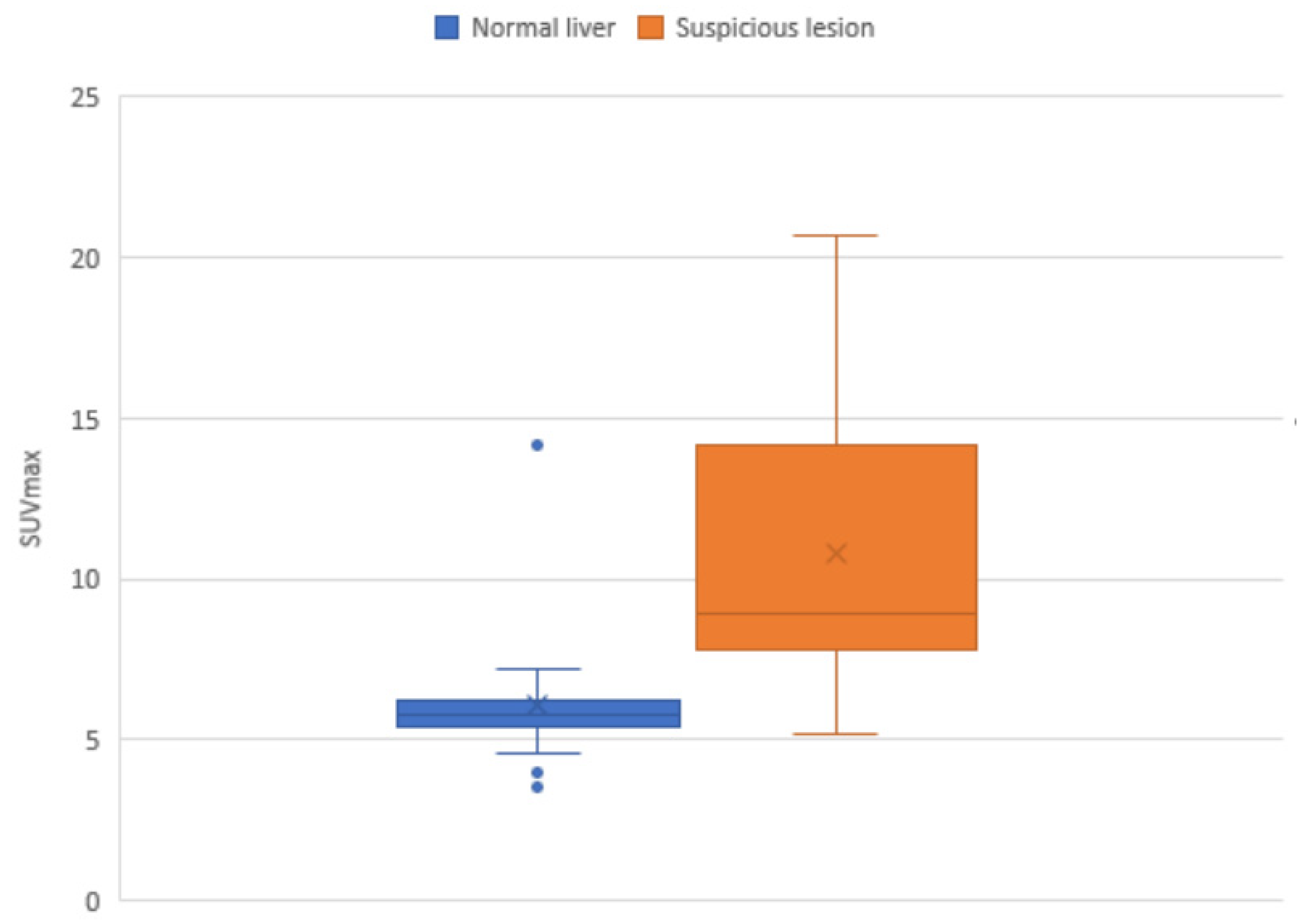
3. Results
4. Discussion
5. Conclusions
Author Contributions
Funding
Institutional Review Board Statement
Informed Consent Statement
Data Availability Statement
Conflicts of Interest
References
- Forner, A.; Reig, M.; Bruix, J. Hepatocellular carcinoma. Lancet 2018, 391, 1301–1314. [Google Scholar] [CrossRef] [PubMed]
- International Agency for Research on Cancer 2020. Available online: https://gco.iarc.fr/ (accessed on 5 November 2020).
- Villanueva, A. Hepatocellular Carcinoma. N. Engl. J. Med. 2019, 380, 1450–1462. [Google Scholar] [CrossRef] [PubMed] [Green Version]
- Kansagara, D.; Papak, J.; Pasha, A.S.; O’Neil, M.; Freeman, M.; Relevo, R.; Quiñones, A.; Motu’apuaka, M.; Jou, J.H. Screening for Hepatocellular Carcinoma in Chronic Liver Disease. Ann. Intern. Med. 2014, 161, 261–269. [Google Scholar] [CrossRef] [PubMed] [Green Version]
- Sherman, M. Surveillance for hepatocellular carcinoma. Best Pract. Res. Clin. Gastroenterol. 2014, 28, 783–793. [Google Scholar] [CrossRef] [PubMed]
- Zhang, B.-H.; Yang, B.-H.; Tang, Z.-Y. Randomized controlled trial of screening for hepatocellular carcinoma. J. Cancer Res. Clin. Oncol. 2004, 130, 417–422. [Google Scholar] [CrossRef]
- Patel, D.; Loh, H.; Le, K.; Stevanovic, A.; Mansberg, R. Incidental Detection of Hepatocellular Carcinoma on Ga-68-Labeled Prostate-Specific Membrane Antigen PET/CT. Clin. Nucl. Med. 2017, 42, 881–884. [Google Scholar] [CrossRef]
- Kesler, M.; Levine, C.; Hershkovitz, D.; Mishani, E.; Menachem, Y.; Lerman, H.; Zohar, Y.; Shibolet, O.; Even-Sapir, E. 68 Ga-labeled prostate-specific membrane antigen is a novel PET/CT tracer for imaging of hepatocellular carcinoma: A prospective pilot study. J. Nucl. Med. 2019, 60, 185–191. [Google Scholar] [CrossRef] [Green Version]
- Berger, I.; Annabattula, C.; Lewis, J.; Shetty, D.V.; Kam, J.; Maclean, F.; Arianayagam, M.; Canagasingham, B.; Ferguson, R.; Khadra, M.; et al. (68)Ga-PSMA PET/CT vs. mpMRI for locoregional prostate cancer staging: Correlation with final histopathology. Prostate Cancer Prostatic Dis. 2018, 21, 204–211. [Google Scholar] [CrossRef]
- Sinn, D.H.; Choi, G.-S.; Park, H.C.; Kim, J.M.; Kim, H.; Song, K.D.; Kang, T.W.; Lee, M.W.; Rhim, H.; Hyun, D.; et al. Multidisciplinary approach is associated with improved survival of hepatocellular carcinoma patients. PLoS ONE 2019, 14, e0210730. [Google Scholar] [CrossRef] [Green Version]
- Zhou, J.; Sun, H.; Wang, Z.; Cong, W.; Wang, J.; Zeng, M.; Zhou, W.; Bie, P.; Liu, L.; Wen, T.; et al. Guidelines for the Diagnosis and Treatment of Hepatocellular Carcinoma (2019 Edition). Liver Cancer 2020, 9, 682–720. [Google Scholar] [CrossRef]
- Shetty, D.; Patel, D.; Le, K.; Bui, C.; Mansberg, R. Pitfalls in Gallium-68 PSMA PET/CT Interpretation—A Pictorial Review. Tomography 2018, 4, 182–193. [Google Scholar] [CrossRef]
- Jiao, D.; Li, Y.; Yang, F.; Han, D.; Wu, J.; Shi, S.; Tian, F.; Guo, Z.; Xi, W.; Li, G.; et al. Expression of Prostate-Specific Membrane Antigen in Tumor-Associated Vasculature Predicts Poor Prognosis in Hepatocellular Carcinoma. Clin. Transl. Gastroenterol. 2019, 10, e00041. [Google Scholar] [CrossRef] [PubMed]
- Tolkach, Y.; Goltz, D.; Kremer, A.; Ahmadzadehfar, H.; Bergheim, D.; Essler, M.; Lam, M.; de Keizer, B.; Fischer, H.P.; Kristiansen, G. Prostate-specific membrane antigen expression in hepatocellular carcinoma: Potential use for prognosis and diagnostic imaging. Oncotarget 2019, 10, 4149–4160. [Google Scholar] [CrossRef] [Green Version]
- Chen, L.X.; Zou, S.J.; Li, D.; Zhou, J.Y.; Cheng, Z.T.; Zhao, J.; Zhu, Y.L.; Kuang, D.; Zhu, X.H. Prostate-specific membrane antigen expression in hepatocellular carcinoma, cholangiocarcinoma, and liver cirrhosis. World J. Gastroenterol. 2020, 26, 7664–7678. [Google Scholar] [CrossRef] [PubMed]
- Kuyumcu, S.; Has-Simsek, D.; Iliaz, R.; Sanli, Y.; Buyukkaya, F.; Akyuz, F.; Turkmen, C. Evidence of Prostate-Specific Membrane Antigen Expression in Hepatocellular Carcinoma Using 68Ga-PSMA PET/CT. Clin. Nucl. Med. 2019, 44, 702–706. [Google Scholar] [CrossRef]
- Roberts, L.R.; Sirlin, C.B.; Zaiem, F.; Almasri, J.; Prokop, L.J.; Heimbach, J.K.; Murad, M.H.; Mohammed, K. Imaging for the diagnosis of hepatocellular carcinoma: A systematic review and meta-analysis. Hepatology 2018, 67, 401–421. [Google Scholar] [CrossRef] [PubMed] [Green Version]
- Chernyak, V.; Fowler, K.J.; Kamaya, A.; Kielar, A.Z.; Elsayes, K.M.; Bashir, M.R.; Kono, Y.; Do, R.K.; Mitchell, D.G.; Singal, A.G.; et al. Liver Imaging Reporting and Data System (LI-RADS) Version 2018: Imaging of Hepatocellular Carcinoma in At-Risk Patients. Radiology 2018, 289, 816–830. [Google Scholar] [CrossRef]
- Hong, T.P.; Gow, P.J.; Fink, M.; Dev, A.; Roberts, S.K.; Nicoll, A.; Lubel, J.S.; Kronborg, I.; Arachchi, N.; Ryan, M.; et al. Surveillance improves survival of patients with hepatocellular carcinoma: A prospective population-based study. Med. J. Aust. 2018, 209, 348–354. [Google Scholar] [CrossRef]
- Sacks, A.; Peller, P.J.; Surasi, D.S.; Chatburn, L.; Mercier, G.; Subramaniam, R.M. Value of PET/CT in the Management of Primary Hepatobiliary Tumors, Part 2. Am. J. Roentgenol. 2011, 197, W260–W265. [Google Scholar] [CrossRef]
- Yamamoto, Y.; Nishiyama, Y.; Kameyama, R.; Okano, K.; Kashiwagi, H.; Deguchi, A.; Kaji, M.; Ohkawa, M. Detection of Hepatocellular Carcinoma Using 11C-Choline PET: Comparison with 18F-FDG PET. J. Nucl. Med. 2008, 49, 1245. [Google Scholar] [CrossRef]
- Filippi, L.; Schillaci, O.; Bagni, O. Recent advances in PET probes for hepatocellular carcinoma characterization. Expert Rev. Med. Devices 2019, 16, 341–350. [Google Scholar] [CrossRef] [PubMed]
- Haug, A.R. Imaging of primary liver tumors with positron-emission tomography. Q. J. Nucl. Med. Mol. Imaging 2017, 61, 292–300. [Google Scholar] [CrossRef]
- Goenka, A. 68 Ga PSMA PET/MRI for Hepatocellular Carcinoma—Phase 2 Study Protocol. 2020. Available online: https://clinicaltrials.gov/ct2/show/NCT03982407 (accessed on 1 December 2021).
- Dondi, F.; Albano, D.; Cerudelli, E.; Gazzilli, M.; Giubbini, R.; Treglia, G.; Bertagna, F. Radiolabelled PSMA PET/CT or PET/MRI in hepatocellular carcinoma (HCC): A systematic review. Clin. Transl. Imaging 2020, 8, 461–467. [Google Scholar] [CrossRef]
- Lu, Q.; Long, Y.; Fan, K.; Shen, Z.; Gai, Y.; Liu, Q.; Jiang, D.; Cai, W.; Wan, C.; Lan, X. PET imaging of hepatocellular carcinoma by targeting tumor-associated endothelium using [68Ga]Ga-PSMA-617. Eur. J. Nucl. Med. Mol. Imaging 2022, 49, 4000–4013. [Google Scholar] [CrossRef] [PubMed]


| Mean age (years) | 65 (SD 7.2) |
| Male | 18/19 (95%) |
| Average BMI (kg/m2) | 31.5 (SD 6.7) |
| Aetiology of liver disease (/19 patients) | Hepatitis C (HCV): 5 (26%) Non-alcoholic steatohepatosis (NASH): 4 (21%) Alcoholic liver disease: 1 (5%) Mixed (HCV and NASH): 1 (5%) Mixed (HCV and alcoholic liver disease): 2 (11%) Unknown: 6 (32%) |
| Lesions | Total assessed: 49 Average lesion/patient: 2.6 * |
| Average PSMA activity administered (MBq) | 285.7 |
| Median AFP at time of study (normal reference ≤ 8 IU/mL) | 5 |
| Previously treated patients | 12/19 (63%) |
| Median follow-up period | 204 days |
| (n) | PSMA (49) | MRI (30) * | CT (49) | Serum AFP (19) * | Average AFP Measurement (19) * |
|---|---|---|---|---|---|
| True negative | 19 | 11 | 27 | 7 | |
| True positive | 20 | 13 | 7 | 5 | |
| False negative | 2 | 2 | 15 | 6 | |
| False positive | 8 | 4 | 0 | 1 | |
| Sensitivity | 91 | 87 | 32 | 45 | |
| Specificity | 70 | 73 | 100 | 88 | |
| PPV | 71 | 76 | 100 | 83 | |
| NPV | 90 | 85 | 64 | 54 |
| Lesion/Patient Initials | Size (Segment) | Previous Treatment | PSMA Uptake | MRI (Performed within 3 Months) | CT | AFP at Time of Scan (Peak) (RR ≤ 8) | Diagnosis (Obtained at MDT) | Progress (Months of Follow-Up Post Study) |
|---|---|---|---|---|---|---|---|---|
| 1 BB | 34 mm (7) | MWA | N | - | A+, PV+, D+ | 4 (30) | SD | SD (11.3) |
| 2 BB | <5 mm (5) | N | N | - | A+, PV+, D+ | SD | ||
| 3 GB | 9 mm (7) | N | N | A+, DWI− | NS | 6 (511) | SD | SD (0.5) |
| 4 GB | 9 mm (5/8) | MWA | N | A-, DWI− | NS | SD | ||
| 5 GB | 19 mm (3) | MWA | N | A−, PV− | Hypodense | SD | ||
| 6 GB | <5 mm (7) | N | N | A+ (sus) | NS | SD | ||
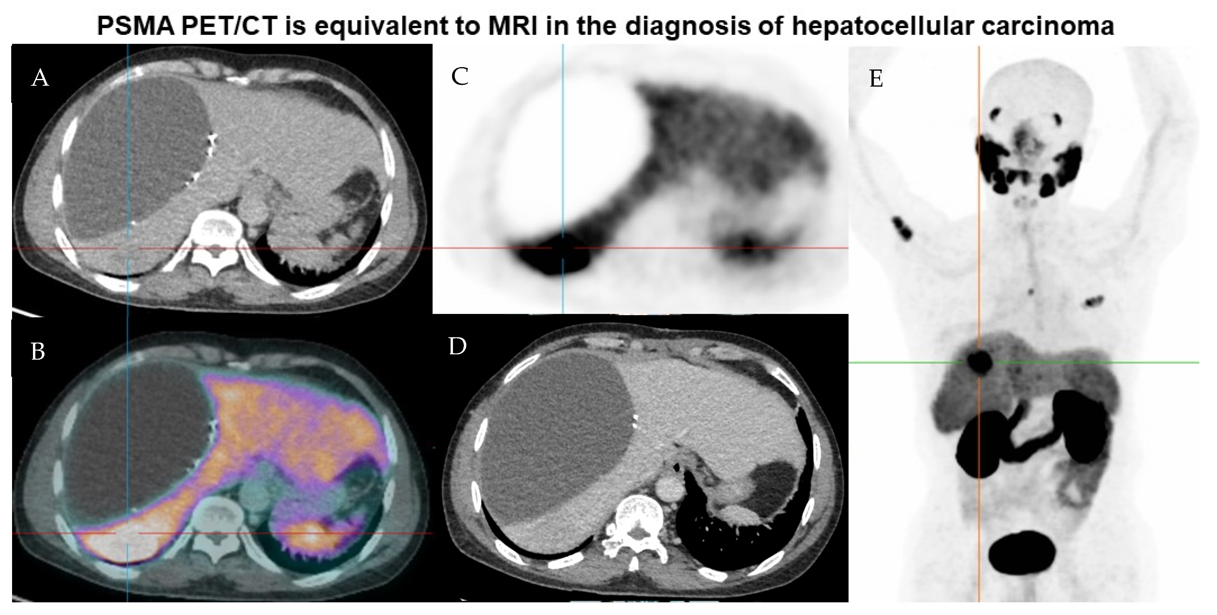
| 7 HB | 47 mm (8) | TACE | Heterogeneous | T2+, T1−, DWI+, A+, progressive washout (sus) | Hypodense, mild PV enhancement (sus) | 197 (214) | PD | PD, palliative, deceased (12.5) |
| 8 HB | 17 mm (2) | N | Y (SUVmax 8.3) | T2+, progressive washout (sus) | NS | PD | ||
| 9 HB | 11 mm (4A) | N | Y (SUVmax 8.9) | T2+, progressive washout (sus) | NS | PD | ||
| 10 HB | 7 mm (6) | N | Y (SUVmax 10.1) | T2+, progressive washout (sus) | NS | PD | ||
| 11 HB | 6 mm (8) | N | Heterogeneous | T2+, progressive washout (sus) | NS | PD | ||
| 12 JB | 17 mm (7/8) | TACE | Y (SUVmax 5.2) | - | Isodense, areas of PV washout (sus) | 23 (44) | PD | PD (9.6), retreated |
| 13 KC | 10 mm (4A/B) | Resected | Y (SUVmax 9.3) | - | NS | 2 (3) | ? PD | SD, false positive (6.4) |
| 14 RC | 38 mm (2) | MWA | N | - | Hypodense | 7 (18) | SD | PD, false negative (6.9) |
| 15 RC | 30 mm (2/3) | TACE | Heterogeneous, SUVmax up to 14.2 | - | Hypodense | SD | ||
| 16 DD | 25 mm (4A) | TACE | Y (SUVmax 8.0) | T1−, T2+, A+, progressive washout (sus) | Hyperdense | 326 (443) | ? PD | PD (6.8) |
| 17 DD | 33 mm (7) | TACE | N | Heterogeneous, T1+, T2+, progressive washout, some peripheral enhancement (sus) | Hypodense | SD | ||
| 18 DD | 37 mm (6) | - | Y (SUVmax 20.5) | T1−, T2+, DWI+, A+, progressive washout (sus) | NS | PD | ||
| 19 DD | 12 mm (4B) | - | Y (SUVmax 5.9) | T1−, T2+, DWI+, A+, progressive washout (sus) | NS | PD | ||
| 20 GE | 31 mm (5) | TACE | N | T1+, T2−, DWI−, A− | Hypodense, subtle arterial enhancement, no significant washout | 1 (2) | SD | SD (3.5) |
| 21 GE | Adjacent to lesion above (5/8) | - | Y (SUVmax 7.2) | T1+, T2−, DWI−, A− | Hypodense, subtle arterial enhancement, no significant washout | PD | ||
| 22 JF | 20 mm (7) | TACE | N | No significant contrast enhancement | Hyperdense, A− | 2 (7) | SD | PD (11.9) |
| 23 JF | 17 mm (1) | - | Y (SUVmax 8.1) | A+ (sus) | NS | PD | ||
| 24 JF | 5 mm (5) | - | N | A+, rapid washout | NS | SD | ||
| 25 JF | 22 mm (6) | MWA | N | A+, rapid washout | A+, no significant washout | SD | ||
| 26 JF | 5 mm (7) | - | N | A+, rapid washout | NS | SD | ||
| 27 RH | 25 mm (7) | SBRT | Y (SUVmax 12.7) | - | Hypodense | 35 (1196) | SD | SD, false positive (2.3) |
| 28 NK | 37 mm (3) | MWA | Y (SUVmax 11.0) | - | A−, no significant portal venous enhancement | 3 (3) | SD | SD, false positive (6.0) |
| 29 NK | 10 mm (7) | - | Y (SUVmax 10.7) | - | NS | SD | ||
| 30 RL | 50 mm (8) | MWA | N | Areas of peripheral T1+ with central T1 isointensity, central T2+ and peripheral T2−, subtle restricted diffusion (sus) | Low attenuation | 2 (4) | SD | SD (6.3) |
| 31 RM | 23 mm (6) | TACE | Y (SUVmax 6.4) | No enhancement | No significant enhancement | 3 (220) | SD | SD, false positive (12.5) |
| 32 RM | 8.4 mm (6/7) | - | N | A+, washout with normalisation | No significant enhancement | SD | ||
| 33 JM | 18 mm (8) | MWA | N | - | No significant enhancement | 15 (15) | SD | PD, false negative (4.5) |
| 34 JM | 13 mm (6) | TACE | N | - | NS | PD | ||
| 35 JM | 12 mm (2) | - | N | - | NS | SD | ||
| 36 JM | 5 mm (4A) | - | N | - | NS | SD | ||
| 37 JM | 5 mm (4B) | - | N | - | NS | PD | ||
| 38 JM | 5 mm (4A/8) | MWA | Y (SUVmax 8.4) | Delayed hypointensity | NS | 5 (7) | PD | PD (9.9) |
| 39 JM | 32 mm (7) | MWA | N | Small T1+ hyperintensity, A- | NS | SD | ||
| 40 FP | 33 mm (8) | MWA | Y (SUVmax 11.6) | A−, hypointense on PV and hepatocyte phase imaging | No significant enhancement | 3 (4) | PD | PD (7.0) |
| 41 RS | 19 mm (2) | - | Y (SUVmax 10.0) | Mildly T2−, isointense T1 FS, DWI+, peripheral A+ and early washout (sus) | Enlarging, low attenuation with A+ and washout (sus) | 18 (30) | PD | PD (8.1) |
| 42 RS | 38 mm (5) | - | Y (SUVmax 16.7) | Mildly T2-, isointense T1 FS, DWI+, peripheral A+ and early washout (sus) | Low attenuation with A+ and washout (sus) | PD | ||
| 43 RS | 19 mm (6) | - | Y (SUVmax 9.6) | Mildly T2−, isointense T1 FS, DWI+, peripheral A+ and early washout (sus) | Low attenuation with A+ and washout (sus) | PD | ||
| 44 RS | 27 mm (5/8) | - | Y (SUVmax 17.4) | Mildly T2−, isointense T1 FS, DWI+, peripheral A+ and early washout (sus) | Enlarging, low attenuation with A+ and washout (sus) | PD | ||
| 45 KU | 10 mm (6) | - | Y (SUVmax 7.6) | - | Subtle arterial enhancement with washout on delayed phase (sus) | 2 (32) | PD | PD (3.6) |
| 46 KU | 19 mm (6) | MWA | Y (SUVmax 8.1) | - | Hypointense, non-enhancing | SD | ||
| 47 KU | 20 mm (4A) | - | Y (SUVmax 7.5) | - | Hypointense, non-enhancing | SD | ||
| 48 KU | 34 mm (5/8) | MWA | Y (SUVmax 7.4) | - | Hypointense, non-enhancing | SD | ||
| 49 SW | 29 mm (7) | MWA | Y (SUVmax 20.7) | - | Non-enhancing | 1 (2) | PD | PD (5.7) |
Disclaimer/Publisher’s Note: The statements, opinions and data contained in all publications are solely those of the individual author(s) and contributor(s) and not of MDPI and/or the editor(s). MDPI and/or the editor(s) disclaim responsibility for any injury to people or property resulting from any ideas, methods, instructions or products referred to in the content. |
© 2023 by the authors. Licensee MDPI, Basel, Switzerland. This article is an open access article distributed under the terms and conditions of the Creative Commons Attribution (CC BY) license (https://creativecommons.org/licenses/by/4.0/).
Share and Cite
Wong, V.C.K.; Yip, J.; Fragomeli, V.; Weltman, M.; Loh, H.; Le, K.; Nguyen, D.; Bui, C.; Mansberg, R. Comparison between PSMA PET/CT and MRI for Characterizing Hepatocellular carcinoma: A Real-World Study. Tomography 2023, 9, 130-138. https://doi.org/10.3390/tomography9010011
Wong VCK, Yip J, Fragomeli V, Weltman M, Loh H, Le K, Nguyen D, Bui C, Mansberg R. Comparison between PSMA PET/CT and MRI for Characterizing Hepatocellular carcinoma: A Real-World Study. Tomography. 2023; 9(1):130-138. https://doi.org/10.3390/tomography9010011
Chicago/Turabian StyleWong, Veronica Chi Ken, Joshua Yip, Vincenzo Fragomeli, Martin Weltman, Han Loh, Ken Le, Diep Nguyen, Chuong Bui, and Robert Mansberg. 2023. "Comparison between PSMA PET/CT and MRI for Characterizing Hepatocellular carcinoma: A Real-World Study" Tomography 9, no. 1: 130-138. https://doi.org/10.3390/tomography9010011