

3D Electrospun Polycaprolactone Scaffolds to Assess Human Periodontal Ligament Cells Mechanobiological Behaviour
 , , , , , and
Abstract
:1. Introduction
2. Materials and Methods
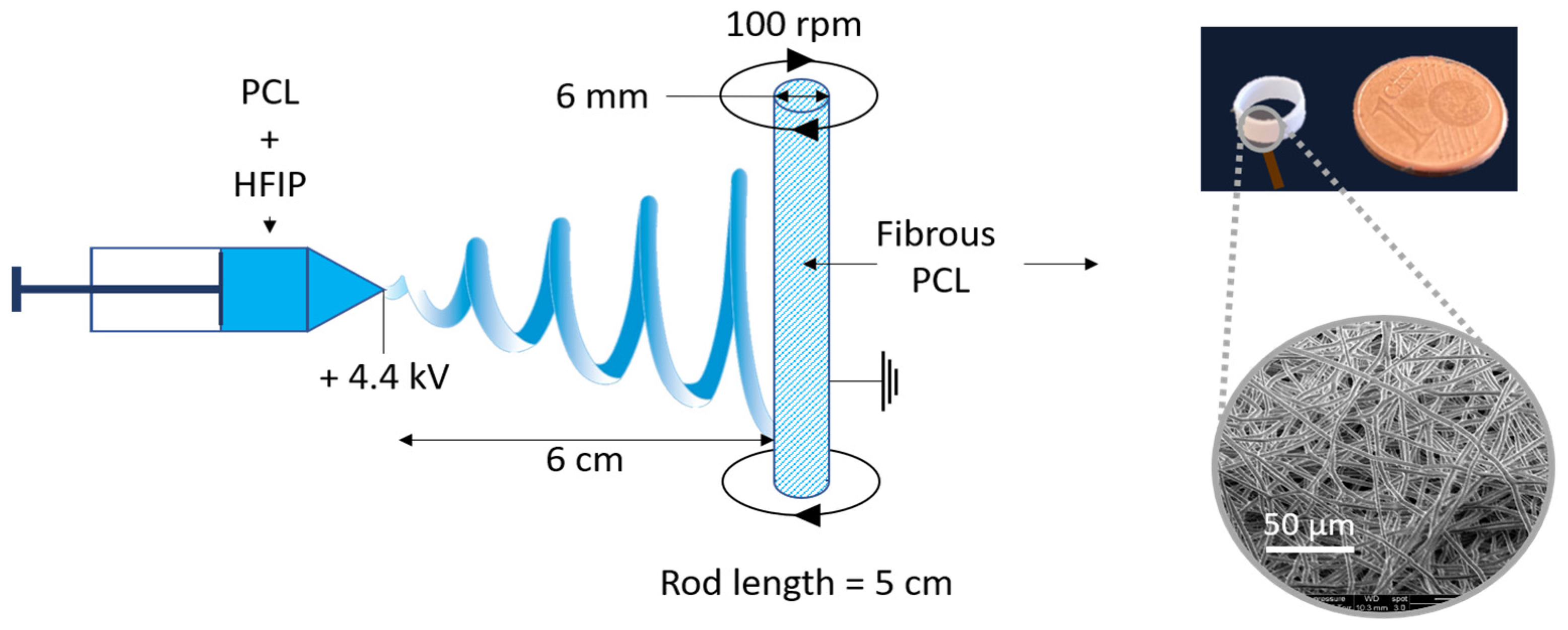
- Scaffold preparation
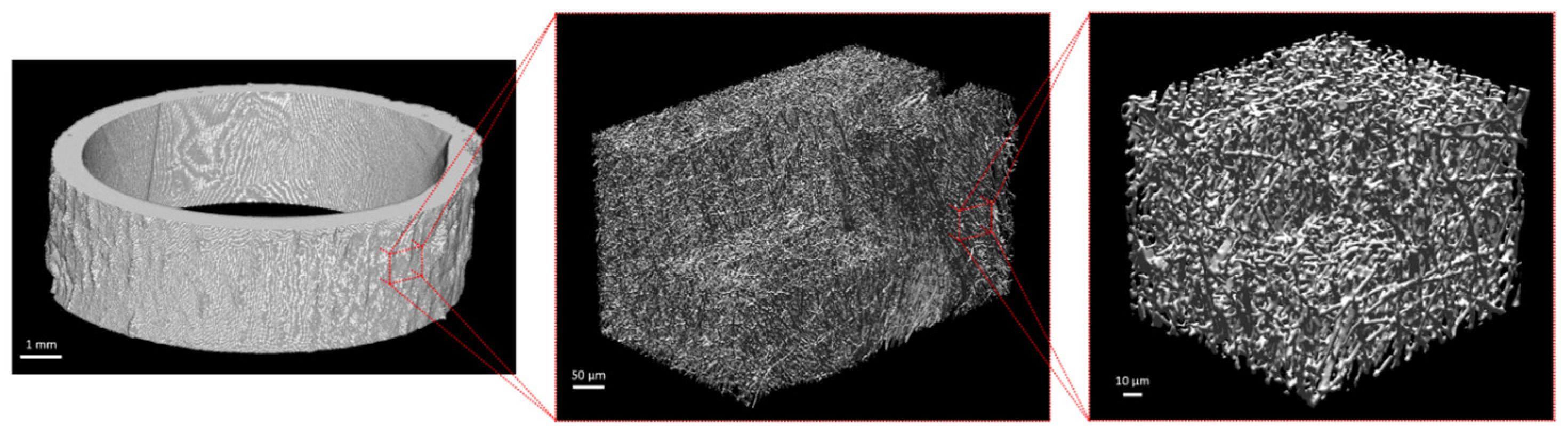
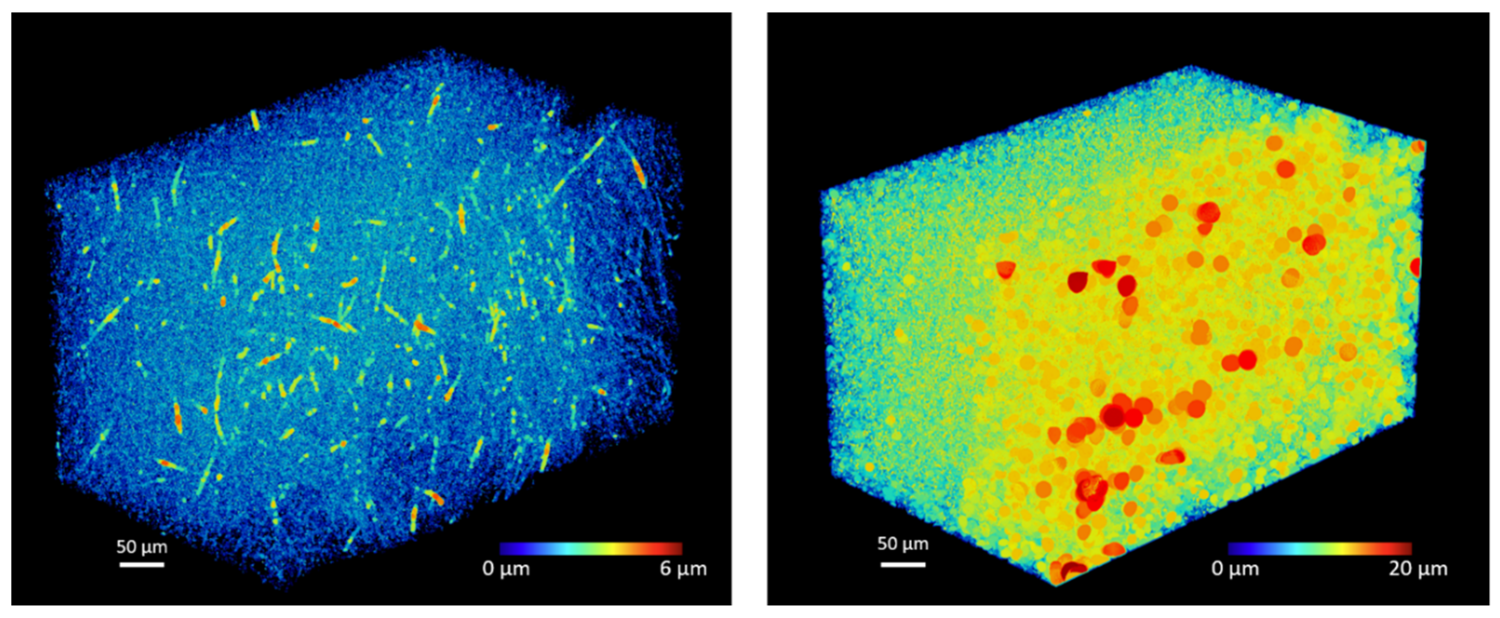
- Scaffold characterization
- Cell culture
- Dynamic bioreactor and loading sequence.
- Mechanical characterization
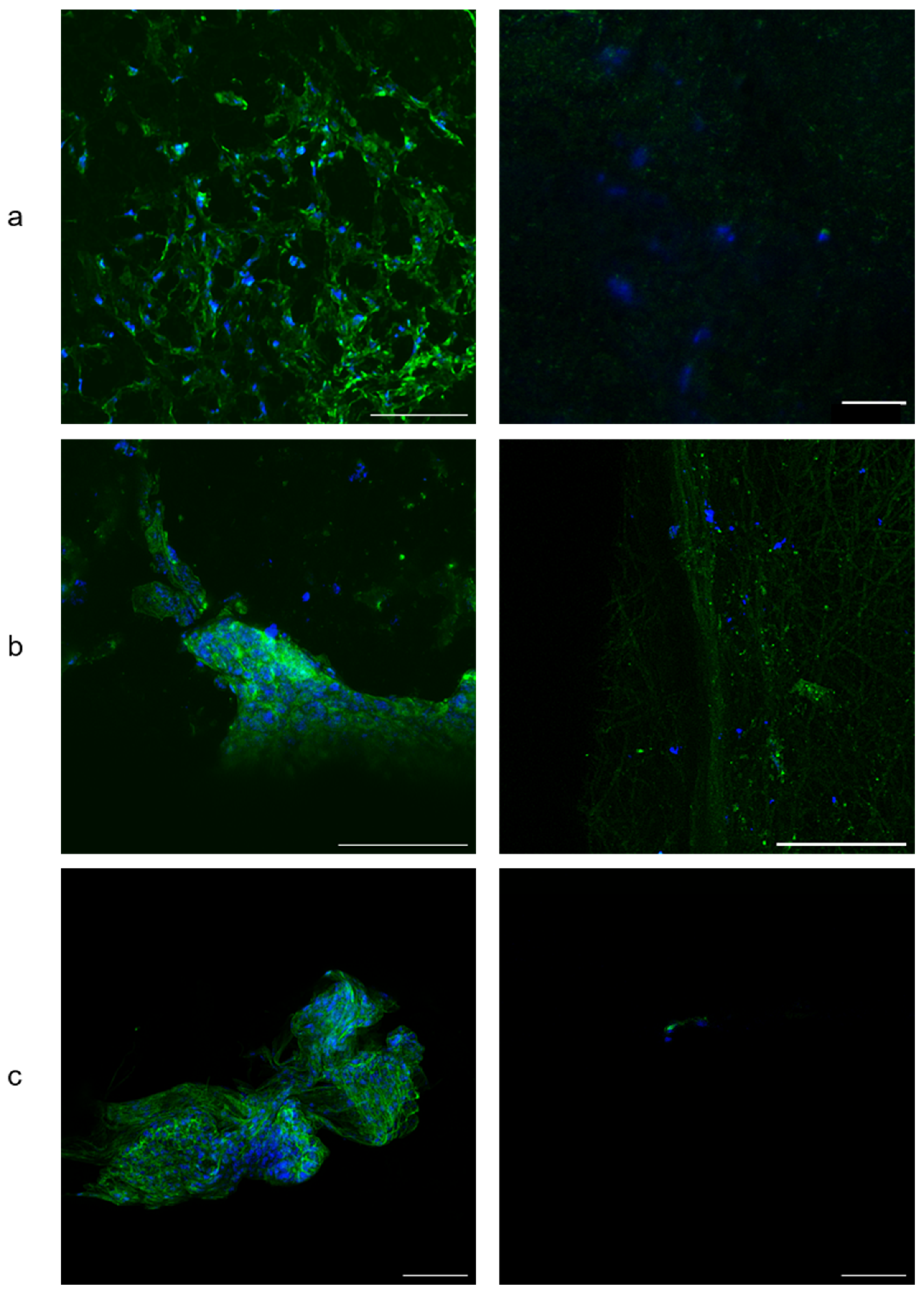
- Cell morphology by Confocal Laser Scanning Microscopy (CLSM)
- Extracellular quantitative assay of ALP activity and inflammatory biomarkers
- Statistical analyses
3. Results
- Scaffold characterization
- Mechanical characterization
- Cell morphology by CLSM
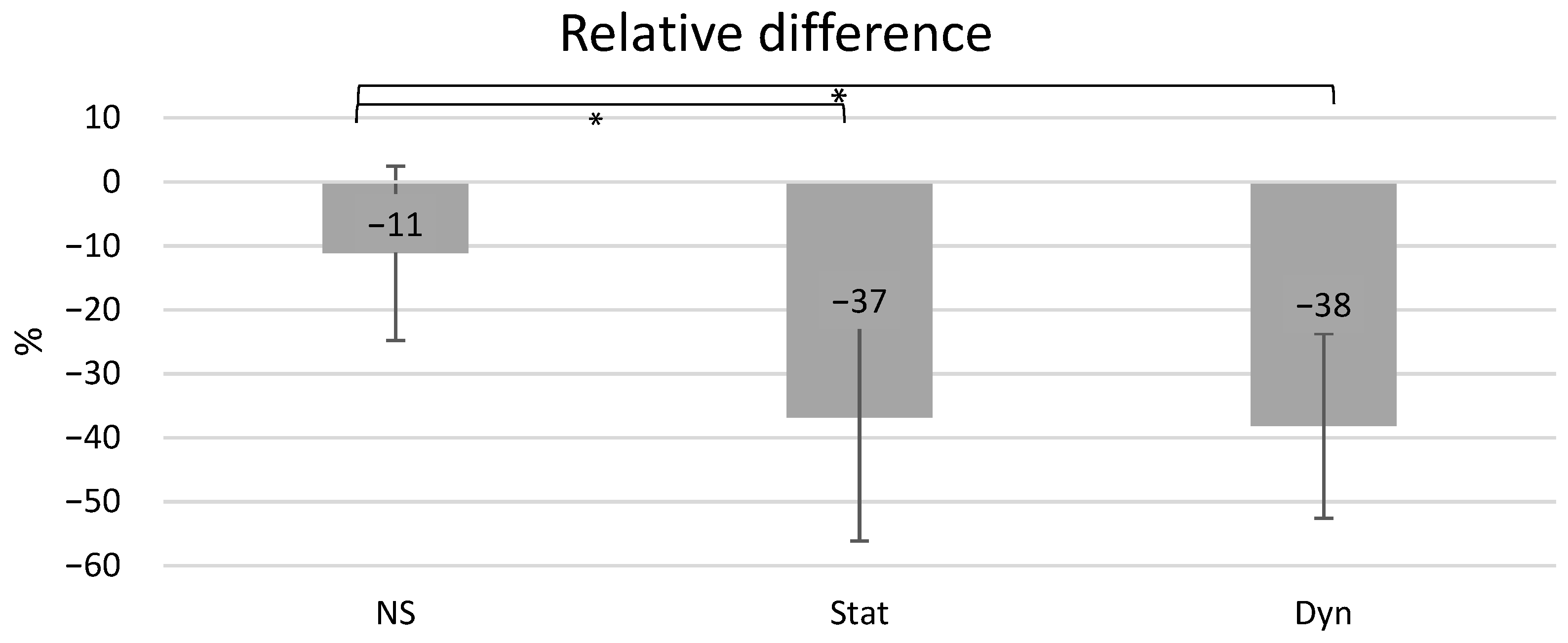
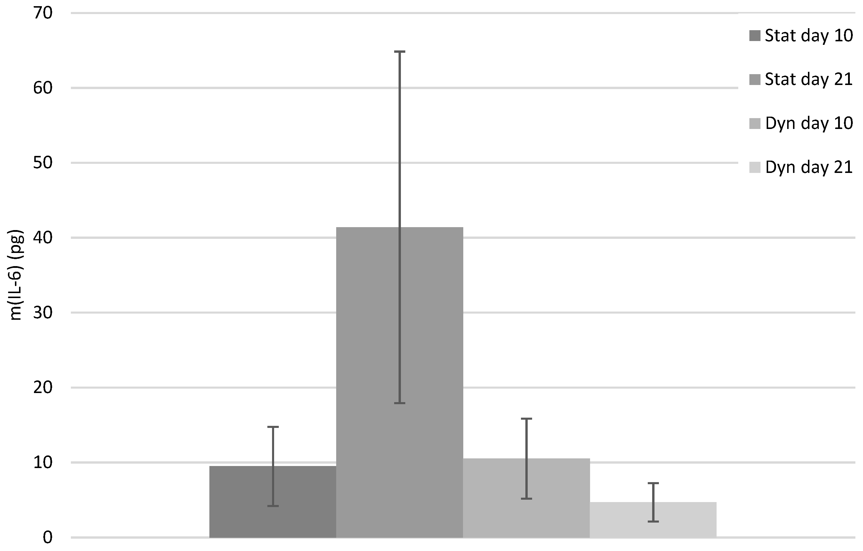
- Extracellular quantitative assay of ALP activity and inflammatory biomarkers
4. Discussion
5. Conclusions
Supplementary Materials
Author Contributions
Funding
Informed Consent Statement
Data Availability Statement
Acknowledgments
Conflicts of Interest
References
- Frencken, J.E.; Sharma, P.; Stenhouse, L.; Green, D.; Laverty, D.; Dietrich, T. Global Epidemiology of Dental Caries and Severe Periodontitis—A Comprehensive Review. J. Clin. Periodontol. 2017, 44, S94–S105. [Google Scholar] [CrossRef] [Green Version]
- Peres, M.A.; Macpherson, L.M.D.; Weyant, R.J.; Daly, B.; Venturelli, R.; Mathur, M.R.; Listl, S.; Celeste, R.K.; Guarnizo-Herreño, C.C.; Kearns, C.; et al. Oral Diseases: A Global Public Health Challenge. Lancet 2019, 394, 249–260. [Google Scholar] [CrossRef] [PubMed]
- Kinane, D.F.; Stathopoulou, P.G.; Papapanou, P.N. Periodontal Diseases. Nat. Rev. Dis. Prim. 2017, 3, 17038. [Google Scholar] [CrossRef] [PubMed]
- Gauthier, R.; Jeannin, C.; Attik, N.; Trunfio-Sfarghiu, A.-M.; Gritsch, K.; Grosgogeat, B. Tissue Engineering for Periodontal Ligament Regeneration: Biomechanical Specifications. J. Biomech. Eng. 2021, 143, 030801. [Google Scholar] [CrossRef]
- Caffesse, R.G.; Nasjleti, C.E.; Morrison, E.C.; Sanchez, R. Guided Tissue Regeneration: Comparison of Bioabsorbable and Non-Bioabsorbable Membranes. Histologic and Histometric Study in Dogs. J. Periodontol. 1994, 65, 583–591. [Google Scholar] [CrossRef] [PubMed]
- Villar, C.C.; Cochran, D.L. Regeneration of Periodontal Tissues: Guided Tissue Regeneration. Dent. Clin. N. A. 2010, 54, 73–92. [Google Scholar] [CrossRef]
- Cho, Y.D.; Kim, K.H.; Lee, Y.M.; Ku, Y.; Seol, Y.J. Periodontal Wound Healing and Tissue Regeneration: A Narrative Review. Pharmaceuticals 2021, 14, 456. [Google Scholar] [CrossRef]
- Zhang, H.; Wang, K.; Gao, T.; Zhang, R.; Cai, Z.; Liu, J.; Ma, H.; Zhang, W. Controlled Release of BFGF Loaded into Electrospun Core–Shell Fibrous Membranes for Use in Guided Tissue Regeneration. Biomed. Mater. 2020, 15, 035021. [Google Scholar] [CrossRef]
- Zhang, E.; Zhu, C.; Yang, J.; Sun, H.; Zhang, X.; Li, S.; Wang, Y.; Sun, L.; Yao, F. Electrospun PDLLA/PLGA Composite Membranes for Potential Application in Guided Tissue Regeneration. Mater. Sci. Eng. C 2016, 58, 278–285. [Google Scholar] [CrossRef]
- Liang, Y.; Luan, X.; Liu, X. Recent Advances in Periodontal Regeneration: A Biomaterial Perspective. Bioact. Mater. 2020, 5, 297–308. [Google Scholar] [CrossRef]
- Discher, D.E.; Janmey, P.; Wang, Y.L. Tissue Cells Feel and Respond to the Stiffness of Their Substrate. Science 2005, 310, 1139–1143. [Google Scholar] [CrossRef] [Green Version]
- Ippolito, A.; Deshpande, V.S. Contact Guidance via Heterogeneity of Substrate Elasticity. Acta Biomater. 2021. [Google Scholar] [CrossRef]
- Hadden, W.J.; Young, J.L.; Holle, A.W.; McFetridge, M.L.; Kim, D.Y.; Wijesinghe, P.; Taylor-Weiner, H.; Wen, J.H.; Lee, A.R.; Bieback, K.; et al. Stem Cell Migration and Mechanotransduction on Linear Stiffness Gradient Hydrogels. Proc. Natl. Acad. Sci. USA 2017, 114, 5647–5652. [Google Scholar] [CrossRef] [Green Version]
- Yamada, S.; Shanbhag, S.; Mustafa, K. Scaffolds in Periodontal Regenerative Treatment. Dent. Clin. N. A. 2022, 66, 111–130. [Google Scholar] [CrossRef] [PubMed]
- McCulloch, C.; Lekic, P.; McKee, M.D. Role of Physical Forces in Regulating the Form and Function of the Periodontal Ligament. Periodontology 2000 2000, 24, 56–72. [Google Scholar] [CrossRef]
- Lin, J.D.; Ryder, M.; Kang, M.; Ho, S.P. Biomechanical Pathways of Dentoalveolar Fibrous Joints in Health and Disease. Periodontology 2000 2020, 82, 238–256. [Google Scholar] [CrossRef]
- Dutra, E.H.; Nanda, R.; Yadav, S. Bone Response of Loaded Periodontal Ligament. Curr. Osteoporos. Rep. 2016, 14, 280–283. [Google Scholar] [CrossRef] [PubMed]
- Berkowitz, K. The Structure of the Periodontal Ligament: An Update. Eur. J. Orthod. 1990, 12, 51–76. [Google Scholar] [CrossRef] [PubMed]
- Attik, N.; Garric, X.; Bethry, A.; Subra, G.; Chevalier, C.; Bouzouma, B.; Verdié, P.; Grosgogeat, B.; Gritsch, K. Amelogenin-Derived Peptide (ADP-5) Hydrogel for Periodontal Regeneration: An In Vitro Study on Periodontal Cells Cytocompatibility, Remineralization and Inflammatory Profile. J. Funct. Biomater. 2023, 14, 53. [Google Scholar] [CrossRef]
- Berendsen, A.D.; Smit, T.H.; Walboomers, X.F.; Everts, V.; Jansen, J.A.; Bronckers, A.L.J.J. Three-Dimensional Loading Model for Periodontal Ligament Regeneration in Vitro. Tissue Eng. Part C Methods 2009, 15, 561–570. [Google Scholar] [CrossRef] [Green Version]
- Bergomi, M.; Cugnoni, J.; Botsis, J.; Belser, U.C.; Anselm Wiskott, H.W. The Role of the Fluid Phase in the Viscous Response of Bovine Periodontal Ligament. J. Biomech. 2010, 43, 1146–1152. [Google Scholar] [CrossRef]
- Komatsu, K.; Chiba, M. Synchronous Recording of Load-Deformation Behaviour and Polarized Light-Microscopic Images of the Rabbit Incisor Periodontal Ligament during Tensile Loading. Arch. Oral Biol. 2001, 46, 929–937. [Google Scholar] [CrossRef] [PubMed]
- Pini, M.; Zysset, P.; Botsis, J.; Contro, R. Tensile and Compressive Behaviour of the Bovine Periodontal Ligament. J. Biomech. 2004, 37, 111–119. [Google Scholar] [CrossRef] [PubMed]
- Hirashima, S.; Ohta, K.; Kanazawa, T.; Okayama, S.; Togo, A.; Miyazono, Y.; Kusukawa, J.; Nakamura, K. ichiro Three-Dimensional Ultrastructural Analysis and Histomorphometry of Collagen Bundles in the Periodontal Ligament Using Focused Ion Beam/Scanning Electron Microscope Tomography. J. Periodontal Res. 2020, 55, 23–31. [Google Scholar] [CrossRef]
- Ullrich, N.; Schröder, A.; Jantsch, J.; Spanier, G.; Proff, P.; Kirschneck, C. The Role of Mechanotransduction versus Hypoxia during Simulated Orthodontic Compressive Strain—An in Vitro Study of Human Periodontal Ligament Fibroblasts. Int. J. Oral Sci. 2019, 11, 33. [Google Scholar] [CrossRef] [PubMed] [Green Version]
- Li, S.; Li, Q.; Zhu, Y.; Hu, W. GDF15 Induced by Compressive Force Contributes to Osteoclast Differentiation in Human Periodontal Ligament Cells. Exp. Cell Res. 2020, 387, 111745. [Google Scholar] [CrossRef]
- Long, P.; Liu, F.; Piesco, N.P.; Kapur, R.; Agarwal, S. Signaling by Mechanical Strain Involves Transcriptional Regulation of Proinflammatory Genes in Human Periodontal Ligament Cells in Vitro. Bone 2002, 30, 547–552. [Google Scholar] [CrossRef] [Green Version]
- Sun, C.; Janjic Rankovic, M.; Folwaczny, M.; Otto, S.; Wichelhaus, A.; Baumert, U. Effect of Tension on Human Periodontal Ligament Cells: Systematic Review and Network Analysis. Front. Bioeng. Biotechnol. 2021, 9, 695053. [Google Scholar] [CrossRef]
- Wenger, K.H.; El-Awady, A.R.; Messer, R.L.W.W.; Sharawy, M.M.; White, G.; Lapp, C.A. Pneumatic Pressure Bioreactor for Cyclic Hydrostatic Stress Application: Mechanobiology Effects on Periodontal Ligament Cells. J. Appl. Physiol. 2011, 111, 1072–1079. [Google Scholar] [CrossRef]
- Wu, J.; Li, Y.; Fan, X.; Zhang, C.; Wang, Y.; Zhao, Z. Analysis of Gene Expression Profile of Periodontal Ligament Cells Subjected to Cyclic Compressive Force. DNA Cell Biol. 2011, 30, 865–873. [Google Scholar] [CrossRef] [PubMed] [Green Version]
- Liu, M.; Dai, J.; Lin, Y.; Yang, L.; Dong, H.; Li, Y.; Ding, Y.; Duan, Y. Effect of the Cyclic Stretch on the Expression of Osteogenesis Genes in Human Periodontal Ligament Cells. Gene 2012, 491, 187–193. [Google Scholar] [CrossRef] [PubMed]
- Ma, J.; Zhao, D.; Wu, Y.; Xu, C.; Zhang, F. Cyclic Stretch Induced Gene Expression of Extracellular Matrix and Adhesion Molecules in Human Periodontal Ligament Cells. Arch. Oral Biol. 2015, 60, 447–455. [Google Scholar] [CrossRef] [PubMed]
- Van Der Pauw, M.T.M.; Klein-Nulend, J.; Van Den Bos, T.; Burger, E.H.; Everts, V.; Beertsen, W. Response of Periodontal Ligament Fibroblasts and Gingival Fibroblasts to Pulsating Fluid Flow: Nitric Oxide and Prostaglandin E2 Release and Expression of Tissue Non-Specific Alkaline Phosphatase Activity. J. Periodontal Res. 2000, 35, 335–343. [Google Scholar] [CrossRef] [PubMed]
- Tang, M.; Peng, Z.; Mai, Z.; Chen, L.; Mao, Q.; Chen, Z.; Chen, Q.; Liu, L.; Wang, Y.; Ai, H. Fluid Shear Stress Stimulates Osteogenic Differentiation of Human Periodontal Ligament Cells via the Extracellular Signal-Regulated Kinase 1/2 and P38 Mitogen-Activated Protein Kinase Signaling Pathways. J. Periodontol. 2014, 85, 1806–1813. [Google Scholar] [CrossRef]
- Walker, T.W.; Ng, G.C.; Burke, P.S. Fluid Pressures in the Periodontal Ligament of the Mandibular Canine Tooth in Dogs. Arch. Oral Biol. 1978, 23, 753–765. [Google Scholar] [CrossRef]
- Zonderland, J.; Moroni, L. Steering Cell Behavior through Mechanobiology in 3D: A Regenerative Medicine Perspective. Biomaterials 2021, 268, 120572. [Google Scholar] [CrossRef]
- Yang, L.; Yang, Y.; Wang, S.; Li, Y.; Zhao, Z. In Vitro Mechanical Loading Models for Periodontal Ligament Cells: From Two-Dimensional to Three-Dimensional Models. Arch. Oral Biol. 2015, 60, 416–424. [Google Scholar] [CrossRef]
- Ivanovski, S.; Vaquette, C.; Gronthos, S.; Hutmacher, D.W.; Bartold, P.M. Multiphasic Scaffolds for Periodontal Tissue Engineering. J. Dent. Res. 2014, 93, 1212–1221. [Google Scholar] [CrossRef]
- Doube, M.; Klosowski, M.M.; Arganda-Carreras, I.; Cordelières, F.P.; Dougherty, R.P.; Jackson, J.S.; Schmid, B.; Hutchinson, J.R.; Shefelbine, S.J. BoneJ: Free and Extensible Bone Image Analysis in ImageJ. Bone 2010, 47, 1076–1079. [Google Scholar] [CrossRef] [PubMed] [Green Version]
- Blaudez, F.; Ivanovski, S.; Ipe, D.; Vaquette, C. A Comprehensive Comparison of Cell Seeding Methods Using Highly Porous Melt Electrowriting Scaffolds. Mater. Sci. Eng. C 2020, 117, 111282. [Google Scholar] [CrossRef]
- Hannoun, A.; Perrier-Groult, E.; Cureu, L.; Pasdeloup, M.; Berthier, Y.; Mallein-Gerin, F.; Trunfio-Sfarghiu, A.M. New “Tribo-Bioreactor” for In-Situ Monitoring of the Mechanical Stress Transmission at the Cellular Level: Application to Cartilage Tissue Engineering. Biotribology 2021, 25, 100158. [Google Scholar] [CrossRef]
- Ben-Zvi, Y.; Maria, R.; Pierantoni, M.; Brumfeld, V.; Shahar, R.; Weiner, S. Response of the Tooth-Periodontal Ligament-Bone Complex to Load: A MicroCT Study of the Minipig Molar. J. Struct. Biol. 2019, 205, 155–162. [Google Scholar] [CrossRef] [PubMed]
- Ortún-Terrazas, J.; Cegoñino, J.; Pérez del Palomar, A. In Silico Study of Cuspid’ Periodontal Ligament Damage under Parafunctional and Traumatic Conditions of Whole-Mouth Occlusions. A Patient-Specific Evaluation. Comput. Methods Programs Biomed. 2020, 184, 105107. [Google Scholar] [CrossRef]
- Kaneko, D.; Sasazaki, Y.; Kikuchi, T.; Ono, T.; Nemoto, K.; Matsumoto, H.; Toyama, Y. Temporal Effects of Cyclic Stretching on Distribution and Gene Expression of Integrin and Cytoskeleton by Ligament Fibroblasts In Vitro. Connect. Tissue Res. 2009, 50, 263–269. [Google Scholar] [CrossRef]
- Zhu, W.; Liang, M. Periodontal Ligament Stem Cells: Current Status, Concerns, and Future Prospects. Stem Cells Int. 2015, 2015, 318269. [Google Scholar] [CrossRef] [Green Version]
- Klouda, L.; Vaz, C.M.; Mol, A.; Baaijens, F.P.T.; Bouten, C.V.C. Effect of Biomimetic Conditions on Mechanical and Structural Integrity of PGA/P4HB and Electrospun PCL Scaffolds. J. Mater. Sci. Mater. Med. 2008, 19, 1137–1144. [Google Scholar] [CrossRef]
- Panadero, J.A.; Vikingsson, L.; Gomez Ribelles, J.L.; Lanceros-Mendez, S.; Sencadas, V. In Vitro Mechanical Fatigue Behavior of Poly-ɛ-Caprolactone Macroporous Scaffolds for Cartilage Tissue Engineering: Influence of Pore Filling by a Poly(Vinyl Alcohol) Gel. J. Biomed. Mater. Res. Part B Appl. Biomater. 2015, 103, 1037–1043. [Google Scholar] [CrossRef] [PubMed] [Green Version]
- Rampichová, M.; Chvojka, J.; Buzgo, M.; Prosecká, E.; Mikeš, P.; Vysloužilová, L.; Tvrdík, D.; Kochová, P.; Gregor, T.; Lukáš, D.; et al. Elastic Three-Dimensional Poly (ε-Caprolactone) Nanofibre Scaffold Enhances Migration, Proliferation and Osteogenic Differentiation of Mesenchymal Stem Cells. Cell Prolif. 2013, 46, 23–37. [Google Scholar] [CrossRef]
- Cukierman, E.; Pankov, R.; Stevens, D.R.; Yamada, K.M. Taking Cell-Matrix Adhesions to the Third Dimension. Science 2001, 294, 1708–1712. [Google Scholar] [CrossRef] [PubMed]
- Hirashima, S.; Kanazawa, T.; Ohta, K.; Nakamura, K. Three-Dimensional Ultrastructural Imaging and Quantitative Analysis of the Periodontal Ligament. Anat. Sci. Int. 2020, 95, 1–11. [Google Scholar] [CrossRef] [PubMed]
- Vashaghian, M.; Diedrich, C.M.; Zandieh-Doulabi, B.; Werner, A.; Smit, T.H.; Roovers, J.P. Gentle Cyclic Straining of Human Fibroblasts on Electrospun Scaffolds Enhances Their Regenerative Potential. Acta Biomater. 2019, 84, 159–168. [Google Scholar] [CrossRef] [PubMed]
- Wang, Y.; Zhao, Z.; Li, Y.; Li, Y.; Wu, J.; Fan, X.; Yang, P. Up-Regulated α-Actin Expression Is Associated with Cell Adhesion Ability in 3-D Cultured Myocytes Subjected to Mechanical Stimulation. Mol. Cell. Biochem. 2010, 338, 175–181. [Google Scholar] [CrossRef] [PubMed]
- Ho, F.C.; Zhang, W.; Li, Y.Y.; Chan, B.P. Mechanoresponsive, Omni-Directional and Local Matrix-Degrading Actin Protrusions in Human Mesenchymal Stem Cells Microencapsulated in a 3D Collagen Matrix. Biomaterials 2015, 53, 392–405. [Google Scholar] [CrossRef]
- Cai, X.; Zhang, Y.; Yang, X.; Grottkau, B.E.; Lin, Y. Uniaxial Cyclic Tensile Stretch Inhibits Osteogenic and Odontogenic Differentiation of Human Dental Pulp Stem Cells. J. Tissue Eng. Regen. Med. 2011, 5, 347–353. [Google Scholar] [CrossRef] [PubMed]
- Yamaguchi, M.; Shimizu, N.; Shibata, Y.; Abiko, Y. Effects of Different Magnitudes of Tension-Force on Alkaline Phosphatase Activity in Periodontal Ligament Cells. J. Dent. Res. 1996, 75, 889–894. [Google Scholar] [CrossRef] [PubMed]
- Elliott, D.S.; Newman, K.J.H.; Forward, D.P.; Hahn, D.M.; Ollivere, B.; Kojima, K.; Handley, R.; Rossiter, N.D.; Wixted, J.J.; Smith, R.M.; et al. A Unified Theory of Bone Healing and Nonunion. Bone Jt. J. 2016, 98B, 884–891. [Google Scholar] [CrossRef] [Green Version]
- Wutticharoenmongkol, P.; Sanchavanakit, N.; Pavasant, P.; Supaphol, P. Novel Bone Scaffolds of Electrospun Polycaprolactone Fibers Filled with Nanoparticles. J. Nanosci. Nanotechnol. 2006, 6, 514–522. [Google Scholar] [CrossRef] [PubMed]
- Bergomi, M.; Wiskott, H.W.A.; Botsis, J.; Mellal, A.; Belser, U.C. Load Response of Periodontal Ligament: Assessment of Fluid Flow, Compressibility, and Effect of Pore Pressure. J. Biomech. Eng. 2010, 132, 014504. [Google Scholar] [CrossRef]
- Vaquette, C.; Fan, W.; Xiao, Y.; Hamlet, S.; Hutmacher, D.W.; Ivanovski, S. A Biphasic Scaffold Design Combined with Cell Sheet Technology for Simultaneous Regeneration of Alveolar Bone/Periodontal Ligament Complex. Biomaterials 2012, 33, 5560–5573. [Google Scholar] [CrossRef]
- Bouet, G.; Cruel, M.; Laurent, C.; Vico, L.; Malaval, L.; Marchat, D. Validation of an in Vitro 3D Bone Culture Model with Perfused and Mechanically Stressed Ceramic Scaffold. Eur. Cells Mater. 2015, 29, 250–267. [Google Scholar] [CrossRef]


| NS-Stat1 | NS-Stat2 | NS-Stat3 | Stat1 | Stat2 | Stat3 | Dyn1 | Dyn2 | Dyn3 | |
|---|---|---|---|---|---|---|---|---|---|
| 1st series | −20 | −1 | −5 | −60 | −34 | −37 | −38 | −35 | −49 |
| 2nd series | −25 | −35 | 11 | −73 | −36 | −19 | −22 | −58 | −7 |
| 3rd series | −8 | 63 | −7 | −33 | −2 | −8 | −41 | −51 | −43 |
| 104 mL−1 | Stat | Dyn |
|---|---|---|
| 1st series | 17.9 | 2.6 |
| 2nd series | 7.5 | 7.2 |
| 3rd series | 19.7 | 7.6 |
| Mean | 15.0 | 5.8 |
| SD | 5.4 | 2.3 |
Disclaimer/Publisher’s Note: The statements, opinions and data contained in all publications are solely those of the individual author(s) and contributor(s) and not of MDPI and/or the editor(s). MDPI and/or the editor(s) disclaim responsibility for any injury to people or property resulting from any ideas, methods, instructions or products referred to in the content. |
© 2023 by the authors. Licensee MDPI, Basel, Switzerland. This article is an open access article distributed under the terms and conditions of the Creative Commons Attribution (CC BY) license (https://creativecommons.org/licenses/by/4.0/).
Share and Cite
Gauthier, R.; Attik, N.; Chevalier, C.; Salles, V.; Grosgogeat, B.; Gritsch, K.; Trunfio-Sfarghiu, A.-M. 3D Electrospun Polycaprolactone Scaffolds to Assess Human Periodontal Ligament Cells Mechanobiological Behaviour. Biomimetics 2023, 8, 108. https://doi.org/10.3390/biomimetics8010108
Gauthier R, Attik N, Chevalier C, Salles V, Grosgogeat B, Gritsch K, Trunfio-Sfarghiu A-M. 3D Electrospun Polycaprolactone Scaffolds to Assess Human Periodontal Ligament Cells Mechanobiological Behaviour. Biomimetics. 2023; 8(1):108. https://doi.org/10.3390/biomimetics8010108
Chicago/Turabian StyleGauthier, Rémy, Nina Attik, Charlène Chevalier, Vincent Salles, Brigitte Grosgogeat, Kerstin Gritsch, and Ana-Maria Trunfio-Sfarghiu. 2023. "3D Electrospun Polycaprolactone Scaffolds to Assess Human Periodontal Ligament Cells Mechanobiological Behaviour" Biomimetics 8, no. 1: 108. https://doi.org/10.3390/biomimetics8010108