Article

Chinese Cabbage BrCAP Has Potential Resistance against Plasmodiophora brassicae

Liaoning Key Laboratory of Genetics and Breeding for Cruciferous Vegetable Crops, College of Horticulture, Shenyang Agricultural University, Shenyang 110866, China
*
Author to whom correspondence should be addressed.
Horticulturae 2023, 9(4), 517; https://doi.org/10.3390/horticulturae9040517
Submission received: 8 March 2023 / Revised: 29 March 2023 / Accepted: 18 April 2023 / Published: 21 April 2023
(This article belongs to the Special Issue Abiotic Stress Responses of Vegetable Crops)

Abstract

:
Clubroot disease caused by Plasmodiophora brassicae Woronin (P. brassicae) has seriously influenced the production of Brassica rapa crops, but the interaction mechanism between P. brassicae and Brassica rapa is not clear. In our previous study, a differentially expressed protein, G15, was found between the roots of Chinese cabbage inoculated and un-inoculated with P. brassicae through two-dimensional electrophoresis (2-DE) and mass spectrometry, and G15 was matched with Bra011464. In this study, Bra011464 was found to have a 94% percent identity with Arabidopsis thaliana CAP, named BrCAP. The expression of BrCAP was the highest in the root compared with the stems and leaves of Chinese cabbage, and its expression in the roots of Chinese cabbage inoculated with P. brassicae was significantly higher than that in the control. The results were verified by real-time quantitative polymerase chain reaction (RT-qPCR) and in situ hybridization. Subcellular localization showed that BrCAP was localized on chloroplasts of leaf epidermal cells. To verify the function of BrCAP, it was found that the Arabidopsis thaliana mutant cap was more susceptible to infection with P. brassicae than the wild type (WT), which suggested that BrCAP has a potential role in the resistance progress of Chinese cabbage to P. brassicae.

1. Introduction

Chinese cabbage (Brassica rapa L. ssp. pekinensis) is a subspecies of B. rapa and is considered an economically important cruciferous vegetable in Asia, particularly in China, Korea, and Japan [1]. Clubroot is a serious soil-borne disease of cruciferous crops caused by the biotrophic pathogen Plasmodiophora brassicae Woronin [2,3]. P. brassicae spores are mononuclear and their outer wall is composed of chitin. P. brassicae can host exclusively on cruciferous plants. The infection can be carried out at any growth period of cruciferous plants. In the early stage of infection, there is no obvious difference in the above-ground parts of host plants. In the late stage of infection, the leaf edges of plants gradually turn yellow and wither, until the whole plant dies. When the whole plant dies, dormant spores are formed in the cells of the diseased part, and the spores of P. brassicae in the tissues can be released into the soil, seriously harming the next crop [4]. Dormant spores of P. brassicae have strong survivability in soil. Studies have shown that the dormant spores of P. brassicae in soil can still be induced by the external environment and are still capable of causing disease after induction [5]. P. brassicae is highly infectious. A low concentration of P. brassicae spores can cause disease, with high transmission speed and multiple modes of transmission, as well as insufficient understanding of the pathogenesis of clubroot disease, making clubroot disease a devastating soil-borne disease worldwide [6]. At present, there are no effective disease resistance measures for clubroot disease, so it is necessary to start from the excavation of disease resistance genes. Many scholars have found some disease-resistant genes by transcriptome [7,8], proteome [9,10], molecular markers [11,12], and other methods, but the role of these genes in the disease resistance mechanism is not clear enough.
Plants defend themselves against biotic stresses through a variety of local, constitutive, and inducible mechanisms [13]. These defense responses are stimulated by the systemic acquired resistance (SAR), induced systemic resistance, and so on [14,15]. Salicylic acid (SA) is a key factor in resistance against specific pathogens [16]. When a plant is infected by a pathogen, the SA content can increase, and the expression of genes encoding PR proteins can be activated by the SA signal pathway [17]. Cysteine-rich secreting protein, Antigen 5, and disease-related Protein 1 (CAP), were identified to play a role in the regulation of host immune attack and infection [18]. Chen and colleagues found that the CAP gene (named PROAtCAPE1) in Arabidopsis thaliana was a salt-responsive gene that was reduced in expression during salt treatment [19]. In addition, many studies have shown that the CAP gene was highly expressed in roots of Arabidopsis thaliana [20], and that its expression could be induced by various external stresses, such as drought stress [21,22], injury induction [19], salt stress induction [21,23], arsenic treatment [24], and iron deficiency [25]. PR-1 protein was a member of a broader family of proteins, named the CAP superfamily. Disease-course related proteins were first identified in TMV-infected tobacco leaves in 1970. At least 17 families have been identified, PR-1 to PR-17 [26], and PR-1 was one of the most produced proteins in the defense response [27]. The significance of PR-1 protein in plant-microbial interactions is now recognized, and an increasing number of identified pathogen-effector proteins interact directly with PR-1 during infection [28,29]. It can be seen that the regulation mechanism of CAP in the defense signal network is complex. At present, the relationship between CAP and clubroot disease is not clear.
In our previous study, a differentially expressed protein, G15, was found between the roots of Chinese cabbage inoculated and un-inoculated with P. brassicae through two-dimensional electrophoresis (2-DE) and mass spectrometry. These techniques found that G15 can be matched with Bra011464 [30]. In this study, Bra011464 was found to have a 94% percent identity with Arabidopsis thaliana CAP, named BrCAP. This research aims to study the role of BrCAP in the infection process of P. brassicae, and provide clues for Brassica crops to resist P. brassicae infection.

2. Materials and Methods

2.1. Plant Materials

The plant material used in the experiment was Chinese cabbage ‘SN742’. A. thaliana mutant cap was purchased from the Arabidopsis thaliana Information Resource website (TAIR), and tobacco (Nicotiana benthamiana) was provided by Shenyang Agricultural University. The P. brassicae used in all the experiments was the physiological race No. 4 and was provided by the vegetable genetics breeding laboratory of Shenyang Agricultural University.

2.2. Preparation of P. brassicae Suspension

An amount of 20 g of freshly clubbed roots, collected from harvested Chinese cabbage ‘SN742’, was cut into small pieces. An amount of 100 mL of sterile water was added and homogenized in a blender (JYL-C022E, Joyoung, China) and then filtered through 8 layers of cheesecloth into the bottle. The concentration of spores was adjusted to 1 × 10⁷ /mL using a hemacytometer under an ordinary light microscope (Nikon Eclipse 80i, Tokyo, Japan). Finally, the suspension was stored at 4 °C for standby.

2.3. Plant Materials Cultivation Method

The ‘SN742’ seeds of Chinese cabbage were washed with 70% ethanol and sterile water for 1 min each, and then evenly spread in the sterilized Petri dish lined with wet filter paper, placed in the constant temperature incubator at 25 °C in the dark environment for 24 h to promote germination. The germinated seeds were transferred to a MLR-350H incubator (SANYO, Osaka, Japan) to be cultivated for 2–3 days under the conditions of light 16 h/dark 8 h, 25 °C, 60% humidity. When the seeding grew two true leaves, treatment group plants were cultured with Hongland solution containing 1 × 10⁷/mL suspension of P. brassicae, and the control group plants were cultured with Hongland solution without P. brassicae. The culture environment of the two groups was consistent. The growth of the plants was observed under the ordinary light microscope (Nikon Eclipse 80i, Tokyo, Japan) daily, until the root hair was found to be infected by P. brassicae spores (around the 14th after inoculation with P. brassicae). Then, the seedlings were transferred into a medium composed of soil and matrix (1:1, vol/vol) for cultivation, respectively. About 40 days after inoculation, clubbed roots could be found in the roots of Chinese cabbage. It can be used for subsequent experiments. The infected and uninfected roots caused by P. brassicae at different periods after inoculation were sampled for further experimentation.
Thirty seeds, each of wild type Columbia (WT), and the cap mutant of A. thaliana, were placed in a refrigerator for 3–5 days and vernalized at 4 °C. They were transferred into a sterilized medium composed of peat, perlite, and vermiculite (3:2:1, vol/vol/vol). They were covered with a transparent plastic film and placed at 25 °C; when the seeds germinated and grew 1–2 young leaves, the film was removed. When the seedlings grew about 8 true leaves after 20 days, they were watered with 1/2 MS nutrient solution every other week. The water and nutrient solution should be recycled until the flowering of A. thaliana.

2.4. Cloning of BrCAP and Bioinformatics Analysis

In our previous study, a candidate protein BrCAP was obtained by two-dimensional electrophoresis (2-DE) and mass spectrometry [29]. Its cDNA sequence was obtained from the Chinese cabbage database according to the obtained amino acid sequence. The full-length cDNA of BrCAP was cloned using the primers (P1 in Table S1), designed based on the most similar sequence of BrCAP (XM_009121709.2) in the Brassica database (www.brassicadb.cn/#/ (accessed on 23 March 2021)). The full length of the BrCAP was amplified from the roots’ cDNA of Chinese cabbage, and the product was connected to the PGEM-T-Easy vector by T4 ligase. The connected production was sent to Sangon Biotech. (Sangon Biotech, Shanghai, China) for sequencing.
The TMHMM website (http://www.cbs.dtu.dk/services/TMHMM/ (accessed on 4 May 2021)) was used to predict transmembrane domain structure, and NCBI (https://www.Ncbi.nlm.nih.gov/structure/CDD/WRPSB.Cgi (accessed on 4 May 2021)) was used to predict the function structure domain.

2.5. Real-Time Quantitative Polymerase Chain Reaction (RT-qPCR)

RNA was extracted from the roots, stems, and leaves of Chinese cabbage ‘SN742’ after inoculation with Plasmodiophora brassicae Woronin (P. brassicae) on the 14th and 40th days using pure Total RNA Kit (Tiangen, Beijing, China) according to the manufacturer’s instructions. Additionally, un-inoculated materials during the same periods were used as the control. The quality of extracted RNA was detected by 1% agarose gel electrophoresis and reversed into cDNA (Vazyme, Nanjing, China). The expression of BrCAP was analyzed by RT-qPCR using QuantStudio6 Real-Time PCR (Thermo Fisher, Waltham, MA, USA), with the kit of UltraSYBR Mixture (CWBIO, Jiangsu, China) and P1/P2 primers (in Table S1). Three replicates were performed for different treatments, and 3 seedlings per experimental unit. Data were calculated by using the 2−∆∆ct method, and the significance analysis was performed by Student’s test (* p ≤ 0.05) or Duncan’s multivariate interval test in SPSS 11.5 (IBM, Armonk, NY, USA). Origin Pro 7.5 (Origin Lab Corp., Northampton, MA, USA) was used to produce the graphics.

2.6. In Situ Hybridization

On the 14th day and 40th day after inoculation with P. brassicae, the roots inoculated with P. brassicae and control roots were taken for an in situ hybridization test. The samples were fixed with 4% FAA fixing solution, and then dehydrated in gradient ethanol (70%, 80%, 90%, 95% and 100%), cleared in ethanol: xylene mixtures (3:1, 1:1 and 1:3 [v/v]) and embedded in 100% paraffin. A BrCAP-specific probe fragment was amplified using primer P4 containing PstI and KpnI enzyme restriction sites (Table S1). Five replicates were performed for each different treatment (five slices were used for each treatment, and each slice had three to five tissues). The vector pSPT18 were digested with PstI and KpnI, purified and connected with a BrCAP-specific probe fragment to obtain pSPT18-BrCAP recombinant vector. pSPT18-BrCAP was purified and digested with PstI or KpnI, and labeled with digoxigenin (DIG) using a SP6/T7 Transcription Kit (Roche, Basle, Switzerland) to synthesize the sense or antisense probes. Sample sections were hybridized with specific DIG-labeled RNA probes (DIG RNA Labeling Kit (SP6/T7), Roche) and were observed under a microscope (Nikon Eclipse 80i, Tokyo, Japan). Detailed steps were referred to Zhang et al. [31].

2.7. Subcellular Localization

The target fragment was amplified using primer P5 containing BamHI and XhoI enzyme restriction sites (Table S1). The amplified fragment was connected to pBWA(V)BS-GFP vector using an In-Fusion cloning kit (Vazyme, Nanjing, China). After successful cloning, a strain containing pBWA(V) BS-BrCAP-GFP was injected into 4-week-old tobacco leaves. There were three tobacco plants for each treatment, and three to five leaves were treated for each plant. Then, a 24 h dark culture and 24 h light culture was conducted in an MLR-350H incubator (SANYO, Osaka, Japan). The transfected plants were observed using a laser confocal microscope (TCS SP8, Leica, Wetzlar, Germany).

2.8. Identification of Disease Resistance of A. thaliana Mutant cap to P. brassicae

The seeds of A. thaliana wild type (WT) and mutant CAP were sterilized and spread evenly in Petri dishes containing moist filter paper. The seeds were cultured at 25 °C for 2–3 d in the dark until germination. The germinated seeds were cultured in an incubator (60% humidity, 16 h light/8 h dark) for about 3–4 d until the root hair had grown. The seedlings were inoculated with a 1 × 10⁷/mL P. brassicae suspension being dropped on the base of plant roots. WT was served as the control and CAP as the treatment. Three plants were randomly selected from WT and CAP, respectively, every 24 h after inoculation until the root hairs were found under a microscope (Nikon Eclipse 80i, Tokyo, Japan) to be infected, and the infection rate was investigated.

3. Results

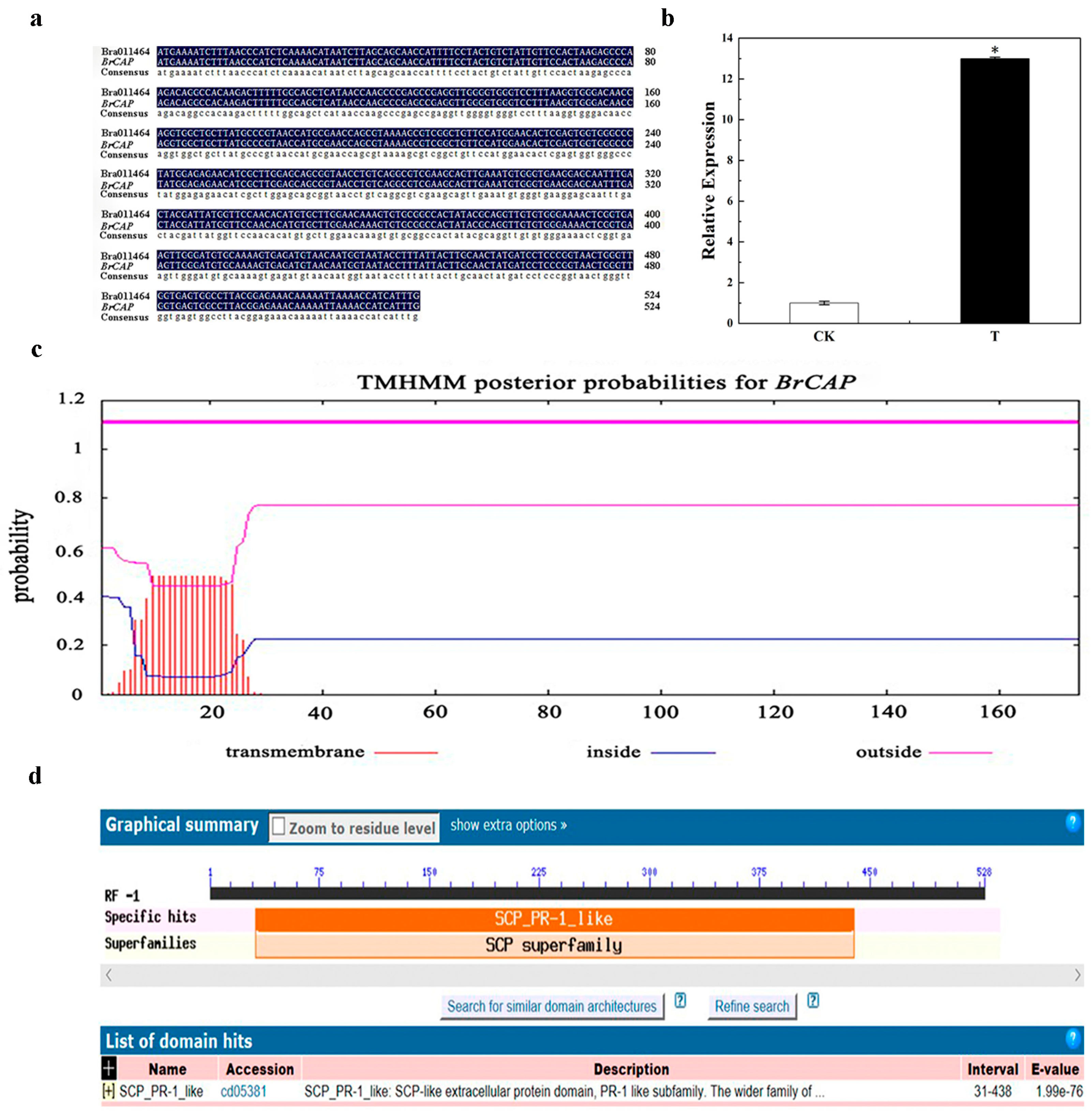
3.1. Obtainment and Prediction of BrCAP Gene

In our previous study, the results of two-dimensional (2-DE) electrophoresis showed that the expression of G15 protein in the roots of Chinese cabbage inoculated with P. brassicae was significantly higher than that in the control roots. Mass spectrometry analysis showed that Bra011464 was highly matched to protein G15 [29]. A BLASTN search found that Bra011464 has a 94% percent identity with Arabidopsis thaliana CAP. Thus, this gene was named BrCAP (Table 1). The full-length 525 bp coding sequence (CDS) of Bra011464 was cloned from the cDNA of roots from Chinese cabbage, which includes the start codon ATG and stop codon TGA. Sequence analysis found that it is completely consistent with the reference sequence (NC_024795.2) in the Brassica database (Figure 1a). RT-qPCR showed that the expression of BrCAP in the roots of Chinese cabbage inoculated with P. brassicae was 12 times higher than that in the control root (Figure 1b, Table 2). The results showed that the expression of BrCAP was significantly changed when Chinese cabbage was infected by P. brassicae. The TMHMM was used to analyze the amino acid sequence of BrCAP, and the results showed that it had an obvious transmembrane domain (Figure 1c), indicating it might be a membrane protein. A CD Search in NCBI prediction showed that BrCAP contained a conserved domain, which was named SCP_PR-1_LIKE (Figure 1d). Therefore, BrCAP may play a role in the resistance against clubroot disease, which provides impetus for our subsequent research.

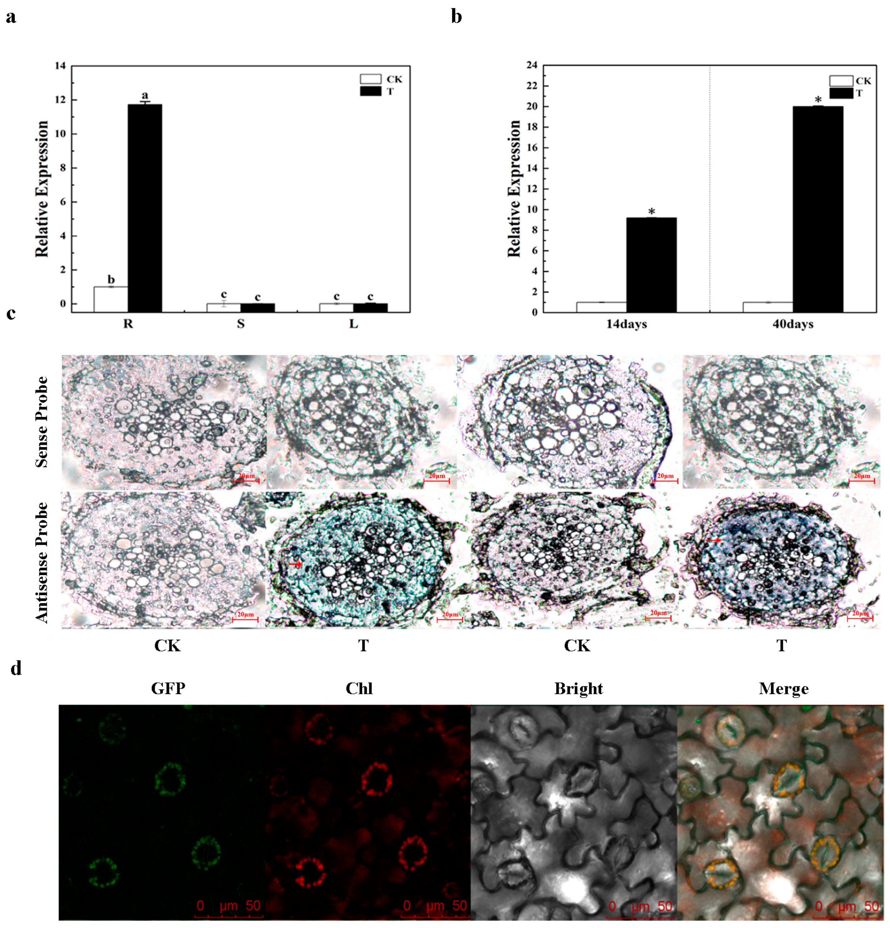
3.2. Expression Mode Analysis of BrCAP

The expression of BrCAP in roots, stems, and leaves of Chinese cabbage ‘SN742’ after inoculation and un-inoculation with P. brassicae was analyzed by RT-qPCR. The results showed that the expression level of BrCAP in the roots inoculated with P. brassicae was 10 times higher than that in un-inoculated roots. However, the expression of BrCAP in stem and leaf were extremely low (Figure 2a, Table 3). This indicated that BrCAP was a root expression gene. Similarly, the expression level of BrCAP gene was detected on the 14th and 40th days in roots inoculated with P. brassicae by RT-qPCR. The results showed that the expression of BrCAP was more than eight times higher in the inoculated roots than that in un-inoculated roots on the 14th day after inoculation. Additionally, its expression was increased 20 times more in the P. brassicae-inoculated roots than that in un-inoculated roots on the 40th day (Figure 2b, Table 4). In conclusion, with the accumulation of time, the expression of BrCAP in the roots after inoculation with P. brassicae was gradually up-regulated and significantly higher than that in the un-inoculated control. The results of RT-PCR were further verified by in situ hybridization, which showed that all the tissue slices of root hybridized with sense probes have no hybridization signal. However, when hybridizing root tissue sections with antisense probes, blue hybridization signals were present on both the 14th and 40th days in the inoculated treatment group, and the signal was stronger on the 40th day. The above results further indicated that BrCAP might be related to P. brassicae infection (Figure 2c). The results of subcellular localization showed that pBWA(V)BS-BrCAP-GFP could produce the green fluorescence signal and co-localize with the red auto-fluorescence signal on chloroplasts of leaf epidermal cells (Figure 2d).

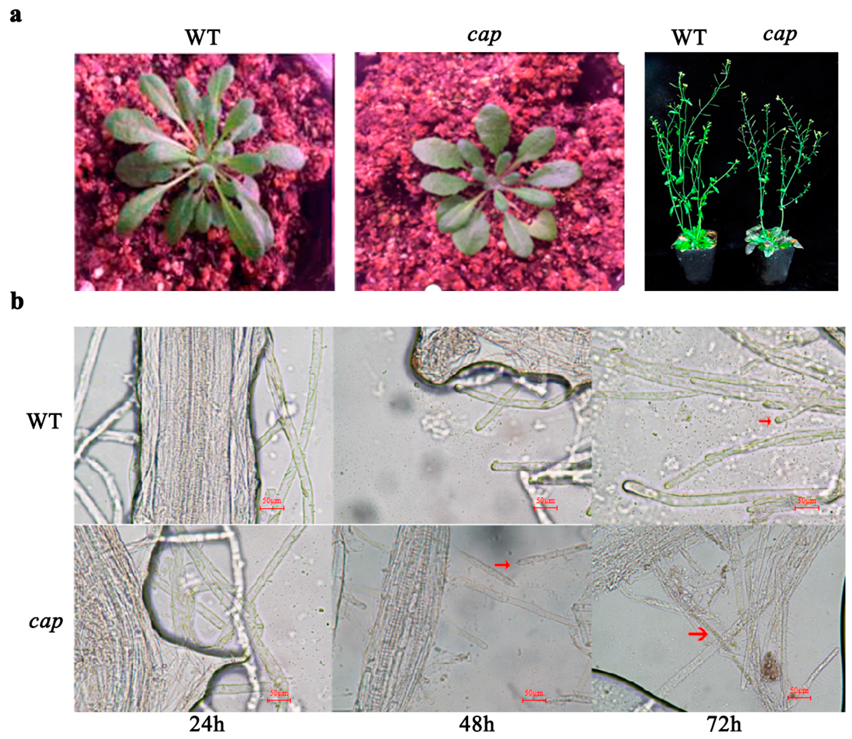
3.3. Resistance Identification of Arabidopsis Thaliana Mutant CAP

Phenotypic observation showed that the wild type (WT) grows better than the mutant CAP during both growth and bolting stages (Figure 3a). Microscopic observation of root hairs of WT and CAP at the 24 h, 48 h, and 72 h after inoculation with P. brassicae showed that the spores of P. brassicae first infected the fibrous roots of CAP at the 48 h, while the fibrous roots of WT were first infected at 72 h. Compared with WT plants, the pathogenic process of P. brassicae was accelerated in CAP. Thus, CAP may play a key role in the progress of plants’ resistance against the infection of P. brassicae (Figure 3b).

4. Discussion

4.1. The Expression of BrCAP in Chinese Cabbage Is Related to the Infection of P. brassicae

Plants exposed to biotic and abiotic stresses can produce a series of responses, such as the expression changes of defense-related genes. The expression level of PR-1 protein was significantly increased after abiotic stress [32]. Most of the GmPR-1 genes of soybean were up-regulated in response to abiotic stress such as salt and drought [33]. Drought stress caused the up-regulation of all SlPR-1 genes of up to 50 times, indicating that SlPR-1 could respond to drought stress [34]. PR-1 protein also responses to biological stress: the expression level of CsPR-1 of a tea plant was significantly up-regulated under tea blister-blight stress [35]. ZmPR-1 of maize was significantly up-regulated after infection with Setosphearia turcica [36]. Thus, PR-1 may play a role in the resistance of abiotic stress and biological stress. In our previous study, 16 differential expressed proteins (DEPs) in clubroot-diseased and control roots of Chinese cabbage were screened and identified by two-dimensional electrophoresis and matrix-assisted laser desorption/ionization-time of fight/mass spectrometry. One of them was Bra011464 (PREDICTED: pathogenesis-related protein 1, Brassica rapa) [30]. In this study, Bra011464 was found to have a 94% percent identity with Arabidopsis thaliana CAP, named BrCAP (Table 1). RT-qPCR analysis showed that the expression level of BrCAP in roots was significantly higher than that in stems and leaves (Figure 2a), which indicated that BrCAP was a root expression gene. The results of RT-qPCR and in situ hybridization showed that the expression level of BrCAP was significantly higher in inoculated roots than that in the control (Figure 2a). Its expression difference was more significant on the 40th day after inoculation (Figure 2b,c). Therefore, the expression of BrCAP in Chinese cabbage is related to the infection of P. brassicae.

4.2. BrCAP was Localized on Chloroplasts

In cells, the subcellular localization of a protein is closely related to its molecular functions. Determining the subcellular localization of a protein provides valuable clues with which to trace its molecular function [37]. Currently, the commonly methods of subcellular localization mainly include agrobacterium-mediated transient transformation [38], particle bombardment [39], and protoplast transformation [40]. PR-1 was a member of CAP superfamily proteins [26]. The study showed PR1-positive compartments were found to be highly mobile and of variable shape [41,42]. When the infusion protein pCamA-TaLr35PR1-GFP was introduced into onion epidermal cells by particle bombardment method, the green fluorescence was mainly concentrated outside the cells, indicating that TaLr35PR1 gene encodes an extracellular protein. Additionally, TaLr35PR1 mainly plays an extracellular role [43]. Tamara found the AtPR1 protein is localized on a vesicle-like intracellular compartment [27]. However, there are no reports about the location of the CAP family. In this study, we found that BrCAP was localized on chloroplasts by agrobacterium-mediated method (Figure 2d), which can be used to trace its molecular function.

4.3. BrCAP May Play a Key Role in the Resistance of Chinese Cabbage against Clubroot Disease

CAP (Cysteine-rich secreting protein, Antigen 5, and disease-related Protein 1) is a key gene in response to attack and infection [18], and PR-1 was a member of CAP superfamily proteins [26]. PR-1 is a disease-course related protein that has been studied in tomatoes, bananas, soybeans, and tea trees [34,35]. PR-1 is an SA receptor that is involved in the SA-dependent defense response [44]. Studies have shown that transgenic tobacco plants with the PR-1A gene introduced had a high level of compositional expression and strong resistance against tobacco black shank disease and downy mildew [45]. In addition, PR-1a (from tobacco) inhibited the growth of the oomycete pathogen Phytophthora brassicae, which proved that PR-1 proteins have anti-oomycete properties [46]. cap1-1 mutants exhibit elevated levels of reactive oxygen species (ROS) under NH4+ stress, and increased the expression of respiratory burst oxidase homologous genes and decreased the expression of catalase gene compared with the wild type. In addition, cap1-1 mutants produce smaller branches and smaller epidermal cells in response to NH4+ emphasis [47,48,49]. However, no relevant studies have shown that BrCAP can play a role in resistance against clubroot disease. In this study, we compared the infection rate of wild type (WT) and mutant CAP, and it was found that CAP was infected with P. brassicae during the 48 h and WT was infected during the 72 h, indicating that CAP was more susceptible to infection than WT and the absence of BrCAP could positively regulate the infection of P. brassicae (Figure 3b). Our previous study showed that the interactions between Chinese cabbage and P. brassicae stimulate the SA signaling pathway and the content of SA and the expression of genes in the SA signaling pathway were altered in the clubroot diseased roots [29]. These results suggested that BrCAP may play a role in the resistance response of Chinese cabbage against clubroot disease by inducing the SA signal pathway.

5. Conclusions

In a previous study, we found the up-regulated gene BrCAP. In this study, RT-qPCR and in situ hybridization showed that the expression level of BrCAP was higher in the roots than in stems and leaves of Chinese cabbage. The roots after inoculation with P. brassicae were further found to be more significant than the control. These results indicated that BrCAP was a root expression gene, and its expression in roots was up-regulated after inoculation with P. brassicae. Subcellular localization showed BrCAP located on chloroplasts of leaf epidermal cells. In addition, the A. thaliana deletion mutant cap can be infected more easily by P. brassicae than by Arabidopsis wild type (WT), which indicated that the deletion of the CAP gene could promote the infection of P. brassicae. Therefore, it was suggested that BrCAP plays a key role in the resistance response of Chinese cabbage against clubroot disease.

Supplementary Materials

The following supporting information can be downloaded at: https://www.mdpi.com/article/10.3390/horticulturae9040517/s1, Table S1: Primers for all manuscript.

Author Contributions

J.Z. (Jiawei Zou): conceptualization, methodology, writing—original draft, formal analysis, investigation, writing—review and editing. S.G.: methodology, formal analysis, data curation, investigation. B.Z.: formal analysis, data curation. W.G.: resources; formal analysis. J.Z. (Jing Zhang): visualization, software; R.J.: funding acquisition, conceptualization, formal analysis, supervision, resources, writing—review and editing. All authors have read and agreed to the published version of the manuscript.

Funding

This research was funded by [The National Natural Science Foundation of China] grant number [31972412, 32272717].

Data Availability Statement

No new data were created.

Acknowledgments

The authors would like to thank Xiangqun Shen for providing technical guidance.

Conflicts of Interest

We declare that we have no known competing financial interest or personal relationships that could have appeared to influence the work reported in this paper.

Abbreviations

P. brassicaePlasmodiophora brassicae Woronin
RT-qPCRReal-time quantitative polymerase chain reaction
2-DEtwo-dimensional electrophoresis
CAPCysteine-rich secreting protein, Antigen 5, and disease-related Protein 1
A. thalianaArabidopsis thaliana
WTwild type

References

  1. Eom, S.H.; Baek, S.A.; Kim, J.K.; Hyun, T.K. Transcriptome analysis in Chinese cabbage (Brassica rapa ssp. pekinensis) provides the role of glucosinolate metabolism in response to drought stress. Molecules 2018, 23, 1186. [Google Scholar] [PubMed]
  2. Luo, H.; Chen, G.; Liu, C. An improved culture solution technique for Plasmodiophora brassicae infection and the dynamic infection in the root hair. Australas. Plant Pathol. 2014, 43, 53–60. [Google Scholar] [CrossRef]
  3. Li, J.; Philp, J.; Li, J.; Wei, Y.; Li, H.; Yang, K.; Ryder, M.; Toh, R.; Zhou, Y.; Denton, M.D.; et al. Trichoderma harzianum inoculation reduces the incidence of clubroot disease in Chinese cabbage by regulating the rhizosphere microbial community. Microorganisms 2020, 8, 1325. [Google Scholar] [CrossRef] [PubMed]
  4. Johannes, S.; Maria-Cruz, G.; Sebastian, W. Extracellular invertase is involved in the regulation of clubroot disease in Arabidopsis thaliana. Mol. Plant Pathol. 2011, 12, 247–262. [Google Scholar]
  5. Ahmed, H.U.; Hwang, S.F.; Strelkov, S.E.; Gossen, S.E.; Peng, G. Assessment of bait crops to reduce inoculum of clubroot (Plasmodiophora brassicae) of canola. Can. J. Plant Sci. 2011, 91, 545–551. [Google Scholar] [CrossRef]
  6. Hatakeyama, K.; Suwabe, K.; Tomita, R.N. Identification and characterization of Crr1a, a gene for resistance to clubroot disease (Plasmodiophora brassicae Woronin) in Brassica rapa L. PLoS ONE 2013, 8, e54745. [Google Scholar] [CrossRef]
  7. Luo, Y.; Dong, D.; Su, Y.; Wang, X.; Peng, Y.; Peng, J.; Zhou, C. Transcriptome analysis of Brassica juncea var. tumida tsen responses to Plasmodiophora brassicae primed by the biocontrol strain zhihengliuella aestuarii. Funct. Integr. Genom. 2018, 18, 301–314. [Google Scholar] [CrossRef]
  8. Daval, S.; Gazengel, K.; Belcour, A.; Linglin, J.; Guillerm-Erckelboudt, A.Y.; Sarniguet, A.; Manzanares-Dauleux, M.J.; Lebreton, L.; Mougel, C. Soil microbiota influences clubroot disease by modulating Plasmodiophora brassicae and Brassica napus transcriptomes. Microb. Biotechnol. 2020, 13, 1648–1672. [Google Scholar] [CrossRef]
  9. Feng, Y.; Cui, R.; Wang, S.; He, M.; Hua, Y.; Shi, L.; Ye, X.; Xu, F. Transcription factor BnaA9.WRKY47 contributes to the adaptation of Brassica napus to low boron stress by up-regulating the boric acid channel gene BnaA3.NIP5;1. Plant Biotechnol. J. 2020, 18, 1241–1254. [Google Scholar] [CrossRef]
  10. Chen, W.; Li, Y.; Yan, R.; Ren, L.; Liu, F.; Zeng, L.; Sun, S.; Yang, H.; Chen, K.; Xu, L.; et al. SnRK1.1-mediated resistance of Arabidopsis thaliana to clubroot disease is inhibited by the novel Plasmodiophora brassicae effector PBZF1. Mol. Plant Pathol. 2021, 22, 1057–1069. [Google Scholar] [CrossRef]
  11. Karim, M.M.; Dakouri, A.; Zhang, Y.; Chen, Q.; Peng, G.; Strelkov, S.E.; Gossen, B.D.; Yu, F. Two clubroot-resistance genes, Rcr3 and Rcr9wa, mapped in Brassica rapa using bulk segregant RNA sequencing. Int. J. Mol. Sci. 2020, 21, 5033. [Google Scholar] [CrossRef] [PubMed]
  12. Ce, F.; Mei, J.; He, H.; Zhao, Y.; Hu, W.; Yu, F.; Li, Q.; Ren, X.; Si, J.; Song, H.; et al. Identification of candidate genes for clubroot-resistance in Brassica oleracea using quantitative trait loci-sequencing. Front. Plant Sci. 2021, 12, 703520. [Google Scholar] [CrossRef] [PubMed]
  13. Wielkopolan, B.; Obrepalska-Steplowska, A. Three-way interaction among plants, bacteria, and coleopteran insects. Planta 2016, 244, 313–332. [Google Scholar] [CrossRef] [PubMed]
  14. Ryals, J.A.; Neuenschwander, U.H.; Willits, M.G.; Molina, A.; Steiner, H.Y.; Hunt, M.D. Systemic acquired resistance. Plant Cell 1996, 8, 1809–1819. [Google Scholar] [CrossRef] [PubMed]
  15. Nie, P.; Li, X.; Wang, S.; Guo, J.; Zhao, H.; Niu, D. Induced systemic resistance against botrytis cinerea by bacillus cereus AR156 through a JA/ET- and NPR1-dependent signaling pathway and activates PAMP-triggered immunity in Arabidopsis. Front. Plant Sci. 2017, 8, 238. [Google Scholar] [CrossRef]
  16. Li, T.; Huang, Y.; Xu, Z.S.; Wang, F.; Xiong, A.S. Salicylic acidinduced diferential resistance to the tomato yellow leaf curl virus among resistant and susceptible tomato cultivars. BMC Plant Biol. 2019, 19, 173. [Google Scholar] [CrossRef]
  17. Koornneef, A.; Pieterse, C.M. Cross talk in defense signaling. Plant Physiol. 2008, 146, 839–844. [Google Scholar] [CrossRef]
  18. Fang, K.S.; Vitale, M.; Fehlner, P.; King, T.P. cDNA cloning and primary structure of a white-face hornet venom allergen, antigen 5. PNAS 1988, 85, 895–899. [Google Scholar] [CrossRef]
  19. Chen, Y.L.; Lee, C.Y.; Cheng, K.T.; Chang, W.H.; Huang, R.N.; Nam, H.G.; Chen, Y.R. Quantitative peptidomics study reveals that a wound-induced peptide from PR-1 regulates immune signaling in tomato. Plant Cell 2014, 26, 4135–4148. [Google Scholar] [CrossRef]
  20. Fizames, C.; Muños, S.; Cazettes, C.; Nacry, P.; Boucherez, J.; Gaymard, F.; Piquemal, D.; Delorme, V.; Commes, T.; Doumas, P.; et al. The Arabidopsis root transcriptome by serial analysis of gene expression. Plant Pathol. 2004, 134, 67–80. [Google Scholar]
  21. Ambrosone, A.; Batelli, G.; Nurcato, R.; Aurilia, V.; Punzo, P.; Bangarusamy, D.K. The Arabidopsis RNA-binding protein AtRGGA regulates tolerance to salt and drought stress. Plant Physiol. 2015, 168, 292–306. [Google Scholar] [CrossRef] [PubMed]
  22. Harshavardhan, V.T.; Van, S.L.; Seiler, C.; Junker, A.; Weigelt-Fischer, K.; Klukas, C.; Altmann, T.; Sreenivasulu, N.; Bäumlein, H.; Kuhlmann, M. AtRD22 and AtUSPL1, members of the plant-specific BURP domain family involved in Arabidopsis thaliana drought tolerance. PLoS ONE 2014, 9, e110065. [Google Scholar] [CrossRef] [PubMed]
  23. Chien, P.S.; Nam, H.G.; Chen, Y.R. A salt-regulated peptide derived from the CAP superfamily protein negatively regulates salt-stress tolerance in Arabidopsis. J. Exp. Bot. 2015, 66, 5301–5313. [Google Scholar] [CrossRef] [PubMed]
  24. Paulose, B.; Kandasamy, S.; Dhankher, O.P. Expression profiling of Crambe abyssinica under arsenate stress identifies genes and gene networks involved in arsenic metabolism and detoxification. BMC Plant Biol. 2010, 10, 108. [Google Scholar] [CrossRef] [PubMed]
  25. Lan, P.; Li, W.; Wen, T.N.; Shiau, J.Y.; Wu, Y.C.; Lin, W.D.; Schmidt, W.S. iTRAQ protein profile analysis of Arabidopsis roots reveals new aspects critical for iron homeostasis. Plant Physiol. 2011, 155, 821–834. [Google Scholar] [CrossRef]
  26. Van, L.C.; Rep, M.; Pieterse, C.M. Significance of inducible defense-related proteins in infected plants. Annu. Rev. Phytopathol. 2006, 44, 135–162. [Google Scholar]
  27. Pecenkova, T.; Pleskot, R.; Zarsky, V. Subcellular localization of Arabidopsis pathogenesis-related 1 (PR1) protein. Int. J. Mol. Sci. 2017, 18, 825. [Google Scholar] [CrossRef]
  28. Breen, S.; Williams, S.J.; Outram, M.; Kobe, B.; Solomon, P.S. Emerging insights into the functions of pathogenesis-related protein 1. Trends Plant Sci. 2017, 22, 871–879. [Google Scholar] [CrossRef]
  29. Choudhary, V.; Schneiter, R. Pathogen-Related Yeast (PRY) proteins and members of the CAP superfamily are secreted sterol-binding proteins. Proc. Natl. Acad. Sci. USA 2012, 109, 16882–16887. [Google Scholar] [CrossRef]
  30. Ji, R.; Gao, S.; Bi, Q.; Wang, Y.; Lv, M.; Ge, W.; Feng, H. The Salicylic acid signaling pathway plays an important role in the resistant process of Brassica rapa L. ssp. pekinensis to Plasmodiophora brassicae Woronin. J. Plant Growth Regul. 2020, 40, 405–422. [Google Scholar] [CrossRef]
  31. Zhang, J.; Ge, W.; Chang, H.; Xin, X.; Ji, R. Discovery of BrATG6 and its potential role in Brassica rapa L. resistance to infection by Plasmodiophora brassicae. Gene 2021, 791, 145711. [Google Scholar] [CrossRef]
  32. Wangorsch, A.; Scheurer, S.; Blanca, M.; Blanca-Lopez, N.; Somoza, M.L.; Martín-Pedraza, L. Allergenic Properties and Molecular Characteristics of PR-1 Proteins. Front. Allergy 2022, 3, 824717. [Google Scholar] [CrossRef]
  33. Almeida-Silva, F.; Venancio, T.M. Pathogenesis-related protein 1 (PR-1) genes in soybean: Genome-wide identification, structural analysis and expression profiling under multiple biotic and abiotic stresses. Gene 2022, 809, 146013. [Google Scholar] [CrossRef]
  34. Akbudak, M.A.; Yildiz, S.; Filiz, E. Pathogenesis related protein-1 (PR-1) genes in tomato (Solanum lycopersicum L.): Bioinformatics analyses and expression profiles in response to drought stress. Genomics 2020, 112, 4089–4099. [Google Scholar] [CrossRef]
  35. Zhang, Q.; Guo, N.; Zhang, Y.; Yu, Y.; Liu, S. Genome-Wide characterization and expression analysis of pathogenesis-related 1 (PR-1) gene family in tea plant (Camellia sinensis (L.) O. Kuntze) in response to blister-blight disease stress. Int. J. Mol. Sci. 2022, 23, 1292. [Google Scholar] [CrossRef]
  36. Ma, L.; Meng, Q.; Shi, F.; Liu, J.; Li, Y.; Liu, C.; Liu, X.; Su, B.; He, C.; Ji, C. Genome-wide analysis of maize PR-1 gene family and expression profiles induced by plant hormones and fungal phytopathogens. Am. J. Transl. 2022, 14, 8315–8331. [Google Scholar]
  37. Liu, J.; Wang, H.; Huang, C.; Qian, H. Subcellular localization of MTA proteins in normal and cancer cells. Cancer Metastasis Rev. 2014, 33, 843–856. [Google Scholar] [CrossRef]
  38. Yang, Y.; LI, R.; QI, M. In vivo analysis of plant promoters and transcription factors by agroinfiltration of tobacco leaves. Plant J. 2000, 22, 543–551. [Google Scholar] [CrossRef]
  39. Christou, P. Strategies for variety-independent genetic transformation of important cereals, legumes and woody species utilizing particle bombardment. Euphytica 1995, 85, 13–27. [Google Scholar] [CrossRef]
  40. Sheen, J. Signal transduction in maize and Arabidopsis mesophyll protoplasts. Plant Physiol. 2001, 127, 1466–1475. [Google Scholar] [CrossRef]
  41. Gu, Y.; Innes, R.W. The keep on going protein of Arabidopsis regulates intracellular protein trafficking and is degraded during fungal infection. Plant Cell 2012, 24, 4717–4730. [Google Scholar] [CrossRef]
  42. Watanabe, S.; Shimada, T.L.; Hiruma, K.; Takano, Y. Pathogen infection trial increases the secretion of proteins localized in the endoplasmic reticulum body of Arabidopsis. Plant Physiol. 2013, 163, 659–664. [Google Scholar] [CrossRef]
  43. Zhang, J.R.; Sun, X.; Liang, F.; Wang, F.; Zhang, Y.J.; Wang, H.Y.; Liu, D.Q. Identification of the signal peptide and subcellular localization of pathogenesis-related protein1 gene from wheat. J Hebei Agric Univ. 2017, 40, 1–7. [Google Scholar]
  44. Wu, Y.; Zhang, D.; Chu, J.Y.; Boyle, P.; Wang, Y.; Brindle, I.D.; De Luca, V.; Despres, C. The Arabidopsis NPR1 protein is a receptor for the plant defense hormone salicylic acid. Cell Rep. 2012, 1, 639–647. [Google Scholar] [CrossRef]
  45. Matthews, B.F.; Beard, H.; Brewer, E.; Kabir, S.; MacDonald, M.H.; Youssef, R.M. Arabidopsis genes, AtNPR1, AtTGA2 and AtPR-5, confer partial resistance to soybean cystnematode (Heterodera glycines) when over expressed in transgenic soybean roots. BMC Plant Biol. 2014, 14, 96. [Google Scholar] [CrossRef]
  46. Gamir, J.; Darwiche, R.; Van’t Hof, P.; Choudhary, V.; Stumpe, M.; Schneiter, R.; Mauch, F. The sterol-binding activity of pathogenesis-related protein 1 reveals the mode of action of an antimicrobial protein. Plant J. 2017, 89, 502–509. [Google Scholar] [CrossRef]
  47. Bai, L.; Ma, X.; Zhang, G.; Song, S.; Zhou, Y.; Gao, L.; Miao, Y.; Song, C.P. A receptor-like kinase mediates ammonium homeostasis and is important for the polar growth of root hairs in Arabidopsis. Plant Cell 2014, 26, 1497–1511. [Google Scholar] [CrossRef]
  48. You, Q.; Dong, N.; Yang, H.; Feng, F.; Xu, Y.; Wang, C.; Yang, Y.; Ma, X.; Bai, L. The Arabidopsis receptor-like kinase CAP1 promotes shoot growth under ammonium stress. Biology 2022, 11, 1452. [Google Scholar] [CrossRef]
  49. Yu, J.; Ma, X.; Wang, L.; Dong, N.; Wang, K.; You, Q.; Xu, Y.; Wang, C.; Dong, Z.; Shi, Z.; et al. Arabidopsis CAP1 mediates ammonium-regulated root hair growth by influencing vesicle trafficking and the cytoskeletal arrangement in root hair cells. J. Genet. Genom. 2022, 49, 986–989. [Google Scholar] [CrossRef]
Figure 1. Cloning and bioinformatics analysis of BrCAP. (a). Comparison of full-length CDS clone sequences of BrCAP. (b). Relative expression of BrCAP in the roots inoculated and un-inoculated with P. brassicae (Note: CK: Control; T: Treatment (inoculated with P. brassicae). Data represent the mean ± standard deviation (n = 3). * indicates significant differences at p ≤ 0.05, according to a Student’s t-test. (c). Transmembrane domain of BrCAP. (d). Prediction of conservative domain in BrCAP.
Horticulturae 09 00517 g001
Figure 2. Expression mode analysis of BrCAP. (a). Relative expression of BrCAP in different tissues of Chinese cabbage. (R: Root; S: Stem; L: Leaf; CK: control; T: Treatment (the roots inoculated with P. brassicae). The data represent the mean ± standard deviation (n = 3). a–c means in the same row with different letters differ significantly, p ≤ 0.05, according to Duncan’s multiple range test. (b). Relative expression of BrCAP on different days after inoculation with P. brassicae. The data represent the mean ± standard deviation (n = 3). * indicates the significance difference at p ≤ 0.05, according to Student’s t-test. (c). In situ hybridization of roots on the 40th day after inoculation with P. brassicae. The data represent the mean ± standard deviation (n = 3). Red arrows show stained tissue. (d). Subcellular localization of BrCAP. (Green fluorescence represents GFP signal. Red fluorescence represents Chl autofluorescence signal. Bright stands for control and Merge stands for Fusion signal).
Horticulturae 09 00517 g002
Figure 3. Phenotypic comparison and resistance identification for mutant CAP and WT. (a). Phenotypic comparison. (b). Infection rate of the spores of P. brassicae for the roots of WT and CAP at 24 h, 48 h, and 72 h. The red arrow indicates the site of spore infection.
Horticulturae 09 00517 g003
Table 1. NCBI Blast Result of Bra011464 cDNA.
Comparison ResultPercent Identity
Brassica rapa (XM_009121709.2)100%
Brassica napus (XM_013810782.3)99%
Brassica oleracea var (XM_013730593.1)98%
Arabidopsis thaliana CAP (NM_119530.3)94%
Table 2. Expression level of BrCAP in P. brassicae-inoculated and un-inoculated plants.
Expression Level of BrCAPp
CK1 ± 0.130.000
T11.73 ± 0.18 *
Note: * indicates that the significance was p = 0.000, which was at p ≤ 0.05, according to a Student’s t-test.
Table 3. Statistical analysis of the expression level of BrCAP among roots, stems, and leaves.
TreatmentExpression Level of BrCAPStatistical Data
RootStemLeafFp
CK1 ± 0.13 b0.01 ± 0.0006 c0.01 ± 0.0013 c262.9170.000
T11.73 ± 0.18 a0.01 ± 0.0003 c0.01 ± 0 c
Note: a–c indicates that the significance was p = 0.000, which was at p ≤ 0.05, according to Duncan’s multiple range test.
Table 4. Statistical data of the expression level of BrCAP between inoculated treatment with P. brassicae and un-inoculated control at different infection periods.
Period after InoculationExpression Level of BrCAPStatistical Data
CKTFp
14 days1 ± 09.2 ± 0.072.6290.00
40 days1 ± 0.06 *20 ± 1.33 *0.1300.00
Note: * indicates that the significance was p = 0.000, which was at p ≤ 0.05, according to a Student’s t-test.
Disclaimer/Publisher’s Note: The statements, opinions and data contained in all publications are solely those of the individual author(s) and contributor(s) and not of MDPI and/or the editor(s). MDPI and/or the editor(s) disclaim responsibility for any injury to people or property resulting from any ideas, methods, instructions or products referred to in the content.

