

High Frequency Direct Organogenesis in Five Romanian Tomato (Lycopersicon esculentum Mill.) Cultivars

NIRDBS, Institute of Biological Research Cluj-Napoca, 48 Republicii Street, 400015 Cluj-Napoca, Romania
*
Author to whom correspondence should be addressed.
Horticulturae 2023, 9(3), 411; https://doi.org/10.3390/horticulturae9030411
Submission received: 10 February 2023
/
Revised: 20 March 2023
/
Accepted: 20 March 2023
/
Published: 22 March 2023
(This article belongs to the Special Issue Micropropagation and In Vitro Techniques: Theory, Methods and Applications)
Abstract
:Tomato (Lycopersicon esculentum Mill.) as the most economically important vegetable crop worldwide has been investigated intensively for the development of new and improved varieties. Most of these technologies require efficient protocols for in vitro regeneration and propagation of plant material. In the present study, an efficient and reproducible in vitro regeneration system for five Romanian tomato genotypes (cvs. ‘Capriciu’, ‘Darsirius’, ‘Kristin’, ‘Pontica’ and ‘Siriana’) has been established. The tomato genotypes were selected based on their horticultural and economically valuable traits. To study the in vitro morphogenic response, various explants, such as cotyledons, cotyledonary nodes, hypocotyls, leaf explants, internodes, stem nodes and apical buds have been selected. The highest efficiency in terms of direct shoot organogenesis was obtained in cv. ‘Capriciu’ (98% for apical buds and 94% for stem nodes) on culture media with zeatin and indole-3-butyric acid. One advantage of this regeneration procedure is beside its feasibility in handling, the high percentage of regenerated shoots and their rooting. The present protocol contributes to the existing information regarding the response of tomato cultivars to in vitro culture conditions.
1. Introduction
Plant tissue cultures are used for various purposes among them clonal multiplication, conservation, international germplasm exchange or to create improved commercial cultivars [1]. Tissue culture technology is an important requisite in breeding programs for development and selection of new cultivars with improved horticultural traits. In recent decades, genetically uniform varieties have replaced cultivars and landraces in traditional agro-ecosystems well adapted to local conditions, therefore it is important to preserve these cultivars which may be further used for selection of quality features [2].
Tomato (Lycopersicon esculentum Mill.), an important horticultural crop cultivated all over the world is known as a major source of essential nutrients [3]. It is one of the most extensively studied species not only for their importance as crop species but also as model system for molecular, physiological [4], genetic integrity [5,6], Raman spectroscopy [7], cryopreservation [6], ultrastructural studies [8], genetic transformation [9], and genome editing [10]. In the last decade the interest in tomato research has significantly increased especially due to its anti-cancer and anti-oxidative properties [11]. It is known that the improvement of various traits by conventional breeding requires long-lasting activity, while in vitro techniques can be a potential solution to assist breeding by manipulating desired traits.
In vitro tomato cultures have been successfully used in some biotechnological applications [12,13,14], for breeding purposes by somatic embryos [15] and to obtain virus-free high-value commercial cultivars [16]. In vitro regeneration of tomato shoots was induced by direct and indirect organogenesis [17,18,19] or through somatic embryogenesis [20]. There are many factors that affect the in vitro regeneration capacity of tomato plants, of which the most important are: the genotype, explant type, culture media composition, concentration of plant growth regulators, intensity and quality of light, photoperiod and temperature [14]. Considering the diversity of parameters, optimization of tissue culture system key factors is essential to achieve high efficiency and reproducibility of a certain approach.
Thus, the aim of this research was to establish an efficient direct organogenesis protocol for five Romanian tomato genotypes starting from seeds.
2. Materials and Methods
2.1. Plant Material
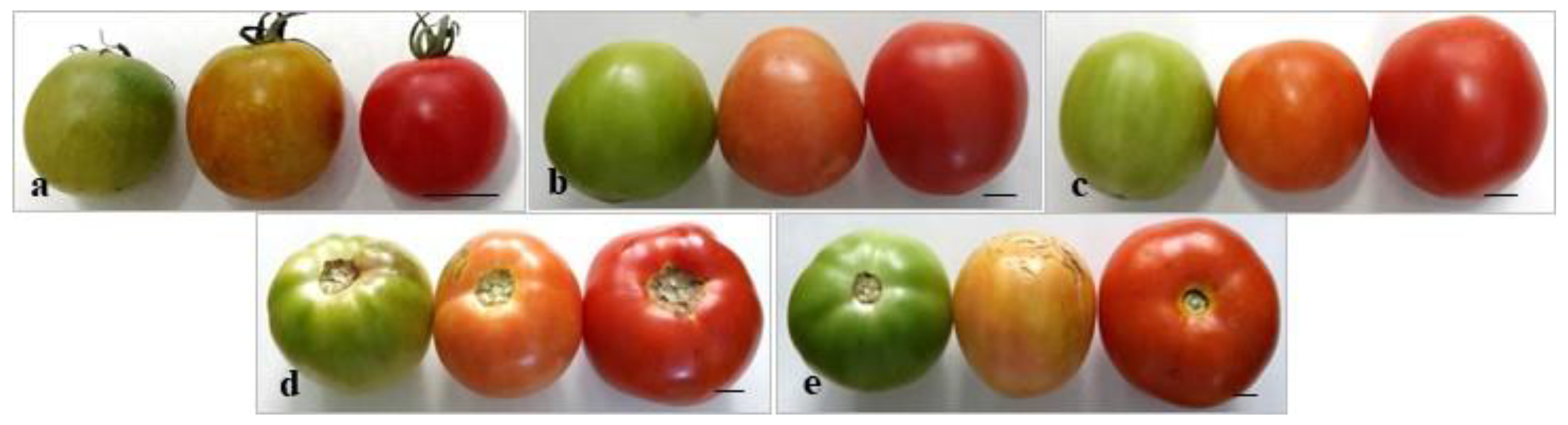
Five tomato (Lycopersicon esculentum Mill., cvs. ‘Capriciu’, ‘Darsirius’, ‘Kristin’, ‘Pontica’ and ‘Siriana’) genotypes (Figure 1a–e) created and approved in Romania have been selected to study the morphogenic response to in vitro cultures of various explant types based on their horticultural and economically valuable traits:
- cv. ‘Capriciu’—created at S.C.D.L. Buzău (Romania) and approved in 2007. Is drought tolerant and has good storage resistance. Has indetermined growth is suitable for fresh consumption or trade.
- cv. ‘Darsirius’—created at S.C.D.L. Buzău and approved in 2009. Has reduced number of seeds and is resistant to specific tomato diseases and nematodes. Has determined growth, intended for processing.
- cv. ‘Kristin’—created at S.C.D.L. Buzău and approved in 2006. Is resistant to transport and storage, has a determined growth and is suited for processing.
- cv. ‘Pontica’—created at I.C.D.L.F. Vidra (Romania) approved in 1988 and re-homologated in 2009. Has reduced number of seeds and is considered highly productive. Has a determined growth and is suitable for processing.
- cv. ‘Siriana’ (F1 hybrid)—created at S.C.D.L. Buzău and approved in 2006. Is a hybrid with high adaptability to environmental conditions and resistant to transport and storage. Has indetermined growth and is suitable for fresh consumption.
The seeds have been provided by the Research and Development Institute for Vegetable and Flower Growing Vidra, Romania.
Figure 1.
Tomato genotypes used for initiation of in vitro cultures starting from seeds. (a) cv. ‘Capriciu’; (b) cv. ‘Darsirius’; (c); cv. ‘Kristin’; (d) cv. ‘Pontica’; (e) cv. ‘Siriana’. Size bars = 1.0 cm.
Figure 1.
Tomato genotypes used for initiation of in vitro cultures starting from seeds. (a) cv. ‘Capriciu’; (b) cv. ‘Darsirius’; (c); cv. ‘Kristin’; (d) cv. ‘Pontica’; (e) cv. ‘Siriana’. Size bars = 1.0 cm.

2.2. Seed Germination—Establishment of In Vitro Cultures
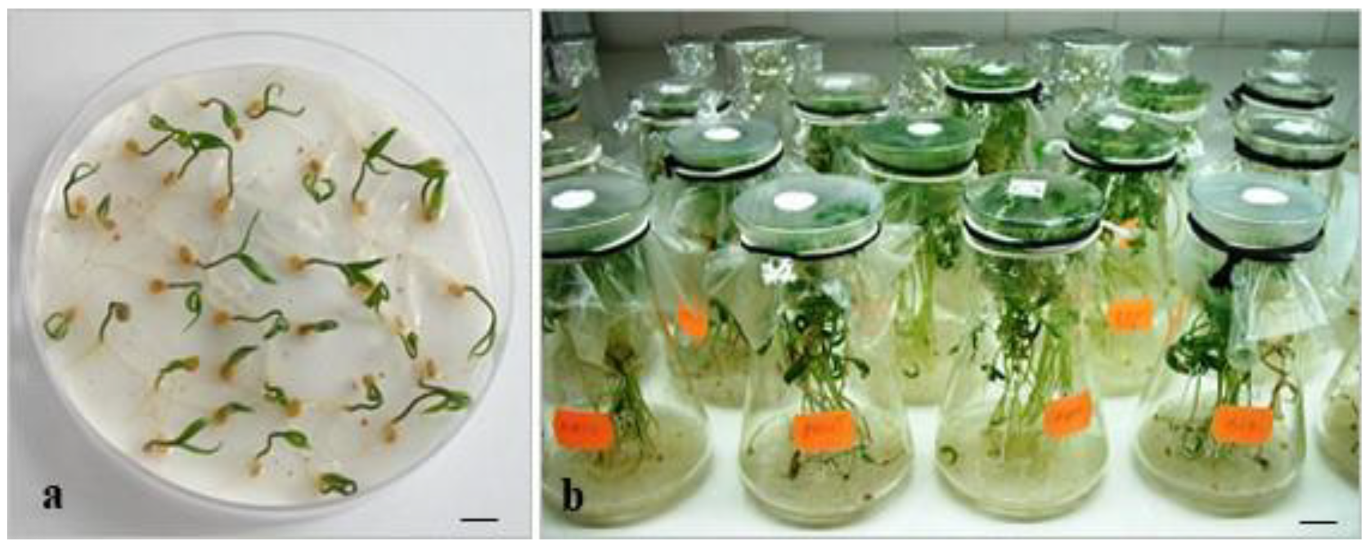
Seed health status was assessed using a stereomicroscope in order to remove empty, small or damaged seeds. To test the germination capacity, two years old mature seeds were germinated under ex vitro conditions. Seeds were first washed under tap water and transferred to Petri dishes (25 seeds/dish with 10 cm diameter) on moistened filter paper. The seeds in covered Petri dishes were randomly stored in darkness for 5 days (at 24 ± 1 °C) and then transferred (dish lid was removed) under light conditions (16 h light/8 h dark photoperiod and 36 µmol m−2 s−1 photosynthetic active radiation) at 24 ± 1 °C (Figure 2a).
For the in vitro germination tests, seed surface disinfection was carried out as follows: washing under tap water (1 h), dipping in 75% commercial Clorox (active chlorine content 5%) solution for 15 min and thorough rinsing with sterile distilled water. The seeds were then transferred for germination in 300 mL Erlenmeyer flasks (10 seeds/flask) on half-strength culture medium (1/2 MS) [21] supplemented with 20 g L−1 sucrose and solidified with 7.6 g L−1 agar (Figure 2b). The medium pH was adjusted to 5.7 before autoclaving (20 min at 121 °C). The in vitro germination conditions were similar to those described for ex vitro germination.
2.3. Morphogenic Response of Various Explants
Based on preliminary experiments related to the efficiency of various plant growth regulators on shoot regeneration from various explants, a MS medium supplemented with 1.5 mg L−1 zeatin (Z) and 0.2 mg L−1 indole-3-butyric acid (IBA), 20 g L−1 sucrose and 7.6 g L−1 agar (pH as mentioned above) was selected for further studies (unpublished data).
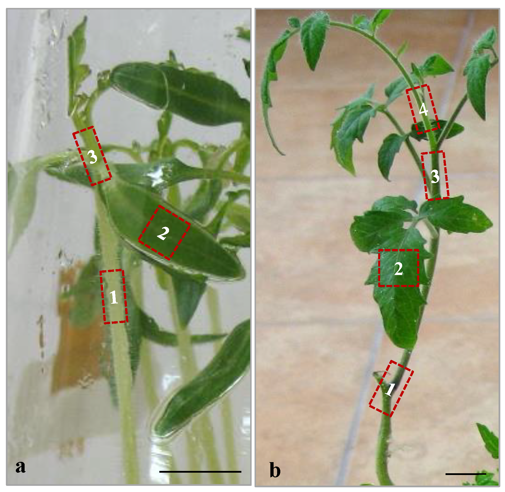
To identify the highest in vitro regeneration frequency the following explants have been studied: (a) cotyledons, (b) cotyledonary nodes, (c) hypocotyls, (d) leaf explants, (e) internodes, (f) stem nodes and (g) apical buds. The explants were excised on 25th day from seedlings resulted from in vitro germinated seeds. The length of cotyledonary nodes, hypocotyls, internodes, stem nodes and apical buds was approximately 1–1.5 cm, while cotyledons and leaf explants (both excised from the central part) had approximately 1 cm2 (Figure 3). Leaf explants and cotyledons were placed with the abaxial side towards the medium surface, hypocotyls, internodes were placed parallel to the medium surface, while cotyledonary nodes, stem nodes and apical buds were placed vertically into the solid culture medium.
2.4. Direct Shoot Organogenesis from Stem Nodes and Apical Buds
Due to the promising results obtained in shoot regeneration from stem nodes and apical buds, the effects of various cytokinins on these explants were further studied. For this purpose, seedlings (resulted from in vitro germinated seeds) were micropropagated (two successive subcultures each at 30 days) to serve as source of explants. Stem nodes and apical buds (both approximately 1–1.5 cm in length) were transferred to 100 mL Erlenmeyer flasks on MS culture medium supplemented with the following cytokinins: 1.5 mg L−1 thidiazuron (TDZ) (V1), 1.5 mg L−1 Z (V2), 1.5 mg L−1 kinetin (K) (V3) or 1.5 mg L−1 N6-benzyladenine (BA) (V4), each in combination with 0.2 mg L−1 IBA, 20 g L−1 sucrose and 7.6 g L−1 agar at pH 5.7. MS culture medium without PGRs was used as control (V0). For induction of shoots the samples were transferred and maintained in light and temperature conditions as mentioned for seed germination, throughout the experiments.
2.5. Assessment of Germination and Regeneration
The following characteristics have been assessed:
- (a)
- ex vitro and in vitro germination percentage and the mean germination time;
The total germination percentage (TGP) was calculated using the following equation:
where G = the number of germinated seeds by the end of the experiment; n = the total number of tested seeds.
TGP (%) = G/n × 100
Mean germination time (MGT) was calculated using the equation:
where n = number of seeds germinated every five days for a period up to 25 days; d = number of days from the beginning of the experiment; N = total number of seeds germinated at the end of the experiment [22].
MGT = ∑ (n × d)/N
The germination experiments followed a completely randomized design, with five replications of 10 seeds each (50 seeds/cultivar). Germination was considered complete once the protruded radicle reached 1 cm in length [23].
- (b)
- in vitro regeneration rates of various explants;
- (c)
- in vitro shoot regeneration percentages of stem nodes and apical buds on culture media with various cytokinins;
- (d)
- height of shoots (resulted from stem nodes and apical buds) and length of primary roots. The measurements were made using a ruler.
- (e)
- explants (%) forming callus. Visual observations on callus morphology, color, texture were recorded and the callus diameter was measured.
Parameters (b–e) were assessed on the 45th day of culture. For each explant type and culture medium variant, 5 jars (3 explants/jar, 15 explants in total) were used in each of the three replicates per treatment. The experiments were repeated twice. The results were expressed as mean values ± standard deviation (SD).
2.6. Statistical Analyses
Data expressed as percentages were arcsin transformed prior to statistical analysis. Data were subjected to one-way analysis of variance (ANOVA) using SPSS (v.11.5 for Windows). Duncan multiple-range test was used to compare differences between means.
The Student t-test was applied to evaluate significant differences between ex vitro and in vitro germination of seeds, and differences in the MGT between the two procedures within a cultivar. Differences were considered statistically significant at p ≤ 0.05. In addition to the statistical analyses the Pearson’s correlation coefficient was determined between the height of shoots and the length of roots for each cultivar on the four culture medium variants using the Excel spreadsheet software (v16.0 Microsoft).
3. Results
3.1. Seed Germination—Establishment of In Vitro Cultures
3.2. Morphogenic Response of Various Explant Types
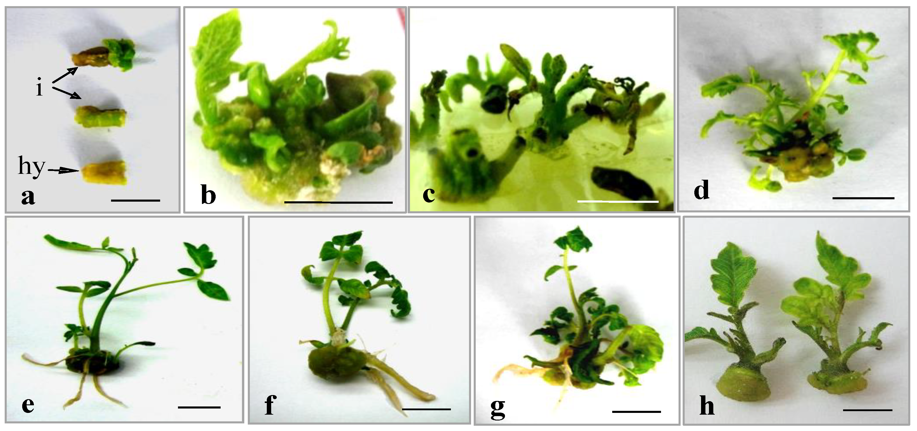
All genotypes showed indirect shoot regeneration from hypocotyls, internodes and leaf explants. Hypocotyls and internodes displayed swollen edges with yellowish and respectively green callus formed after approximately 10–15 days (Figure 4a). Leaf explants started to form shoots after 25 days in culture (Figure 4b). In cotyledonary nodes (Figure 4c), stem nodes (Figure 4d–g), and apical buds (Figure 4h) direct shoot regeneration was recorded whereas callus was induced at the base of explants.
After 45 days in culture the various explants showed significant differences in regeneration percentages within a cultivar (Table 2). Thus, apical buds displayed significantly higher regeneration percentages in all cultivars compared to most of the explants tested. The cultivar with the highest regeneration rates was cv. ‘Kristin’ (90% apical buds and 84% stem nodes). The lowest shoot formation rates in all cultivars were obtained for cotyledons (between 8% in cv. ‘Siriana’ and 11% in cv. ‘Capriciu’) (Table 2).
3.3. Direct Shoot Organogenesis from Stem Nodes and Apical Buds
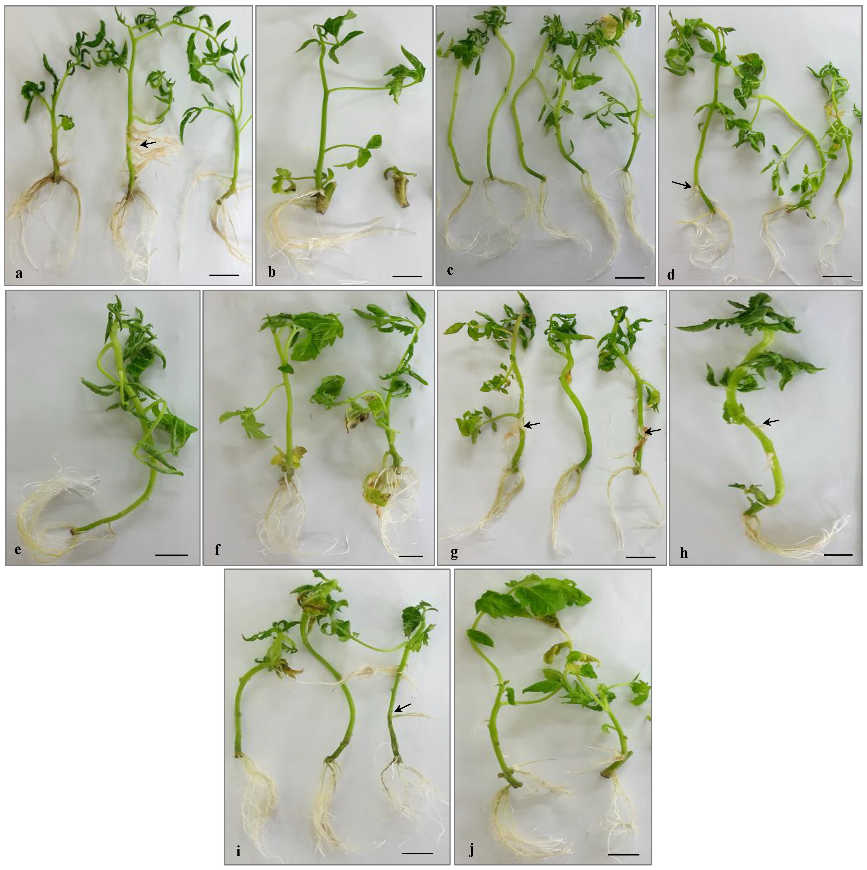
Due to the convincing positive results obtained with stem nodes and apical buds in terms of shoot regeneration, these two explant types were further used to study the effects of various cytokinins on direct shoot organogenesis. A comparison of stem nodes and apical buds grown on various culture media variants showed significant differences regarding the effects of cytokinins on the two explant types (Table 3 and Table 4). For instance, in all cultivars the cytokinin that lead to the highest frequency of regenerating stem nodes and apical buds was Z, although, the differences were not always significantly different (Table 3 and Table 4), (Figure 5a–j). The height of shoots and length of primary roots varied significantly according to the cytokinin used. The Pearson’s correlation coefficients between height of shoot and length of roots are shown in Table 5. In stem nodes, the height of shoots had positive correlation with the length of roots in cv. ‘Darsirius’ (0.84), and a negative correlation in cv. ‘Pontica’ (−0.25). In apical buds the correlation was strong positive cv. ‘Capriciu’ (0.96), and cv. ‘Kristin’ (0.98) (Table 5). Rooting was observed in all genotypes (for both explant types) even on medium without PGRs (V0) (Figure 5a,b). In both explants a high number of lateral branches was observed in all cultivars; these roots could not be counted and their length could not be measured (Figure 5a–j). The roots were formed regardless of genotype and hormonal treatment always from the explants and not from the callus developed at explant base (Figure 6a,b,h,i). Some cultivars showed adventitious root formation in particular on stems developed from apical buds explants (Figure 6a,g,i).
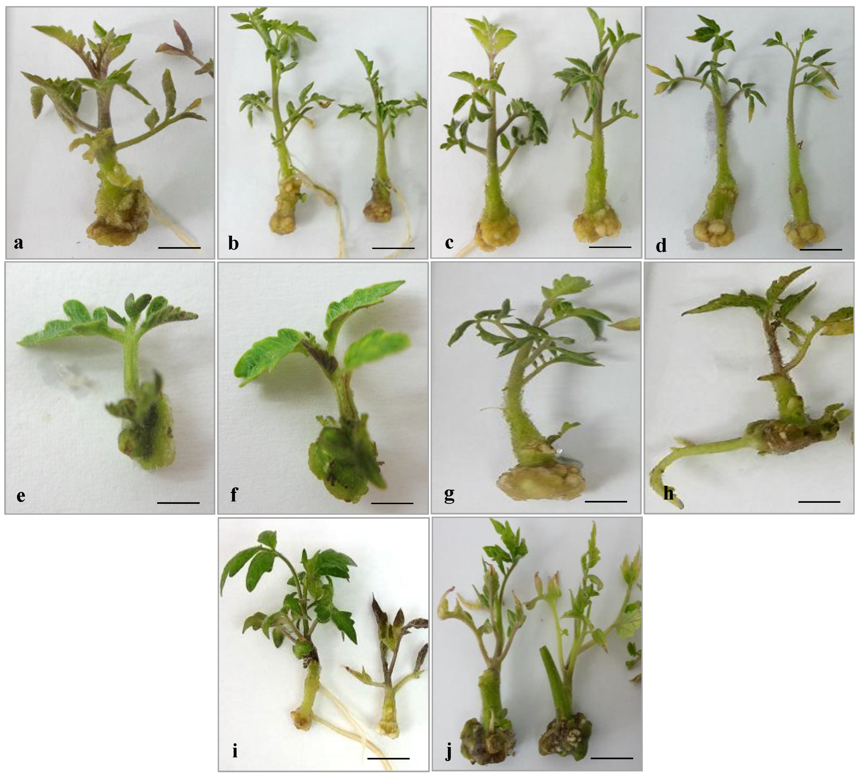
A common issue observed in both explant types was spontaneous callus formation at the base of the explants (Figure 6a–j). Stem nodes showed a higher percentage of callus formation compared to apical buds (Table 6). The percentage of apical buds forming callus ranged between 9% on medium with K (V3) (cv. ‘Pontica’) and 35% on medium with BA (V4) (cv. ‘Kristin’) (Table 6). For stem nodes the highest percentage of callus induction was 40% on V4 (cv. ‘Kristin’). The nature and size of callus varied widely according to cultivar and medium composition (Table 6).
A common issue observed in both explant types (on media with cytokinins) was spontaneous callus formation at the explant base (Figure 6a–j). No callus development was observed in none of the tested explant types on culture medium without PGRs (V0). Various cytokinins lead to significant differences regarding the percentages of explants forming callus (Table 6). In stem nodes of cv. ‘Kristin’ 39% of explants showed callus formation on medium with BA (V4), whereas in apical buds the percentage of explants forming callus ranged between 9% on medium with K (V3) (cv. ‘Pontica’) and 34% on medium with BA (V4) (cv. ‘Kristin’) (Table 6). The nature and size of callus varied widely according to cultivar and medium composition (Table 6). Especially on medium with TDZ (V1), calluses of cv. ‘Capriciu’ were nodular, extremely compact, almost lignified with a yellow-green colour (Figure 6a,b). Callus with the same appearance but less compact was present in cv. ‘Siriana’ on culture medium with K (V3) (Figure 6i,j). Cultivars ‘Kristin’ and ‘Pontica’ displayed friable callus with a dark-brown colour (Figure 6e–h).
4. Discussion
Considering the economic value of the targeted Romanian cultivars that had not been previously studied, the development of an efficient regeneration protocol is essential. Therefore, in the present study, the morphogenic response (shoot, root, and callus formation) of various explants (cotyledons, cotyledonary nodes, hypocotyls, leaf explants, internodes, stem nodes and apical buds) to in vitro culture conditions was investigated. Based on the obtained results, subsequently a direct shoot organogenesis protocol from stem nodes and apical buds was established after testing the explants reaction on culture media with various cytokinins. Development of effective in vitro micropropagation protocols to obtain high-quality tomato plants could significantly reduce the market value of seedlings, which became expensive, especially for the valuable cultivars. Such plant biotechnology tools opened great opportunities for genetic engineering of tomatoes [24]. Plant regeneration from cultured tissues is genetically controlled and factors such as the age and physiological condition influences the response of explants to in vitro culture conditions [25]. It was shown that high germination rates are important to obtain homogenous sets of shoots used for the induction of tissue cultures [26]. There were no significant differences in TGP and MGT between ex vitro and in vitro germination conditions within the same cultivar. However, overall high germination percentages were registered, varying between 68–91% (cvs. ‘Darsirius’ and ‘Capriciu’) for ex vitro conditions and 71–89% (cv. ‘Darsirius’ and cv. ‘Pontica’ for in vitro germination tests. The shortest MGT was obtained in cv. ‘Capriciu’ for both ex vitro (9 days) and in vitro (11 days) germination procedures. It was reported that the MGT in tomato under ex vitro conditions was 20% lower when zinc oxide nanoparticles with a certain size and concentration were used [27] or when priming treatments were applied [28].
There are a broad range of studies showing that in vitro tomato regeneration was dependent on the genotype, type of explants, age, type and concentration of plant growth regulators, and growth conditions [29,30,31]. For example, high proliferation efficiency was reported for hypocotyls [4,32,33], nodal explants [34], cotyledonary leaves and nodes [35,36], and petiole explants [37]. Our results confirmed that among the explant types studied, apical buds and stem nodes displayed the highest percentage of response in all cultivars. On the other hand, we obtained lower regeneration percentages, for hypocotyls (between 10% in cv. ‘Pontica’ and 16% cv. in ‘Kristin’), internodes (between 8% cv. ‘Siriana’ and 21% cv. ‘Capriciu’), and cotyledonary nodes (up to 19% cv. ‘Capriciu’), results that confirm that the explant type influences the in vitro regeneration efficiency. A successful plant regeneration system has been reported from different tomato explant types on medium supplemented with TDZ and auxin [38]. In contrast, the medium fortified with TDZ did not improve regeneration in various explants in tomato, while Z showed the best results concerning multiple shoot induction [39].
In our study, we obtained the highest efficiency in terms of direct shoot organogenesis from apical buds (98% cv. ‘Capriciu’) on culture media with Z and IBA. Some authors reported 80% direct shoot regeneration from tomato shoot tips grown on MS medium with Z and IAA [40]. Other studies showed high shoot regeneration of apical buds on media supplemented with K and BA [41]. It was underlined that TDZ inhibits root formation in in vitro grown tomato [36]. In the studied cultivars rooting occurred for stem nodes and apical buds on media containing TDZ and IBA. Some authors reported high number of roots per shoot on culture media without PGRs [42]. Similarly, we obtained rooting in all genotypes (for both explant types) even on medium without PGRs. Some cultivars showed adventitious root formation mainly on stems developed from apical buds. It was shown that IBA, the most common exogenously applied plant growth regulator, has a greater ability to promote adventitious root formation than IAA [43].
Indirect organogenesis via callus in tomato was reported [30]. Callus induction for various purposes from different explants, such as internodes [44], cotyledon explants [45], and hypocotyls [46] was studied. In the present study, callus formation at the explants base was a spontaneous effect of the in vitro culture (on media with a cytokinin and IBA), which did not influence direct shoot organogenesis. It was shown that callus formed at the base of nodal explants did not suppress regeneration of shoots and had no further consequences on growth of tomato plants [17]. Nevertheless, direct organogenesis from explants is the best option for multiplication as it leads to the generation of true-to-type plants [47]. To preserve all the traits of the cultivars genetic uniformity is of paramount importance especially for micropropagation. However, further studies are necessary to assess if morphological changes that could arise following ex vitro acclimatization of seedlings till ripening.
5. Conclusions
The present study describes a direct organogenesis protocol from stem nodes and apical buds in five Romanian tomato cultivars. It can be concluded that the response of different tomato genotypes and explants on various culture media was different in terms of organogenesis. Optimal direct shoot bud induction was obtained in cv. ‘Capriciu’ using a solid MS medium supplemented with 1.5 mg L−1 Z and 0.2 mg L−1 IBA for both apical buds and stem nodes. Establishment of highly efficient regeneration protocol for these commercially valuable cultivars is definitely justified. Based on the promising results in all mentioned parameters we anticipate that these five Romanian tomato genotypes will represent new resources for improved breeding varieties and approaches and research towards preserving local cultivars.
Author Contributions
Conceptualization: A.H., A.C., C.D. and I.B.; Methodology: A.C., A.H. and C.D.; Validation: A.H. and A.C.; Formal analysis: A.C., A.H. and I.B.; Investigation: A.H., A.C., C.D. and I.B.; Data curation: A.H. and A.C.; Original draft preparation: A.H. and A.C.; Writing—review and editing: A.H., A.C., C.D. and I.B. Funding acquisition: A.H. All authors have read and agreed to the published version of the manuscript.
Funding
This work was supported by partial funding from the Ministry of Research, Innovation and Digitization through the Core Project BIORESGREEN, subproject BioClimpact no. 7/30.12.2022, code 23020401 and Projects for Excellence Financing in RDI, project 2PFE/2021.
Data Availability Statement
Not applicable.
Acknowledgments
The authors are grateful to Gabriela Șovărel (Research and Development Institute for Vegetable and Flower Growing Vidra, Romania) for providing the tomato seeds.
Conflicts of Interest
The authors declare no conflict of interest related to this article. The funders had no role in the design of the study, analyses, data interpretation, manuscript writing or in the decision to publish the results.
References
- Larkin, P. Somaclonal variation: Origin and causes. In Encyclopedia of Plant and Crop Science; Goodman, R.M., Ed.; Marcel Dekker: New York, NY, USA, 2004; pp. 1158–1161. [Google Scholar]
- Rao, K.V.; Kiranmayee, K.; Pavan, U.; Sree, T.J.; Rao, A.V.; Sadanandam, A. Induction of multiple shoots from leaf segments, in vitro-flowering and fruiting of a dwarf tomato (Lycopersicon esculentum). J. Plant Physiol. 2005, 162, 959–962. [Google Scholar] [CrossRef]
- Story, E.N.; Kopec, R.E.; Schwartz, S.J.; Harris, G.K. An update on the health effects of tomato lycopene. Ann. Rev. Food Sci. Technol. 2010, 1, 189–210. [Google Scholar] [CrossRef] [PubMed] [Green Version]
- Gubiš, J.; Lajchová, Z.; Faragó, J.; Jureková, Z. Effect of growth regulators on shoot induction and plant regeneration in tomato (Lycopersicon esculentum Mill.). Biologia 2004, 59, 405–408. [Google Scholar]
- Coste, A.; Suteu, D.; Bacila, I.; Deliu, C.; Valimareanu, S.; Halmagyi, A. Genetic stability monitoring in micropropagated tomato cultivars. Olten. Stud. Comun. Stiintele Nat. 2014, 30, 38–43. [Google Scholar]
- Coste, A.; Şuteu, D.; Băcilă, I.; Deliu, C.; Vălimăreanu, S.; Halmagyi, A. Genetic integrity assessment of cryopreserved tomato (Lycopersicon esculentum Mill.) genotypes. Turk. J. Biol. 2015, 39, 638–648. [Google Scholar] [CrossRef] [Green Version]
- Muntean, C.M.; Leopold, N.; Tripon, C.; Coste, A.; Halmagyi, A. Surface-enhanced raman spectroscopy of genomic DNA from in vitro grown tomato (Lycopersicon esculentum Mill.) cultivars before and after plant cryopreservation. Spectrochim. Acta Part A Mol. Biomol. Spect. 2015, 144, 107–114. [Google Scholar] [CrossRef] [PubMed]
- Halmagyi, A.; Coste, A.; Tripon, S.; Craciun, C. Low temperature induced alterations in tomato (Lycopersicon esculentum Mill.) shoot apex cells. Sci. Hortic. 2017, 222, 22–31. [Google Scholar] [CrossRef]
- Di Matteo, A.; Rigano, M.M.; Sacco, A.; Frusciante, L.; Barone, A. Genetic transformation in tomato: Novel tools to improve fruit quality and pharmaceutical production. In Genetic Transformation; Alvarez, M., Ed.; InTech Europe: London, UK, 2011; pp. 55–80. [Google Scholar]
- Xia, X.; Cheng, X.; Li, R.; Yao, J.; Li, Z.; Cheng, Y. Advances in application of genome editing in tomato and recent development of genome editing technology. Theor. Appl. Gen. 2021, 134, 2727–2747. [Google Scholar] [CrossRef]
- Ali, M.Y.; Sina, A.A.I.; Khandker, S.S.; Neesa, L.; Tanvir, E.M.; Kabir, A.; Khalil, M.I.; Gan, S.H. Nutritional composition and bioactive compounds in tomatoes and their impact on human health and disease: A review. Foods 2021, 10, 45. [Google Scholar] [CrossRef]
- Raza, M.A.; Nawaz, A.; Ali, M.; Zaynab, M.; Muntha, S.T.; Zaidi, S.H.R.; Khan, A.R.; Zheng, X.L. In-vitro regeneration and development for the conservation and propagation of tomato plant (Solanum lycopersicum) and currant tomato (S. pimpinellifolium) from two different explants. Appl. Ecol. Environ. Res. 2020, 18, 879–888. [Google Scholar] [CrossRef]
- Magdoleen, G.; Osman, M.G.; Mutasim, M.; Khalafalla, M.M. Promotion of in vitro shoot formation from shoot tip of tomato (Lycopersicon esculentum Mill. cv. Omdurman) by ethylene inhibitors. Int. J. Curr. Res. 2010, 4, 82–86. [Google Scholar]
- Bhatia, P.; Ashwath, N.; Senaratna, T.; Midmore, D. Tissue culture studies of tomato (Lycopersicon esculentum). Plant Cell Tissue Organ Cult. 2004, 78, 1–21. [Google Scholar] [CrossRef]
- Saeed, W.; Naseem, S.; Gohar, D.; Ali, Z. Efficient and reproducible somatic embryogenesis and micropropagation in tomato via novel structures—Rhizoid tubers. PLoS ONE 2019, 14, e0215929. [Google Scholar] [CrossRef] [PubMed] [Green Version]
- Koeda, S.; Takisawa, R.; Nabeshima, T.; Tanaka, Y.; Kitajima, A. Production of tomato yellow leaf curl virus-free parthenocarpic tomato plants by leaf primordia-free shoot apical meristem culture combined with in vitro grafting. Hortic. J. 2015, 84, 327–333. [Google Scholar] [CrossRef] [Green Version]
- Rashid, R.; Bal, S.S. Effect of hormones on direct shoot regeneration in hypocotyl explants of tomato. Not. Sci. Biol. 2010, 2, 70–73. [Google Scholar] [CrossRef] [Green Version]
- Mohamed, A.N.; Ismail, M.R.; Rahman, M.H. In vitro response from cotyledon and hypocotyls explants in tomato by inducing 6-benzylaminopurine. Afr. J. Biotech. 2010, 9, 4802–4807. [Google Scholar]
- Chaudhary, Z.; Afroz, A.; Rashid, H. Effect of variety and plant growth regulators on callus proliferation and regeneration response of three tomato cultivars (Lycopersicon esculentum). Pak. J. Bot. 2007, 39, 857–869. [Google Scholar]
- Godishala, V.; Mangamoori, L.; Nanna, R. Plant regeneration via somatic embryogenesis in cultivated tomato (Solanum lycopersicum L.). J. Cell Tissue Res. 2011, 11, 2521–2528. [Google Scholar]
- Murashige, T.; Skoog, F. A revised medium for rapid growth and bioassays with tobacco tissue cultures. Physiol. Plant. 1962, 15, 473–497. [Google Scholar] [CrossRef]
- Ellis, R.H.; Roberts, E.H. The quantification of ageing and survival in orthodox seeds. Seed Sci. Technol. 1981, 9, 373–409. [Google Scholar]
- International Seed Testing Association. Rules Proposal for the International Rules for Seed Testing; International Seed Testing Association (ISTA): Zurich, Switzerland, 2016. [Google Scholar]
- Gerszberg, A.; Hnatuszko-Konka, K.; Kowalczyk, T.; Kononowicz, A.K. Tomato (Solanum lycopersicum L.) in the service of biotechnology. Plant Cell Tissue Organ Cult. 2015, 120, 881–902. [Google Scholar] [CrossRef] [Green Version]
- Delporte, F.; Pretova, A.; Du Jardin, P.; Watillon, B. Morpho-histology and genotype dependence of in vitro morphogenesis in mature embryo cultures of wheat. Protoplasma 2014, 251, 1455–1470. [Google Scholar] [CrossRef] [PubMed] [Green Version]
- Ishag, S.; Osman, M.G.; Khalafalla, M.M. Effects of growth regulators, explant and genotype on shoot regeneration in tomato (Lycopersicon esculentum cv. Omdurman). Int. J. Sustain. Crop Prod. 2009, 4, 7–13. [Google Scholar]
- Włodarczyk, K.; Smolińska, B. New method of tomato fertilization with the use of chosen nanoparticles. Chem. Proc. 2022, 10, 92. [Google Scholar] [CrossRef]
- Delian, E.; Lupu, C.; Săvulescu, E. Effect of different priming treatments on seeds germination and early seedlings growth of tomato. Curr. Trends Nat. Sci. 2018, 7, 38–46. [Google Scholar]
- Mamidala, P.; Nanna, R.S. Effect of genotype, explant source and medium on in vitro regeneration of tomato. Int. J. Gen. Mol. Biol. 2011, 3, 5–50. [Google Scholar]
- Gerszberg, A.; Hnatuszko-Konka, K.; Kowalczyk, T.; Kononowicz, A.K. Efficient in vitro callus induction and plant regeneration protocol for different polish tomato cultivars. Not. Bot. Horti Agrobot. 2016, 44, 452–458. [Google Scholar] [CrossRef] [Green Version]
- Titeli, V.S.; Zafeiriou, I.; Laskaridou, A.; Menexes, G.; Madesis, P.; Stavridou, E.; Nianiou-Obeidat, I. Development of a simple and low-resource regeneration system of two greek tomato varieties. Agriculture 2021, 11, 412. [Google Scholar] [CrossRef]
- Gubiš, J.; Lajchová, Z.; Faragó, J.; Jureková, Z. Effect of genotype and explant type on shoot regeneration in tomato (Lycopersicon esculentum Mill.) in vitro. Czech J. Genet. Plant Breed. 2003, 39, 9–14. [Google Scholar] [CrossRef] [Green Version]
- Hanur, V.S.; Krishnareddy, B. In vitro organogenesis in tomato (Solanum lycopersicum) using kinetin. Adv. Plants Agric. Res. 2016, 4, 397–401. [Google Scholar] [CrossRef]
- Soressi, G.P.; Cammareri, G.; Picarella, M.E. Improvement of in vitro vegetative propagation technique in tomato (Solanum lycopersicum). Acta Hortic. 2009, 812, 283–288. [Google Scholar] [CrossRef]
- Alatar, A.A.; Faisal, M.; Abdel-Salam, E.M.; Canto, T.; Saquib, Q.; Javed, S.B.; El-Sheikh, M.A.; Al-Khedhairy, A.A. Efficient and reproducible in vitro regeneration of Solanum lycopersicum and assessment genetic uniformity using flow cytometry and SPAR methods. Saudi J. Biol. Sci. 2017, 24, 1430–1436. [Google Scholar] [CrossRef] [Green Version]
- Ashakiran, K.; Sivankalyani, V.; Jayanthi, M.; Govindasamy, V.; Girija, S. Genotype specific shoots regeneration from different explants of tomato (Solanum lycopersicum L.) using TDZ. Asian J. Plant Sci. Res. 2011, 1, 107–113. [Google Scholar]
- Sharma, C.; Srivastava, D.K. In vitro plant regeneration from seedlings-derived explants of tomato (Lycopersicon esculentum Mill.). Int. J. Agric. Environ. Biotechnol. 2014, 7, 421–432. [Google Scholar] [CrossRef]
- Vinoth, R.; Kumaravel, S.; Ranganathan, R. Anatomical and physiological adaptation of mangrove wetlands in east coast of Tamil Nadu. World Sci. News 2019, 129, 161–179. [Google Scholar]
- Sandhya, D.; Jogam, P.; Venkatapuram, A.P.; Savitikadi, P.; Peddaboina, V.; Allini, V.R.; Abbagani, S. Highly efficient Agrobacterium-mediated transformation and plant regeneration system for genome engineering in tomato. Saudi J. Biol. Sci. 2022, 29, 103292. [Google Scholar] [CrossRef]
- Jabeen, N.; Chaudhry, Z.; Rashid, H.; Mirza, B. Effect of genotype and explant type on in vitro shoot regeneration of tomato (Lycopersicon esculentum Mill.). Pak. J. Bot. 2005, 37, 899–903. [Google Scholar]
- Jamous, F.; Abu-Qaoud, H. In vitro regeneration of tomato (Lycopersicon esculentum Mill). Plant Cell Biotech. Mol. Biol. 2015, 16, 181–190. [Google Scholar]
- Baye, E.; Matewos, T.; Belew, D. Optimization of in vitro rooting protocol for tomato (Lycopersicon esculentum Mill.) varieties. J. App. Nat. Sci. 2020, 12, 365–371. [Google Scholar] [CrossRef]
- De Klerk, G.J.; Van der Krieken, W.; De Jong, J.C. The formation of adventitious roots: New concepts, new possibilities. In Vitro Cell Dev. Biol. Plant 1999, 35, 189–199. [Google Scholar] [CrossRef]
- Karim, M.A.; Kayuml, M.A. In vitro regeneration of tomato plant from leaf and internode segments. J. Bangladesh Agric. Univ. 2007, 5, 213–216. [Google Scholar] [CrossRef]
- Arulananthu, G.; Bhat, S.G.; Ramesh, N. Callus induction and in-vitro regeneration of tomato (Lycopersicon esculentum Mill.). Res. J. Life Sci. Bioinform. Pharm. Chem. Sci. 2019, 5, 491. [Google Scholar]
- Manawadu, I.P.; Nilanthi, D.; Senanayake, S.G.J.N. Callus formation and organogenesis of tomato (Lycopersicon esculentum Mill variety Thilina). Trop. Agric. Res. Ext. 2014, 17, 86–94. [Google Scholar] [CrossRef] [Green Version]
- Verma, V.; Zinta, G.; Kanwar, K. Optimization of efficient direct organogenesis protocol for Punica granatum L. cv. Kandhari Kabuli from mature leaf explants. In Vitro Cell. Dev. Biol. Plant 2021, 57, 48–59. [Google Scholar] [CrossRef]
Figure 2.
Tomato seed germination. (a) ex vitro seed germination (cv. ‘Kristin’); (b) in vitro seed germination (cv. ‘Kristin’). Size bars = 1.0 cm.
Figure 2.
Tomato seed germination. (a) ex vitro seed germination (cv. ‘Kristin’); (b) in vitro seed germination (cv. ‘Kristin’). Size bars = 1.0 cm.

Figure 3.
Explants used for in vitro regeneration experiments. (a) 1—hypocotyl; 2—cotyledon; 3—cotyledonary node; (b) 1—stem node; 2—leaf explant; 3—internode; 4—apical bud. Bars 1 cm.
Figure 3.
Explants used for in vitro regeneration experiments. (a) 1—hypocotyl; 2—cotyledon; 3—cotyledonary node; (b) 1—stem node; 2—leaf explant; 3—internode; 4—apical bud. Bars 1 cm.

Figure 4.
Morphogenic response of various explant types after 45 days in culture. (a) internodes (i) and hypocotyl (hy) (cv. ‘Kristin’); (b) leaf explant (cv. ‘Kristin’); (c) cotyledonary nodes (cv. ‘Kristin’); (d) stem nodes (cv. ‘Kristin’); (e–g) stem nodes (cvs. ‘Capriciu’, ‘Darsirius’ and ‘Pontica’); (h) apical buds (cv. ‘Siriana’). Explants were grown on MS with 1.5 mg L−1 Z + 0.2 mg L−1 IBA. Size bars = 1.0 cm.

Figure 5.
Shoot regeneration and rooting of stem nodes and apical buds on culture media with various cytokinins. (a) cv. ‘Pontica’ apex (V0); (b) cv. ‘Pontica’ nodes (V0); (c) cv. ‘Capriciu’ apex (V1); (d) cv. ‘Capriciu’ nodes (V1); (e) cv. ‘Siriana’ apex (V2); (f) cv. ‘Siriana’ nodes (V2); (g) cv. ‘Kristin’ apex (V3); (h) cv. ‘Kristin’ nodes (V3); (i) cv. ‘Darsirius’ apex (V4); (j) cv. ‘Darsirius’ nodes (V4). Arrows represent adventitious roots. Size bars = 1 cm.

Figure 6.
Callus development on culture media with different cytokinins. (a) cv. ‘Capriciu’ apical bud (V1); (b) cv. ‘Capriciu’ stem node (V1); (c) cv. ‘Darsirius’ apical bud (V2); (d) cv. ‘Darsirius’ stem node (V2); (e) cv. ‘Kristin’ apical bud (V4); (f) cv. ‘Kristin’ stem node (V4); (g) cv. ‘Pontica’ apical bud (V4); (h) cv. ‘Pontica’ stem node (V4); (i) cv. ‘Siriana’ apical bud (V3); (j) cv. ‘Siriana’ stem node (V3). Bars 1 cm.


Table 1.
Ex vitro and in vitro germination of tomato seeds, 25 days after transfer to germination substrate.
Table 1.
Ex vitro and in vitro germination of tomato seeds, 25 days after transfer to germination substrate.
| Germination | TGP (% ± SD) * | MGT (Days) | |
|---|---|---|---|
| ‘Capriciu’ | ex vitro | 91.0 ± 3.5 | 9.6 |
| in vitro | 87.0 ± 1.7 | 11.5 | |
| ‘Darsirius’ | ex vitro | 68.0 ± 4.8 | 13.1 |
| in vitro | 71.6 ± 3.5 | 15.4 | |
| ‘Kristin’ | ex vitro | 77.3 ± 5.5 | 15.3 |
| in vitro | 78.6 ± 2.1 | 15.6 | |
| ‘Pontica’ | ex vitro | 91.3 ± 2.1 | 12.3 |
| in vitro | 89.0 ± 2.7 | 14.8 | |
| ‘Siriana’ | ex vitro | 75.0 ± 3.9 | 17.9 |
| in vitro | 72.6 ± 3.8 | 15.7 |
* Data represent mean values (±SD); TGP—Total germination percentage; MGT—Mean germination time; No significant differences were obtained between treatments within a cultivar (Student t-test, p ≤ 0.05).
Table 2.
In vitro shoot regeneration of various explants after 45 days in culture.
| Cultivar | Cotyledons | Cotyledonary Nodes | Hypocotyls | Leaf Explants | Internodes | Stem Nodes | Apical Buds |
|---|---|---|---|---|---|---|---|
| ‘Capriciu’ | 11.6 ± 1.6 e,* | 19.6 ± 2.6 d | 15.3 ± 3.5 de | 36.6 ± 3.0 c | 21.3 ± 2.1 d | 73.0 ± 3.5 b | 84.3 ± 3.1 a |
| ‘Darsirius’ | 9.00 ± 1.6 e | 10.3 ± 1.6 e | 11.6 ± 2.3 e | 34.0 ± 3.0 c | 20.3 ± 2.3 d | 68.3 ± 3.6 b | 78.3 ± 1.9 a |
| ‘Kristin’ | 10.3 ± 1.7 c | 14.3 ± 1.4 c | 16.3 ± 2.1 c | 25.6 ± 3.1 b | 10.6 ± 1.9 c | 84.6 ± 4.1 a | 90.0 ± 2.1 a |
| ‘Pontica’ | 9.30 ± 1.6 e | 15.0 ± 1.8 cde | 10.6 ± 2.2 de | 21.3 ± 3.1 c | 16.6 ± 1.7 cd | 70.0 ± 3.8 b | 82.3 ± 3.1 a |
| ‘Siriana’ | 8.30 ± 2.0 d | 14.6 ± 1.9 d | 11.0 ± 2.8 d | 22.3 ± 2.9 c | 8.00 ± 2.2 d | 67.3 ± 4.8 b | 77.3 ± 2.3 a |
* Data represent mean values (% ± SD). Values followed by the same letter, within a row, are not significantly different (p ≤ 0.05). Explants were grown on MS medium with 1.5 mg L−1 Z + 0.2 mg L−1 IBA.
Table 3.
Direct shoot organogenesis from stem nodes on culture media with different cytokinins.
| Cultivar | Medium | Explants Regenerating Shoots (% ± SD) * | Height of Shoots (cm ± SD) | Length of Primary Roots (cm ± SD) |
|---|---|---|---|---|
| ‘Capriciu’ | V0 | 65.5 ± 1.4 b | 8.7 ± 0.5 a | 6.1 ± 0.6 b |
| V1 | 78.8 ± 2.3 b | 7.8 ± 0.7 b | 6.4 ± 0.6 b | |
| V2 | 94.4 ± 1.3 a | 6.2 ± 0.7 c | 3.0 ± 0.7 c | |
| V3 | 73.3 ± 1.6 b | 4.0 ± 0.4 d | 6.2 ± 0.7 b | |
| V4 | 77.7 ± 1.7 b | 7.5 ± 0.5 b | 10.8 ± 0.9 a | |
| ‘Darsirius’ | V0 | 63.3 ± 1.4 c | 5.7 ± 0.5 b | 3.8 ± 0.5 d |
| V1 | 71.1 ± 1.4 bc | 9.3 ± 0.6 a | 15.6 ± 0.7 a | |
| V2 | 82.2 ± 1.1 a | 5.2 ± 0.5 bc | 3.5 ± 0.4 d | |
| V3 | 72.2 ± 0.8 abc | 9.1 ± 0.5 a | 9.8 ± 0.5 b | |
| V4 | 76.7 ± 1.5 ab | 4.5 ± 0.6 c | 5.7 ± 0.8 c | |
| ‘Kristin’ | V0 | 52.2 ± 1.0 c | 8.4 ± 0.7 a | 6.6 ± 0.8 a |
| V1 | 65.6 ± 1.5 b | 4.8 ± 0.5 c | 3.7 ± 0.3 c | |
| V2 | 84.4 ± 1.3 a | 2.7 ± 0.5 e | 3.0 ± 0.3 d | |
| V3 | 68.9 ± 1.8 b | 5.6 ± 1.0 b | 4.4 ± 0.4 b | |
| V4 | 70.0 ± 1.6 b | 3.7 ± 0.2 d | 2.8 ± 0.5 d | |
| ‘Pontica’ | V0 | 58.9 ± 0.8 b | 4.7 ± 0.6 c | 2.7 ± 0.8 d |
| V1 | 81.1 ± 1.5 a | 2.5 ± 0.5 e | 18.2 ± 1.5 a | |
| V2 | 88.9 ± 2.1 a | 3.8 ± 0.4 d | 7.2 ± 0.5 c | |
| V3 | 62.2 ± 1.3 b | 5.9 ± 0.6 b | 11.4 ± 0.6 b | |
| V4 | 80.0 ± 1.6 a | 8.5 ± 0.9 a | 10.3 ± 0.9 b | |
| ‘Siriana’ | V0 | 56.7 ± 1.0 b | 9.5 ± 0.6 b | 5.5 ± 0.4 a |
| V1 | 82.2 ± 1.6 a | 6.5 ± 0.3 c | 6.0 ± 0.4 a | |
| V2 | 84.4 ± 2.2 a | 3.5 ± 0.6 e | 2.4 ± 0.9 b | |
| V3 | 73.3 ± 1.5 a | 12.5 ± 0.6 a | 5.4 ± 0.7 a | |
| V4 | 74.4 ± 1.8 a | 4.8 ± 0.6 d | 6.1 ± 0.7 a |
* Data represent mean values (±SD). Values followed by the same letter, within the same row and studied aspect, were not significantly different (p ≤ 0.05).
Table 4.
Direct shoot organogenesis from apical buds on culture media with different cytokinins.
| Cultivar | Medium | Explants Regenerating Shoots (% ± SD) * | Height of Shoots (cm ± SD) | Length of Primary Roots (cm ± SD) |
|---|---|---|---|---|
| ‘Capriciu’ | V0 | 68.8 ± 1.8 b | 12.0 ± 0.4 a | 7.2 ± 0.5 a |
| V1 | 80.0 ± 1.4 b | 2.6 ± 0.3 bc | 5.8 ± 0.4 b | |
| V2 | 97.7 ± 0.5 a | 2.4 ± 1.7 bc | 5.1 ± 1.0 b | |
| V3 | 81.1 ± 0.7 b | 1.5 ± 1.3 c | 1.1 ± 0.9 d | |
| V4 | 80.0 ± 2.0 b | 3.0 ± 0.4 b | 3.2 ± 0.6 c | |
| ‘Darsirius’ | V0 | 64.4 ± 1.6 c | 10.0 ± 1.3 b | 6.6 ± 0.9 b |
| V1 | 76.7 ± 1.0 abc | 4.4 ± 1.2 c | 5.7 ± 2.5 b | |
| V2 | 88.9 ± 2.1 a | 15.7 ± 2.1 a | 4.9 ± 1.2 b | |
| V3 | 74.4 ± 1.2 bc | 8.8 ± 2.4 b | 14.8 ± 4.1 a | |
| V4 | 78.8 ± 2.1 ab | 3.9 ± 1.0 c | 7.6 ± 0.9 b | |
| ‘Kristin’ | V0 | 61.1 ± 1.3 c | 10.4 ± 0.9 a | 6.2 ± 0.7 a |
| V1 | 71.1 ± 1.2 bc | 3.2 ± 1.0 b | 2.3 ± 0.9 b | |
| V2 | 90.0 ± 1.4 a | 3.1 ± 0.6 bc | 2.6 ± 0.7 b | |
| V3 | 76.7 ± 1.4 b | 11.1 ± 1.4 a | 6.2 ± 1.5 a | |
| V4 | 75.5 ± 1.2 b | 2.0 ± 0.4 c | 1.9 ± 0.8 b | |
| ‘Pontica’ | V0 | 68.9 ± 2.1 b | 6.3 ± 0.5 a | 6.5 ± 1.2 c |
| V1 | 84.4 ± 0.8 a | 6.9 ± 0.9 a | 9.5 ± 1.3 a | |
| V2 | 91.1 ± 1.2 a | 5.0 ± 1.4 b | 1.8 ± 0.6 d | |
| V3 | 68.9 ± 1.6 b | 6.7 ± 0.6 a | 7.8 ± 1.1 b | |
| V4 | 81.1 ± 1.7 ab | 6.0 ± 0.9 ab | 2.1 ± 0.7 d | |
| ‘Siriana’ | V0 | 62.2 ± 1.2 b | 6.2 ± 0.8 a | 3.9 ± 0.5 b |
| V1 | 85.6 ± 2.3 a | 4.0 ± 0.7 bc | 1.1 ± 0.6 c | |
| V2 | 87.8 ± 1.7 a | 4.7 ± 1.2 b | 3.4 ± 0.5 b | |
| V3 | 76.7 ± 1.6 ab | 3.2 ± 0.7 cd | 4.9 ± 0.6 a | |
| V4 | 78.8 ± 2.2 a | 2.8 ± 0.8 d | 5.5 ± 0.7 a |
* Data represent mean values (±SD). Values followed by the same letter, within the same row and studied aspect, were not significantly different (p ≤ 0.05).
Table 5.
Correlation coefficients on shoots height and roots length of analyzed tomato genotypes.
| Cultivar | Explants | Pearson’s Coefficient |
|---|---|---|
| ‘Capriciu’ | stem nodes | 0.25 |
| apical buds | 0.96 | |
| ‘Darsirius’ | stem nodes | 0.84 |
| apical buds | −0.55 | |
| ‘Kristin’ | stem nodes | 0.78 |
| apical buds | 0.98 | |
| ‘Pontica’ | stem nodes | −0.25 |
| apical buds | 0.87 | |
| ‘Siriana’ | stem nodes | 0.50 |
| apical buds | −0.12 |
Table 6.
Callus formation from stem nodes and apical buds grown on culture media with different cytokinins.
Table 6.
Callus formation from stem nodes and apical buds grown on culture media with different cytokinins.
| Cultivar | Medium | Stem Nodes | Apical Buds | ||
|---|---|---|---|---|---|
| Callus Formation (% ± SD) * | Callus Diameter (cm ± SD) | Callus Formation (% ± SD) * | Callus Diameter (cm ± SD) | ||
| ‘Capriciu’ | V0 | 0 e | 0 e | ||
| V1 | 14.0 ± 2.2 d | <0.5 | 10.4 ± 2.10 d | <0.5 | |
| V2 | 38.8 ± 2.6 a | 0.5–1.0 | 25.4 ± 4.6 b | 0.5–1.0 | |
| V3 | 25.8 ± 4.9 b | 0.5–1.0 | 21.0 ± 2.9 bc | 0.5–1.0 | |
| V4 | 25.6 ± 3.6 b | ˃1.0 | 19.6 ± 5.1 c | ˃1.0 | |
| ‘Darsirius’ | V0 | 0 f | 0f | ||
| V1 | 22.6 ± 3.2 bc | <0.5 | 13.2 ± 3.3 e | <0.5 | |
| V2 | 18.6 ± 4.0 cd | 0.5–1.0 | 12.6 ± 3.0 e | 0.5–1.0 | |
| V3 | 18.4 ± 4.3 cd | <0.5 | 15.2 ± 3.3 de | <0.5 | |
| V4 | 26.0 ± 3.7 a | 0.5–1.0 | 18.6 ± 2.7 cd | 0.5–1.0 | |
| ‘Kristin’ | V0 | 0 f | 0f | ||
| V1 | 20.4 ± 3.3 d | <0.5 | 17.6 ± 4.3 de | <0.5 | |
| V2 | 21.4 ± 3.9 d | 0.5–1.0 | 15.6 ± 3.8 e | <0.5 | |
| V3 | 31.4 ± 3.8 c | <0.5 | 20.6 ± 3.2 d | 0.5–1.0 | |
| V4 | 39.6 ± 3.4 a | ˃1.0 | 34.8 ± 4.4 ab | ˃1.0 | |
| ‘Pontica’ | V0 | 0 e | 0 e | ||
| V1 | 30.4 ± 4.3 a | <0.5 | 25.8 ± 3.5 ab | <0.5 | |
| V2 | 30.0 ± 3.1 a | 0.5–1.0 | 22.0 ± 4.1 b | 0.5–1.0 | |
| V3 | 22.0 ± 4.7 b | ˃1.0 | 9.80 ± 1.9 d | 0.5–1.0 | |
| V4 | 20.6 ± 5.1 b | ˃1.0 | 14.4 ± 2.1 c | ˃1.0 | |
| ‘Siriana’ | V0 | 0 f | 0 f | ||
| V1 | 17.6 ± 4.7 d | <0.5 | 12.8 ± 3.2 e | <0.5 | |
| V2 | 29.6 ± 2.3 b | ˃1.0 | 24.6 ± 4.2 c | 0.5–1.0 | |
| V3 | 35.2 ± 3.5 a | 0.5–1.0 | 24.4 ± 3.4 c | 0.5–1.0 | |
| V4 | 22.4 ± 3.8 c | ˃1.0 | 16.0 ± 2.1 de | ˃1.0 | |
* Data represent mean values (±SD). Values followed by the same letter, within the same row were not significantly different (p ≤ 0.05) (comparison was performed between stem nodes and apical buds within the same cultivar).
Disclaimer/Publisher’s Note: The statements, opinions and data contained in all publications are solely those of the individual author(s) and contributor(s) and not of MDPI and/or the editor(s). MDPI and/or the editor(s) disclaim responsibility for any injury to people or property resulting from any ideas, methods, instructions or products referred to in the content. |
© 2023 by the authors. Licensee MDPI, Basel, Switzerland. This article is an open access article distributed under the terms and conditions of the Creative Commons Attribution (CC BY) license (https://creativecommons.org/licenses/by/4.0/).
Share and Cite
MDPI and ACS Style
Halmagyi, A.; Coste, A.; Deliu, C.; Băcilă, I. High Frequency Direct Organogenesis in Five Romanian Tomato (Lycopersicon esculentum Mill.) Cultivars. Horticulturae 2023, 9, 411. https://doi.org/10.3390/horticulturae9030411
AMA Style
Halmagyi A, Coste A, Deliu C, Băcilă I. High Frequency Direct Organogenesis in Five Romanian Tomato (Lycopersicon esculentum Mill.) Cultivars. Horticulturae. 2023; 9(3):411. https://doi.org/10.3390/horticulturae9030411
Chicago/Turabian StyleHalmagyi, Adela, Ana Coste, Constantin Deliu, and Ioan Băcilă. 2023. "High Frequency Direct Organogenesis in Five Romanian Tomato (Lycopersicon esculentum Mill.) Cultivars" Horticulturae 9, no. 3: 411. https://doi.org/10.3390/horticulturae9030411
Note that from the first issue of 2016, this journal uses article numbers instead of page numbers. See further details here.