Article

Antioxidant, Antimicrobial, and Anti-Insect Properties of Boswellia carterii Essential Oil for Food Preservation Improvement

by
Petra Borotová
1,2,
Natália Čmiková
3,
Lucia Galovičová
3,
Nenad L. Vukovic
4,
Milena D. Vukic
3,4,
Eva Tvrdá
1,
Przemysław Łukasz Kowalczewski
5,
Maciej Ireneusz Kluz
6,
Czeslaw Puchalski
6,
Marianna Schwarzová
3,
Ladislav Bakay
7 and
Miroslava Kačániová
3,6,*
1
Institute of Biotechnology, Faculty of Biotechnology and Food Sciences, Slovak University of Agriculture, Tr. A. Hlinku 2, 94976 Nitra, Slovakia
2
AgroBioTech Research Centre, Slovak University of Agriculture, Tr. A. Hlinku 2, 94976 Nitra, Slovakia
3
Institute of Horticulture, Faculty of Horticulture and Landscape Engineering, Slovak University of Agriculture, Tr. A. Hlinku 2, 94976 Nitra, Slovakia
4
Department of Chemistry, Faculty of Science, University of Kragujevac, P.O. Box 12, 34000 Kragujevac, Serbia
5
Institute of Food Technology of Plant Origin, Poznań University of Life Sciences, 31 Wojska Polskiego St., 60-624 Poznań, Poland
6
Department of Bioenergetics and Food Analysis, Institute of Food Technology and Nutrition, University of Rzeszow, Zelwerowicza 4, 35-601, Rzeszow, Poland
7
Institute of Landscape Architecture, Faculty of Horticulture and Landscape Engineering, Slovak University of Agriculture, Tr. A. Hlinku 2, 94976 Nitra, Slovakia
*
Author to whom correspondence should be addressed.
Horticulturae 2023, 9(3), 333; https://doi.org/10.3390/horticulturae9030333
Submission received: 31 January 2023 / Revised: 24 February 2023 / Accepted: 27 February 2023 / Published: 3 March 2023

Abstract

:
Boswellia carterii, known as frankincense, is a fragrant medicinal plant. The essential oil from this plant is often used in traditional medicine or aromatherapy. Due to its positive properties, it has potential applications as an antimicrobial agent in medicine and the food industry. The aim of this study was to evaluate the antimicrobial, anti-insect, and antioxidant activities of frankincense essential oil (FEO). The composition of volatile compounds was determined by GC/MS, and the main components were found to be α-pinene (37.0%), α-limonene (19.8%), and p-cymene (6.3%). The antioxidant activity was evaluated with DPPH and ABTS methods with the resulting inhibition of 73.88 ± 0.35% of DPPH radical (6.27 ± 0.17 TEAC) and 97.09 ± 0.48% of ABTS radical cation (5.21 ± 0.07 TEAC). The antimicrobial activity was the strongest against Candida species with inhibition zones in the range of 38.00–44.00 mm and MIC 50 and MIC 90 values of 11.72 and 12.58 μL/mL, respectively. The inhibition activity of the vapor phase was the highest against G+ and G bacteria growing on a carrot with inhibition ranging from 65.14 to 93.67%. The anti-insect activity against O. lavaterae was determined as 100% at 100% concentration and 50% at 25% concentration. The degradation of biofilm was tested with MALDI-TOF MS, and changes in the protein profile were observed.

1. Introduction

Frankincense essential oil (FEO) is produced from trees of the genus Boswellia that are native to the Arabian Peninsula, northeast Africa, and India. The Boswellia genus contains about 20 different species [1]. Popular types include B. carterii, B. sacra, B. serrata, or B. neglecta which occur in different locations and show different compositions of oils [2]. Some authors claim B. carterii and B. sacra to be the same species, but differences in chemical composition and the ratio of some enantiomers between these plants have been observed [3].
Most of the FEOs are obtained by steam distillation of an oleo gum resin of Boswellia species. They contain volatile compounds that cause a characteristic woody and spicy aroma [4]. FEO often contains components such as α-pinene, α-thujene, β-pinene, limonene, p-cymene, myrcene, or sabinene [4,5,6]. The resin has a protective role in plants; thus, the essential oil also manifests many positive activities. Its good antimicrobial and anti-insect activity protects the tree from [2]. FEO can inhibit the growth of human pathogens such as S. aureus or P. aeruginosa [7,8]. FEO has good antimicrobial properties against bacterial (P. acnes) and fungal (C. albicans, Malassezia spp., and Trichophyton spp.) pathogens that cause skin-associated infections [9]. Moreover, FEO prevents the formation of biofilms by Staphylococcus and Candida microorganisms [10]. The antiparasitic activity of FEO against Leishmania was also reported [11].
Frankincense has been used in traditional medicine due to its positive medicinal properties [12]. The resin from Boswellia has a positive impact on patients with diseases of the gastrointestinal tract [13]. Due to the specific aroma, FEO has stress-relieving properties [14]. FEO also shows potential for beneficial effects in neurodegenerative disorders [15]. Moreover, the anticancer activity of FEO towards pancreatic cancer cells was reported in in vitro studies [16]. FEO was also found to induce tumor cell-specific apoptosis in the cell lines of bladder [17] and breast cancer [18].
Nowadays, an increasing trend to substitute artificial substances with natural alternatives is observed. Due to the overuse of antibiotics, some microorganisms have become more resistant. Research has thus become more focused on the search for bioactive compounds that inhibit pathogenic microorganisms in medicine or the food industry. Moreover, the extended use of synthetic pesticides has led to increased concerns related to the condition of the environment and human health. This has promoted research on the anti-insect properties of natural substances.
According to previous research, FEOs have antimicrobial and anti-insect potential. On the other hand, there is not enough research that would provide information about the use of FEO as an antimicrobial agent in food preservation against pathogenic microorganisms or insects. These potential positive properties of FEO have resulted in its selection for analysis from among the plethora of readily available essential oils.
The aim of this study was to evaluate the positive properties of FEO. The antioxidant potential of this substance was determined. Antimicrobial and antibiofilm activities were tested in order to assess the potential of this oil in inhibiting pathogenic bacteria which are common risk factors in the food industry. Moreover, anti-insect activity against Oxycarenus lavaterae was tested and the chemical composition of volatile compounds of FEO was determined.

2. Materials and Methods

2.1. Essential Oil

Frankincense essential oil (FEO) was purchased from Slovak company Hanus s.r.o. The provider stated that essential oil was extracted by steam distillation from Boswellia carterii resin obtained from Somalia. The essential oil was stored in the dark at 4 °C during the analyses.

2.2. Gas Chromatography–Mass Spectrometry and Gas Chromatography Analyses

The identification of volatile compounds in the FEO sample was performed using an Agilent Technologies (Palo Alto, Santa Clara, CA, USA) 6890 N gas chromatograph equipped with a quadrupole mass spectrometer 5975 B (Agilent Technologies, Santa Clara, CA, USA). The Agilent Technologies gas chromatograph was operated by an interfaced HP Enhanced ChemStation software (Agilent Technologies, Santa Clara, CA, USA). Using an HP-5MS (30 m × 0.25 mm × 0.25 µm) capillary column separation of volatiles has been achieved. The essential oil was diluted in hexane (10% solution) prior to analysis and injected in a volume of 1 µL. With the flow rate of 1 mL/min as a carrier gas helium 5.0 was used. The temperature of the split/splitless injector was set at 280 °C, while the temperature of the MS source and MS quadruple were set at 230 °C and 150 °C, respectively. The mass scan was in the range of 35–550 amu at 70 eV. The solvent delay time was 3.00 min. The temperature program of GC and GC-MS analysis was as follows: temperature program of 60 °C to 150 °C (rate of increase 3 °C/min), and 150 °C to 280 °C (rate of increase 5 °C/min), held 4 min at 280 °C. The total run time was 60 min. The split ratio was 40.8:1.
The identification of volatile constituents was performed by the comparison of their retention indices (RI) as well as the reference spectra reported in the literature and the ones stored in the MS library (Wiley7Nist) [19,20]. Using the data collected with GC-FID equipped with the same HP-5MS capillary column, semiquantification of the components was performed. Only compounds in amounts higher than 0.1% were taken into consideration.

2.3. Antioxidant Activity

2.3.1. DPPH Assay

The antioxidant potential of FEO was determined using the 2,2-diphenyl-1-picrylhydrazyl (DPPH) method. An aliquot of 190 μL of DPPH radical solution in methanol (prepared so absorbance is adjusted at 0.8 at 515 nm) was mixed with 10 μL FEO in a 96-well microtiter plate. The reaction mixture was incubated at room temperature for 30 min in darkness by continuous shaking at 1000 rpm, after which the absorbance of the sample was measured spectrophotometrically at 517 nm. All measurements were performed in triplicate. Methanol was used as the control solution, whereas Trolox (1–5 mg/L in methanol) was used as the reference compound. Antioxidant activity was expressed as a percentage of DPPH inhibition. The calculation was performed using the equation:
(A0 − AA)/A0 × 100
where A0 was the absorbance of DPPH and AA was the absorbance of the sample.
The total radical scavenging capacity was expressed according to the calibration curve of Trolox (TEAC). The results were presented as mean values ± standard deviation (SD) of three independent measurements.

2.3.2. ABTS Assay

ABTS [2,20-azinobis(3-ethylbenzothiazoline-6-sulfonic acid) diammonium] radical cation was generated according to an already described procedure [21]. The prepared radical cation was diluted prior to the analysis up to an absorbance value of 0.7 at 744 nm. An aliquot of 190 μL of this solution was mixed with 10 μL of FEO (in a 96-well microtiter plate). This was followed by a 30 min incubation at room temperature, in darkness, and with shaking at 1000 rpm. A decrease in absorbance at 744 nm was registered and the results are presented as a percentage of ABTS inhibition using the previous Equation (1). All measurements were performed in triplicate. Methanol was used as the blank sample, and Trolox was the standard reference substance. The results were expressed as the percent of inhibition as well as an equivalent of Trolox (TEAC) calculated based on the standard curve. The results were presented as mean values ± standard deviation (SD) of three independent measurements.

2.4. Antimicrobial Activity

The antimicrobial activity of FEO was tested against the following microorganisms Pseudomonas aeruginosa CCM 3955, Yersinia enterocolitica CCM 7204, Salmonella enterica subsp. enterica ser. Enteritidis CCM 4420, Bacillus subtilis CCM 1999, Staphylococcus aureus subsp. aureus CCM 2461, Enterococcus faecalis CCM 4224, Candida krusei CCM 8271, Candida albicans CCM 8261, Candida tropicalis CCM 8223, Candida glabrata CCM 8270 purchased from the Czech Collection of Microorganisms (Brno, Czech Republic).
Three microscopic filamentous fungi (Aspergillus flavus, Botrytis cinerea, and Penicillium citrinum) previously obtained from grapes and identified with MALDI-TOF MS Biotyper and 16S rRNA sequencing were used for the analyses of antifungal activity. A biofilm-forming S. enterica was obtained and cultivated from chicken meat, analyzed by 16S rRNA sequencing, and identified using MALDI-TOF MS with a score higher than 2.0.
The inocula of bacteria and microscopic filamentous fungi were incubated for 24 h. Bacterial samples were incubated in Mueller Hinton Broth (MHB, Oxoid, Basingstoke, UK) at 37 °C and microscopic filamentous fungi samples were incubated in Sabouraud dextrose broth (SDB, Oxoid, Basingstoke, UK) at 25 °C.

2.4.1. Disk Diffusion Method

The disc diffusion method was used for the analyses of the antimicrobial activity of FEO. The inoculum was diluted to 0.5 McFarland (1.5 × 10⁸ CFU/mL) and 100 μL of it was spread onto a Petri dish with Mueller Hinton agar (MHA, Oxoid, Basingstoke, UK) for bacteria and Sabouraud dextrose agar (SDA, Oxoid, Basingstoke, UK) for microscopic filamentous fungi. Discs with a 6 mm diameter were placed in a Petri dish and 10 μL of non-diluted FEO was added. Petri dishes were incubated for 24 h at 37 °C (bacteria) or 25 °C (microscopic filamentous fungi).
The antimicrobial activity was evaluated according to the Clinical and Laboratory Standards Institute—Performance Standards for Antimicrobial Susceptibility Testing [22]. Antibiotics (Oxoid, Basingstoke, UK) were used as a control: cefoxitin for G bacteria, gentamicin for G+ bacteria, and fluconazole for microscopic filamentous fungi [23]. Analyses were performed in triplicate.

2.4.2. Minimum Inhibitory Concentration

The Minimum Inhibitory Concentration (MIC) was evaluated by the broth microdilution method. The inoculum was adjusted to 0.5 McFarland and an aliquot of 50 μL was added to a well in a 96-well plate. FEO was distributed into the wells by two-fold serial dilution to final concentration values from 500 μL/mL to 0.2 μL/mL. Samples were incubated for 24 h at 37 °C (bacteria) and 25 °C (yeasts). As a negative control, MHB with FEO was used and MHB with inoculum served as a positive control. Absorbance was measured at 570 nm in time 0 h and after 24 h. Analyses were prepared in triplicate [23].
The minimal inhibition concentration of FEO against fungi was measured in four concentrations (500, 250, 125, and 62.5 μL/mL) of the oil diluted in 0.1% DMSO solution. The inoculum was diluted to 0.5 McFarland (1.5 × 10⁸ CFU/mL). A total of 100 μL of inoculum was spread to SDA. Discs with 6 mm were placed on the Petri dish and 10 μL of the appropriate concentration of FEO was added. Petri dishes were incubated for 5 days at 25 °C.

2.4.3. In Situ Inhibition of Vapor Phase FEO on Carrot

An antimicrobial analysis in situ was performed on the carrot. A 0.5 mm slice of carrot was washed in distilled water, left to dry, and subsequently transferred to a Petri dish with MHA. Bacteria and microscopic filamentous fungi were diluted to 0.5 McFarland and the carrot was inoculated with three stabs. The FEO was diluted with ethyl acetate to final concentrations of 500, 250, 125, and 62.5 μL/L. A total of 100 µL of each diluted solution was added to filter paper and ethyl acetate was allowed to evaporate. A total of 100% ethyl acetate was used as the negative control. The samples were incubated for 7 days at 37 °C (bacteria) and 25 °C (microscopic filamentous fungi). The inhibitory activity was calculated using the stereological method. The bulk density was calculated according to the formula Vv = P/p × 100 (P = stereological lattice of the colonies, p = substrate). Growth inhibition was expressed as GI = [(C − T)/C] × 100 (C = growth density of control group, T = growth density in the group with FEO) [24]. The activity was measured in triplicate.

2.4.4. Antibiofilm Activity

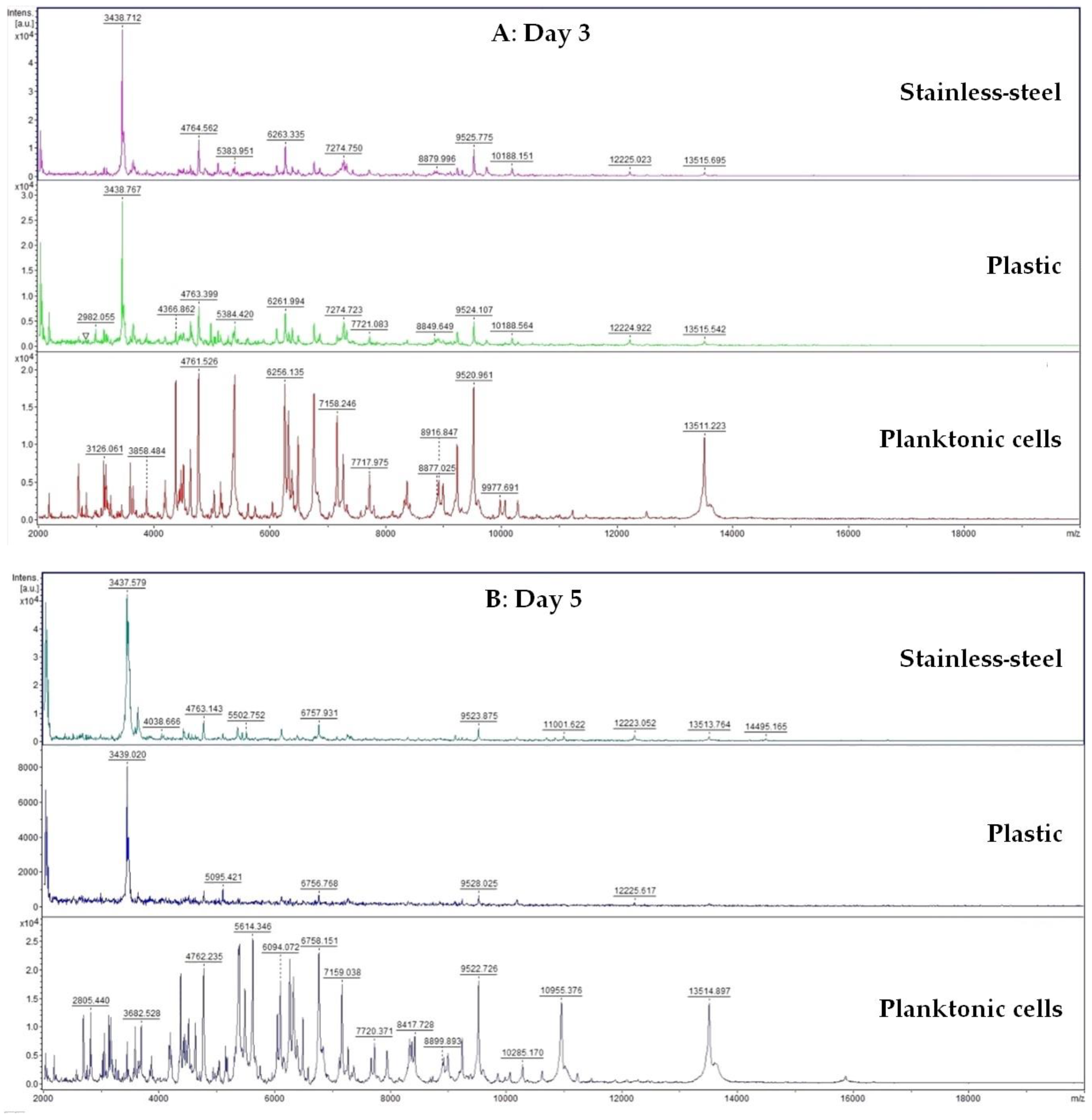
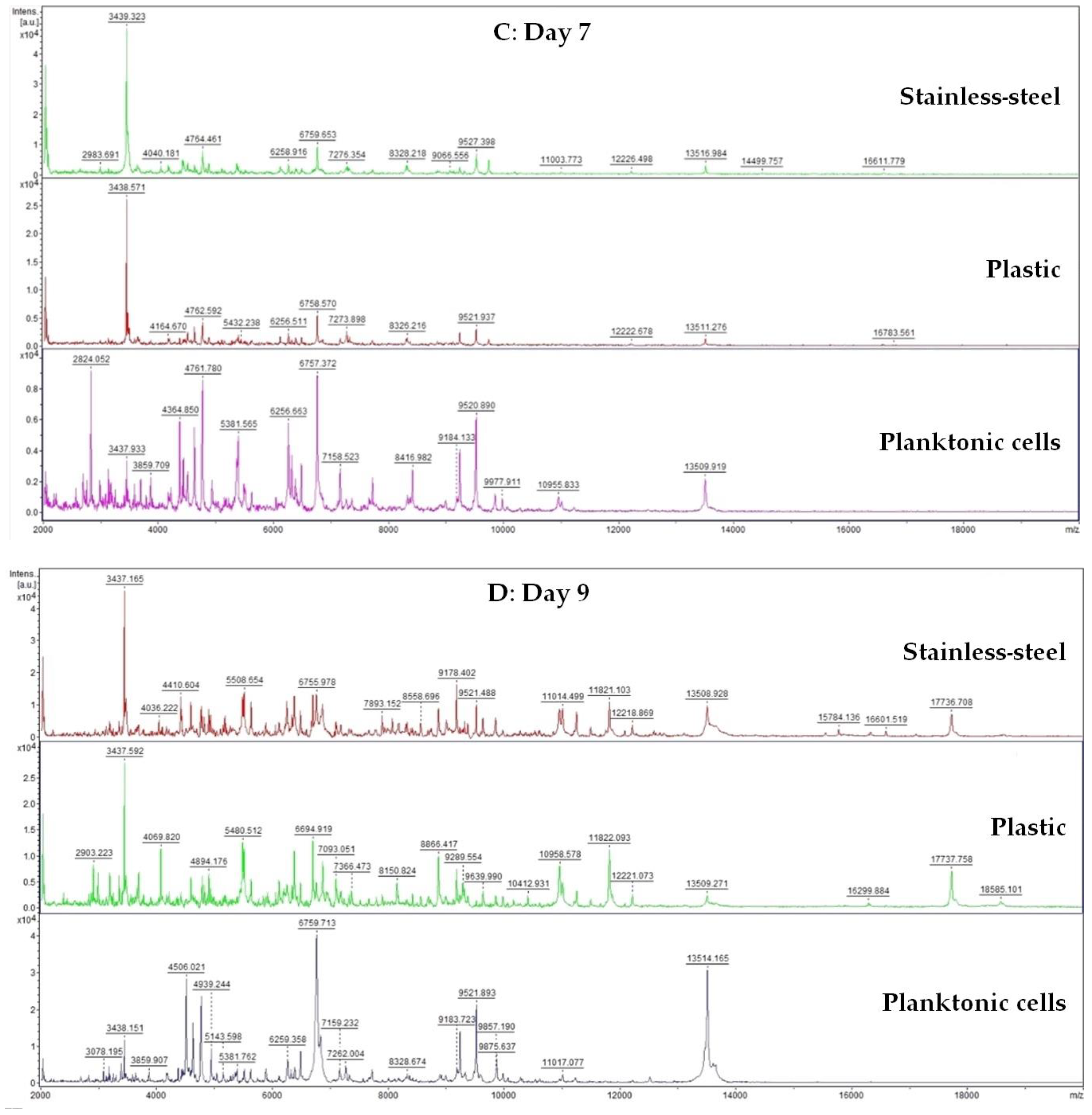
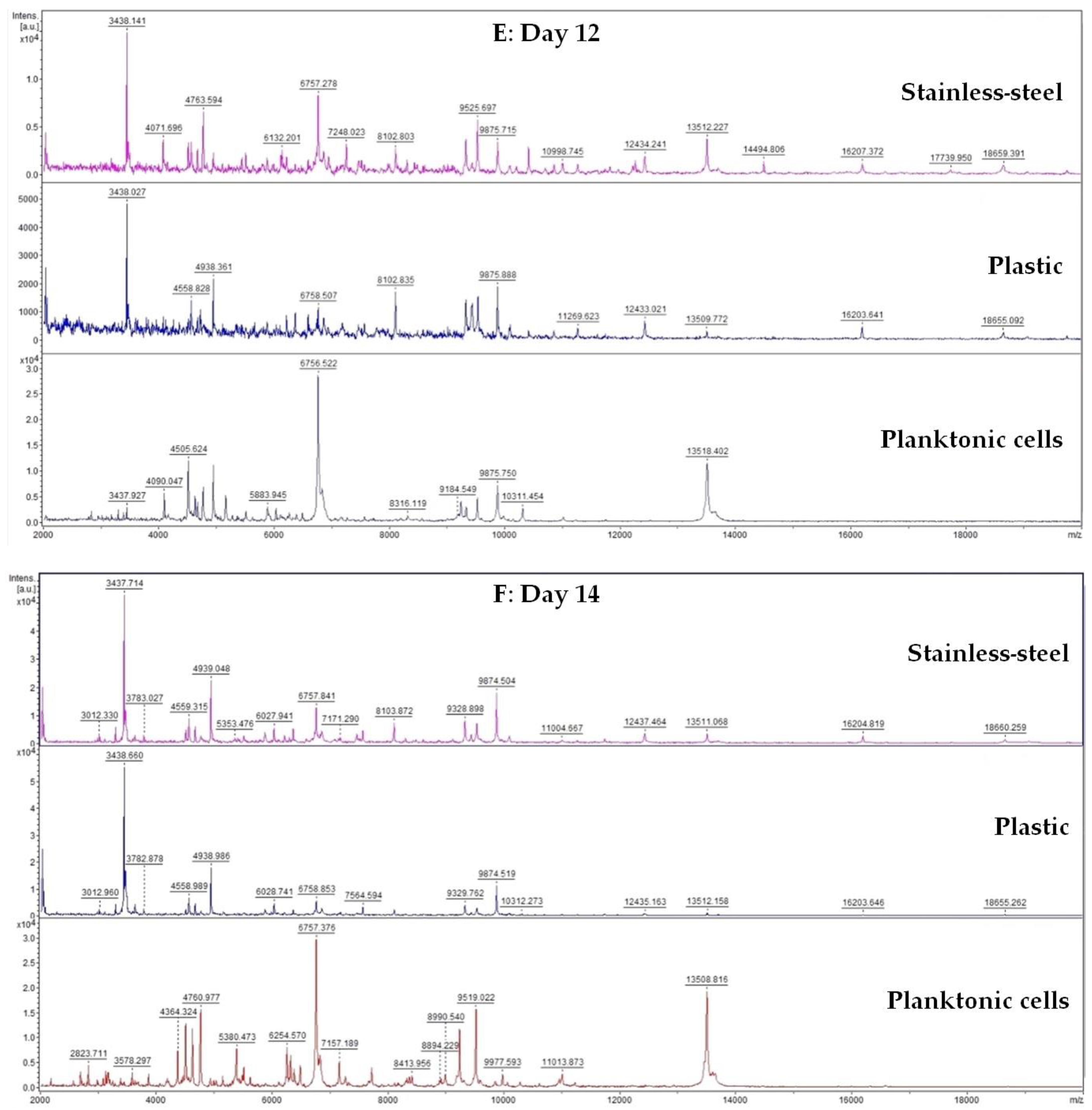
A 50 mL tube was filled with 20 mL of MHB and 100 μL of biofilm-forming S. enterica inoculum. Subsequently, a plastic and stainless-steel piece were added to the tube. FEO was added to experimental groups in the final concentration of 0.1%. Control samples were left untreated. The incubation was carried out at 37 °C with shaking at 170 rpm. Samples were analyzed on days 3, 5, 7, 9, 12, and 14.
Changes in protein spectra during biofilm development in the presence of FEO were monitored using MALDI-TOF MS Biotyper. Biofilm samples were taken from plastic and stainless-steel surfaces by a sterile cotton swab and were directly transferred to a MALDI-TOF plate. A 300 µL of culture medium was taken for analysis of planktonic cell spectra. A suspension with planktonic cells was centrifuged for 1 min at 12,000 rpm. The pellet was washed three times in ultrapure water and resuspended. A total of 1 μL was applied to a target plate.
A matrix α-Cyano-4-hydroxycinnamic acid matrix (10 mg/mL) was applied to the dried target plate. MALDI-TOF MicroFlex (Bruker Daltonics) was used for the analysis of biofilm protein spectra. Spectra in the range of mass to charge ratio 200–2000 were recorded in the linear and positive mode. The protein spectra were obtained, and the similarities of the spectra were used to generate the standard global spectrum (MSP). Nineteen MSP were generated from the spectra with MALDI Biotyper 3.0 and were grouped into dendrograms using Euclidean distance [25].

2.5. Insecticidal Activity of FEO Vapor Phase

Oxycarenus lavaterae was used for the analysis of insecticidal activity. Thirty specimens were added to Petri dishes with vented lids. Subsequently, 100 μL was added to filter paper which was placed inside the Petri. Petri plates were sealed with parafilm. Concentrations of 100%, 50%, 25%, 12.5%, 6.25%, and 3.125% of FEO were prepared in 0.1% polysorbate. A negative control sample was treated with 100 μL of 0.1% polysorbate. Specimens were exposed for 24 h at room temperature. Live and dead subjects were counted, and the percentage of insecticidal activity was calculated [24]. The insecticidal activity was measured in triplicate.

2.6. Statistical Analysis

One-way analysis of variance (ANOVA) was performed with Prism 8.0.1 (GraphPad Software, San Diego, CA, USA) and by Tukey’s test at p < 0.05. SAS® version 8 software (SAS Institute, Cary, NC, USA) was used to process the data. MIC 50 and MIC 90 values (50% and 90% inhibition of microbial growth) were determined by logit analysis.

3. Results

3.1. Volatile Composition of Examined Essential Oil

The results of the chemical composition analysis of FEO are presented in Table 1. In Table 2, percentage amounts of each class of identified compounds are presented. Overall, this essential oil is characterized by monoterpene hydrocarbons that constitute 79.3% of the total. Among this class of compounds, major detected were α-pinene (37.0%) and α-limonene (19.8%), followed by a notable amount of p-cymene (6.3%), α-thujene (4.2%), sabinene (4.1%), and β-myrcene (2.6%). Sesquiterpene hydrocarbons (10.4%) were the second most represented class of compounds, with β-elemene (2.2%) and (E)-caryophyllene (2.5%) found in significant quantities. Other identified volatiles were found in amounts smaller than 2%.

3.2. Antioxidant Activity

The antioxidant potential of FEO was determined by the means of neutralization of the stable DPPH radical and the ABTS radical cation. The obtained results are presented in Table 3. As can be seen, the radical scavenging capacity of the tested FEO is found to be stronger compared to the IC50 value of the reference compound Trolox. A total of 10 μL of this FEO was able to neutralize 73.88 ± 0.35% of DPPH radical, which is equivalent to the 6.27 ± 0.17 TEAC, and 97.09 ± 0.48% of ABTS radical cation (5.21 ± 0.07 TEAC).

3.3. Antimicrobial Activity

Overall, the FEO showed good antimicrobial activity against the tested microorganisms (Table 4). The evaluation was performed according to the CLSI-M100 guideline. The microorganisms for which an inhibition zone larger than 20 mm in diameter was observed were considered susceptible to FEO. All the chosen microorganisms were thus susceptible to the tested essential oil. The most pronounced activity was noted for yeasts. The inhibition zone diameters for C. glabrata, C. tropicalis, and C. albicans were 38.00, 40.67, and 44.00 mm, respectively. High activity was also observed against Gram-positive bacteria B. subtilis and S. aureus with inhibition zones of 37.34 and 31.34 mm, respectively. Strong activity was also determined for Gram-negative bacteria Y. enterocolitica and biofilm-forming S. enterica. In the case of filamentous fungi, the biggest inhibition zone was noted for A. flavus.
The microdilution method (Table 5) also showed the highest activity against the yeasts of Candida spp. The lowest MIC 50 and MIC 90 were determined for C. krusei with values of 11.72 and 12.58 μL/mL, respectively. MIC 50 and MIC 90 values for C. tropicalis were 11.83 and 28.19 μL/mL, respectively, while for C. albicans 50% and 90% inhibition was found at 23.44 and 25.10 μL/mL, respectively. Among bacteria, the best results were observed against S. aureus with MIC 50 and MIC 90 values of 23.44 and 25.10 μL/mL, respectively.
The antifungal activity was performed against microscopic filamentous fungi B. cinerea, A. flavus, and P. citrinum for four different concentrations of FEO (Table 6). The strongest antifungal activity of FEO against all three tested microorganisms was observed at the highest concentration of 500 μL/mL. The effect of FEO against B. cinerea appeared to be dependent on the concentration as the size of inhibition increased with it. In contrast, the inhibitory activity against A. flavus was lower at the concentrations of 125 and 250 μL/mL compared to the value observed for the concentration of 62.5 μL/mL.

3.4. In Situ Inhibition of Vapor Phase FEO on Carrot

The activity of the vapor phase of FEO was evaluated on carrots inoculated with selected microorganisms (Table 7). Compared to the untreated control, the inhibition of bacteria was the strongest at the highest concentration of 500 μL/L of FEO for which inhibition percentage values ranged between 65.14 and 93.67%. The highest bacterial inhibition was observed for B. subtilis at the mentioned concentration. Strong inhibition of the bacterial growth was also noticeable at 250 μL/L against Y. enterocolitica and B. subtilis with percentages of inhibition of 74.82 and 75.92%, respectively. Moreover, E. faecalis was strongly inhibited at the concentration of 125 μL/L.
The antifungal activity (Table 8) was not as strong as the antibacterial activity. The percentual inhibition of all the tested fungi at the highest concentration of 500 μL/L ranged from 15.23 to 95.63%. Strong inhibition was noted only for C. krusei and C. glabrata with values of 76.33 and 95.63%, respectively.

3.5. Antibiofilm Activity

The inhibitory effect of FEO against S. enterica biofilm developing on plastic and stainless-steel surfaces was evaluated with a MALDI-TOF MS Biotyper mass spectrometer. Control mass spectra for both tested surfaces were developed identically with control planktonic spectra. For better comprehensibility of the results, a planktonic spectrum was used as the control for each day of the experiment. Experimental spectra of stainless-steel and plastic surfaces, and control planktonic spectra are grouped in the individual images prepared on selected days of the experiment (day 3, 5, 7, 9, 12, and 14).
Since day 3 (Figure 1A), differences in the development of the experimental and control groups were noticed, while similarity was preserved between experimental groups treated with FEO. On the 5th day of the experiment (Figure 1B), a difference in biofilm development was recorded between the control and experimental groups. However, a more pronounced effect of FEO was visible against the biofilm developing on the plastic surface as reflected by the lower number of peaks in the mass spectra compared to the mass spectra of biofilm developing on stainless steel. This trend persisted until the 7th day of the experiment (Figure 1C), which suggests that the antibiofilm activity of FEO on S. enterica with has a stronger effect on a biofilm created on a plastic surface. During the following days of the experiment (Figure 1D–F), the differences in mass spectra of the experimental and control groups persisted. This suggests that FEO affects the creation of S. enterica biofilm and causes deterioration of homeostasis, thus inhibiting biofilm development.
To express the similarity of mass spectra profiles of biofilm after FEO addition, a dendrogram was constructed based on the main peaks of the mass spectra (MSP) (Figure 2). The shortest distances in MSP were visible for the control groups (CSE) and biofilms of the experimental group during days 3 and 5 (SES 3, SEP 3, SES 5, and SEP 5). During the following days of the experiment, the MSP were extended, with the most significant changes on the 12th and 14th days of the experiment (SES 12, SEP 12, SES 14, SEP 14). During the whole experiment, the distance of the control group MSP was clearly shorter, compared to the distance of the experimental groups. Based on these observations, we can suggest that the addition of FEO resulted in changes of the protein profile, leading to the deterioration of biofilm homeostasis and the inhibition of the development of S. enterica biofilm. These findings are in accordance with the results of the mass spectra analyses.

3.6. Insecticidal Activity

The activity of FEO against insects was evaluated in triplicate by testing with Oxycarenus lavaterae (Table 9). With a growing concentration of FEO, the mortality of individuals increased. At a 100% concentration of FEO, 100% of the tested subjects were found to be dead. At a concentration of 25% of FEO, only 50% of the insects seemed to be alive. The concentration of 3.125% of FEO did not affect the viability of tested specimens.

4. Discussion

The findings observed in the present study show that commercial FEO is characterized by a high abundance of monoterpene hydrocarbons (79.3% of the total), with α-pinene (37.0%) and α-limonene (19.8%) being the major compounds. These results are in agreement with a previously published study on the chemical composition of 21 samples of commercial FEO as well as two laboratory-distilled oils [6]. Moreover, a report by DeCarlo et al. [26] indicates at least three different chemotypes defined by a high abundance of α-pinene, α-thujene, and methoxydecan. According to the same report, as well as many others, the α-pinene chemotype is also rich in limonene [4,6,26,27,28]. We can thus conclude that FEO investigated in this study belonged to the previously described α-pinene chemotype. Moreover, some previous examinations showed FEOs rich in octyl acetate, a compound not identified in the sample investigated in this study [29,30]. Observed differences in the chemical composition of this FEO could be explained by distinct environmental factors, such as harvesting season, geographical location, climate, as well as the part of the plant and the method used for FEO extraction, etc. Even though there are a number of previously published results on the biological activity of this species, the variations in chemical composition can seriously influence the observed results.
DPPH and ABTS assays are commonly used for the evaluation of the antioxidant potential of essential oils. Our study showed that 10 μL of FEO can neutralize 73.88 ± 0.35% of DPPH radical and 97.09 ± 0.48% of ABTS radical cation. Considering these results, we can imply a high potential of this FEO as an antioxidant. The observed differences in the results of the two performed assays can be attributed to the differences in the mechanisms involved in the reactions responsible for neutralization. It is well known that ABTS radical cation reacts much faster, including the electron transfer reaction, compared to DPPH radical, whose neutralization is dependent on the antioxidant’s ability to donate hydrogen. In previously published results, this FEO showed a weak to moderate antioxidant potential [31,32,33]. Essential oils are known to be complex mixtures of compounds with different functional groups, and mainly the synergistic effect of two or more of its components is responsible for the observed effects. With that in mind, the diversity of the results obtained in various studies can be explained. Nevertheless, the results can also vary due to the assay employed in the determination of the antioxidant activity.
In our study, the antimicrobial activity of FEO was the strongest against Candida species. Strong activity of FEO was also determined against G+ and G bacteria. Abers et al. [34] analyzed the antimicrobial activity by disc diffusion method and determined a moderate activity of FEO with inhibition zone diameters exceeding 30 mm against G+ bacteria S. pyogenes and M. smegmatis and G bacteria P. aeruginosa. Al-Saidi et al. [35] determined the activity against G P. aeruginosa to result in diameters of 23.3–29.7 mm. Among G+ bacteria, a comparable activity was determined for B. subtilis and S. aureus with ranges of inhibition zones 14.7–16.0 mm and 7.0–24.3 mm, respectively. Almutairi et al. [36] observed the antimicrobial properties of FEO against antibiotic-resistant strains and showed mild activity against methicillin-resistant S. aureus and multi-drug-resistant P. aeruginosa.
Di Stefano et al. [7] compared the growth-inhibition activity of three FEOs. They determined MIC value against bacteria S. epidermidis, S. hominis, and P. acnes ranging from 0.264 to 6.16 mg/mL. MIC for S. aureus and P. aeruginosa ranged from 52 to 400 mg/mL, which appeared to reflect the weak activity. Notable activity was determined against C. albicans and M. furfur with a MIC range of 0.03–3.10 mg/mL. On the other hand, Man et al. [37] tested six essential oils (Frankincense, myrtle, thyme, lemon, oregano, and lavender) and determined the weakest activity compared to the essential oil with MIC of 50% v/v or higher against G+ and G bacteria. De Rapper et al. [38] tested the activity of various Boswellia species and found MIC values to be the lowest against fungi C. neoformans (0.8 mg/mL) and the highest against S. aureus (6.0 mg/mL). Van Vuuren et al. [4] tested various FEOs and reported mean MIC values against G+ bacteria, S. aureus and B. cereus, of 8.1 and 3.4 mg/mL, respectively. The reported MIC values against G bacteria, E. coli and P. vulgaris, were 6.2 and 4.0 mg /mL, respectively, and against the yeast C. albicans, the value was 7.4 mg/mL. These values were lower compared to our study. Bogavac et al. [39] determined the activity of FEO against a C. albicans strain at 12.5 μL/mL which suggest a stronger activity compared to our study.
The activity of FEO together with other essential oils against fungal pathogens was evaluated as mild. Against A. niger, an MIC value of 625 μg/mL was determined [40]. Antifungal activity of FEO against Aspergillus, Penicillium, Fusarium, or Alternaria species was found at low concentrations, which are suitable for the inhibition of pathogenic fungi to preserve plant-based products [32]. FEO showed promising antifungal properties against 15 different fungal species with a value of mycelial inhibition ranging from 15.9 to 56.3% at 1 µL/mL. MIC was determined in a range between 0.039 and 0.625 µL/mL [41]. Ljaljević Grbić et al. [42] determined MIC for Aspergillus species in the range from 10.0 to 120.0 mg/mL and for Penicillium species, in the range from 10 to 30 mg/mL. These results on the antifungal properties of FEO are in agreement with our study.
Many factors affect the activity of the FEOs. Generally, the changes in biological activity are a result of differences in chemical composition [4]. Alpha-pinene, which was the most abundant molecule of our FEO, did not show any significant activity alone, but the antimicrobial activity was stronger when combined with other substances, which suggests synergism with other components in FEO [43,44]. On the contrary, limonene, the second most abundant compound, has shown better activity than the essential oil containing it [45,46]. Overall, no correlation was observed between the number of major constituents in essential oils [47]. Therefore, the antimicrobial activity cannot be attributed only to the major constituents of essential oils, as minor compounds contribute to activity against potentially pathogenic microorganisms as well [48].
FEO showed good inhibitory activity against G+ and G bacteria on the carrot used as a model food. For microscopic filamentous fungi, determined activity was weaker compared to bacteria. Prakash et al. [32] evaluated the potential of FEO for the preservation of Piper nigrum L. fruits and reported that the growth of a storage mold, A. flavus, was decreased by 65.38%. Not enough research has been focused on the application of FEO for the preservation of fruits or vegetables from potential pathogenic bacteria. On the other hand, some research has been conducted in our laboratories which aimed at the observation of the inhibition activity of essential oils against pathogenic microorganisms that grew on fruits, vegetables, and bread samples [23,49,50]. In other reports the inhibitory activity of eucalyptus essential oils on sweet potatoes and bread [51], or thyme oil against Botrytis in grapes [52] were investigated. Generally, essential oil components are considered to be good preservatives of food products [53]. Various mechanisms of action have been described, including interactions of compounds with cell membranes, cell walls, intracellular proteins, enzymes, or nucleic acids [54]. Some components of FEO have been described as sufficient inhibitors of pathogenic microorganisms. For example, α-pinene was used in the prolongation of the storage of wine [55]. Limonene coating was found to decrease fungal growth on cucumbers [56]. We can suggest that the chemical composition of FEO increases the potential for its use as a food preservative.
Bacteria in the form of biofilm have a better ability to survive compared to single-cell forms due to the ability to adhere and the quorum sensing mechanisms. Analyses of biofilm degradation showed the ability of FEO to inhibit growth and deteriorate the formation of biofilm. There has not been enough research on the changes in biofilm resulting from the addition of FEO. Moreover, this is the first time that biofilm degradation was analyzed on different surfaces. The antibiofilm activity of the essential oils isolated from Boswellia against Staphylococcus species and Candida was reported, with FEO showing the ability to prevent the adhesion of biofilm [10]. The properties of terpenes, which are found in essential oils, were described with regard to biofilm formation [57]. Boudiba et al. [58] and Soyocak et al. [59] observed the disruption of biofilms by the inhibition of quorum sensing in bacteria after the treatment with essential oils with a similar composition.
The activity of FEO against Oxycarenus lavaterae showed 50% mortality at the concentration of 25%. The anti-insect activity of FEO against female forms of the fly Musca domestica was tested by Pavela et al. [60] who rported the toxicity values of LD50 and LD90 of 72.5 and 269.3 μg/individual, respectively. The activity against the mosquito C. quinquefasciatus larvae was found to be significant. Yang et al. [61] observed insecticidal activity of various essential oils against the weevil Sitophilus zeamais. Even the contact and fumigant activity of FEO was not significant, the repellent activity of FEO was moderate even after 24 h of incubation. Metayi et al. [62] tested a nanoemulsion of FEO against the cotton bollworm Earias insulana and found out that 80% mortality of larvae was reached at a concentration of 1800 μL. Due to the activity of FEO against pests, it can potentially serve as a repellent or pesticide. More research is necessary in order to determine the mechanisms of action.

5. Conclusions

The FEO tested in our study showed good properties that enable it to be used as an antimicrobial agent. The main volatile compounds were α-pinene, α-limonene, and p-cymene. The antioxidant activity was evaluated as strong. Antimicrobial activity determined by the disk diffusion method was the strongest against Candida species. The inhibition activity of the vapor phase on carrot was more pronounced against G+ and G bacteria than against microscopic filamentous fungi. Visible changes in the protein profile suggest the degradation of bacterial biofilm forming on plastic and stainless-steel surfaces. The anti-insect activity against O. lavaterae was found to increase with increasing concentration of FEO. We can thus conclude that the analyzed essential oil could serve as an agent against pathogenic microorganisms and can be used as a potential preservative of food and agricultural crops.

Author Contributions

Conceptualization, P.B., N.Č., L.G., N.L.V., M.D.V. and M.K.; Data curation, P.B., N.Č., L.G., N.L.V., M.D.V., E.T., L.B., M.I.K., C.P. and M.K.; Methodology, P.B., M.K., N.Č., L.G., N.L.V., M.D.V., M.S. and M.K.; Supervision, P.B., M.K., N.L.V., P.Ł.K. and M.D.V.; Writing—original draft, P.B., N.Č., L.G., N.L.V., M.D.V., E.T., M.S., P.Ł.K., M.I.K., C.P., L.B. and M.D.V. All authors have read and agreed to the published version of the manuscript.

Funding

This research was funded by the grant APVV-20-0058 “The potential of the essential oils from aromatic plants for medical use and food preservation”.

Institutional Review Board Statement

Not applicable.

Informed Consent Statement

Not applicable.

Data Availability Statement

All data generated or analyzed during this study are included in this published article.

Acknowledgments

This work has been supported by the grants of the VEGA no. 1/0180/20, and by the Serbian Ministry of Education, Science, and Technological Development (Agreement Nos. 451-03-68/2023-01/200122).

Conflicts of Interest

The authors declare no conflict of interest.

References

  1. Mertens, M.; Buettner, A.; Kirchhoff, E. The Volatile Constituents of Frankincense—A Review. Flavour Fragr. J. 2009, 24, 279–300. [Google Scholar] [CrossRef]
  2. Almeida-da-Silva, C.L.C.; Sivakumar, N.; Asadi, H.; Chang-Chien, A.; Qoronfleh, M.W.; Ojcius, D.M.; Essa, M.M. Effects of Frankincense Compounds on Infection, Inflammation, and Oral Health. Molecules 2022, 27, 4174. [Google Scholar] [CrossRef] [PubMed]
  3. Woolley, C.L.; Suhail, M.M.; Smith, B.L.; Boren, K.E.; Taylor, L.C.; Schreuder, M.F.; Chai, J.K.; Casabianca, H.; Haq, S.; Lin, H.-K.; et al. Chemical Differentiation of Boswellia sacra and Boswellia carterii Essential Oils by Gas Chromatography and Chiral Gas Chromatography-Mass Spectrometry. J. Chromatogr. A 2012, 1261, 158–163. [Google Scholar] [CrossRef] [PubMed]
  4. Van Vuuren, S.F.; Kamatou, G.P.P.; Viljoen, A.M. Volatile Composition and Antimicrobial Activity of Twenty Commercial Frankincense Essential Oil Samples. S. Afr. J. Bot. 2010, 76, 686–691. [Google Scholar] [CrossRef]
  5. Kieliszek, M.; Edris, A.; Kot, A.M.; Piwowarek, K. Biological Activity of Some Aromatic Plants and Their Metabolites, with an Emphasis on Health-Promoting Properties. Molecules 2020, 25, 2478. [Google Scholar] [CrossRef]
  6. Ojha, P.K.; Poudel, D.K.; Rokaya, A.; Satyal, R.; Setzer, W.N.; Satyal, P. Comparison of Volatile Constituents Present in Commercial and Lab-Distilled Frankincense (Boswellia carteri) Essential Oils for Authentication. Plants 2022, 11, 2134. [Google Scholar] [CrossRef]
  7. Di Stefano, V.; Schillaci, D.; Cusimano, M.G.; Rishan, M.; Rashan, L. In Vitro Antimicrobial Activity of Frankincense Oils from Boswellia sacra Grown in Different Locations of the Dhofar Region (Oman). Antibiotics 2020, 9, 195. [Google Scholar] [CrossRef] [Green Version]
  8. Camarda, L.; Dayton, T.; Di Stefano, V.; Pitonzo, R.; Schillaci, D. Chemical Composition and Antimicrobial Activity of Some Oleogum resin Essential Oils from Boswellia Spp. (Burseraceae). Ann. Chim. 2007, 97, 837–844. [Google Scholar] [CrossRef]
  9. Sadhasivam, S.; Palanivel, S.; Ghosh, S. Synergistic Antimicrobial Activity of Boswellia serrata Roxb. Ex Colebr. (Burseraceae) Essential Oil with Various Azoles against Pathogens Associated with Skin, Scalp and Nail Infections. Lett. Appl. Microbiol. 2016, 63, 495–501. [Google Scholar] [CrossRef]
  10. Schillaci, D.; Arizza, V.; Dayton, T.; Camarda, L.; Stefano, V.D. In Vitro Anti-Biofilm Activity of Boswellia Spp. Oleogum resin Essential Oils. Lett. Appl. Microbiol. 2008, 47, 433–438. [Google Scholar] [CrossRef]
  11. Monzote, L.; Herrera, I.; Satyal, P.; Setzer, W.N. In-Vitro Evaluation of 52 Commercially-Available Essential Oils Against Leishmania amazonensis. Molecules 2019, 24, 1248. [Google Scholar] [CrossRef] [Green Version]
  12. Al-Yasiry, A.R.M.; Kiczorowska, B. Frankincense—Therapeutic Properties. Postep. Hig. Med. Dosw. Online 2016, 70, 380–391. [Google Scholar] [CrossRef] [PubMed]
  13. Gupta, I.; Parihar, A.; Malhotra, P.; Gupta, S.; Lüdtke, R.; Safayhi, H.; Ammon, H.P.T. Effects of Gum Resin of Boswellia serrata in Patients with Chronic Colitis. Planta Med. 2001, 67, 391–395. [Google Scholar] [CrossRef] [PubMed]
  14. Okano, S.; Honda, Y.; Kodama, T.; Kimura, M. The Effects of Frankincense Essential Oil on Stress in Rats. J. Oleo Sci. 2019, 68, 1003–1009. [Google Scholar] [CrossRef] [Green Version]
  15. Rajabian, A.; Sadeghnia, H.; Fanoudi, S.; Hosseini, A. Genus Boswellia as a New Candidate for Neurodegenerative Disorders. Iran. J. Basic Med. Sci. 2020, 23, 277–286. [Google Scholar] [CrossRef]
  16. Ni, X.; Suhail, M.M.; Yang, Q.; Cao, A.; Fung, K.-M.; Postier, R.G.; Woolley, C.; Young, G.; Zhang, J.; Lin, H.-K. Frankincense Essential Oil Prepared from Hydrodistillation of Boswellia sacra Gum Resins Induces Human Pancreatic Cancer Cell Death in Cultures and in a Xenograft Murine Model. BMC Complement. Altern. Med. 2012, 12, 253. [Google Scholar] [CrossRef] [Green Version]
  17. Frank, M.B.; Yang, Q.; Osban, J.; Azzarello, J.T.; Saban, M.R.; Saban, R.; Ashley, R.A.; Welter, J.C.; Fung, K.-M.; Lin, H.-K. Frankincense Oil Derived from Boswellia carteri Induces Tumor Cell Specific Cytotoxicity. BMC Complement. Altern. Med. 2009, 9, 6. [Google Scholar] [CrossRef] [Green Version]
  18. Suhail, M.M.; Wu, W.; Cao, A.; Mondalek, F.G.; Fung, K.-M.; Shih, P.-T.; Fang, Y.-T.; Woolley, C.; Young, G.; Lin, H.-K. Boswellia sacra Essential Oil Induces Tumor Cell-Specific Apoptosis and Suppresses Tumor Aggressiveness in Cultured Human Breast Cancer Cells. BMC Complement. Altern. Med. 2011, 11, 129. [Google Scholar] [CrossRef] [Green Version]
  19. Adams, R.P. Identification of Essential Oil Components by Gas Chromatography/Mass Spectrometry; Allured Publishing Corporation: Carol Stream, IL, USA, 2007. [Google Scholar]
  20. van Den Dool, H.; Kratz, P.D. A Generalization of the Retention Index System Including Linear Temperature Programmed Gas—Liquid Partition Chromatography. J. Chromatogr. A 1963, 11, 463–471. [Google Scholar] [CrossRef] [PubMed]
  21. Proestos, C.; Lytoudi, K.; Mavromelanidou, O.; Zoumpoulakis, P.; Sinanoglou, V. Antioxidant Capacity of Selected Plant Extracts and Their Essential Oils. Antioxidants 2013, 2, 11–22. [Google Scholar] [CrossRef]
  22. CLSI. Clinical and Laboratory Standards Institute: M100—Performance Standards for Antimicrobial Susceptibility Testing, 31st ed.; CLSI: Wayne, PA, USA, 2021. [Google Scholar]
  23. Galovičová, L.; Borotová, P.; Valková, V.; Vukovic, N.L.; Vukic, M.; Terentjeva, M.; Štefániková, J.; Ďúranová, H.; Kowalczewski, P.Ł.; Kačániová, M. Thymus serpyllum Essential Oil and Its Biological Activity as a Modern Food Preserver. Plants 2021, 10, 1416. [Google Scholar] [CrossRef]
  24. Galovičová, L.; Borotová, P.; Valková, V.; Ďúranová, H.; Štefániková, J.; Vukovic, N.L.; Vukic, M.; Kačániová, M. Biological Activity of Pogostemon cablin Essential Oil and Its Potential Use for Food Preservation. Agronomy 2022, 12, 387. [Google Scholar] [CrossRef]
  25. Pereira, F.D.E.S.; Bonatto, C.C.; Lopes, C.A.P.; Pereira, A.L.; Silva, L.P. Use of MALDI-TOF Mass Spectrometry to Analyze the Molecular Profile of Pseudomonas aeruginosa Biofilms Grown on Glass and Plastic Surfaces. Microb. Pathog. 2015, 86, 32–37. [Google Scholar] [CrossRef] [PubMed] [Green Version]
  26. DeCarlo, A.; Johnson, S.; Poudel, A.; Satyal, P.; Bangerter, L.; Setzer, W.N. Chemical Variation in Essential Oils from the Oleo-Gum Resin of Boswellia carteri: A Preliminary Investigation. Chem. Biodivers. 2018, 15, e1800047. [Google Scholar] [CrossRef] [PubMed]
  27. Hamm, S.; Bleton, J.; Connan, J.; Tchapla, A. A Chemical Investigation by Headspace SPME and GC–MS of Volatile and Semi-Volatile Terpenes in Various Olibanum Samples. Phytochemistry 2005, 66, 1499–1514. [Google Scholar] [CrossRef] [PubMed]
  28. Nikolic, M.; Smiljkovic, M.; Markovic, T.; Cirica, A.; Glamoclija, J.; Markovic, D.; Sokovic, M. Sensitivity of Clinical Isolates of Candida to Essential Oils from Burseraceae Family. EXCLI J. 2016, 15, 280–289. [Google Scholar] [CrossRef] [PubMed]
  29. Mikhaeil, B.R.; Maatooq, G.T.; Badria, F.A.; Amer, M.M.A. Chemistry and Immunomodulatory Activity of Frankincense Oil. Z. Nat. C 2003, 58, 230–238. [Google Scholar] [CrossRef] [PubMed]
  30. Marongiu, B.; Piras, A.; Porcedda, S.; Tuveri, E. Extraction of Santalum album and Boswellia carterii Birdw. Volatile Oil by Supercritical Carbon Dioxide: Influence of Some Process Parameters. Flavour Fragr. J. 2006, 21, 718–724. [Google Scholar] [CrossRef]
  31. Amin Mohamed, A.; Ali, S.; Kabiel, H.; Hegazy, A.; Kord, M.; El-Baz, F. Assessment of Antioxidant and Antimicrobial Activities of Essential Oil and Extracts of Boswellia carteri Resin. Int. J. Pharmacogn. Phytochem. Res. 2015, 7, 502–509. [Google Scholar]
  32. Prakash, B.; Mishra, P.K.; Kedia, A.; Dubey, N.K. Antifungal, Antiaflatoxin and Antioxidant Potential of Chemically Characterized Boswellia carterii Birdw Essential Oil and Its in Vivo Practical Applicability in Preservation of Piper nigrum L. Fruits. LWT-Food Sci. Technol. 2014, 56, 240–247. [Google Scholar] [CrossRef]
  33. Mothana, R.A.A.; Hasson, S.S.; Schultze, W.; Mowitz, A.; Lindequist, U. Phytochemical Composition and in Vitro Antimicrobial and Antioxidant Activities of Essential Oils of Three Endemic Soqotraen Boswellia Species. Food Chem. 2011, 126, 1149–1154. [Google Scholar] [CrossRef]
  34. Abers, M.; Schroeder, S.; Goelz, L.; Sulser, A.; St. Rose, T.; Puchalski, K.; Langland, J. Antimicrobial Activity of the Volatile Substances from Essential Oils. BMC Complement. Med. Ther. 2021, 21, 124. [Google Scholar] [CrossRef] [PubMed]
  35. Al-Saidi, S.; Rameshkumar, K.B.; Hisham, A.; Sivakumar, N.; Al-Kindy, S. Composition and Antibacterial Activity of the Essential Oils of Four Commercial Grades of Omani Luban, the Oleo-Gum Resin of Boswellia sacra Flueck. Chem. Biodivers. 2012, 9, 615–624. [Google Scholar] [CrossRef] [PubMed]
  36. Almutairi, M.B.F.; Alrouji, M.; Almuhanna, Y.; Asad, M.; Joseph, B. In-Vitro and In-Vivo Antibacterial Effects of Frankincense Oil and Its Interaction with Some Antibiotics against Multidrug-Resistant Pathogens. Antibiotics 2022, 11, 1591. [Google Scholar] [CrossRef] [PubMed]
  37. Man, A.; Santacroce, L.; Jacob, R.; Mare, A.; Man, L. Antimicrobial Activity of Six Essential Oils Against a Group of Human Pathogens: A Comparative Study. Pathogens 2019, 8, 15. [Google Scholar] [CrossRef] [PubMed] [Green Version]
  38. de Rapper, S.; Van Vuuren, S.F.; Kamatou, G.P.P.; Viljoen, A.M.; Dagne, E. The Additive and Synergistic Antimicrobial Effects of Select Frankincense and Myrrh Oils—A Combination from the Pharaonic Pharmacopoeia. Lett. Appl. Microbiol. 2012, 54, 352–358. [Google Scholar] [CrossRef] [PubMed]
  39. Bogavac, M.A.; Perić, T.M.; Mišković, J.; Karaman, M. Antimicrobial and Toxic Effects of Boswellia serrata Roxb. and Mentha piperita Linn. Essential Oils on Vaginal Inhabitants. Medicines 2022, 9, 62. [Google Scholar] [CrossRef]
  40. Powers, C.N.; Osier, J.L.; McFeeters, R.L.; Brazell, C.B.; Olsen, E.L.; Moriarity, D.M.; Satyal, P.; Setzer, W.N. Antifungal and Cytotoxic Activities of Sixty Commercially-Available Essential Oils. Mol. J. Synth. Chem. Nat. Prod. Chem. 2018, 23, 1549. [Google Scholar] [CrossRef] [Green Version]
  41. Venkatesh, H.N.; Sudharshana, T.N.; Abhishek, R.U.; Thippeswamy, S.; Manjunath, K.; Mohana, D.C. Antifungal and Antimycotoxigenic Properties of Chemically Characterised Essential Oil of Boswellia serrata Roxb. Ex Colebr. Int. J. Food Prop. 2017, 20, 1856–1868. [Google Scholar] [CrossRef] [Green Version]
  42. Ljaljević Grbić, M.; Unković, N.; Dimkić, I.; Janaćković, P.; Gavrilović, M.; Stanojević, O.; Stupar, M.; Vujisić, L.; Jelikić, A.; Stanković, S.; et al. Frankincense and Myrrh Essential Oils and Burn Incense Fume against Micro-Inhabitants of Sacral Ambients. Wisdom of the Ancients? J. Ethnopharmacol. 2018, 219, 1–14. [Google Scholar] [CrossRef]
  43. Yang, C.; Hu, D.-H.; Feng, Y. Antibacterial Activity and Mode of Action of the Artemisia Capillaris Essential Oil and Its Constituents against Respiratory Tract Infection-Causing Pathogens. Mol. Med. Rep. 2015, 11, 2852–2860. [Google Scholar] [CrossRef] [PubMed] [Green Version]
  44. Dhar, P.; Chan, P.; Cohen, D.T.; Khawam, F.; Gibbons, S.; Snyder-Leiby, T.; Dickstein, E.; Rai, P.K.; Watal, G. Synthesis, Antimicrobial Evaluation, and Structure–Activity Relationship of α-Pinene Derivatives. J. Agric. Food Chem. 2014, 62, 3548–3552. [Google Scholar] [CrossRef]
  45. Sreepian, A.; Popruk, S.; Nutalai, D.; Phutthanu, C.; Sreepian, P.M. Antibacterial Activities and Synergistic Interaction of Citrus Essential Oils and Limonene with Gentamicin against Clinically Isolated Methicillin-Resistant Staphylococcus aureus. Sci. World J. 2022, 2022, e8418287. [Google Scholar] [CrossRef] [PubMed]
  46. Pathirana, H.N.K.S.; Wimalasena, S.H.M.P.; Silva, B.C.J.D.; Hossain, S.; Heo, G.-J. Antibacterial Activity of Lime (Citrus aurantifolia) Essential Oil and Limonene against Fish Pathogenic Bacteria Isolated from Cultured Olive Flounder (Paralichthys olivaceus). Fish. Aquat. Life 2018, 26, 131–139. [Google Scholar] [CrossRef]
  47. Cimanga, K.; Kambu, K.; Tona, L.; Apers, S.; De Bruyne, T.; Hermans, N.; Totté, J.; Pieters, L.; Vlietinck, A.J. Correlation between Chemical Composition and Antibacterial Activity of Essential Oils of Some Aromatic Medicinal Plants Growing in the Democratic Republic of Congo. J. Ethnopharmacol. 2002, 79, 213–220. [Google Scholar] [CrossRef]
  48. Ambrosio, C.M.S.; Ikeda, N.Y.; Miano, A.C.; Saldaña, E.; Moreno, A.M.; Stashenko, E.; Contreras-Castillo, C.J.; Da Gloria, E.M. Unraveling the Selective Antibacterial Activity and Chemical Composition of Citrus Essential Oils. Sci. Rep. 2019, 9, 17719. [Google Scholar] [CrossRef] [Green Version]
  49. Kačániová, M.; Galovičová, L.; Borotová, P.; Vukovic, N.L.; Vukic, M.; Kunová, S.; Hanus, P.; Bakay, L.; Zagrobelna, E.; Kluz, M.; et al. Assessment of Ocimum basilicum Essential Oil Anti-Insect Activity and Antimicrobial Protection in Fruit and Vegetable Quality. Plants 2022, 11, 1030. [Google Scholar] [CrossRef]
  50. Valková, V.; Ďúranová, H.; Galovičová, L.; Borotová, P.; Vukovic, N.L.; Vukic, M.; Kačániová, M. Cymbopogon citratus Essential Oil: Its Application as an Antimicrobial Agent in Food Preservation. Agronomy 2022, 12, 155. [Google Scholar] [CrossRef]
  51. Marcel, S. Use of Essential Oils as New Food Preservatives (Case: Eucalyptus grandis and Eucalyptus crebra). J. Plant Sci. Phytopathol. 2018, 2, 083–090. [Google Scholar]
  52. Walter, M.; Jaspers, M.V.; Eade, K.; Frampton, C.M.; Stewart, A. Control of Botrytis Cinerea in Grape Using Thyme Oil. Australas. Plant Pathol. 2001, 30, 21–25. [Google Scholar] [CrossRef]
  53. Masyita, A.; Mustika Sari, R.; Dwi Astuti, A.; Yasir, B.; Rahma Rumata, N.; Emran, T.B.; Nainu, F.; Simal-Gandara, J. Terpenes and Terpenoids as Main Bioactive Compounds of Essential Oils, Their Roles in Human Health and Potential Application as Natural Food Preservatives. Food Chem. X 2022, 13, 100217. [Google Scholar] [CrossRef]
  54. Hyldgaard, M.; Mygind, T.; Meyer, R.L. Essential Oils in Food Preservation: Mode of Action, Synergies, and Interactions with Food Matrix Components. Front. Microbiol. 2012, 3, 12. [Google Scholar] [CrossRef] [Green Version]
  55. Shih, M.-K.; Lai, Y.-H.; Lin, C.-M.; Chen, Y.-W.; Hou, Z.-T.; Hou, C.-Y. A Novel Application of Terpene Compound α-Pinene for Alternative Use of Sulfur Dioxide-Free White Wine. Int. J. Food Prop. 2020, 23, 520–532. [Google Scholar] [CrossRef] [Green Version]
  56. Maleki, G.; Sedaghat, N.; Woltering, E.J.; Farhoodi, M.; Mohebbi, M. Chitosan-Limonene Coating in Combination with Modified Atmosphere Packaging Preserve Postharvest Quality of Cucumber during Storage. J. Food Meas. Charact. 2018, 12, 1610–1621. [Google Scholar] [CrossRef]
  57. Gómez-Sequeda, N.; Cáceres, M.; Stashenko, E.E.; Hidalgo, W.; Ortiz, C. Antimicrobial and Antibiofilm Activities of Essential Oils against Escherichia coli O157:H7 and Methicillin-Resistant Staphylococcus aureus (MRSA). Antibiotics 2020, 9, 730. [Google Scholar] [CrossRef]
  58. Boudiba, S.; Tamfu, A.N.; Berka, B.; Hanini, K.; Hioun, S.; Allaf, K.; Boudiba, L.; Ceylan, O. Anti-Quorum Sensing and Antioxidant Activity of Essential Oils Extracted From Juniperus Species, Growing Spontaneously in Tebessa Region (East of Algeria). Nat. Prod. Commun. 2021, 16, 1934578X211024039. [Google Scholar] [CrossRef]
  59. Soyocak, A.; Ak, A.; Önem, E. Anti-Quorum Sensing and Cytotoxic Activity of Elemi Essential Oil. Int. J. Second. Metab. 2022, 9, 258–267. [Google Scholar] [CrossRef]
  60. Pavela, R.; Maggi, F.; Giordani, C.; Cappellacci, L.; Petrelli, R.; Canale, A. Insecticidal Activity of Two Essential Oils Used in Perfumery (Ylang Ylang and Frankincense). Nat. Prod. Res. 2021, 35, 4746–4752. [Google Scholar] [CrossRef] [PubMed]
  61. Yang, Y.; Isman, M.B.; Tak, J.-H. Insecticidal Activity of 28 Essential Oils and a Commercial Product Containing Cinnamomum cassia Bark Essential Oil against Sitophilus Zeamais Motschulsky. Insects 2020, 11, 474. [Google Scholar] [CrossRef] [PubMed]
  62. Metayi, M.H.; Abd El-Naby, S.S.; El-Habal, N.A.; Fahmy, H.H.; Abdou, M.S.; Ali, B.; Abdel-Rheim, K.H.; Abdel-Megeed, A. Omani frankincense Nanoemulsion Formulation Efficacy and Its Latent Effects on Biological Aspects of the Spiny Bollworm Earias insulana (Boisd.). Front. Physiol. 2022, 13, 1001136. [Google Scholar] [CrossRef] [PubMed]
Figure 1. Changes in MALDI-TOF mass spectra of S. enterica biofilm after FEO addition: (A) 3rd day; (B) 5th day; (C) 7th day; (D) 9th day; (E) 12th day; (F) 14th day.
Horticulturae 09 00333 g001aHorticulturae 09 00333 g001bHorticulturae 09 00333 g001c
Figure 2. Dendrogram of S. enterica biofilm progress after FEO exposition. SE—S. enterica; C—control; S—stainless steel; P—plastic; PC—planktonic cells.
Horticulturae 09 00333 g002
Table 1. Chemical composition of FEO.
NoRI aCompound b% c
1926α-thujene4.2
2938α-pinene37.0
3948camphene1.3
4969verbenene0.4
5977sabinene4.1
6980β-pinene1.9
7992β-myrcene2.6
81004α-phellandrene0.8
91009δ-3-carene0.9
101023p-cymene6.3
111028α-limonene19.8
121098Linalool0.3
131101α-thujonetr
141140trans-pinocarveol1.0
151145trans-verbenol0.9
1611784-terpinenol0.6
171183p-cymen-8-ol0.5
181208verbenone0.6
191217trans-carveol0.4
201229cis-carveoltr
211241Carvone0.4
221286bornyl acetate1.0
261353α-cubebene0.4
271379α-copaene0.7
281385β-bourbonenetr
291388β-elemene2.2
301408α-gurjunene0.4
311422(E)-caryophyllene2.5
321437α-trans-bergamotenetr
331443aromadendrenetr
341485α-amorphene1.1
351490β-selinene1.5
361492α-selinene0.7
371525δ-cadinene0.9
381542α-cadinenetr
391545α-calacorenetr
401583caryophyllene oxide1.6
411593viridiflorol0.3
total 97.3
a Values of retention indices on HP-5MS column; b Identified compounds; c tr—compounds identified in amounts less than 0.1%.
Table 2. The volatiles presented in percentage for each class of compounds.
Class of Compounds%
Monoterpenes85.0
monoterpene hydrocarbons 79.3
oxygenated monoterpenes 5.7
monoterpene alcohols3.7
monoterpene ketones1.0
monoterpene esters1.0
Sesquiterpenes12.3
sesquiterpene hydrocarbons 10.4
oxygenated sesquiterpenes 1.9
sesquiterpene alcohols0.3
sesquiterpene epoxides1.6
Total97.3
Table 3. In vitro antioxidant activity of FEO.
% of InhibitionTEAC (mg/L)Trolox (IC50) (mg/L)
DPPH73.88 ± 0.356.27 ± 0.174.39 ± 0.13
ABTS•+97.09 ± 0.485.21 ± 0.072.96 ± 0.01
Table 4. Antimicrobial activity of FEO measured by disk diffusion method.
MicroorganismInhibition Zone Diameter (mm)Control (mm)Activity of FEO
G bacteriaP. aeruginosa26.00 ± 2.0025.00 ± 0.03***
Y. enterocolitica28.00 ± 2.0024.00 ± 0.08***
S. enterica ser. Enteritidis26.00 ± 2.0028.00 ± 0.06***
S. enterica biofilm28.00 ± 2.0025.00 ± 0.02***
G+ bacteriaB. subtilis37.34 ± 1.1526.00 ± 0.05***
S. aureus subsp. aureus31.34 ± 1.1524.00 ± 0.08***
E. faecalis21.34 ± 1.1525.00 ± 0.08***
YeastsC. krusei23.34 ± 1.1524.00 ± 0.09***
C. albicans38.00 ± 2.0026.00 ± 0.08***
C. tropicalis40.67 ± 4.1625.00 ± 0.02***
C. glabrata44.00 ± 2.0028.00 ± 0.04***
FungiA. flavus29.34 ± 1.1531.50 ± 0.58***
B. cinerea24.66 ± 1.1532.00 ± 1.00***
P. citrinum25.34 ± 1.1533.00 ± 0.58***
*** susceptible ≥ 20 mm; antibiotics used as a control: cefoxitin for G bacteria, gentamicin for G+ bacteria, fluconazole for microscopic filamentous fungi.
Table 5. Minimum inhibitory concentration of FEO against microorganisms.
MicroorganismMIC 50 (μL/mL)MIC 90 (μL/mL)
G bacteriaP. aeruginosa93.80 ± 0.6499.91 ± 1.41
Y. enterocolitica46.89 ± 0.5650.07 ± 1.23
S. enterica ser. Enteritidis39.69 ± 1.6479.18 ± 4.98
S. enterica biofilm374.02 ± 3.42397.64 ± 4.68
G+ bacteriaB. subtilis108.98 ± 0.14347.46 ± 1.56
S. aureus subsp. aureus23.44 ± 0.1425.10 ± 0.90
E. faecalis248.24 ± 2.98413.93 ± 3.72
YeastsC. krusei11.72 ± 0.3212.58 ± 0.53
C. albicans23.44 ± 1.3025.10 ± 2.10
C. tropicalis11.83 ± 0.1928.19 ± 0.21
C. glabrata245.02 ± 0.58295.79 ± 1.81
Table 6. Antifungal activity of FEO. Inhibition zones measured in mm.
Concentration (µL/L)B. cinereaA. flavusP. citrinum
5008.33 ± 0.58 a9.33 ± 0.58 b6.67 ± 0.58 a
2507.67 ± 0.58 a6.00 ± 1.00 a6.33 ± 1.15 a
1255.00 ± 1.00 b6.67 ± 1.15 a6.33 ± 0.58 a
62.54.33 ± 0.58 b9.00 ± 1.00 b4.33 ± 0.58 b
One-Way ANOVA, Individual letters (a,b) in upper case indicate the statistical differences between the concentrations; p < 0.05.
Table 7. Bacterial growth inhibition after FEO addition (%).
Concentration (µL/L) P. aeruginosaY. enterocoliticaS. entericaS. enterica BiofilmB. subtilisE. faecalisS. aureus
62.545.87 ± 2.04 b4.64 ± 1.32 a73.63 ± 0.95 c56.17 ± 2.42 b7.76 ± 1.11 a15.63 ± 2.07 a6.63 ± 0.97 a
1257.14 ± 1.68 a13.44 ± 2.07 b14.74 ± 1.01 a5.33 ± 0.87 a14.30 ± 2.18 b74.39 ± 2.56 c13.10 ± 1.93 b
25054.00 ± 2.67 c74.82 ± 2.06 c36.83 ± 2.14 b56.25 ± 2.03 b75.92 ± 2.90 c25.82 ± 1.00 b57.14 ± 1.68 c
50075.73 ± 2.87 d86.14 ± 0.54 d85.33 ± 2.14 d65.14 ± 2.09 c93.67 ± 2.11 d85.70 ± 2.68 d75.22 ± 1.53 d
One-Way ANOVA, Individual letters (a–d) in upper case indicate the statistical differences between the concentrations; p ≤ 0.05.
Table 8. Microscopic filamentous fungi growth inhibition after FEO addition (%).
Concentration (µL/L)C. albicansC. glabrataC. kruseiC. tropicalisA. flavusB. cinereaP. citrinum
62.515.10 ± 2.15 a13.74 ± 2.83 a54.17 ± 0.52 c15.04 ± 2.51 a8.66 ± 1.07 a43.77 ± 2.10 d23.75 ± 1.78 b
12534.55 ± 0.99 b36.83 ± 1.03 b14.07 ± 1.57 a24.14 ± 2.47 b15.84 ± 1.00 b34.37 ± 2.24 c46.03 ± 2.68 c
25046.26 ± 1.74 c47.17 ± 1.20 c24.14 ± 3.32 b55.67 ± 1.65 d23.68 ± 0.99 c24.11 ± 0.51 b17.37 ± 0.70 a
50056.29 ± 2.62 d76.33 ± 2.71 d95.63 ± 2.69 d45.85 ± 2.05 c34.77 ± 1.91 d15.23 ± 1.57 a23.22 ± 2.26 b
One-Way ANOVA, Individual letters (a–d) in upper case indicate the statistical differences between the concentrations; p ≤ 0.05.
Table 9. Anti-insect activity of FEO.
Concentration (%)Number of Living IndividualsNumber of Dead IndividualsInsecticidal Activity (%)
100030100.00
5072376.66
25151550.00
12.522826.66
6.2527310.00
3.1253000.00
control3000.00
Disclaimer/Publisher’s Note: The statements, opinions and data contained in all publications are solely those of the individual author(s) and contributor(s) and not of MDPI and/or the editor(s). MDPI and/or the editor(s) disclaim responsibility for any injury to people or property resulting from any ideas, methods, instructions or products referred to in the content.

