
Fusarium Species Associated with Diseases of Major Tropical Fruit Crops

School of Biological Sciences, Universiti Sains Malaysia, USM, Pulau Pinang 11800, Malaysia
Horticulturae 2023, 9(3), 322; https://doi.org/10.3390/horticulturae9030322
Submission received: 27 December 2022
/
Revised: 30 January 2023
/
Accepted: 17 February 2023
/
Published: 1 March 2023
(This article belongs to the Special Issue Pathogens and Disease Control of Fruit Trees)
Abstract
:Mango, banana, papaya, pineapple, and avocado are categorized as major tropical fruits grown for local consumption, export, and sources of income to the growers. These fruit crops are susceptible to infection by Fusarium in the field, and after harvest, it causes root rot, vascular wilt, stem rot, and fruit rot. Among the most common and economically important Fusarium species associated with diseases of major fruit are F. oxysporum and F. solani, which are prevalent in tropical regions. Other species include F. incarnatum, F. proliferatum, and F. verticilliodes. Most of these species have a wide host range and infect different parts of the plant. Due to the economic importance of these fruit crops, this review highlights the diseases and Fusarium species that infect fruit crops in the field as well as after harvest. Updated information on Fusarium species infecting major tropical fruit crops is important as disease management in the field and after harvest often relies on the causal pathogens. Moreover, major fruit crops are traded worldwide, and newly recorded species associated with these fruit crops are important for biosecurity purposes. Information on the diseases and causal pathogens may help to facilitate routine diagnosis and planning of suitable plant disease management methods.
1. Introduction
Tropical environments exist at latitudes between the Tropics of Cancer and Capricorn, covering equatorial areas in Oceania, Asia, Africa, Central and South America, and the Caribbean. In these hot and humid regions, diverse edible fruits are grown for consumption, and as a result, agricultural cultivation linked to the fruit trade forms a significant and unique ecological niche [1,2]. Major tropical fruits, including mango, banana, papaya, pineapple, and avocado, are popular throughout the world and are cultivated in many tropical countries. Moreover, where weather permits, fruit crops such as avocados are also cultivated in sub-tropical countries [3].
Tropical fruits are commonly grown by subsistence farmers and are consumed locally. However, as processing technology, post-harvest handling, and refrigerated storage develop, the production of major tropical fruit crops increases, facilitating export to America and to European countries [4]. As the awareness of the health benefits of tropical fruits has increased, so has the global demand and consumption of tropical fruit crops.
Four major tropical fruits, mango, pineapple, avocado, and papaya, are among the main agricultural crops which have the fastest average annual growth. It was estimated in 2017, the total combined export of tropical fruit crops was about USD10 billion [5]. The largest producer of major tropical fruit crops is India, which is estimated to account for 30% of global production, mainly due to extensive mango and papaya production for the domestic market. Other notable producers include China, Indonesia, the Philippines, and Thailand. Production in China and Brazil exists mainly for the domestic market [6]. Latin American and Caribbean countries account for approximately 26% of tropical fruit production, and the largest producers in this region are Brazil, Ecuador, Mexico, and Costa Rica. Costa Rica is the largest exporter of major topical fruit crops, followed by Mexico [6].
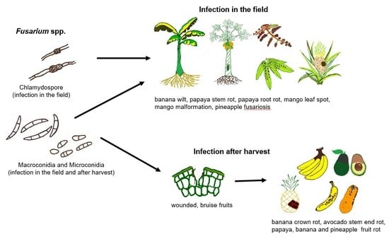
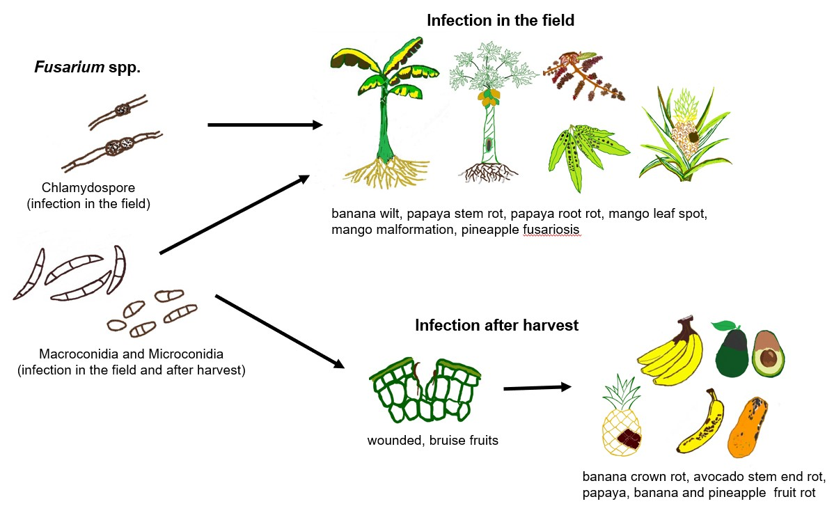
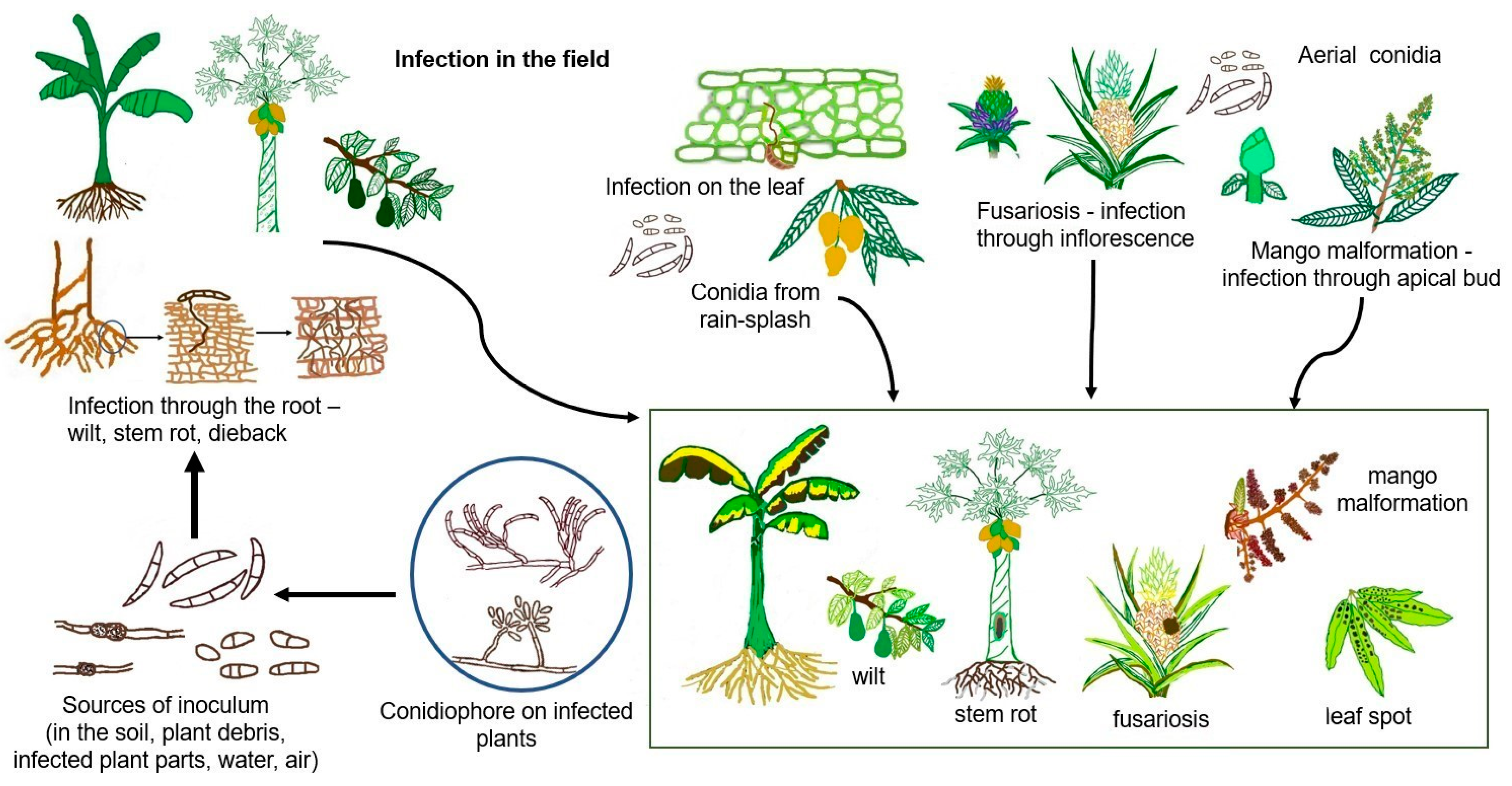
Many tropical crops are susceptible to infection by Fusarium, one of the most significant agricultural plant pathogens. The majority of Fusarium species are soil inhabitants, as saprophytes and pathogens. As a pathogen, Fusarium infects above-ground and below-ground plant parts, either as the main or secondary pathogen [7]. Fusarium species produce conidia which can disseminate through the air, rain splash, and irrigation water. Chlamydospores produced by some of the species can remain in the soil and plant debris for a long period of time [8,9] and become a source of inoculum.
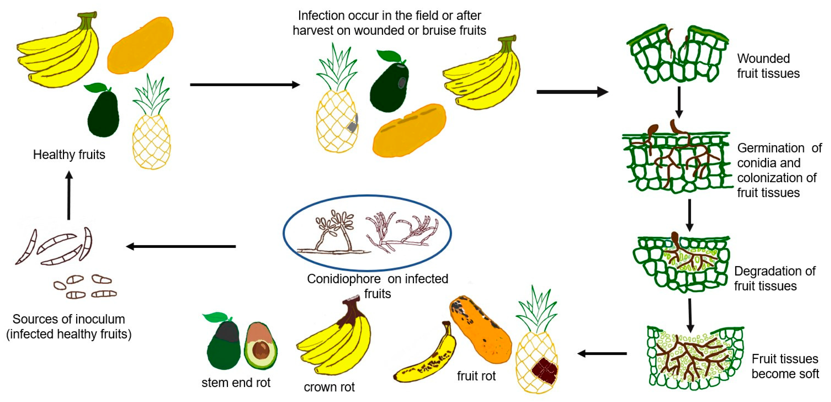
Plant-pathogenic Fusarium species have a range of infection strategies, of which most of the species are hemibiotrophs, as, during the early infection stage, the pathogen depends on the living host for growth and development. The pathogen turns into a necrotroph which eventually kills the plant host [10]. Root infection by Fusarium originates from inoculum in the soil, and infection on above-ground plant parts is from water (rain splash) and air. Some species can directly infect the plant host, and there are species that infect the plants through a wound. A wound on the plant and plant parts predisposes the plant to infection by Fusarium. Fusarium species cause plant diseases spread by soil, seed, and air and can adhere to residual plant mass; hence species of Fusarium infect a wide range of host plants. Infection on plants cause various diseases, including vascular wilt, root and stem rot, crown rot, damping off, and canker [7]. Harvested fruit crops can also be infected with Fusarium, which can cause fruit rot and stem end rot. The general disease cycle of Fusarium disease infection of major tropical fruit crops in the field is shown in Figure 1, and infection after harvest is shown in Figure 2.
Fusarium oxysporum and F. solani are the most commonly associated with tropical fruit crops. Both species mainly inhabit the soil and are widespread throughout tropical regions. The most well-known disease associated with these species is vascular wilt [7]. Currently, both species are species complexes, comprised of numerous cryptic species or phylogenetically similar species. In addition to F. oxysporum and F. solani, F. incarnatum (syn. F. semitectum), F. proliferatum, and F. verticilliodes are also commonly reported to be associated with major tropical fruits.
Plant-pathogenic Fusarium species, particularly members of the Fusarium fujikuroi species complex (FFSC), are mycotoxin producers, such as F. proliferatum, F. fujikuroi, and F. verticillioides. The main mycotoxins produced that are of major agricultural concern include fumonisins, zearalenone, and trichothecenes [11]. These mycotoxins are mainly produced by Fusarium species infecting cereal grains, but mycotoxigenic species can also affect tropical fruit yields; up to 50% losses have been reported in banana and pineapple, and losses have also been reported for other crops such as pea, tomato, and lentils [12]. Mycotoxins can be produced in growing field crops as well as after harvest and during storage. Infection at any stage may lead to mycotoxin contamination of numerous plant tissues [13].
Since the development of improved molecular methods applicable to fungal taxonomy, taxonomic changes in the genus Fusarium cannot be avoided. The generation of molecular data has resulted in major changes to both the taxonomic and systematic systems and has led to the introduction of the “one fungus, one name” concept that denotes the species name in the genus Fusarium. Many new species have also been described, as the existence of several species within species complexes. At present, molecular tools are widely used for phylogenetic analysis of Fusarium species, and often used markers are translation elongation factor 1-α (TEF-1α), β-tubulin, RNA polymerase II largest protein subunit (RPB1), and RNA polymerase II second largest protein subunit (RPB2) [14]. Morphological characterization is used mainly to sort or categorize isolates into groups before performing molecular identification. Thus, the taxonomy and nomenclature of tropical, plant-pathogenic Fusarium species have also changed. New species and diseases have both been identified by using these molecular markers.
To date, only one review article reported the association between tropical crops and Fusarium; however, this review, published by Ploetz [15], did not specifically focus on fruit crops. Given the present improvement in our understanding of Fusarium species associated with major tropical fruit crops, as well as the economic importance of these crops, a new review is necessary. This review which will describe the current state of knowledge regarding Fusarium species associated with diseases of major fruit crops may help plant pathologists and quarantine authorities to recognize the importance of the genus Fusarium and to facilitate better routine diagnosis of fruit crop diseases.
2. Fusarium Species Associated with Banana Diseases
Banana is a large, perennial, and monocotyledonous herb assigned to the genus Musa and family Musaceae [15]. Bananas are thought to have originated in Southeast Asia (including Malaysia, Indonesia, and the Philippines), where many wild banana varieties grow in the jungle. From this region, bananas have spread to other parts of the world, including South and Central America, India, Africa, and China [16].
Banana is an important cash crop planted on both large and small scales for local consumption and export. In many developing countries, particularly in Central Africa, bananas are an essential staple food. Asia is the largest producer of bananas, accounting for 54% of global production, with India being the largest producer, followed by China, Indonesia, the Philippines, and Thailand. The largest producers in Latin America and the Caribbean include Brazil, Ecuador, Mexico, and Costa Rica [5]. The Latin America and Caribbean region leads in banana exports, with Ecuador being the largest exporting country, followed by Colombia and Costa Rica. The largest importer of bananas from this region is the European Union. In Africa, almost all countries produce bananas, with Côte d’Ivoire representing the main banana-exporting country; these bananas also go primarily to the European Union, particularly to France [17]. The total global export of bananas was about 21.5 million tonnes in 2020 [17].
The most serious disease afflicting banana crops is Fusarium wilt or Panama disease, which infects bananas in almost all banana-producing countries. Furthermore, Fusarium spp. also causes post-harvest diseases such as crown and fruit rot.
2.1. Fusarium Wilt
Fusarium wilt is regarded as the most destructive disease of bananas, caused by Fusarium oxysporum f. sp cubense (Foc), and affects crops in many banana-producing countries. The disease may have originated in Southeast Asia but was first reported in Australia in 1874 [18]. In 1890, a wilt disease outbreak of unknown cause destroyed the “Gros Michel” banana plantations in Panama, and thereafter the disease was also known as Panama disease. Later, the banana wilt disease spread to Costa Rica. The pathogen responsible was identified in 1910 from a Cuban sample as F. oxysporum; it was later designated as F. oxysporum f. sp. cubense [19,20].
Fusarium oxysporum f. sp cubense strains are classified into races based on their pathogenic ability. The four main races include Race 1, which caused the outbreak of Fusarium wilt in Panama; these infect the Gros Michel and Silk cultivars. Race 2 affects the Bluggoe subgroup, which is a cooking banana [21]. Race 3 infects Heliconia spp., known as “wild banana” in Central and South America, but is no longer considered part of the Foc species complex [22]. Race 4 infects all cultivars in the Cavendish subgroup, as well as cultivars susceptible to Races 1 and 2 [23,24]. Race 4 is divided into tropical race 4 (Foc-TR4) and sub-tropical race 4 (Foc-SR4). Foc-TR4 affects Cavendish cultivars in both tropical and sub-tropical areas, whereas Foc-SR4, which is adapted to colder conditions that predispose bananas to infection, affects bananas cultivated in sub-tropical areas such as Australia, South Africa, and the Canary Islands [18,25,26].
Foc-TR4 was first detected in Taiwan, where it infected the Cavendish banana [27]. In 1990, Foc-TR4 was found in two Cavendish banana plantations in Malaysia; neither plantation had previously recorded any Fusarium wilt infection. In the same year, Foc-TR4 infection was also reported in banana plantations in southern Sumatra (Indonesia), which featured the Valery cultivar from the Cavendish subgroup [28]. According to Buddenhagen [26], Foc-TR4 may have existed in village banana plants in Malaysia and Indonesia before the Cavendish banana had become established. However, at that time, the pathogenic race did not cause serious infection and did not cause significant losses. The losses that were observed may have been due to the presence of other Foc races and/or the planting of various other banana cultivars in the same area.
Banana Fusarium wilt is a polycyclic disease whereby carry-over of inoculum from previous plantings can occur, causing infection during a current planting; thus, the disease is referred to as a “compound interest” disease [29,30]. New inocula are produced from current banana plantings, and these are added to existing inocula from previous plantings, causing a steady accumulation of inocula [30]. This is a serious problem since Fusarium wilt infection can result from even small amounts of inoculum. Infected banana plants exhibit typical wilt disease symptoms, including leaf-yellowing and discoloration of vascular tissues. Internal symptoms develop initially at the site of infection, which is where the feeder roots; here, the xylem becomes reddish-brown and discolored. The disease further develops by moving to the rhizome and the pseudostem. Mature leaves then turn yellow and wilt. The disease then progresses to the younger leaves that surround the pseudostem [19,20]. The vascular tissues of the infected banana plant become generally discolored and clogged with the pathogen, which results in disruptions to nutrient and water transport.
In many banana-producing countries, the Fusarium-susceptible Gros Michel cultivar has been replaced by the resistant Cavendish, thereby facilitating increased banana cultivation, both for export and for local consumption. With this replacement, it was first thought that Fusarium wilt would no longer be a threat to banana cultivation. However, the emergence of Foc-TR4 in Southeast Asia has led scientists to believe that Fusarium wilt will become a major threat to Cavendish bananas as well as other varieties, such as plantains [31,32,33]. Foc-TR4 has already become a threat to banana cultivation in several parts of Southeast Asia, including Malaysia, Indonesia, and The Philippines, as well as in Northern Territory and Queensland, Australia [34].
After 20 years, Foc-TR4 has only infected bananas in Southeast Asia and Australia but has recently been detected in other banana-producing countries, including Jordan [35], China [36], Pakistan and Lebanon [33], Queensland, Australia [37], Israel [38], Laos [39], Myanmar [40], Vietnam [41], India [42], Colombia [43], Mayotte [44], Turkey [45], and Peru [46].
In Turkey, banana is cultivated in open fields as well as in plastic greenhouses, a method known as protected cultivation [47]. Foc-TR4 was detected in Turkey in banana greenhouses in three cities located along the Mediterranean coast. Since Foc-TR4 was detected in the greenhouse, it is also possible that it can be introduced to open field areas. The presence of Foc-TR4 on the Island of Mayotte posed a serious threat to banana cultivation in surrounding countries [44]. The Island of Mayotte is located in the Comoros archipelago, which borders the East African coast, including the waters of northwestern Madagascar and northeastern Mozambique. The spreading of pathogenic Foc-TR4 may have a significant effect on the livelihoods of people in these countries since banana is an economically important crop in both countries. In South America, Foc-TR4 has been identified in Colombia and Peru, indicating that the pathogenic race has spread to this region [43,46]. The presence of Foc-TR4 in Colombia and Peru has caused neighboring countries, particularly Ecuador, to be on high alert since Ecuador is the leading banana exporter to the European market. Moreover, the threat of Foc-TR4 is of great concern in banana-producing countries in both South America and the Caribbean due to the close proximity of these regions.
Dissemination of Foc-T4 into new areas, either from one farm to another or between countries, likely occurs via the movement of infected planting materials related to banana propagation, such as suckers or rhizomes [29,48]. Other key factors of infection include infested soil, which is used to grow planting materials before field cultivation [26,49], contaminated surface or irrigation water, and contaminated farm tools, footwear, and clothing [20,35]. According to Dita et al. [48], anthropogenic factors play an important role in disseminating Foc due to the increase in cross-border activities related to banana cultivation. Anthropogenic factors related to the international banana trade may have also played a role in dispersing Foc-T4 from Southeast Asia to Africa and the Middle East [48] as well as within the same region.
Phylogeographical data indicate that the global dispersal of Foc-TR4 represents the colonization of a single clone and that the introduction into new areas likely occurs from infected planting materials [33,40]. Several phylogeographical analyses have analyzed the dispersal patterns of Foc-TR4 among related banana-producing countries. Foc-TR4 found in Laos, Vietnam, and Myanmar may have originated from Yunnan, China. In addition, Foc-TR4 detected in Pakistan likely originated from the Philippines since the pathogens found in these countries are closely related [33,40]. Isolates from Colombia were found to be associated with isolates from Indonesia [38], suggesting a possible source of pathogen introduction to South America. Middle Eastern Foc-TR4 isolates are also closely related. A study by García-Bastidas et al. [41] on Foc-TR4 in Lebanon and Jordan showed that pathogenic races found in both countries were related. A similar finding has also been demonstrated by Maymon et al. [38], who reported that Foc-TR4 isolates from Israel, Jordan, and Lebanon were grouped in the same clade, thereby indicating close relatedness.
Recently, Maryani et al. [50] renamed Foc-TR4 as F. odoratissimum based on lineages formed from a phylogeny plotted using three loci, RPB1, RPB2, and TEF-1α. However, according to Torres Bedoya et al. [51], the phylogenetic inference contained notable flaws. Most importantly, data generated by this phylogenetic analysis were not robust enough to support the monophyly of a new species. Even the TEF-1α tree lacked robust support of monophyly for the proposed taxonomic redesignation. A detailed explanation of the ambiguity of the proposed name is described in greater detail by Torres Bedoya et al. [51].
2.2. Crown Rot
Crown rot infects the banana crown, which is the area that is connected to the peduncles. Wounds created during fruit removal from the fruit bunch are the entry point for crown rot pathogens, and this occurs mainly during harvest. Symptoms appear later, either during shipping or when the fruits ripen. Moreover, physiological changes in banana fruits expedite pathogen development. Infected tissues become soft and black before withering, at which point the rot lesion progresses into the fruit pulp [52].
Crown rot is regarded as the main post-harvest disease of exported bananas since the rot lesion develops rapidly during shipping. Early ripening is often triggered by the ethylene released caused by pathogen infection and is followed by tissue necrosis. Thus, fruit quality is affected by the formation of rot lesions or by necrosis in the crown area [52,53].
Diverse fungal pathogens cause banana crown rot, and therefore this disease is thought to be caused by a fungal complex rather than a specific species. The crown rot fungal complex consists of different fungal species of which the presence and prevalence vary according to the area in which bananas are cultivated as well as due to seasonal variation. Fruit contamination by crown rot pathogens can occur in the field as well as after harvest. In the latter case, contamination can spread from infected washing tanks, which also affects pathogen composition [53,54]. Some of the fungal species recovered from crown rot lesions include Acremonium spp., Arthrinium phaeospermum, Aspergillus spp., Ceratocystis paradoxa, Cladosporium spp., Colletotrichum musae, Curvularia lunata, Lasiodiplodia theobromae; Musicillium theobromae, Nigrospora sphaerica, Penicillium spp., Phomopsis spp., Phyllosticta musarum, Thielaviopsis paradoxa, and many Fusarium species [55,56,57]. The co-inoculation of crown rot pathogens, including some Fusarium species, showed highly severe infections compared to plants inoculated with individual pathogens separately. Anthony et al. [58] reported that co-inoculation of Lasiodiplodia theobromae, F. verticillioides, and Colletotrichum musae caused more severe crown rot infection than when pathogens were inoculated separately.
The main source of inoculum of Fusarium spp. infection are conidia from decomposing leaves and banana flowers [59]. Contamination by Fusarium conidia primarily occurs within the first 40 days after bunch emergence, and the Fusarium slowly replaces other crown rot fungi [60]. Conidia can also be spread from detached bananas after dehanding from the fruit bunch, of which infections occur when bananas are exposed to the washing tank in the packing house, which is another source of inoculum [54,61].
Fusarium-causing banana crown rot has been reported since the 1960s. Studies performed from the 1960s to the present have indicated that multiple species of Fusarium can cause crown rot; these include F. camptoceras, F. chlamydosporum, F. concentricum, F. dimerum, F. equiseti, F. semitectum (syn. F. incarnatum), F. verticillioides, F. sacchari, F. sporotrichoides, F. oxysporum, F. solani, F. subglutinans sensu lato, F. graminearum, F. musae, F. musarum, F. proliferatum, and F. pseudocircinatum [53,62,63,64,65,66]. Most Fusarium spp. associated with banana crown rot have a wide host range. Exceptions include F. musae and F. musarum, which are only found on diseased bananas in certain countries. Fusarium musarum was reported in Panama, while F. musae was recovered in Ecuador, Mexico, Panama, the Canary Islands, and the Philippines [67,68].
Despite various pathogenic Fusarium spp. causing banana crown rot, the virulence of each species also varies. In the Windward Islands, three species, F. oxysporum, F. verticillioides, and F. graminearum are regarded as the main pathogens since these species have been the most often recovered [69]. In the Caribbean, two species, F. semitectum and F. graminearum, are the major banana crown rot pathogens [70], while in Costa Rica, F. verticillioides and F. semitectum are the most common pathogenic species [71]. Other Fusarium species may not be major pathogens of banana crown rot but may nevertheless be a part of a fungal complex causing this disease, therefore also affecting disease occurrence and severity.
Crown rot fungal composition and banana cultivars present in organic farms may differ from those in conventional farms since the use of fungicides is restricted in organic farms [64]. In symptomless organic banana hands from the Dominican Republic, fungal isolates from the genus Fusarium were found to be the most prevalent isolates recovered, representing 55% of all fungi. The species identified included F. incarnatum (53%), F. verticillioides (12%), F. sacchari (12%), F. proliferatum (7%), and F. solani (6%). Fusarium dimerum, F. musae, and F. pseudocircinatum were also isolated but were less abundant [64]. Pathogenicity tests indicated that although F. verticillioides and F. sacchari were more virulent than F. incarnatum, a higher percentage of F. incarnatum was isolated. This suggests that F. incarnatum is the main pathogen associated with banana crown rot in the Dominican Republic [64].
In another study on organic Cavendish and Gros Michel cultivars in Costa Rica, the most common crown rot fungi isolated were F. subglutinans, but the most aggressive were F. verticillioides. Other species of Fusarium recovered included F. proliferatum, F. sacchari, F. semitectum, and F. graminearum [72,73]. Moreover, studies by Kamel et al. [64] and Umana-Rojas and Garcia [72,73] showed that the Fusarium species isolated from organic banana crown rot samples were similar to Fusarium species found in crown rot samples in non-organic bananas.
2.3. Fruit Rot
Fruit rot appears as black or dark brown rot lesions that form on the fruit surface or epidermis. Smaller lesions may merge and form larger ones. As rot lesions progress, fungal mycelia develop within the lesion and can cause extensive rotting [74,75]. As with crown rot, fruit rot may also affect bananas destined for export, and symptoms may appear during shipping or marketing.
Banana fruit rot is often confused with banana crown rot since some Fusarium species can cause both diseases. As with crown rot, many Fusarium species have been reported to be associated with banana fruit rot, and these species have been referred to as contaminating fungi. The Fusarium species and their associated diseases reported here are based on the species reported and the diseases described in these publications.
In samples of bananas imported to Spain and Italy, 12 species, one of which was unidentified, were isolated and identified from fruit rot lesions. These species included F. acuminatum, F. camptoceras, F. dimerum, F. equiseti, F. graminearum, F. moniliforme, F. oxysporum, F. proliferatum, F. semitectum var. majus, F. solani, F. subglutinans, and Fusarium sp. Of these species, F. subsglutinans, F. acuminatum, and F. graminearum were distinctly more pathogenic than other species [76]. Based on toxicity assessments performed using brine shrimp assays, the most pathogenic species, which showed a larval mortality rate of more than 70%, include F. camptoceras, Fusarium sp., F. moniliforme, F. proliferatum, F. oxysporum, F. graminearum, F. acuminatum, and F. equiseti. These findings showed that numerous species could produce toxic metabolites in banana fruit [76]. Two similar studies were conducted by Alghuthaymi et al. [77,78] on banana fruits imported to Saudi Arabia. In the study, seven Fusarium species were isolated from rotted bananas, with the most prevalent being F. semitectum, followed by F. proliferatum, F. circinatum, F. chlamydosporum, F. solani, F. oxysporum, and F. thapsinum. Except for F. oxysporum, these species all produced fumonisin B1, deoxynivalenol, and zearalenone. These results were in contrast to an earlier study of banana fruit rot in Riyadh, where only F. thapsinum was identified as a causal pathogen [79]. A study by Molnár et al. [80] on imported bananas in Hungary identified two species present in rotten bananas, F. verticillioides and F. musae. Only F. verticillioides was found to produce high amounts of fumonisin B. Fusarium verticillioides has also been identified in other studies of banana fruit rot in different countries, including where bananas were imported and others where bananas were cultivated locally [74,75,76,81,82]. Another three species that have been commonly recovered from banana fruit rot include F. semitectum (syn. F. incarnatum), F. oxysporum, and F. solani; these are also associated with imported and locally cultivated bananas [76,78,81,82,83,84]. Other Fusarium species reported include F. sacchari, F. concolor, F. concentricum, and F. fujikuroi [82,85].
Table 1 summarizes the occurrence of Foc-TR4 in banana-producing countries worldwide, as well as Fusarium spp. associated with banana crown rot and fruit rot. Although Fusarium is often associated with banana wilt, crown rot, and fruit rot caused by various species of Fusarium also have serious effects on banana, especially since they are post-harvest diseases that affect bananas destined for import.
3. Fusarium spp. Associated with Papaya Diseases
Papaya (Carica papaya L.) is also known as papaw or pawpaw. It is a perennial herbaceous plant belonging to the Caricaceae. It is among the most important tropical fruits, is widely cultivated in tropical areas, and is marketed worldwide. Papaya is also cultivated in several Mediterranean countries, including Italy and Spain, under greenhouse conditions [86]. The top five papaya-producing countries include India (44.05%), followed by the Dominican Republic, Brazil, Mexico, and Indonesia. India is both the top producer and the top consumer of this fruit [87]. Although the most serious diseases of papaya are papaya ringspot virus and bacterial dieback, other diseases can also negatively affect yield. Some diseases known to affect papaya include root and stem rot, post-harvest fruit rot, and stem end rot, and are caused by Fusarium spp.
3.1. Root Rot
Most reports on root rot of papaya are related to Phytophthora spp. However, based on a study by Singh and Kumar [88] in Bihar, India, root rot of papaya caused by F. solani is a newly emerging disease of papaya cultivation. Fusarium solani is a soil-borne pathogen that infects seedlings and causes rotting and wilting of the young papaya plant. Development of Fusarium solani root rot is more rapid after rain, and the disease affects all growth stages and all papaya varieties. The pathogen can cause up to 95% damage to papaya cultivation in Bihar [88]. Later, using molecular methods, Gupta et al. [89] and Vega-Gutiérrez [90] reported F. falciforme as a causal pathogen of papaya root rot in India and Mexico, respectively. Fusarium falciforme is a phylogenetic species member of the Fusarium solani species complex.
3.2. Stem Rot
Similar to root rot, stem rot also tends to infect young papaya plants. The infected stem becomes tender, and black or brown lesions then develop. These lesions eventually become the focal point of the rot. Rotting roots often progress to the trunk, and sometimes symptoms also include leaf drooping and yellowing [89]. Although the major pathogenic causes of papaya stem rot are Phytophthora palmivora and Pythium aphanidermatum, this disease can also be caused by F. solani. In Brazil, one report of papaya stem rot described the emergence of reddish-brown to dark-brown lesions, of which the causal pathogen was a species of the Fusarium solani species complex [91]. Gupta et al. [89] and Vega-Gutiérrez [90] reported that F. falciforme was also a causal pathogen involved in papaya stem rot.
3.3. Stem End Rot
Stem end rot infects the tissues at the stem or peduncle attached to the papaya fruit. This disease infects harvested papaya and is caused by a complex of fungal pathogens, including Fusarium spp. The infection often occurs during the flowering stage via wounds at the peduncle and/or natural openings. The pathogen then remains latent until the ripening stage, when soluble sugar content and phytoalexin levels are reduced. These conditions favor pathogen growth, and symptoms begin with slight browning at the peduncle. After a few days, rotting appears, and the peduncle area becomes blackened and soft [92,93]. Among the fungal species causing papaya stem end rot is F. solani, which has been identified in the Philippines, Japan, and Brazil [94,95,96]. To date, F. solani is the only species from the genus Fusarium that has been reported as a causal pathogen of papaya stems end rot. Such pathogens are likely to be similar to those that cause mango stem end rot; for this reason, in addition to F. solani, other pathogens considered to cause stem end rot include Lasiodiplodia theobromae, Phoma caricae-papayae, Colletotrichum gloeosporioides, and Ascochyta caricae [95,96].
3.4. Fruit Rot
Fruit rot is another post-harvest disease of papaya fruit. It is usually caused by inappropriate handling after harvest, including unsuitable storage and/or transportation conditions as well as improper handling at point-of-sale [97]. Fusarium spp. are important pathogens associated with papaya fruit rot but are regarded as secondary pathogens since they often require some other infection or stress to establish themselves in a host [98]. As such, Fusarium infects papaya fruits through cuts or abrasions created during harvesting and handling or via injuries caused by insects. Symptoms of Fusarium rot on papaya emerge as rounded, tender areas that later become small depressions. As these lesions develop, rot and mycelia appear on the surface [99]. Unlike stem end rot, which is thus far thought to be caused only by F. solani, several species of Fusarium are associated with papaya fruit rot. Fusarium solani is the most reported, having been identified in Hawaii, India, the Philippines [98], Malaysia [81,99], Allahabad, India [100], and Nigeria [101]. The dry rot of papaya fruits is also known to be caused by F. solani [93]. Other Fusarium spp. implicated in cases of papaya fruit rot include F. acuminatum [102], F. equiseti [103], F. semitectum (syn. F. incarnatum) [81], F. nivale [104], F. oxysporum [101,105], F. thapsinum, and F. chlamydosporum [106].
Table 2 shows Fusarium spp. associated with papaya diseases reported in several countries, which indicate a possible threat to papaya cultivation. Although Fusarium diseases of papaya are not as serious as ringspot virus and anthracnose, any diseases that affect the fruit crop lessen yield and are therefore important. Post-harvest diseases such as stem end rot, fruit rot, and anthracnose can also reduce the marketability of produce, which also has a significant economic impact.
4. Fusarium Species Associated with Mango Diseases
Mango (Mangifera indica L.), which belongs to the family Anacardiaceae, is grown for its edible fruit, which is consumed fresh. Mango is known as the “king of fruits” due to its nutritional value, taste, flavor, texture, and aroma [107]. Mango is cultivated in both tropical and sub-tropical countries. India is the largest mango-producing country, followed by China, Thailand, Indonesia, Pakistan, Mexico, Brazil, Bangladesh, Nigeria, and the Philippines. At present, mango production has also expanded to Australia, the Middle East (especially Egypt, Israel, and Oman), as well as in several countries in Central and South America (including Brazil, Venezuela, and El Salvador) [108].
Due to the economic importance of mango, diseases affecting yield can lead to serious economic loss for mango growers. Several mango diseases are caused by Fusarium spp., including mango malformation, dieback, leaf spot, and gall. Of these diseases, mango malformation is the most serious, and it affects mango production in both tropical and sub-tropical areas of mango production. Table 3 summarizes the Fusarium spp. associated with mango diseases in several countries as well as the symptoms reported.
4.1. Mango Malformation
Mango malformation was first detected in India, and this disease has now been found in many mango cultivation areas. This disease affects both vegetative and floral tissues, causing malformation of the tissue itself. Vegetative malformation affects nursery seedlings and young mango plants. Symptoms include hypertrophic growth of vegetative buds, swollen axillary buds, and disturbed apical dominance [109,110]. The growing buds produce distorted shoots bearing small leaves; these appear as crowded, unhealthy masses, known as “witches’ broom” [110]. The malformed seedlings become stunted and eventually die.
Floral or inflorescence malformation develops on mature mango trees during flowering. The inflorescence becomes enlarged while at the same time, the axes branch abundantly, become short and thick, and produce sterile flowers. The formation of leaves (phyllody) on the inflorescence may ensue [111]. Inflorescence malformation is more significant since infected trees do not set fruit, thereby affecting fruit production [109]. As mango malformation affects the inflorescence, the fruit set stage does not occur; this leads directly to yield loss. A significant number of Fusarium spp. have been reported to be associated with mango malformation, and their presence in most mango-producing countries indicates that this disease is widespread.
Fusarium moniliforme var. subglutinans was initially identified as the causal pathogen of both vegetative and inflorescence malformations [112,113]. The species was later recognized as F. subglutinans. However, based on histone H3 and β-tubulin gene phylogenies, F. subglutinans was found to form two phylogenetic groups; these groups were later identified as two separate species, F. mangiferae and F. sterilihyphosum [114,115].
Fusarium mangiferae is the most common species associated with mango malformation. To date, it has been identified in South Africa, India, China, Egypt, Malaysia, Oman, Sri Lanka, Spain, and the United States [110,116]. Fusarium sterilihyphosum is less common and has been identified only in South Africa and Brazil [115,117]. However, other Fusarium spp. have also been reported to be associated with mango malformation. Fusarium tupiense has been found in Brazil [118], Senegal [119], and Spain [120]. Two species, F. mexicanum and F. pseudocircinatum have been detected in Mexico [121]. Fusarium pseudocircinatum was also found in the Dominican Republic [122]. Another well-known species, F. proliferatum, has been isolated from malformed mango tissue in Malaysia [123], China [124], and Egypt [125]. Other species, including F. anthophilum, F. fujikuroi, F. incarnatum, F. oxysporum, F. parvisorum, F. scirpi, F. solani, F. verticillioides, and three undescribed species, were found to be associated with malformation-like symptoms in Australia [126]. Finally, Molina-Cárdenas [127] described F. neocosmosporiellum as a causal pathogen of mango malformation in Mexico.
4.2. Mango Decline and/or Dieback
Mango decline and/or dieback are complex diseases that often involve a combination of causal pathogens, including Fusarium spp. The symptoms of decline and dieback are similar and only differ subtly. Infected mango trees show a uniform pattern of dying back from the crown downwards, accompanied by leaf discoloration and death. Severe infections lead to the death of whole branches, which can result in the death of the infected tree. Most other common symptoms, such as blight, tip dieback, blights, cankers, gummosis, and stem bleeding, also fall into the general description of “decline” as the result of this infection [128]. Once mango trees show symptoms of decline and/or dieback, it can be very difficult to treat the tree [129].
Although Lasiodiplodia theobromae is the most common pathogen associated with mango decline and/or dieback, several Fusarium spp. have also been found to be associated with this disease. Among these species are F. oxysporum, F. semitectum, and F. solani [130]. In one location in Pakistan, F. solani was found to cause mango decline with disease severity of 62.50–78.75% [131]. In addition, dieback caused by F. decemcellulare has been identified as an emerging disease infecting mango trees in Sichuan province, China [132].
Another disease that is similar to mango decline/dieback is known as sudden death or sudden decline. This disease manifests decline symptoms such as wilting; this starts on one side of the tree and then spreads to the whole tree. Gummosis exudes from the bark, and vascular tissues become discolored [133]. Sudden death syndrome is likely spread by the ambrosia bark beetle (Hypocryphalus mangiferae), and the causal pathogens have been isolated from the bark beetles themselves. Fusarium spp. are among the most frequently isolated fungi from both mango trees showing sudden death symptoms and from bark beetles from affected the trees [134,135].
The main pathogens associated with mango tree sudden death syndrome are reportedly Ceratocystis fimbriata and Lasiodiplodia theobromae [133]. Nevertheless, Fusarium spp. may also contribute to the occurrence and severity of mango tree sudden death syndrome since many decline-like diseases are caused by a complex of causal pathogens. Two species, F. oxysporum and F. solani have been recovered from the root collar and bark of an infected mango tree [136]. Moreover, F. equiseti has been isolated from the dried stems, branches, and roots of different varieties of mango trees [137]. Finally, a study by Abbasi et al. [135] showed that F. solani was present in bark beetles. Using a pathogenicity test, the authors found that this fungus was a causal pathogen of mango tree sudden death syndrome.
To date, most studies related to mango decline/dieback diseases have been performed in India, Pakistan, and Oman. More studies are needed to generate information on how Fusarium spp. are associated with this disease, and more studies should be performed in other mango-producing countries
4.3. Gall
Mango gall disease has been reported in the mainland USA, Mexico, Dominican Republic, Brazil, and Puerto Rico. In the USA, large galls with a rough and scaly exterior were found on the main trunks of mango trees at the USDA-ARS germplasm collection in Miami, Florida [138]. The causal pathogen was identified as F. decemcellulare, which is also known to cause mango tree gall in Mexico [139]. The latest report of mango tree gall disease was in the Dominican Republic, in which the causal pathogen was also F. decemcellulare [140].
4.4. Leaf Spot
Leaf spot is a common disease in many plants and is often characterized by discolored lesions caused by tissue necrosis [141]. Long periods of wet and humid conditions usually promote leaf spot disease, and most pathogens associated with leaf spots are spread by precipitation, irrigation water, and/or wind [142]. An abundance of leaf spot lesions can lead to the disruption of photosynthesis and will consequently affect nutrient transportation [143]. Thus, plant growth is reduced, which increases susceptibility to infection by secondary pathogens.
Fusarium spp. associated with mango leaf spot have been reported in Malaysia and China. In Malaysia, F. proliferatum, F. semitectum, and F. chlamydosporum were found to cause mango leaf spot. In addition, two species, F. solani and F. mangiferae were also isolated from leaf spot lesions on mango leaves, but a pathogenicity test showed that both species were not pathogenic [144]. In China, seven species, including F. concentricum, F. hainanense, F. mangiferae, F. pernambucanum, F. proliferatum, F. sulawesiense, and F. verticillioides, were found to be able to cause leaf spot on mango leaves [145]. To date, ten species of Fusarium have been reported to be associated with mango leaf spot. However, the Fusarium spp. reported are likely secondary pathogens since pathogenicity tests were performed using a wounded treatment. Moreover, the pathogenicity test of Omar et al. [144] showed that Fusarium spp. was associated with low to moderate-severity disease.

Table 3.
Fusarium spp. associated with diseases of mango.
| Disease | Symptoms | Fusarium spp. | Country | References |
|---|---|---|---|---|
| Mango malformation | Vegetative malformation—hypertrophic growth of vegetative buds, swollen axillary buds, and disturbed apical dominance. Growing buds produce distorted shoots bearing small leaves that appear as crowded, unhealthy masses or “witches’ broom” appearance. Malformed seedlings become stunted and eventually die. Floral or inflorescence malformation—inflorescence becomes enlarged, and at the same time, the axes branch abundantly becomes short and thick and produces sterile flowers. Formation of leaves (phyllody) on the inflorescence may occur. | Fusarium moniliforme var. subglutinans | India | [112,113] |
| F. mangiferae | China, Egypt, India, Israel, Malaysia, Oman, South Africa, Spain, Sri Lanka, USA, Australia | [110,116,126] | ||
| F. sterilihyphosum | South Africa, Brazil | [115,117] | ||
| F. mexicanum | Mexico | [121] | ||
| F. tupiense | Brazil, Senegal, Spain | [118,119,120] | ||
| F. pseudocircinatum | Mexico, Dominican Republic | [121,122] | ||
| F. proliferatum | Malaysia, China, Egypt | [123,124,125] | ||
| F. anthophilum, F. fujikuroi, F. incarnatum, F. oxysporum, F. parvisorum, F. scirpi, F. solani, F. verticillioides, three undescribed species (associated with malformation-like symptoms) | Australia | [126] | ||
| F. neocosmosporiellum | Mexico | [127] | ||
| Decline/ Dieback | Uniform pattern of dying back from the crown downwards, accompanied by leaf discoloration. Severe infections cause dying of entire branches, resulting in the death of the infected tree. Other symptoms—include blight, tip dieback, blights, cankers, gummosis, and stem bleeding. | F. solani, F. oxysporum, F. solani | Pakistan | [131,135,136] |
| F. decemcellulare | China | [132] | ||
| Gall | Large galls with a rough and scaly exterior on the main trunks. | F. decemcellulare | Miami, Florida; Mexico; Dominican Republic | [134,139,140] |
| Leaf spot | Discolored lesions or spots on the leaves caused by necrosis of the tissues. | F. proliferatum, F. semitectum, F. chlamydosporum | Malaysia | [144] |
| F. concentricum, F. hainanense, F. mangiferae, F. pernambucanum, F. proliferatum, F. sulawesiense, F. verticillioides | China | [145] |
5. Fusarium spp. Associated with Pineapple Diseases
Pineapple (Ananas comosus Merr.) is a mostly epiphytic perennial plant from the family Bromeliaceae. Pineapple is originally from South America and may be native to regions of southern Brazil and Paraguay, where wild relatives of pineapple are still found [146,147]. Pineapple is cultivated in both tropical and sub-tropical countries, where it requires warm and humid conditions and consistent rainfall. The top five pineapple-producing countries are Costa Rica, the Philippines, Brazil, Thailand, and Indonesia [5]. In all these countries, pineapple is a major fruit crop, commonly consumed fresh, but it can also be made into processed food products.
Pineapple plants are also infected by Fusarium, and the most common and serious diseases include fruitlet core rot and fusariosis. Less severe diseases are fruit rot, leaf spot, heart rot, and dieback (Table 4).
5.1. Fruitlet Core Rot
Fruitlet core rot is a serious pineapple disease that affects the internal tissue (flesh) of the fruit. Infection often occurs during the early stages of flowering as well as during fruit maturation. The pathogen enters the fruit either through the stigma during flowering or through wounds [148]. Typical internal symptoms include brown discoloration in the center of the fruitlet, which may spread to the fruit core. The infected flesh looks similar to a black spot. External symptoms include dry rot at the infected site, where the flesh can remain quite firm, and the fruit remains green. When the fruits ripen and the infection is severe, the infected part becomes sunken [149,150].
The causal agents of fruitlet core rot can be either a single pathogen or a group of pathogens; thus, this disease is considered to be a disease complex. Pathogens commonly associated with fruitlet core rot include species from the genera Penicillium and Fusarium, Candida guilliermondii (a round yeast), as well as pineapple red mite [151]. Little information is available on the association between round yeast and mites, but there are many reports related to infection by Penicillium and Fusarium.
Early studies showed that a combination of Talaromyces (previously known as the Penicillium subgenus Biverticillium) and Fusarium were causal pathogens of fruitlet core rot in pineapple [152,153,154]. In Queensland, Australia, Oxenham [154] isolated Penicillium funiculosus (syn. Talaromyces funiculosum) and F. verticillioides from infected fruits. Initially, T. funiculosus was thought to be the main pathogen responsible for fruitlet core rot, but later studies found that Fusarium isolates were also recovered from diseased pineapples, especially from infected flesh tissue [155]. Generally, the appearance of the rot lesion formed can be used to differentiate between infection by Talaromyces and Fusarium. Rot lesions produced by Talaromyces tend to be dark to medium brown with a moist, gray area in the center. In contrast, rot lesions produced by Fusarium infection vary from light to dark brown and spread to the fruitlet core. Moreover, rot lesions produced by Fusarium are normally of a dry rot type [151].
Several Fusarium spp. are associated with pineapple fruitlet core rot. F. subglutinans and F. guttiforme were first reported to be causal pathogens of this disease. However, molecular phylogenies based on TEF-1α, β-tubulin, and RPB2 gene sequences showed that other Fusarium spp. were also associated with the disease. Jacobs et al. [156] reidentified F. guttiforme as F. ananatum, a new species causing pineapple fruitlet core rot in South Africa; it has subsequently been reported in China [150] and Japan [157]. Based on phylogenetic analysis of the RPB2 gene sequence, three species (i.e., F. guttiforme, F. ananatum, and F. oxysporum) were found to be associated with pineapple fruitlet core rot of the “Pérola” cultivar planted in Paraiba, Pernambuco, and Rio Grande do Norte in Brazil [158]. Pathogenicity tests indicated that although rot lesions formed by F. guttiforme and F. ananatum on pineapple fruits were visually similar, those rot lesions caused by F. ananatum were darker and grew deeper into the inner tissue of the fruit, thus forming a V-shaped rot lesion [158].
Barral et al. [159] reported that species from two fungal genera, Talaromyces and Fusarium, were the causal pathogens of pineapple fruitlet core rot on Reunion Island (France). This study supported the findings of previous studies [149,152,153,154] that indicated the species from these two fungal genera were both associated with the disease. In the study of Barral et al. [159], 79% of isolates recovered from both infected and healthy fruitlets were from the genus Fusarium. Three species of Fusarium, F. ananatum, F. oxysporum, and F. proliferatum, were identified using a TEF-1α gene phylogeny. Of these species, F. ananatum (n = 107) was the most common species recovered, followed by F. oxysporum (n = 10) and F. proliferatum (n = 1). Barral et al. [159] also examined the mycotoxigenic ability of these three Fusarium species. Three types of mycotoxins, fumonisins (FB1, FB2, and FB3), moniliformin (MON), and beauvericin (BEA), were detected in artificially inoculated pineapple ruitlets and in naturally infected fruits. Higher levels of mycotoxins were detected in inoculated fruitlets compared to naturally infected fruits. Fusarium ananatum and F. proliferatum were able to produce all three types of mycotoxins tested, but F. oxysporum produced only MON and BEA. The findings of Vignassa et al. [160] agreed with those of Barral et al. [159] in that they found that the pathogens responsible for fruitlet core rot included species from two species complexes, FFSC and the T. purpureogenus species complex. Several FFSC species were isolated from both infected and healthy fruitlet, identified as F. proliferatum, F. ananatum, F. sacchari, F. fujikuroi, F. circinatum, and F. verticillioides. In addition, other species of Fusarium were also recovered from both infected and healthy fruitlets, which included F. oxysporum, F. equiseti, F. solani, F. incarnatum, F. chlamydosporum, and F. napiforme. Other Fusarium species, F. falciforme, F. circinatum, F. cortaderiae, F. graminearum, F. dlamini, and F. ficicrescens were isolated from either healthy or infected fruitlets, but not both [160]. A pathogenicity test was conducted on only F. proliferatum, F. sacchari, and F. oxysporum. These three species were able to produce symptoms similar to fruitlet core rot, including the development of rot lesions or black spots. Fusarium proliferatum and F. sacchari both caused severe rot lesions on the flesh of the fruitlets. However, F. oxysporum showed much milder symptoms [160]. Further pathogenicity tests of other Fusarium spp. should be performed to confirm their pathogenic ability; this is especially important since many species of Fusarium have a wide range of hosts.
The environmental conditions of pineapple-producing countries may contribute to the prevalence of particular pathogens associated with fruitlet core rot [151]. Penicillium and Fusarium species have been reported as the most common causes of pineapple fruitlet core rot in Hawaii. Moreover, in South Africa, Penicillium species are the most prevalent, while in Brazil, Fusarium species are more common [151].
According to Vignassa et al. [160], pineapple fruitlet core rot is a multipartite pathology that involves Fusarium spp. and other microbes found in the mycobiome of healthy and infected fruitlets, as well as environmental factors. Vignassa et al. [160] also considered data related to wind direction and the nearby presence of cultivated crops that can be infected by the same causal pathogens as fruitlet core rot and concluded that fruitlet core rot might be an airborne disease.
Reports on fruitlet core rot have been obtained from Brazil, Japan, China, Reunion Island, India, Hawaii, and Malaysia, all of which are among the top pineapple-producing areas. However, detailed studies of the disease and its causal pathogens have been performed in Brazil and on Reunion Island [158,159,160]. From these reports, F. ananatum (previously known as F. guttiforme) is the most prevalent species known to cause fruitlet core rot. Another important finding was that Fusarium spp. were found to be associated with the ability of the diseased tissues to produce mycotoxin; this information may be useful to mitigate potential risks to food safety since pineapple is often consumed fresh.
5.2. Fusariosis
Fusariosis is also a serious disease of pineapple, and it affects all parts of the plant but most severely infects the fruit. This disease was first reported and described in Argentina in 1954. A decade later, fusariosis was detected in Brazil, where it caused severe infection and affected Brazil’s position as the world’s top producer of pineapples [161,162]. Much later, fusariosis was identified in other pineapple-producing countries, including Cuba, South Africa, India, and Malaysia. Fusariosis infections usually occur from the early flowering stages throughout all stages of fruit development. Fusariosis pathogens enter the inflorescence through wounds, and thus the earliest disease symptoms appear in the fruitlets. Infected planting materials can also spread the disease [162,163]. Finally, infection of the developing fruit can lead to secondary infections, which occur on developing suckers and slips.
Obvious symptoms on affected fruits include discoloration of the infected areas, which have light to dark brown fruitlets. Moreover, these rot lesions may spread to the fruit core. The infected areas eventually become sunken and fungal sporulation, and gum exudation becomes visible [162,164]. Other symptoms of fusariosis on pineapple plants include stunting, chlorosis, shortened stems, bent or dead stems at the apex, and phyllotaxic disruption throughout the plant [162,164].
Although the symptoms of fusariosis are similar to those of fruitlet core rot, there are slight differences between both diseases. At low severity, fruitlet core rot causes dry-type rotting [162,164]. In contrast, pineapple fusariosis lacks the plant-rot phase that is often associated with fruitlet core rot [165]. Previously, the Fusarium species associated with fusariosis was referred to as F. moniliforme var. subglutinans and later as F. subglutinans. The name F. subglutinans has also been used to describe the pathogen of a disease known as “fusariose” in Cuba, but it is not certain if this disease is the same as fusariosis described in South America [166,167]. Due to the pineapple host specificity of this fungus, Ventura et al. [163] suggested that F. subglutinans f. sp. ananas was the causal agent of fusariosis. Later, F. subglutinans f. sp. ananas was renamed as F. guttiforme [168]. Thereafter, F. guttiforme was the primary pathogen associated with pineapple fusariosis, particularly in South and Central America. In South Africa, F. ananatum was identified as a species associated with pineapple fruit rot cases where the plant showed symptoms that resembled fusariosis [156]. From these studies, F. guttiforme and F. ananatum were the two species within FFSC that were consistently associated with pineapple fusariosis. Interestingly, F. ananatum is also a causal pathogen of fruitlet core rot.
In Malaysia, F. semitectum and F. fujikuroi have been recovered from pineapple plants showing fusariosis symptoms that were similar to those symptoms described by Rohrbach and Schmitt [165] and Ploetz [162]. Via pathogenicity testing of fruits and leaves, both species produced symptoms that were similar to those observed in the field [169,170]. It is not surprising that both species were implicated in pineapple fusariosis since both F. semitectum (syn. F. incarnatum) and F. fujikuroi are common species found throughout the tropics and can infect various plant hosts. However, although the primary species associated with pineapple fusariosis are F. guttiforme and F. ananatum, the possibility that other Fusarium species may also cause this disease cannot be ruled out. Moreover, many Fusarium species are secondary pathogens and therefore require predisposing factors before they can cause infection in the host.
5.3. Fruit Rot and Leaf Spot
Pineapple fruit rot refers to the rotting of the flesh, the formation of brown lesions, and sometimes the appearance of visible mycelia in rot lesions. Six Fusarium species have been reported as causing pineapple fruit rot in the Malaysian peninsula; these include F. oxysporum, F. solani, Fusarium sp., F. proliferatum, F. verticillioides, and F. sacchari. Of these species, F. proliferatum was the most common species recovered from fruit rot lesions. Based on pathogenicity testing, F. proliferatum is also regarded as the main causal agent of pineapple fruit rot. Fusarium sp. is phylogenetically related to three other species within the FFSC, F. subglutinans, F. circinatum, and F. guttiforme [171,172]. Several Fusarium species have been isolated from pineapple fruits showing symptoms of fruit rot, including F. ananatum, F. concentricum, F. fujikuroi, F. guttiforme, F. incarnatum, F. oxysporum, F. polyphialidicum, F. proliferatum, F. temperatum and F. verticillioides [13]. Fusarium proliferatum was the most common species recovered from diseased pineapple plants. However, the pathogenic ability of these species remains unknown since pathogenicity tests were not performed. Studies by Stepien et al. [13] and Ibrahim et al. [171,172] also showed that different Fusarium species were associated with pineapple fruit rot, including cosmopolitan species such as F. oxysporum, F. solani, F. incarnatum, F. proliferatum, and F. verticillioides. These species occur worldwide and infect a wide range of crops. Moreover, since many Fusarium species are secondary or opportunistic pathogens, it is not unexpected that several cosmopolitan species cause pineapple diseases.
Some Fusarium species isolated from pineapple fruit rot samples were mycotoxin producers. Analysis of the mycotoxins produced identified FUM, BEA, and MON in the samples. This is important since there is a potential risk of mycotoxin contamination of the pineapple fruit, which is often consumed fresh. Ibrahim et al. [173] used in vitro experiments to identify species of Fusarium that cause pineapple fruit rot which is also capable of producing FB1, MON, and BEA. They found that isolates of F. proliferatum, F. fujikuroi, and F. verticillioides produced FB1, whereas MON was produced by F. proliferatum, F. fujikuroi, F. verticillioides, F. sacchari, and Fusarium sp., and BEA was mainly produced by F. verticillioides isolates, but also by some isolates of F. proliferatum, F. fujikuroi, and Fusarium sp. The in vitro production of mycotoxin by Fusarium spp. and its link to pineapple fruit rot suggested the potential of these species to produce mycotoxins in the field. Moreover, Stepien et al. [13] reported that FB1 was present in pineapple skin (250 μg/g) and juice (20 μg/mL). Of the mycotoxins, FB1 is more toxic than MON or BEA. Nevertheless, mycotoxin production is a major food security risk. When taken together, these findings suggest that mycotoxins are likely produced by mycotoxigenic Fusarium spp. in pineapple fruit and accumulate in fleshy parts that may be directly consumed.
Although Fusarium species are not the primary pathogen involved in leaf spot disease, abundant leaf spot lesions affect photosynthesis, leading to reduced growth and yield. Affected plants are also more susceptible to opportunistic pathogens [174]. Four species, F. proliferatum, F. verticillioides, F. sacchari, and Fusarium sp. recovered from pineapple fruit rot samples, have also been recovered from leaf spot lesions. In addition, F. proliferatum was the most common species recovered in both leaf rot and fruit rot lesions [172].
5.4. Heart Rot
Pineapple heart rot is commonly caused by Phytophthora spp., Pythium spp., and Erwinia chrysanthemi. This disease affects the basal leaf tissue of the youngest leaves, which are located at the heart of the apical meristem. Symptoms include the soft rot of infected leaves and leaf loss [175]. Moreover, from the basal leaf tissue, microbes can easily infect both fruits and roots of the same plant. A Fusarium sp. was isolated from the basal tissues of young pineapple leaves showing symptoms of heart rot [176]. Heart rot symptoms have also been observed in plantations in South Cotabato and Davao City in the Philippines. Pathogenicity tests performed on detached leaves produced rot lesions, and the same Fusarium sp. were reidentified as the causal pathogens [176]. Dionio et al. [176] also revealed that Phytophthora was not recovered from infected leaves; instead, the authors identified Fusarium sp. using molecular data. These results agreed with their comparison of symptoms between Fusarium sp. and Phytophthora spp., as well as with disease incidence and prevalence comparisons. So far, this is the only report of Fusarium sp. associated with pineapple heart rot.
5.5. Dieback
Pineapple dieback disease was reported by Vásquez Jiménez and Mata Granados [177], who detected it in a pineapple plantation in Venecia—San Carlos, Costa Rica. The disease is characterized by the drying and yellowing of leaves from the apex (crown) to the base. In the focal plantation, diseased plants occurred in patches. The causal pathogen has been identified as F. oxysporum. Another fungus, reportedly Botrydiplodia theobromae, was implicated as the causal pathogen of pineapple dieback in Ibadan, Nigeria [178]. Fungal pineapple dieback has not been reported in other pineapple-producing countries.
Table 4.
Fusarium spp. associated with pineapple diseases.
| Disease | Symptoms | Fusarium spp. | Country | References |
|---|---|---|---|---|
| Fruitlet core rot | Internal symptoms—brown discoloration in the center of the fruitlet may spread to the fruit core. Infected flesh looks similar to a black spot. External symptoms—dry rot at the infected site, the flesh can remain quite firm, and the fruit remains green. Infected part becomes sunken when the fruits ripen, and the infection is severe. | F. verticillioides | Queensland, Australia | [154] |
| F. guttiforme (formerly F.moniliforme var. subglutinans, F. subglutinans) | Brazil | [168] | ||
| F. ananatum | South Africa, China, Okinawa Prefecture, Japan | [150,156,157] | ||
| F. guttiforme, F. ananatum, F. oxysporum | Paraiba, Pernambuco and Rio Grande do Norte, Brazil | [158] | ||
| F. ananatum, F. oxysporum, F. proliferatum | Reunion Island, France | [159] | ||
| F. proliferatum, F. sacchari, F. oxysporum | Reunion Island, France | [160] | ||
| Fusariosis | Obvious symptoms on affected fruits— discoloration of the infected areas, fruitlets appearing light to dark brown, and rot lesions may spread to the fruit core. Infected areas become sunken, with visible fungal sporulation and gum exudation. Other symptoms—include stunting, chlorosis, shortened stems, bent or dead stems at the apex, and phyllotaxic disruption throughout the plant. | F. moniliforme var. subglutinans (later identified as F. subglutinans) | Cuba | [166,167] |
| F. guttiforme | South and Central America | [168] | ||
| F. ananatum | South Africa | [156] | ||
| F. semitectum, F. fujikuroi | Malaysia | [169,170] | ||
| Fruit Rot and Leaf spot | Leaf spot —leaf discoloration with spot and necrosis | F. oxysporum, F. solani, Fusarium sp., F. proliferatum, F. verticillioides, F. sacchari | Malaysia | [172] |
| Fruit Rot | Fruit rot— rotting of the flesh, formation of brown lesions, and sometimes mycelia appear in the rot lesions. | F. ananatum, F. concentricum, F. fujikuroi, F. guttiforme, F. incarnatum, F. oxysporum, F. polyphialidicum, F. proliferatum, F. temperatum, F. verticillioides | Poland (imported pineapple) | [13] |
| Heart rot | Basal leaf tissue of the youngest leaves (located at the heart of the apical meristem) is affected. Symptoms of soft rot of infected leaves and leaf loss are visible. | Fusarium sp. | South Cotabato and Davao City, the Philippine | [176] |
| Dieback | Drying and yellowing of leaves from the apex (crown) to the base. Diseased plants occurred in patches. | F. oxysporum | Venecia—San Carlos, Costa Rica | [175] |
6. Fusarium Species Associated with Avocado Diseases
Avocado (Persea americana Mill., Lauraceae) is cultivated throughout the Americas, the Caribbean, Africa, Asia, the Middle East, and Europe. This wide area of cultivation indicates that avocados can be readily adapted to various local climates, which may be due to genetic bottlenecks resulting from population isolation [179]. Currently, Mexico is the largest producer and exporter of avocados, followed by the Dominican Republic, Peru, Indonesia, Colombia, and Brazil. At present, the area of cultivation in Latin America and the Caribbean is expanding due to favorable conditions in these regions [180]. However, diseases remain an important factor limiting avocado production, especially diseases caused by Fusarium. Avocado diseases involving Fusarium include those that affect the entire plant, such as dieback and wilt, as well as those that affect only the fruit, such as stem end rot and fruit rot (Table 5).
6.1. Dieback
Avocado dieback caused by Fusarium is a complex disease since it involves spreading by an ambrosia beetle (Euwallacea spp.) that forms an obligately symbiotic association with Fusarium spp. The ambrosia beetle carries Fusarium conidia in a specialized structure called mycangia. Adult female beetles, which are capable of tree boring, construct a tunnel in the avocado tree to create brood galleries and, at the same time, deposit the conidia within these galleries. The conidia act as food sources for the larvae and the adult ambrosia beetles [181,182]. After ambrosia beetle infestation, the fungus spread from the gallery to vascular tissues, causing necrosis and affecting water and nutrient transportation. Vascular tissue necrosis results in wilting and branch dieback, which are the two most visible symptoms of dieback disease. A white powdery exudate also becomes noticeable, indicating the ambrosia beetle entry and exit holes. Underneath these holes, infected wood becomes discolored and necrotic [183].
Fusarium-associated dieback disease in avocados was initially reported in California and Israel. The fungal species purported to be the causal pathogen in both cases was an undescribed Fusarium spp. [182,183]. In Israel, avocado dieback was found to be caused by the ambrosia beetle, Euwallacea fornicatus, and an undescribed Fusarium spp. [184]. Eskalen et al. [182] detected the disease on avocado trees in residential areas of Los Angeles County, California, and this represents the first report of Fusarium spp. and E. fornicatus in California. Freeman et al. [184] later identified the previously undescribed Fusarium spp. as a species within Clade 3 of the Fusarium solani species complex. Based on multiple markers, including LSU rDNA, TEF-1α, RPB1, and RPB2, isolates of the undescribed Fusarium spp. recovered from both dead and live ambrosia beetles, they seemed to be cloned from the same population [184]. Moreover, E. fornicatus was found to be unable to complete its lifecycle when it fed on other Fusarium species [184]. Marker-based phylogenies showed that the undescribed Fusarium spp. was a monophyletic sister of an ambrosia fungus, F. ambrosium; this species is a symbiont of E. fornicatus that is known to infect tea plants in Sri Lanka [184]. Another related study by Freeman et al. [185] identified the undescribed ambrosia Fusarium spp. as F. euwallaceae, a newly described species. Fusarium euwallaceae is now recognized as an obligate fungal symbiont of the ambrosia beetle that has caused the dieback of avocados in Israel and California. It is closely related morphologically with F. ambrosium but can be differentiated based on bluish to brownish conidia that form in greenish masses on PDA [185].
Pathogenicity tests later revealed that another species of ambrosia fungus, F. kuroshium, was associated with the Kuroshio shot hole borer and was als o found to be pathogenic on avocados [186]. Furthermore, although the severity of F. kuroshium was found to be lower than that of F. euwallaceae, infection of avocado trees still poses a threat to avocado production. Fusarium euwallaceae has also been isolated from infected avocado trees showing dieback symptoms in Palestine and South Africa [187,188]. Another species, F. obliquiseptatum, was found on avocados in Queensland, Australia, and this species was associated with an ambrosia beetle from the E. fornicatus species complex [189].
To date, Fusarium-associated dieback caused by the Euwallacea-Fusarium mutualistic symbiosis has not been reported in other avocado-producing countries, including Mexico, the Dominican Republic, Peru, Indonesia, Colombia, or Brazil. Nevertheless, recent publications have emphasized that this disease poses a serious threat to the avocado industry.
6.2. Avocado Wilt
Avocado wilt (also known as avocado wilt complex) is another complex disease known to be caused by multiple pathogens [190]. Similar to other wilt diseases, avocado wilt affects vascular tissues in roots and stems and produces symptoms on the aerial part of the plant. Common noticeable symptoms of avocado wilt include yellowing of the leaves, loss of vigor, and growth retardation, all of which lead to the production of low-quality fruits. As the wilt disease progresses, defoliation occurs, and dieback symptoms become visible. Internal symptoms follow, including discoloration of the vascular tissues. Avocado wilt thus affects not only the plants in the field but also those in the nursery as well [191]. Since avocado wilt is a disease complex, various fungal pathogens are involved, including Fusarium species. The main causal pathogens associated with avocado wilt include an oomycete, Phytophthora cinnamomi, as well as other Phytophthora species. Other fungal pathogens can be involved, including Lasiodiplodia theobromae, Pythium spp., Fusarium spp., Cylondocarpon spp., and Verticillium spp. [192]. Several Fusarium species are known to be associated with avocado wilt, including F. oxysporum, F. solani, and F. equiseti [191]. Of the Fusarium species, F. oxysporum has been associated with wilts causing a mortality rate of 60.3% of evaluated plants, which was the highest mortality observed [190]. Although Fusarium is not the main pathogen associated with avocado wilt, in combination with other fungal pathogens, it can contribute to the development of wilt disease and can cause severe infections in avocado trees.
6.3. Post-Harvest Diseases
Avocado is a climacteric fruit that ripens after harvest. Thus, similar to any other climacteric fruit crop, avocado is prone to post-harvest diseases. Major avocado post-harvest diseases include anthracnose and stem end rot, although fruit rot is also common. Anthracnose of avocado is mainly caused by various species of Colletotrichum [193,194], and common stem end rot pathogens include various species of Botryosphaeriaceae, although other fungi can also be involved [195]. In addition to Colletotrichum and Botryosphaeriaceae fungi, Fusarium species are among the causal pathogens of stem end rot and fruit rot of avocado.
6.3.1. Stem End Rot
The symptoms and etiology of avocado stem end rot are similar to stem end rots found in other fruit crops, especially mango [195]. Infection by stem end rot pathogens can occur through inoculum present on the stem, branches, leaves, or twigs, as well as via endophytic colonization within plant tissue [196,197]. Stem end rot rarely appears before harvest, and infection often occurs after harvest. Infection may occur during fruit packing, transportation, or during distribution and sale. Symptoms of stem end rot develop as the fruit ripens. The rot lesion emerges as brown to black discoloration at the stem end of the avocado fruit. As the lesion progresses, the entire fruit becomes rotten [198,199]. Since this disease causes discoloration on the surface of the fruit, it affects both the quality and marketability of avocado fruit.
Although Botryosphaeriaceae fungi are the main pathogen associated with avocado stem end rot, several species of Fusarium have also been reported as causal pathogens of this disease. In South Africa, F. sambucinum and F. solani were identified in avocado stem end rot isolates [200], and in the Philippines, three species of Fusarium, F. solani, F. equiseti, and F. moniliforme were identified as causal pathogens [201]. Furthermore, F. oxysporum was found to be a causal pathogen of avocado stem end rot in Sri Lanka [202], although initially, Suratos [201] demonstrated that F. oxysporum was non-pathogenic. In Kenya, Wanjiku et al. [203] found that F. solani, F. oxysporum, and F. equiseti were minor pathogens associated with avocado stem end rot. Thus, many publications suggest that a variety of Fusarium species are associated with avocado stem end rot. However, although Fusarium species are not the major pathogens of avocado stem end rot, combinations involving Fusarium spp. and other stem end rot pathogens can cause severe rot lesions on avocado fruit.
6.3.2. Fruit Rot
Avocado fruit rot pathogens commonly infect fruits through injuries. However, some pathogens can also directly penetrate the fruit skin [204]. Fusarium species associated with avocado fruit rot have been occasionally isolated. Darvas and Kotze [205] demonstrated that F. decemcellulare was a weak pathogen associated with avocado fruit rot and was also among those fungal pathogens that affect the avocado ripening process, thereby leading to the softening of the fruit. Moreover, Hartill and Everett [196] recovered several Fusarium species from rotten avocado fruits, including F. crookwellense, F. pallidoroseum (syn. F. semitectum), F. equiseti, and F. graminearum. These species are regarded as uncommon pathogens of avocado fruit rot. Two more species, F. verticillioides and F. proliferatum have also been isolated from avocado fruit rot samples [206,207]. These studies represent the only scientific reports of Fusarium species associated with avocado fruit rot. This may indicate that Fusarium spp. do not commonly cause avocado fruit rot and may be less common than Colletotrichum, which causes anthracnose.
Table 5.
Fusarium spp. associated with diseases of avocado.
| Disease | Symptoms | Fusarium spp. | Country | References |
|---|---|---|---|---|
| Dieback | Wilting and branch dieback due to necrosis of vascular tissue. A white powdery exudate become visible. Infected wood becomes discolored and necrotic. | Fusarium sp. | California, Israel | [181,182,184] |
| F. euwallaceae | California, Israel, Palestine, South Africa | [185,187,188] | ||
| F. obliquiseptatum | Queensland, Australia | [189] | ||
| Wilt | Yellowing of the leaves, loss of vigor, and stunted growth. Wilt progresses, defoliation occurs, and dieback symptoms become visible. Internal symptoms—discoloration of the vascular tissues. | F. oxysporum, F. solani, F. equiseti | Colombia | [191,192] |
| Stem end rot | On ripe fruits, rot lesion emerges as brown to black discoloration at the stem end. As the lesion developed, entire fruit becomes rotten | F. sambucinum, F. solani | South Africa | [200] |
| F. solani, F. equiseti, ‘F. moniliforme’ | Phillipines | [201] | ||
| F. oxysporum | Sri Lanka | [202] | ||
| F. solani, F. oxysporum, F. equiseti | Kenya | [203] | ||
| Fruit rot | Brown, circular spots are visible on the surface of infected fruits. | F. decemcellulare | South Africa | [205] |
| F. crookwellense, F. pallidoroseum (syn. F. semitectum), F. equiseti, F. graminearum | New Zealand | [196] | ||
| F. verticillioides, F. proliferatum | Khon Kaen, Thailand; Port Harcourt, Rivers State, Nigeria | [206,207] |
7. Control Measures
Fusarium spp. are well-known soil-borne pathogens causing banana and avocado wilt and root and stem rot of papaya. Among these diseases, there are more reports and publications on disease management of banana wilt [29,208,209,210,211] compared to avocado wilt and root and stem rot of papaya. Fusarium wilt of banana cause great losses in almost all banana-producing countries [29], and thus, studies on disease control measures are more focused on banana wilt.
Disease control measures for soil-borne pathogens, including diseases caused by Fusarium, are similar. Among the control measures are field sanitation, crop rotation, decontamination of farming tools, and destroying weeds or volunteer plants. Field sanitation refers to practices that aim to restrict the spread of pathogens, which involves removing infected plant parts, and plant debris that contains the inoculum [212]. Sanitation is also recommended to manage mango malformation of which symptomatic panicles are removed [111] as well as for control measures of leaf spot pathogens.
Other methods to manage Fusarium wilt and root and stem rot include chemical control, biological control, soil amendments, disease-suppressive soils, anaerobic soil infestation, soil sterilization, quarantine or regulation method, and resistant/tolerant cultivars [11,212]. Detailed descriptions of these methods as well as their limitations, are provided in the publication by Ploetz [11] and Panth et al. [212].
Fusarium spp. also causes dieback of mango and avocado, of which the control methods in both tree crops are similar. The control measures recommended are removing or pruning infected branches, reducing plant stress, avoiding mechanical injuries and also injuries by beetles, and disinfecting pruning tools [213,214]. For the dieback of mango, spraying with propoconazole (0.1%) was effective after pruning [215]
Fruit rot, stem end rot, and heart rot are postharvest diseases caused by Fusarium. Most of Fusarium spp. causing fruit rot gains entry through injury or wound. Thus, prevention of injury during harvesting and postharvest handling is vital [97]. Other postharvest control measures of fruit crops include fungicide application, heat treatment, biocontrol, and natural fungicide.
The treatment of tropical fruits using chemical fungicides started in the 1960s as the main control method for reducing postharvest losses. Several fungicides, including thiabendazole, benomyl, carbendazim, imazalil, prochloraz, and propiconazole, are effective against stem rot of banana, papaya, pineapple, mango, and avocado [216]. Other fungicides commonly used to treat fruit rot of mango, papaya, and avocado are cyprodinil, carbendazim, thiabendazole, pyraclostrobin, trifloxystrobin, fludioxonil, and difenoconazole [217]. For fungicides application, it is necessary to attain information on the fungicide mode of action. It is also necessary to know the maximum residual limit of fungicides permitted on various fruits, as different fungicides have to be applied alternately to delay or prevent resistant postharvest fungal pathogens [217].
Nowadays, the trend has shifted towards reducing the use of fungicides and finding alternative methods such as biocontrols, essential oils, and biofungicides. The use of chitosan as a natural fungicide and essential oil for postharvest treatment is gaining popularity due to its biodegradable properties and safety characteristics. Chitosan has not been tested on Fusarium fruit rot but was tested on other fungi causing postharvest disease in bananas, mangos, and avocados [218]. However, chitosan was reported to reduce the severity of tuber dry rot of potatoes caused by Fusarium spp. [219] which demonstrated the effectiveness of chitosan in inhibiting the growth of Fusarium. Thus, treatment with chitosan should also work well on Fusarium fruit rot. Essential oils can be developed into edible coatings to prevent or control postharvest fruit rot pathogens. The application of various essential oils, including cinnamon oil, almond and bitter almond oil, and ginger oil on various types of tropical fruits, including banana, pineapple, papaya, and mango, was highlighted in a review by Mohd Israfi et al. [220]. However, the studies are not specifically on Fusarium spp. causing fruit rot. Nevertheless, these data provide information on the potential uses of essential oil to manage diseases associated with Fusarium fruit rot.
Over the years, the application of biological control agents as part of control measures for postharvest diseases has significantly progressed. However, to ensure effective and consistent performance, the biocontrol agent is combined with other approaches, such as low doses of fungicides, heat treatment, and chitosan [221]. A combination of Pseudomonas syringae with thiabendazole and imazalil was found to be effective against pathogens of banana crown rot, including Fusarium sp. [222]. Govender and Korsten [223] reported Bacillus licheniformis, in combination with prochloraz and stoburilin, was able to reduce the occurrence of mango stem end rot.
Heat treatment comprising hot water dips, hot dry air, and vapor heat has been used to reduce fruit rot caused by postharvest pathogens [224]. Hot water treatment has been applied on bananas, pineapples, mangos, and avocados, to reduce the occurrence of fruit rot as well as to inhibit the growth of postharvest pathogens. On bananas, hot water treatment (40—50 °C) has been found to delay the degreening and ripening of bananas [225] and to improve the external and internal tissues of avocados when treated at 41 °C for 25–30 min, or 42 °C for 25 min [226]. A combination of hot water treatment (48 °C for 20 min) and thiabendazole on papaya reduced the occurrence of stem end rot as well as anthracnose [227,228]. Pineapple fruits soaked at 40 °C for 1 min inhibited the growth of fruit rot pathogens and reduced the rot of internal tissues when immersed at 50 °C for 1 min [229].
Combinations or integrated postharvest disease management practices provide reliable control of postharvest diseases, including fruit rot caused by Fusarium spp. Integrated postharvest disease management is vital to ensure fruit quality, particularly for tropical fruits intended for international markets.
8. Conclusions
Major tropical fruit crops are infected by one or more diseases caused by Fusarium, of which the diseases can be regarded as one of the constraints to the sustainable production of the fruit crops. In the field, Fusarium diseases weaken the crops and affect the quality and the yield before harvest. Fusarium causes postharvest diseases by affecting the marketability of the fruits as the product is not appealing to consumers.
Many Fusarium spp. are soil-borne pathogens; thus, it is commonly spread through contaminated planting materials or farm equipment, particularly vascular wilts, root rot, and stem rot pathogens. The movement of contaminated post-harvest fruit crops can also spread pathogens since some post-harvest diseases, including stem end rot and fruit rot, manifest disease symptoms only after fruit maturation. The long-range dispersal or movement of Fusarium occurs through human activity, air, soil, and infected plants [230]. The long-distance transport of infected fruit can therefore influence patterns of pathogen spread; this is especially true of spread via the global import/export trade of fruit crops.
The impact of Fusarium infection on major fruit crops may differ from one producing country to another as the impact depends on the types of fruit crops cultivated, agronomic practices, environmental conditions, and farming system, which affect the pathogen populations [231]. To minimize the impact of Fusarium infection on the international trade of fruit crops, which influences the socio-economic status of certain producing countries, strict quarantine measures are required.
Not much information is available on the contamination of mycotoxins in tropical fruit crops. Fusarium species producing mycotoxin have been isolated from fruit rot of banana and pineapple, which is the only report of mycotoxin associated with major tropical fruit crops. Some pathogenic species affecting fruit crops, including F. proliferatum, F. verticillioides, F. ananatum, and F. oxysporum, have been found to produce fumonisins, moniliformin, and beauvericin. Mycotoxins have also been detected in naturally infected pineapple fruits. These findings suggest that mycotoxins may play a role in plant pathogenesis, particularly during infection and host colonization. There is also the possibility of mycotoxin contamination within fruit tissue. Fruit contamination by mycotoxigenic fungi can occur in the field, during or after the harvest, during storage, transportation, and at the point of sale.
Plant-pathogenic Fusarium spp. are widely present in the environment and can cause infection in immunocompromised individuals [232], which suggests species-infected fruit crops might also opportunistically infect susceptible humans. Triest and Hendrickx [63] hypothesized that marketed bananas infected by F. musae are most likely to infect susceptible humans. Thus, there are possibilities that other Fusarium spp. infected fruit crops are also able to infect susceptible humans. The ability of clinical isolates of Fusarium to infect corn and Arabidopsis was demonstrated by Meza-Menchaca et al. [233], who demonstrated infection of Fusarium from humans to plants. The study by Meza-Menchaca et al. [233] could provide information related to the infectivity of Fusarium between plants and humans.
Advancing our knowledge of Fusarium species that infect major tropical fruit crops is important for improving the effectiveness of management strategies related to causative pathogens. In addition, the identification of specific pathogenic Fusarium species also provides a basis for proper handling and storage of post-harvest produce since these stages can affect the susceptibility of crops to microbes capable of spoiling harvested fruit.
Funding
Not applicable.
Institutional Review Board Statement
Not applicable.
Informed Consent Statement
Not applicable.
Conflicts of Interest
The author declares no conflict of interest.
References
- Martin, F.W. (Ed.) Handbook of Tropical Food Crops; CRC Press: Boca Raton, FL, USA, 1984. [Google Scholar]
- Ploetz, R.C. Fusarium-induced diseases of tropical, perennial crops. Phytopathology 2006, 96, 648–652. [Google Scholar] [CrossRef] [PubMed] [Green Version]
- Galán Saúco, V. Potential of minor tropical fruits to become important fruit crops. Acta Hortic. 2013, 975, 581–591. [Google Scholar] [CrossRef]
- Underhill, S.J.R. Fruits of tropical climates, commercial and dietary importance. In Encyclopedia of Food Science and Nutrition, 2nd ed.; Caballero, B., Ed.; Academic Press: Oxford, UK, 2003. [Google Scholar]
- FAO. Major Tropical Fruits Statistical Compendium 2019; FAO: Rome, Italy, 2020. [Google Scholar]
- Altendorf, S. Major Tropical Fruits Market review 2017; FAO: Rome, Italy, 2019. [Google Scholar]
- Summerell, B.A.; Salleh, B.; Leslie, J.F. A utilitarian approach to Fusarium identification. Plant Dis. 2003, 87, 117–128. [Google Scholar] [CrossRef] [Green Version]
- Smith, S.N. An overview of ecological and habitat aspects in the genus Fusarium with special emphasis on the soil-borne pathogenic forms. Plant Pathol. Bull. 2007, 16, 97–120. [Google Scholar]
- Lin, Y.H.; Su, C.C.; Chao, C.P.; Chen, C.Y.; Chang, C.J.; Huang, J.W.; Chang, P.F.L. A molecular diagnosis method using real-time PCR for quantification and detection of Fusarium oxysporum f. sp. cubense race 4. Eur. J. Plant Pathol. 2013, 135, 395–405. [Google Scholar] [CrossRef]
- Ma, L.J.; Geiser, D.M.; Proctor, R.H.; Rooney, A.P.; O′Donnell, K.; Trail, F.; Gardiner, D.M.; Manners, J.M.; Kazan, K. Fusarium pathogenomics. Annu. Rev. Microbiol. 2013, 67, 399–416. [Google Scholar] [CrossRef] [Green Version]
- Bakker, M.G.; Brown, D.W.; Kelly, A.C.; Kim, H.S.; Kurtzman, C.P.; Mccormick, S.P.; O’Donnell, K.L.; Proctor, R.H.; Vaughan, M.M.; Ward, T.J. Fusarium mycotoxins: A trans-disciplinary overview. Can. J. Plant Pathol. 2018, 40, 161–171. [Google Scholar] [CrossRef]
- Ploetz, R.C. (Ed.) Diseases of Tropical Fruit Crops; CABI Publishing: Wallingford, UK, 2003. [Google Scholar]
- Stępień, Ł.; Koczyk, G.; Waśkiewicz, A. Diversity of Fusarium species and mycotoxins contaminating pineapple. J. Appl. Genet. 2013, 54, 367–380. [Google Scholar] [CrossRef] [Green Version]
- O’Donnell, K.l.; Sutton, D.A.; Rinaldi, M.G.; Sarver, B.A.; Balajee, S.A.; Schroers, H.J.; Summerbell, R.C.; Robert, V.A.; Crous, P.W.; Zhang, N.; et al. Internet-accessible DNA sequence database for identifying fusaria from human and animal infections. J. Clin. Microbiol. 2010, 48, 3708–3718. [Google Scholar] [CrossRef] [Green Version]
- Lassoudière, A. Le bananier et sa Culture; Editions Quae: Versailles, France, 2007. [Google Scholar]
- Heslop-Harrison, J.S.; Schwarzacher, T. Domestication, genomics and the future for banana. Ann. Bot. 2007, 100, 1073–1084. [Google Scholar] [CrossRef] [Green Version]
- FAO. Banana Market Review—Preliminary Results 2020; FAO: Rome, Italy, 2021. [Google Scholar]
- Ploetz, R.C.; Pegg, K.G. Fusarium Wilt. Diseases of Banana, Abaca and Enset; CABI Publishing: Wallingford, UK, 2000. [Google Scholar]
- Stover, R.H. Fusarial Wilt (Panama Disease) of Bananas and Other Musa Species; Commonwealth Mycological Inst.: Kew, UK, 1962. [Google Scholar]
- Ploetz, R.C. Panama disease: Return of the first banana menace. Int. J. Pest Manag. 1994, 40, 326–336. [Google Scholar] [CrossRef]
- Stover, R.H.; Buddenhagen, I.W. Banana breeding: Polyploidy, disease resistance and productivity. Fruits 1986, 41, 175–191. [Google Scholar]
- Ploetz, R.C. Panama disease, an old nemesis rears its ugly head: Part 1. Plant Health Prog. 2005, 6, 8. [Google Scholar] [CrossRef]
- Waite, B.H.; Stover, R.H. Studies on Fusarium wilt of bananas. VI. variability and the cultivars concept in Fusarium oxysporum f. sp cubense. Can. J. Bot. 1960, 38, 985–994. [Google Scholar] [CrossRef]
- Su, H.; Hwang, S.; Ko, W. Fusarial wilt of Cavendish bananas in Taiwan. Plant Dis. 1986, 70, 814–818. [Google Scholar]
- Moore, N.; Pegg, K.G.; Langdon, P.W.; Smith, M.K.; Whiley, A.W. Current research on Fusarium wilt of banana in Australia. In INIBAP/ASPNET Proceedings of the International Symposium on Recent Developments in Banana Cultivation Technology, Pingtung, Taiwan, 14–18 December 1992; Valmayor, R.V., Hwang, S.C., Ploetz, R.C., Lee, S.W., Roa, V.N., Eds.; Taiwan Banana Research Institute: Pingtung, Taiwan, 1993; pp. 270–284. [Google Scholar]
- Buddenhagen, I. Understanding strain diversity in Fusarium oxysporum f. sp. cubense and history of introduction of “Tropical Race 4” to better manage banana production. Acta Hortic. 2009, 828, 193–204. [Google Scholar] [CrossRef]
- Ploetz, R.C. Fusarium Wilt. Handbook of Diseases of Banana, Abaca and Enset; Jones, D.R., Ed.; CABI: Wallingford, UK, 2018. [Google Scholar]
- Viljoen, A.; Mostert, D.; Chiconela, T.; Beukes, I.; Fraser, C.; Dwyer, J.; Murray, H.; Amisse, J.; Matabuana, E.L.; Tazan, G.; et al. Occurrence and spread of the banana fungus Fusarium oxysporum f. sp. cubense TR4 in Mozambique. S. Afr. J. Sci. 2020, 116, 1–11. [Google Scholar]
- Ploetz, R.C. Management of Fusarium wilt of banana: A review with special reference to tropical race 4. Crop Prot. 2015, 73, 7–15. [Google Scholar] [CrossRef]
- Pegg, K.G.; Coates, L.M.; O’Neill, W.T.; Turner, D.W. The epidemiology of Fusarium Wilt of banana. Front. Plant Sci. 2019, 10, 1395. [Google Scholar] [CrossRef] [Green Version]
- Ploetz, R.C.; Pegg, K.G. Fusarium wilt of banana and Wallace’s line: Was the disease originally restricted to his Indo-Malayan region? Australas. Plant Pathol. 1997, 26, 239–249. [Google Scholar] [CrossRef]
- Ploetz, R. Diseases and pests: A review of their importance and management. Infomusa 2004, 13, 11–16. [Google Scholar]
- Ordoñez, N.; García-Bastidas, F.; Laghari, H.B.; Akkary, M.Y.; Harfouche, E.N.; al Awar, B.N.; Kema, G.H.J. First report of Fusarium oxysporum f. sp. cubense tropical Race 4 causing Panama disease in Cavendish bananas in Pakistan and Lebanon. Plant Dis. 2016, 100, 209. [Google Scholar]
- Bentley, S.; Moore, N.Y.; Pegg, K.G.; Gerlach, K.S.; Smith, L.J. Genetic characterization and detection of Fusarium wilt. In Banana Fusarium Wilt Management: Towards Sustainable Cultivation; Molina, A.B., Masdek, N.H.N., Liew, K.W., Eds.; INIBAP: Los Banños, Philippines, 2001; pp. 143–151. [Google Scholar]
- García-Bastidas, F.; Ordóñez, N.; Konkol, J.; Al-Qasim, M.; Naser, Z.; Abdelwali, M.; Salem, N.; Waalwijk, C.; Ploetz, R.C.; Kema, G.H.J. First Report of Fusarium oxysporum f. sp. cubense Tropical Race 4 associated with Panama disease of banana outside Southeast Asia. Plant Dis. 2014, 98, 694. [Google Scholar] [CrossRef] [PubMed]
- Guo, L.; Yang, L.; Liang, C.; Wang, G.; Dai, Q.; Huang, J. Differential colonization patterns of bananas (Musa spp.) by physiological race 1 and race 4 isolates of Fusarium oxysporum f. sp. cubense. J. Phytopathol. 2015, 163, 807–817. [Google Scholar] [CrossRef]
- O’Neill, W.T.; Henderson, J.; Pattemore, J.A.; O’Dwyer, C.; Perry, S.; Beasley, D.R.; Tan, Y.P.; Smyth, A.L.; Goosem, C.H.; Thomson, K.M.; et al. Detection of Fusarium oxysporum f. sp. cubense tropical race 4 strain in northern Queensland. Australas. Plant Dis. Notes. 2016, 11, 33. [Google Scholar] [CrossRef] [Green Version]
- Maymon, M.; Shpatz, U.; Harel, Y.M.; Levy, E.; Elkind, G.; Teverovsky, E.; Gofman, R.; Haberman, A.; Zemorski, R.; Ezra, N.; et al. First Report of Fusarium oxysporum f. sp. cubense Tropical race 4 Causing Fusarium Wilt of Cavendish Bananas in Israel. Plant Dis. 2018, 98, 694. [Google Scholar] [CrossRef]
- Chittarath, K.; Mostert, D.; Crew, K.S.; Viljoen, A.; Kong, G.; Molina, A.B.; Thomas, J.E. First report of Fusarium oxysporum f. sp. cubense tropical race 4 (VCG01213/16) associated with Cavendish bananas in Laos. Plant Dis. 2018, 102, 449. [Google Scholar] [CrossRef]
- Zheng, S.J.; García-Bastidas, F.A.; Li, X.; Zeng, L.; Bai, T.; Xu, S.; Yin, K.; Li, H.; Fu, G.; Yu, Y.; et al. New geographical insights of the latest expansion of Fusarium oxysporum f.sp. cubense tropical Race 4 into the greater Mekong subregion. Front. Plant Sci. 2018, 9, 457. [Google Scholar] [CrossRef] [Green Version]
- Hung, T.N.; Hung, N.Q.; Mostert, D.; Viljoen, A.; Chao, C.P.; Molina, A.B. First report of Fusarium Wilt on Cavendish bananas, caused by Fusarium oxysporum f. sp. cubense tropical Race 4 (VCG 01213/16), in Vietnam. Plant Dis. 2018, 102, 448. [Google Scholar] [CrossRef]
- Damodaran, T.; Mishra, V.K.; Jha, S.K.; Gopal, R.; Rajan, S.; Ahmed, I. First report of Fusarium Wilt in banana caused by Fusarium oxysporum f. sp. cubense tropical Race 4 in India. Plant Dis. 2019, 3, 1022. [Google Scholar] [CrossRef]
- García-Bastidas, F.A.; Quintero-Vargas, J.C.; Ayala-Vasquez, M.; Schermer, T.; Seidl, M.F.; Santos-Paiva, M.; Noguera, A.M.; Aguilera-Galvez, C.; Wittenberg, A.; Hofstede, R.; et al. First report of Fusarium Wilt Tropical Race4 in Cavendish bananas caused by Fusarium odoratissimum in Colombia. Plant Dis. 2020, 104, 994. [Google Scholar] [CrossRef]
- Aguayo, J.; Cerf, I.; Folscher, A.B.; Fourrier-Jeandel, C.; Ioos, R.; Matthews, M.C.; Mostert, D.; Renault, C.; Wilson, V.; Viljoen, A. First report of Fusarium oxysporum f. sp. cubense tropical race 4 (TR4) causing banana wilt in the Island of Mayotte. Plant Dis. 2020, 10, 1094. [Google Scholar] [CrossRef]
- Özarslandan, M.; Akgül, D.S. First Report of Fusarium oxysporum f. sp. cubense Race 4 Causing Fusarium Wilt Disease of Banana in Turkey. Plant Dis. 2020, 104, 974. [Google Scholar] [CrossRef]
- Olivares, B.O.; Rey, J.C.; Lobo, D.; Navas-Cortés, J.A.; Gómez, J.A.; Landa, B.B. Fusarium Wilt of Bananas: A Review of Agro-Environmental Factors in the Venezuelan Production System Affecting Its Development. Agronomy 2021, 11, 986. [Google Scholar] [CrossRef]
- Gubbuk, H.; Altinkaya, L.; Balkıç, R. Banana: A very profitable tropical crop for Turkey. Chron. Horticult. 2017, 57, 20–25. [Google Scholar]
- Dita, M.; Barquero, M.; Heck, D.; Mizubuti, E.S.G.; Staver, C.P. Fusarium Wilt of Banana: Current Knowledge on Epidemiology and Research Needs Toward Sustainable Disease Management. Front. Plant Sci. 2018, 9, 1468. [Google Scholar] [CrossRef] [PubMed] [Green Version]
- Ploetz, R.C. Fusarium wilt of banana. Phytopathology 2015, 105, 1512–1521. [Google Scholar] [CrossRef] [Green Version]
- Maryani, N.; Lombard, L.; Poerba, Y.S.; Subandiyah, S.; Crous, P.W.; Kema, G.H.J. Phylogeny and genetic diversity of the banana Fusarium wilt pathogen Fusarium oxysporum f. sp. cubense in the Indonesian centre of origin. Stud. Mycol. 2019, 92, 155–194. [Google Scholar] [CrossRef]
- Torres Bedoya, E.; Bebber, D.P.; Studholme, D.J. Taxonomic revision of the banana Fusarium wilt TR4 pathogen is premature. Phytopathology 2021, 111, 2141–2145. [Google Scholar] [CrossRef]
- Krauss, U.; Johanson, A. Recent advances in the control of crown rot of banana in the Windward Islands. Crop Prot. 2000, 19, 151–159. [Google Scholar] [CrossRef]
- Lassois, L.; de Bellaire, D.L. Chapter 3: Crown rot disease of bananas. In Postharvest Decay; Bautista-Baños, S., Ed.; Academic Press: Cambridge, MA, USA, 2014. [Google Scholar]
- Finlay, A.R.; Lubin, C.; Brown, A.E. The banana stalk as a source of inoculum of fungal pathogens which cause crown rot. Trop. Sci. 1992, 32, 343–352. [Google Scholar]
- Alvindia, D.; Kobayashi, T.; Tanda, S. Identification of fungi isolated from nonchemical banana fruits and farms in the Philippines. J. Agric. Sci. Tokyo Univ. Agric. 2002, 47, 78–97. [Google Scholar]
- Lassois, L.; Jijakli, M.H.; Chillet, M.; de Lapeyre de Bellaire, L. Crown rot of bananas: Preharvest factors involved in postharvest disease development and integrated control methods. Plant Dis. 2010, 94, 648–658. [Google Scholar] [CrossRef] [PubMed] [Green Version]
- Chandran, D.R.; Thara Susha, S. Etiology of fungi causing postharvest crown rot of robusta variety banana in Kerala. Trop. Agric. 2021, 59, 124–133. [Google Scholar]
- Anthony, S.; Abeywickrama, K.; Dayananda, R.; Wijeratnam, S.W.; Arambewela, L. Fungal pathogens associated with banana fruit in Sri Lanka, and their treatment with essential oils. Mycopathologia 2004, 157, 91–97. [Google Scholar] [CrossRef]
- de Lapeyre de Bellaire, L.; Chillet, M.; Dubois, C.; Mourichon, X. Importance of different sources of inoculum and dispersal methods of conidia of Colletotrichum musae, the causal agent of banana anthracnose, for fruit contamination. Plant Pathol. 2000, 49, 782–790. [Google Scholar] [CrossRef]
- de Lapeyre de Bellaire, L.; Mourichon, X. The pattern of fungal contamination of the banana bunch during its development and potential influence on incidence of crown-rot and anthracnose diseases. Plant Pathol. 1997, 46, 481–489. [Google Scholar] [CrossRef]
- Shillingford, C.A. Control of banana fruit rots and of fungi that contaminate washing water. Trop. Sci. 1977, 19, 197–203. [Google Scholar]
- Ploetz, R.C.; Thomas, J.E.; Slabaugh, W. Diseases of banana and plantain. In Diseases of Tropical Fruit Crops; Ploetz, R.C., Ed.; CABI Publishing: Wallingford, UK, 2003; pp. 73–134. [Google Scholar]
- Triest, D.; Hendrickx, M. Postharvest disease of banana caused by Fusarium musae: A public health concern? PLoS Pathog. 2016, 12, e1005940. [Google Scholar] [CrossRef] [Green Version]
- Kamel, M.A.M.; Cortesi, P.; Saracchi, M. Etiological agents of crown rot of organic bananas in Dominican Republic. Postharvest Biol. Technol. 2016, 120, 112–120. [Google Scholar] [CrossRef]
- Du, Y.X.; Chen, F.R.; Shi, N.N.; Ruan, H.C. First report of Fusarium chlamydosporum causing banana crown rot in Fujian Province, China. Plant Dis. 2017, 101, 1048. [Google Scholar] [CrossRef]
- Waliullah, S.; Fonsah, E.G.; Brock, J.; Li, Y.; Ali, M.E. First Report of Crown Rot of Banana Caused by Fusarium proliferatum in Georgia, USA. Plant Dis. 2022, 106, 1526. [Google Scholar] [CrossRef] [PubMed]
- Van Hove, F.; Waalwijk, C.; Logrieco, A.; Munaut, F.; Moretti, A. Gibberella musae (Fusarium musae) sp. nov., a recently discovered species from banana is sister to F. verticillioides. Mycologia 2011, 103, 570–585. [Google Scholar] [CrossRef] [PubMed] [Green Version]
- Triest, D.; Piérard, D.; De Cremer, K.; Hendrickx, M. Fusarium musae infected banana fruits as potential source of human fusariosis: May occur more frequently than we might think and hypotheses about infection. Commun. Integr. Biol. 2016, 9, e1162934. [Google Scholar] [CrossRef]
- Knight, C. Pathogenicity of some fungi associated with crown rot of bananas. J. Phytopathol. 1982, 104, 13–18. [Google Scholar] [CrossRef]
- Griffee, P.J. Pathogenicity of some fungi isolated from diseased crowns of banana hands. J. Phytopathol. 1976, 85, 206–216. [Google Scholar] [CrossRef]
- Marin, D.H.; Sutton, T.B.; Blankenship, S.M.; Swallow, W.H. Pathogenicity of fungi associated with crown rot of bananas in Latin America on Grande Naine and disease-resistant hybrid bananas. Plant Dis. 1996, 80, 525–528. [Google Scholar] [CrossRef]
- Umaña-Rojas, G.; García, J. Frequency of organisms associated with crown rot of bananas in integrated and organic production systems. Acta Hortic. 2011, 906, 211–217. [Google Scholar] [CrossRef]
- Umana-Rojas, G.; Garcia, J. Pathogenicity of organisms associated with banana crown rot in two banana cultivars. Acta Hortic. 2011, 906, 219–223. [Google Scholar] [CrossRef]
- Hirata, T.; Kimishima, E.; Aoki, T.; Nirenberg, H.I.; O’Donnell, K. Morphological and molecular characterization of Fusarium verticillioides from rotten banana imported into Japan. Mycoscience 2001, 42, 155–166. [Google Scholar] [CrossRef]
- Salem, N.M.; AlMomany, A.M.; Tahat, M.M.; Aldakil, H. First report of Fusarium verticillioides causing banana fruit rot in Jordan. Plant Dis. 2020, 104, 3255. [Google Scholar] [CrossRef]
- Jiménez, M.; Logrieco, A.; Bottalico, A. Occurrence and pathogenicity of Fusarium species in banana fruits. J. Phytopathol. 1993, 137, 214–220. [Google Scholar] [CrossRef]
- Alghuthaymi, M.A.; Bahkali, A.H. Toxigenic profiles and trinucleotide repeat diversity of Fusarium species isolated from banana fruits. Biotechnol. Biotechnol. Equip. 2015, 29, 324–330. [Google Scholar] [CrossRef] [PubMed] [Green Version]
- Alghuthaymi, M.; Alshehri, W.A.; Al-Maary, K.S.; Bahkali, N.A.; Alkahtani, M.D.F.; Alarfaj, A.A.; AlNadhari, S.; Ameen, F. Mycotoxigenicity of Fusarium isolated from banana fruits: Combining phytopathological assays with toxin concentrations. J. King Saud Univ. Sci. 2020, 32, 1482–1485. [Google Scholar] [CrossRef]
- Abd-Elsalam, K. First report of Fusarium thapsinum on imported banana fruits into Saudi Arabia. Pest Technol. 2009, 3, 25–27. [Google Scholar]
- Molnár, O.; Bartók, T.; Szécsi, Á. Occurrence of Fusarium verticillioides and Fusarium musae on banana fruits marketed in Hungary. Acta Microbiol. Immunol. Hung. 2015, 62, 109–119. [Google Scholar] [CrossRef] [Green Version]
- Latiffah, Z.; Mazzura, W.C.; Heng, K.W.; Baharuddin, S. Fusarium species associated with fruit rot of banana (Musa spp.), papaya (Carica papaya) and guava (Psidium guajava). Malays. J. Microbiol. 2012, 8, 127–130. [Google Scholar]
- Abd Murad, N.B.; Mohamed Nor, N.M.I.; Shohaimi, S.; Mohd Zainudin, N.A.I. Genetic diversity and pathogenicity of Fusarium species associated with fruit rot disease in banana across Peninsular Malaysia. J. Appl. Microbiol. 2017, 123, 1533–1546. [Google Scholar] [CrossRef]
- Supriya, S.; Girisham, S.; Reddy, S.M. Incidence of post-harvest fungal diseases of banana fruit in Warangal market. Indian Phytopathol. 2009, 62, 103–105. [Google Scholar]
- Bashar, M.; Shamsi, S.; Hossain, M. Fungi associated with rotten fruits in Dhaka metropolis. Bangladesh J. Bot. 2012, 41, 115–117. [Google Scholar] [CrossRef]
- Riolo, M.; Aloi, F.; Faedda, R.; Cacciola, S.O.; Pane, A. First Report of Postharvest Fruit Rot Caused by Fusarium sacchari on Lady Finger Banana in Italy. Plant Dis. 2020, 104, 2290. [Google Scholar] [CrossRef] [Green Version]
- Farina, V.; Passafiume, R.; Tinebra, I.; Scuderi, D.; Saletta, F.; Gugliuzza, G.; Gallotta, A.; Sortino, G. Postharvest application of Aloe vera gel-based edible coating to improve the quality and storage stability of fresh-cut papaya. J. Food Qual. 2020, 2020, 8303140. [Google Scholar] [CrossRef] [Green Version]
- Sharma, S.K.; Mitra, S.K.; Saran, S. Papaya production in India—History, present status and future prospects. Acta Hort. 2016, 1111, 87–94. [Google Scholar] [CrossRef]
- Singh, S.K.; Kumar, R. Etiology, symptoms and molecular characterization of papaya root rot-a new and serious threat. Indian Phytopathol. 2015, 68, 348–349. [Google Scholar]
- Gupta, A.K.; Choudhary, R.; Bashyal, B.M.; Rawat, K.; Singh, D.; Solanki, I.S. First report of root and stem rot disease on papaya caused by Fusarium falciforme in India. Plant Dis. 2019, 103, 2676. [Google Scholar] [CrossRef]
- Vega-Gutiérrez, T.A.; Tirado-Ramírez, M.A.; López-Urquídez, G.A.; Angulo-Castro, A.; Martínez-Gallardo, J.A.; López-Orona, C.A. Fusarium falciforme (FSSC 3 + 4) causing root and stem rot in papaya (Carica papaya) in Mexico. Plant Dis. 2019, 103, 2681. [Google Scholar] [CrossRef]
- Correia, K.C.; Souza, B.O.; Câmara, M.P.S.; Michereff, S.J. First report of stem rot of papaya caused by Fusarium solani species complex in Brazil. Plant Dis. 2013, 97, 140. [Google Scholar] [CrossRef]
- Hunter, J.E.; Buddenhagen, I.W. Incidence, epidemiology and control of fruit diseases of papaya in Hawaii. Trop. Agric. 1972, 49, 61–72. [Google Scholar]
- Alvarez, A.M.; Nishijima, W.T. Postharvest diseases of papaya. Plant Dis. 1987, 71, 681–686. [Google Scholar] [CrossRef]
- Manalastas, E.T.; Pordesimo, A.N. Fungal flora of ripening papaya [Carica papaya L.] fruits (Philippines). In Proceedings of the 13th Anniversary and Annual Convention of the Pest Control Council of the Philippines, Baguio, Philippines, 5–8 May 1982. [Google Scholar]
- Yaguchi, Y.; Nakamura, S. Stem-end rot of papaya and its pathogens. Jpn. J. Phytopathol. 1992, 58, 30–36. [Google Scholar] [CrossRef]
- Nery-Silva, F.A.; Machado, J.D.; Resende, M.L.; Lima, L.C. Inoculation methodology of papaya fruits with fungi causing stem-end rot. Ciênc. Agrotec. 2007, 31, 1374–1379. [Google Scholar] [CrossRef] [Green Version]
- Coates, L.; Johnson, G. Chapter 33: Postharvest diseases of fruit and vegetables. In Plant Pathogens and Plant Diseases; Brown, J., Ogle, H., Eds.; Rockvale Publications: Cambridge, UK, 1997; pp. 533–547. [Google Scholar]
- Nishijima, W. Fusarium solani: Fruit and Seedling Rot of Papaya. Crop Knowl. Master. 1993. Available online: http://www.extento.hawaii.edu/kbase/crop/Type/f_solan.htm (accessed on 5 March 2021).
- Rahman, M.A.; Mahmud, T.M.M.; Kadir, J.; Abdul, R.R.; Begum, M.M. Major postharvest fungal diseases of Papaya cv. ‘Sekaki’ in Selangor, Malaysia. Pertanika J. Tropi. Agric. Sci. 2008, 31, 27–34. [Google Scholar]
- Sharddha, G.; Lal, A.A. Eco-friendly management of post harvest fungal pathogen causing Fusarium rot of papaya (Carica papaya L.) in Allahabad. Natl. Acad. Sci. Lett. 2010, 33, 227–233. [Google Scholar]
- Margaret, O.; Egwari, L. Fruit, Leaf and Stem Diseases of Carica papaya. J. Int. Sci. Pub. 2015, 3, 398–407. [Google Scholar]
- Pathak, V.N.; Goyal, J.P.; Bhatanagar, L.G. Effect of chemical and hot water treatment on Fusarium and Rhizopus rots of papaya. Indian Phytopathol. 1976, 29, 210–211. [Google Scholar]
- Gupta, A.K.; Pathak, V.N. Epidemiology and management of papaya fruit rots. Summa Phytopathol. 1990, 16, 92–105. [Google Scholar]
- Helal, R.B.; Hosen, S.; Shamsi, S. Mycoflora associated with post-harvest disease of papaya (Carica papaya l.) and their pathogenic potentiality. Bangladesh J. Bot. 2018, 47, 389–395. [Google Scholar] [CrossRef] [Green Version]
- Oke, O.A.; Banjoko, K.M. The effects of Penicillium digitatum and Fusarium oxysporum rots on nutritional content of pawpaw (Carica papaya L.). Mycopathologia 1991, 116, 199–202. [Google Scholar] [CrossRef]
- Vivek, K.; Prasad, B.; Anuradha Sandhya, S.; Sourabh, C.; Sujit, W.; Rupali, C.; Kanade, M.B. Studies on Post-Harvest Fungal Pathogens of Papaya Fruits (Carica papaya). Int. J. Curr. Microbiol. Appl. Sci. 2019, 8, 2176–2180. [Google Scholar]
- Tharanathan, R.N.; Yashoda, H.M.; Prabha, T.N. Mango (Mangifera indica L.). The king of fruits. Food Rev. Int. 2006, 22, 95–123. [Google Scholar] [CrossRef]
- Saúco, V.G. Trends in world mango production and marketing. Acta Hortic. 2017, 1183, 351–364. [Google Scholar] [CrossRef]
- Ploetz, R.C.; Freeman, S. Foliar, floral and soilborne diseases. In The Mango: Botany, Production and Uses; Litz, R.E., Ed.; CABI: Wallingford, UK, 2009; pp. 231–302. [Google Scholar]
- Freeman, S.; Shtienberg, D.; Maymon, M.; Levin, A.G.; Ploetz, R.C. New insights into mango malformation disease epidemiology lead to a new integrated management strategy for subtropical environments. Plant Dis. 2014, 98, 1456–1466. [Google Scholar] [CrossRef] [PubMed] [Green Version]
- Marasas, W.F.O.; Ploetz, R.C.; Wingfield, M.J.; Wingfield, B.D.; Steenkamp, E.T. Mango malformation disease and the associated Fusarium species. Phytopathology 2006, 96, 667–672. [Google Scholar] [CrossRef] [Green Version]
- Summanwar, A.S.; Raychaudhuri, S.P.; Phatak, S.C. Association of the fungus Fusarium moniliforme Sheld. with the malformation in mango (Mangifera indica L.). Indian Phytopathol. 1966, 19, 227–228. [Google Scholar]
- Varma, A.; Lele, V.C.; Raychaudhuri, S.P.; Ram, A.; Sang, A. Mango malformation: A fungal disease. J. Phytopathol. 1974, 79, 254–257. [Google Scholar] [CrossRef]
- Steenkamp, E.; Britz, H.; Coutinho, T.; Wingfield, B.; Marasas, W.; Wingfield, M. Molecular characterization of Fusarium subglutinans associated with mango malformation. Mol. Plant Pathol. 2000, 1, 187–193. [Google Scholar] [CrossRef] [Green Version]
- Britz, H.; Steenkamp, E.T.; Coutinho, T.A.; Wingfield, B.D.; Marasas, W.F.O.; Wingfield, M.J. Two new species of Fusarium section Liseola associated with mango malformation. Mycologia 2002, 94, 722–730. [Google Scholar] [CrossRef]
- Freeman, S.; Maimon, M.; Pinkas, Y. Use of GUS transformants of Fusarium subglutinans for determining etiology of mango malformation disease. Phytopathology 1999, 89, 456–461. [Google Scholar] [CrossRef] [Green Version]
- Lima, C.S.; Pfenning, L.H.; Costa, S.S.; Campos, M.A.; Leslie, J.F. A new Fusarium lineage within the Gibberella fujikuroi species complex is the main causal agent of mango malformation disease in Brazil. Plant Pathol. 2009, 58, 33–42. [Google Scholar] [CrossRef]
- Lima, C.S.; Pfenning, L.H.; Costa, S.S.; Abreu, L.M.; Leslie, J.F. Fusarium tupiense sp. nov., a member of the Gibberella fujikuroi complex that causes mango malformation in Brazil. Mycologia 2012, 104, 1408–1419. [Google Scholar] [CrossRef]
- Senghor, A.L.; Sharma, K.; Kumar, P.L.; Bandyopadhyay, R. First report of mango malformation disease caused by Fusarium tupiense in Senegal. Plant Dis. 2012, 96, 1582. [Google Scholar] [CrossRef] [PubMed]
- Crespo, M.; Cazorla, F.M.; Hermoso, J.M.; Guirado, E.; Maymon, M.; Toŕes, J.A.; Freeman, S.; de Vicente, A. First report of mango malformation disease caused by Fusarium mangiferae in Spain. Plant Dis. 2012, 96, 286. [Google Scholar] [CrossRef] [PubMed]
- Otero-Colina, G.; Rodríguez-Alvarado, G.; Fernández-Pavía, S.; Maymon, M.; Ploetz, R.C.; Aoki, T.; O’Donnell, K.; Freeman, S. Identification and characterization of a novel etiological agent of mango malformation disease in Mexico. Fusarium mexicanum sp. nov. Phytopathology 2010, 100, 1176–1184. [Google Scholar] [CrossRef] [Green Version]
- Garcia-Lopez, E.L.V.I.S.; Mora-Aguilera, J.A.; Nava-Diaz, C.; Villegas-Monter, A.N.G.E.L.; Tovar-Pedraza, J.M.; Serra, C.; Batista-Marte, C.M. First report of Fusarium pseudocircinatum causing mango malformation disease in Dominican Republic. Plant Dis. 2016, 100, 1501. [Google Scholar] [CrossRef]
- Mohamed Nor, N.M.I.; Salleh, B.; Leslie, J.F. Fusarium species associated with mango malformation in peninsular Malaysia. J. Phytopathol. 2013, 161, 617–624. [Google Scholar] [CrossRef]
- Zhan, R.L.; Yang, S.J.; Ho, H.H.; Liu, F.; Zhao, Y.L.; Chang, J.M.; He, Y.B. Mango malformation disease in South China caused by Fusarium proliferatum. J. Phytopathol. 2010, 158, 721–725. [Google Scholar] [CrossRef]
- Haggag, W.M.; Hazza, M.; Sehab, A.; El-Wahab, M.A. Epidemiology and the association of the Fusarium species with the mango malformation disease in Egypt. Nat. Sci. 2010, 8, 128–135. [Google Scholar]
- Liew, E.C.Y.; Laurence, M.H.; Pearce, C.A.; Shivas, R.G.; Johnson, G.I.; Tan, Y.P.; Edwards, J.; Perry, S.; Cooke, A.W.; Summerell, B.A. Review of Fusarium species isolated in association with mango malformation in Australia. Australas. Plant Pathol. 2016, 45, 547–559. [Google Scholar] [CrossRef]
- Molina-Cárdenas, L.; López-Urquídez, G.A.; Amarillas-Bueno, L.A.; Vega-Gutierrez, T.A.; Tirado-Ramírez, M.A.; Velázquez-Alcaraz, T.D.J.; Velarde-Félix, S.; López-Orona, C.A. Mango malformation disease caused by Fusarium neocosmosporiellum in Mexico. Can. J. Plant Pathol. 2021, 43, 714–721. [Google Scholar] [CrossRef]
- Pernezny, K.; Ploetz, R.C. Some Common Diseases of Mango in Florida. Plant Pathol. Fact Sheet. PP-23. 2000. Available online: http://plantpath.ifas.ufl.edu/takextpub/FactSheets/pp0023.pdf (accessed on 23 May 2022).
- Naqvi, S.A.H.; Perveen, R. Mango quick decline manifestation on various cultivars at plants of particular age in the vicinity of district Multan. Pak. J. Phytopathol. 2015, 27, 31–39. [Google Scholar]
- Kalidindi, U. Mango Decline: Mango Diseases Is a Big Threat to Mango Industry. 2015. Available online: https://www.krishisandesh.com/mango-diseases-a-threat-to-mango-industry (accessed on 10 May 2021).
- Khaskheli, M.I.; Jiskani, M.M.; Soomro, M.H.; Talpur, M.A.; Poussio, G.B. Prevalence of mango sudden decline/death syndrome (msds) on various varieties at the orchards of different age in the vicinity of Tando Qaiser, Syderabad, Sindh. Pak. J. Agric., Agrol. Eng. Vet. Sci. 2011, 27, 160–167. [Google Scholar]
- Qi, Y.; Pu, J.; Zhang, X.; Zhang, H.; Lu, Y.; Yu, Q.; Zhang, H.; Xie, Y. First report of dieback of mango caused by Fusarium decemcellulare in China. J. Phytopathol. 2013, 161, 735–738. [Google Scholar] [CrossRef]
- Al Adawi, A.O.; Deadman, M.L.; Al Rawahi, A.K.; Al Maqbali, Y.M.; Al Jahwari, A.A.; Al Saadi, B.A.; Al Amri, I.S.; Wingfield, M.J. Aetiology and causal agents of mango sudden decline disease in the Sultanate of Oman. Eur. J. Plant Pathol. 2006, 116, 247–254. [Google Scholar] [CrossRef]
- Ploetz, R.C. The major diseases of mango: Strategies and potential for sustainable management. Acta Hortic. 2004, 645, 137–150. [Google Scholar] [CrossRef]
- Abbasi, Q.D.; Jan, N.D.; Mahar, A.N.; Panhwar, A.; Khuhro, R.D. Etiology of mango tree mortality in Pakistan. Int. J. Fruit Sci. 2008, 8, 237–250. [Google Scholar] [CrossRef]
- Mahmood, A.; Gill, M.A. Quick decline of mango and in vitro response of fungicides against the disease. International J. Agric. Biol. 2002, 4, 39–40. [Google Scholar]
- Pathan, M.A.; Nizamani, Z.A.; Jiskani, M.M.; Wagan, K.H. Pathogenicity and control Fusarium equiseti (Corda) Sacc. causing tip dieback disease of mango (Mangifera indica L.). Pak. Agr. J. Agr. Eng. Vet. Sci. 2004, 20, 43–47. [Google Scholar]
- Ploetz, R.C. First Report of Fusarium decemcellulare as a Pathogen of Mango in the United States. Plant Dis. 1996, 80, 1207. [Google Scholar] [CrossRef]
- Angulo, S.M.; Villapudua, J.R. Buba of mango (Mangifera indica L.) in the state of Sinaloa, Mexico. Phytopathology 1982, 72, 171. [Google Scholar]
- García-López, E.; Mora-Aguilera, J.A.; Hernández Castro, E.; Jiménez-Vásquez, C.J.; Batista-Marte, C.M.; Serra, C. First report of gall disease in mango trees caused by Fusarium decemcellulare in Dominican Republic. J. Plant Pathol. 2017, 99, 287–304. [Google Scholar]
- Horst, R. Westcott’s Plant Disease Handbook, 7th ed.; Springer: Dordrecht, The Netherlands, 2008. [Google Scholar]
- Lucas, G.; Campbell, L. Introduction to Plant Diseases Identification and Management, 2nd ed.; Springer: New York, NY, USA, 1992. [Google Scholar]
- Agrios, G. Plant Pathology, 5th ed.; Academic Press: Cambridge, MA, USA, 2015. [Google Scholar]
- Omar, N.H.; Mohd, M.; Mohamed Nor, N.M.I.; Zakaria, L. Characterization and pathogenicity of Fusarium species associated with leaf spot of mango (Mangifera indica L.). Microb. Pathog. 2018, 114, 362–368. [Google Scholar] [CrossRef] [PubMed]
- Guo, Z.; Yu, Z.; Li, Q.; Tang, L.; Guo, T.; Huang, S.; Mo, J.; Hsiang, T.; Luo, S. Fusarium species associated with leaf spots of mango in China. Microb. Pathog. 2021, 150, 104736. [Google Scholar] [CrossRef] [PubMed]
- Morton, J. Fruits of Warm Climates; J.F. Morton: Miami, FL, USA, 1987. [Google Scholar]
- Bartholomew, D.; Paull, R.; Rohrbach, K. The Pineapple Botany, Production, and Uses; CAB International: New York, NY, USA, 2003. [Google Scholar]
- Rohrbach, K.G.; Apt, W.J. Nematode and disease problems of pineapple. Plant Dis. 1986, 70, 81–87. [Google Scholar] [CrossRef]
- Joy, P.P.; Sindhu, G. Diseases of pineapple (Ananas comosus): Pathogen, symptoms, infection, spread & management. Consult. Agosto 2012. pp. 4–5. Available online: https://kau.in/sites/default/files/documents/diseases_of_pineapple.pdf (accessed on 10 August 2022).
- Gu, H.; Zhan, R.-L.; Zhang, L.-B.; Gong, D.-Q.; Jia, Z.-W. First report of Fusarium ananatum causing pineapple fruitlet core rot in China. Plant Dis. 2015, 99, 1653. [Google Scholar] [CrossRef]
- Rohrbach, K.G.; Johnson, M.W. Pests, diseases and weeds. In The Pineapple: Botany, Production and Uses; Bartholomew, D.P., Paull, R.E., Rohrbach, K.G., Eds.; CABI Publishing: Wallingford, UK, 2003; pp. 203–252. [Google Scholar]
- Johnson, M.O. The Pineapple, 1st ed.; Paradise of the Pacific Press: Honolulu, HI, USA, 1935. [Google Scholar]
- Edmonstone-Sammons, C. The Fungal Flora Associated with Black Spot of Pineapples. In Some Aspects of the Microflora of Citrus Soils; Rhodes University: Grahamstown, South Africa, 1955. [Google Scholar]
- Oxenham, B. Etiology of fruitlet core rot of pineapple in Queensland. Qld. J. Agric. Sci. 1962, 19, 27–31. [Google Scholar]
- Petty, G.J.; Tustin, H.A.; Dicks, H.M. Control of black spot disease/fruitlet core rot in queen pineapple with integrated mealybug, pineapple fruit mite and fungus control programmes. Acta Hortic. 2006, 702, 143–149. [Google Scholar] [CrossRef]
- Jacobs, A.; Van Wyk, P.S.; Marasas, W.F.; Wingfield, B.D.; Wingfield, M.J.; Coutinho, T.A. Fusarium ananatum sp. nov. in the Gibberella fujikuroi species complex from pineapples with fruit rot in South Africa. Fungal Biol. 2010, 114, 515–527. [Google Scholar] [CrossRef] [PubMed] [Green Version]
- Yamashiro, M.; Arasaki, C.; Takushi, T.; Ooshiro, A.; Ajitomi, A.; Takeuchi, M.; Moromizato, C.; Aoki, T. Fruitlet core rot of pineapple (Ananas comosus) caused by Fusarium ananatum in Japan. Jpn. J. Phytopathol. 2019, 85, 25–29. [Google Scholar] [CrossRef]
- Souza, W.C.O.; Nascimento, L.C.; Oliveira, M.D.M.; Porcino, M.M.; Silva, H.A.O. Genetic diversity of Fusarium spp. in pineapple ‘Pérola’ cultivar. Eur. J. Plant Pathol. 2018, 150, 853–868. [Google Scholar] [CrossRef]
- Barral, B.; Chillet, M.; Doizy, A.; Grassi, M.; Ragot, L.; Léchaudel, M.; Durand, N.; Rose, L.J.; Viljoen, A.; Schorr-Galindo, S. Diversity and toxigenicity of fungi that cause pineapple fruitlet core rot. Toxins 2020, 12, 339. [Google Scholar] [CrossRef]
- Vignassa, M.; Meile, J.C.; Chiroleu, F.; Soria, C.; Leneveu-Jenvrin, C.; Schorr-Galindo, S.; Chillet, M. Pineapple mycobiome related to fruitlet core rot occurrence and the influence of fungal species dispersion patterns. J. Fungi 2021, 7, 175. [Google Scholar] [CrossRef] [PubMed]
- Rohrbach, K.G. Fusariosis. In Compendium of Tropical Diseases; Ploetz, R.C., Zentmeyer, G.A., Nishijima, W.T., Rohrbach, K.G., Ohr, H.D., Eds.; American Phytopathological Society Press: Saint Paul, MN, USA, 1994; pp. 45–56. [Google Scholar]
- Ploetz, R.C. Significant diseases in the tropics that are caused by species of Fusarium. In Fusarium: Paul Nelson Memorial Symposium; Summerell, B.A., Leslie, J.F., Backhouse, D., Bryden, W.L., Burgess, L.W., Eds.; American Phytopathological Society Press: Saint Paul, MN, USA, 2001; pp. 295–309. [Google Scholar]
- Ventura, J.; Maffia, L.; Chaves, G. Field induction of fusariosis in pineapple fruit with Fusarium moniliforme Sheld. var. subglutinans WR. & RG. Fruits 1981, 36, 707–710. [Google Scholar]
- Matos, A.P. Pathological Aspects of the Pineapple Crop with Emphasis on the Fusariose. Rev. Fac. Agron. 1995, 21, 179–197. [Google Scholar]
- Rohrbach, K.G.; Schmitt, D.P. Fusariosis. In Compendium of Tropical Fruit Diseases; Ploetz, R.C., Zentmyer, G.A., Nishijima, W.T., Rohrbach, K.G., Ohr, H.D., Eds.; American Phytopathological Society Press: Saint Paul, MN, USA, 1998; p. 49. [Google Scholar]
- Hidalgo, O.B.; Pires de Matos, A.; Cabral, R.S.; Tussel, R.T.; Arzola, M.; Santos, R.; Pérez, M.C. Phytotoxic effect of culture filtrate from Fusarium subglutinans the causal agent of fusariose of pineapple (Ananas comosus (L.) Merr. Euphytica 1998, 104, 73–77. [Google Scholar] [CrossRef]
- Borras, O.; Santos, R.; Matos, A.P.; Cabral, R.S.; Arzola, M. A first attempt to use a Fusarium subglutinans culture filtrate for the selection of pineapple cultivars resistant to fusariose disease. Plant Breed. 2001, 120, 435–438. [Google Scholar] [CrossRef]
- Nirenberg, H.I.; O’Donnell, K. New Fusarium species and combinations within the Gibberella fujikuroi species complex. Mycologia 1998, 90, 434–458. [Google Scholar] [CrossRef]
- Ibrahim, N.F.; Mohd, M.H.; Mohamed Nor, N.M.I.; Zakaria, L. Fusarium fujikuroi causing fusariosis of pineapple in peninsular Malaysia. Ibrahim, NF. Australas. Plant Dis. Notes. 2016, 11, 21. [Google Scholar] [CrossRef]
- Ibrahim, N.F.; Mohd, M.H.; Mohamed Nor, N.M.I.; Zakaria, L. Pathogenicity of Fusarium semitectum and Fusarium chlamydosporum associated with pineapple fusariosis. Malays. J. Microbiol. 2016, 12, 164–170. [Google Scholar]
- Ibrahim, N.F.; Mohd, M.H.; Nor, N.M.I.M.; Zakaria, L. First report of Fusarium oxysporum and F. solani associated with pineapple rot in peninsular Malaysia. Plant Dis. 2015, 99, 1650. [Google Scholar] [CrossRef]
- Ibrahim, N.F.; Mohd, M.H.; Mohamed Nor, N.M.I.; Zakaria, L. Characterization of Fusarium spp. associated with pineapple fruit rot and leaf spot in Peninsular Malaysia. J. Phytopathol. 2017, 165, 718–726. [Google Scholar] [CrossRef]
- Ibrahim, N.F.; Mohd, M.H.; Mohamed Nor, N.M.I.; Zakaria, L. Mycotoxigenic potential of Fusarium species associated with pineapple diseases. Arch. Phytopathol. Plant Prot. 2020, 53, 217–229. [Google Scholar] [CrossRef]
- Parry, D.W. Plant Pathology in Agriculture; Cambridge University Press: New York, NY, USA, 1990. [Google Scholar]
- Green, J.; Nelson, S. Heart and root rots of pineapples. UH-CTAHR PD-106 2015, 106, 1–7. [Google Scholar]
- Dionio, B.; Bacoba, C.J.; Puig, C.; Ramos, F.R. Fusarium Heart Rot: First Report on pineapple in South Cotabato and Davao City, Philippines. Southeast. Philipp. J. Res. Dev. 2020, 25, 183–196. [Google Scholar] [CrossRef]
- Vásquez Jiménez, J.; Mata Granados, X. Diagnosis of Fusarium oxysporum in the cultivation of pineapple Ananas comosus (L.) Merr. Net. J. Agric. Sci. 2014, 2, 107–112. [Google Scholar]
- Oyedeji, E.; Kareem, K. In-vitro evaluation of some fungicides against Botrydiplodia theobromae: Causal pathogen of pineapple dieback. Am. J. Exp. Agric. 2016, 11, 1–7. [Google Scholar] [CrossRef]
- Guzmán, L.F.; Machida-Hirano, R.; Borrayo, E.; Cortés-Cruz, M.; Espíndola-Barquera, M.D.C.; Heredia García, E. Genetic Structure and Selection of a Core Collection for Long Term Conservation of Avocado in Mexico. Front. Plant Sci. 2017, 8, 243. [Google Scholar] [CrossRef] [PubMed] [Green Version]
- OECD-FAO. Agricultural Outlook; FAO: Rome, Italy; OECD Publishing: Paris, France, 2021–2029. [Google Scholar]
- Eskalen, A.; Gonzalez, A.; Wang, D.H.; Twizeyimana, M.; Mayorquin, J.S.; Lynch, S.C. First report of a Fusarium sp. and its vector tea shot hole borer (Euwallacea fornicatus) causing Fusarium dieback on avocado in California. Plant Dis. 2012, 96, 1070. [Google Scholar] [CrossRef]
- Eskalen, A.; Stouthamer, R.; Lynch, S.C.; Rugman-Jones, P.F.; Twizeyimana, M.; Gonzalez, A.; Thibault, T. Host Range of Fusarium Dieback and Its Ambrosia Beetle (Coleoptera: Scolytinae) Vector in Southern California. Plant Dis. 2013, 97, 938–951. [Google Scholar] [CrossRef] [Green Version]
- Mendel, Z.; Protasov, A.; Sharon, M.; Zveibil, A.; Ben Yehuda, S.B.; O’Donnell, K.; Rabaglia, R.; Wysoki, M.; Freeman, S. An Asian ambrosia beetle Euwallacea fornicatus and its novel symbiotic fungus Fusarium sp. pose a serious threat to the Israeli avocado industry. Phytoparasitica 2012, 40, 235–238. [Google Scholar] [CrossRef]
- Freeman, S.; Protasov, A.; Sharon, M.; Mohotti, K.; Eliyahu, M.; Okon-Levy, N.; Maymon, M.; Mendel, Z. Obligate feed requirement of Fusarium sp. nov., an avocado wilting agent, by the ambrosia beetle Euwallacea aff. fornicata. Symbiosis 2013, 58, 245–251. [Google Scholar] [CrossRef]
- Freeman, S.; Sharon, M.; Maymon, M.; Mendel, Z.; Protasov, A.; Aoki, T.; Eskalen, A.; O’Donnell, K. Fusarium euwallaceae sp. nov. a symbiotic fungus of Euwallacea sp., an invasive ambrosia beetle in Israel and California. Mycologia 2013, 105, 1595–1606. [Google Scholar] [CrossRef] [PubMed] [Green Version]
- Na, F.; Carrillo, J.D.; Mayorquin, J.S.; Ndinga-Muniania, C.; Stajich, J.E.; Stouthamer, R.; Huang, Y.T.; Lin, Y.T.; Chen, C.Y.; Eskalen, A. Two Novel Fungal Symbionts Fusarium kuroshium sp. nov. and Graphium kuroshium sp. nov. of Kuroshio Shot Hole Borer (Euwallacea sp. nr. fornicatus) Cause Fusarium Dieback on Woody Host Species in California. Plant Dis. 2018, 102, 1154–1164. [Google Scholar] [CrossRef] [PubMed] [Green Version]
- Mazen, S.; Rola, M.; Ziad, F.; Osama, A.; Khadija, N.; Jehad, R.; Ruba, A. First report of Fusarium euwallaceae on avocado trees in Palestine. Arch. Phytopathol. Plant Prot. 2019, 52, 930–937. [Google Scholar]
- Berg, N.; Toit, M.D.; Morgan, S.W.; Fourie, G.; Beer, Z.W. First report of Fusarium euwallaceae causing necrotic lesions on Persea americana in South Africa. Plant Dis. 2019, 103, 1774. [Google Scholar] [CrossRef]
- Aoki, T.; Smith, J.A.; Kasson, M.T.; Freeman, S.; Geiser, D.M.; Geering, A.D.W.; O’Donnell, K. Three novel Ambrosia Fusarium Clade species producing clavate macroconidia known (F. floridanum and F. obliquiseptatum) or predicted (F. tuaranense) to be farmed by Euwallacea spp. (Coleoptera: Scolytinae) on woody hosts. Mycologia 2019, 111, 919–935. [Google Scholar] [CrossRef]
- Ramírez-Gil, J.G.; Morales-Osorio, J. Diseases and disorders associated with different stages of crop development and factors that determine the incidence in Hass avocado crops. Rev. Cer. 2021, 68, 71–82. [Google Scholar] [CrossRef]
- Ramirez Gil, J.G. Avocado wilt complex disease, implications and management in Colombia. Rev. Fac. Nac. Agron. Medellin. 2018, 71, 8525–8541. [Google Scholar] [CrossRef]
- Ramírez-Gil, J.G.; Gilchrist Ramelli, E.; Morales Osorio, J.G. Economic impact of the avocado (cv. Hass) wilt disease complex in Antioquia, Colombia, crops under different technological management levels. Crop Prot. 2017, 101, 103–115. [Google Scholar] [CrossRef]
- Fuentes-Aragón, D.; Silva-Rojas, H.V.; Guarnaccia, V.; Mora-Aguilera, J.A.; Aranda-Ocampo, S.; Bautista-Martínez, N.; Téliz-Ortíz, D. Colletotrichum species causing anthracnose on avocado fruit in Mexico: Current status. Plant Pathol. 2020, 69, 1513–1528. [Google Scholar] [CrossRef]
- Hofer, K.M.; Braithwaite, M.; Braithwaite, L.J.; Sorensen, S.; Siebert, B.; Pather, V.; Goudie, L.; Williamson, L.; Alexander, B.J.R.; Toome-Heller, M. First report of Colletotrichum fructicola, C. perseae, and C. siamense causing anthracnose disease of avocado (Persea americana) in New Zealand. Plant Dis. 2021, 105, 1564. [Google Scholar] [CrossRef]
- Galsurker, O.; Diskin, S.; Maurer, D.; Feygenberg, O.; Alkan, N. Fruit stem-end rot. Horticulturae 2018, 4, 50. [Google Scholar] [CrossRef] [Green Version]
- Hartill, W.F.T.; Everett, K.R. Inoculum sources and infection pathways of pathogens causing stem-end rots of ‘Hass’ avocado (Persea americana). N. Z. J. Crop Hortic. Sci. 2002, 30, 249–260. [Google Scholar] [CrossRef]
- Menge, J.A.; Ploetz, R.C. Diseases of avocado. In Diseases of Tropical Fruit Crops; Ploetz, R.C., Ed.; CABI Publishing: Wallingford, UK, 2003; pp. 35–71. [Google Scholar]
- Twizeyimana, M.; Förster, H.; McDonald, V.; Wang, D.H.; Adaskaveg, J.E.; Eskalen, A. Identification and pathogenicity of fungal pathogens associated with stem-end rot of avocado in California. Plant Dis. 2013, 97, 1580–1584. [Google Scholar] [CrossRef] [PubMed]
- Guarnaccia, V.; Vitale, A.; Cirvilleri, G.; Aiello, D.; Susca, A.; Epifani, F.; Perrone, G.; Polizzi, G. Characterisation and pathogenicity of fungal species associated with branch cankers and stem-end rot of avocado in Italy. Eur. J. Plant Pathol. 2016, 146, 963–976. [Google Scholar] [CrossRef]
- Darvas, J.M.; Kotze, J.M.; Wehnef, F.C. Pathogenicity of fungi causing pre and post-harvest diseases of avocado fruit. Phytophylactica 1987, 19, 489–493. [Google Scholar]
- Suratos, S.C.M. Interaction of molds associated with stem-end rot in avocado (Persea Americana Mill.) fruit. CLSU Sci. J. 2005, 25, 63–64. [Google Scholar]
- Nilmini, R.K.; Panapitiya, D.; Abeywickrama, K.; Kuruppu, M. Morphological and molecular identification of fungal species associated with postharvest stem-end rot disease of avocado in Sri Lanka. Sri Lanka J. Food Agric. 2020, 6, 47–56. [Google Scholar] [CrossRef]
- Wanjiku, E.K.; Waceke, J.W.; Wanjala, B.W.; Mbaka, J.N. Identification and pathogenicity of fungal pathogens associated with stem end rots of avocado fruits in Kenya. Int. J. Microbiol. 2020, 2020, 4063697. [Google Scholar] [CrossRef]
- Yahia, E.M. Avocado. In Crop Post-Harvest: Science and Technology, Perishables; Rees, D., Farrell, G., Orchard, J., Eds.; Wiley-Blackwell: Oxford, UK, 2012; pp. 159–179. [Google Scholar]
- Darvas, J.M.; Kotze, J.M. Post-harvest diseases of avocados. S. Afr. Avocado Grow. Assoc. Yearb. 1981, 4, 63–66. [Google Scholar]
- Jitjak, W.; Sanoamuang, N. Application of cost-effective coating materials supplemented with different types of local essential oil to control Fusarium verticillioides (Sacc.) Nerenberg from post-harvest avocado′ fruits. Int. J. Agric. Tech. 2021, 17, 883–898. [Google Scholar]
- Iyanyi, N.G.; Ataga, A.E.; Rotimi, I.S.; Blessing, I. Molecular identification of fungi associated with avocado (Persea americana Mill.) fruits. Agro-Science 2021, 20, 80–86. [Google Scholar] [CrossRef]
- Nel, B.; Steinberg, C.; Labuschagne, N.; Viljoen, A. Evaluation of fungicides and sterilants for potential application in the management of Fusarium wilt of banana. Crop Prot. 2007, 26, 697–705. [Google Scholar] [CrossRef] [Green Version]
- Jamil, F.N.; Tang, C.N.; Saidi, N.B.; Lai, K.S.; Baharum, N.A. Fusarium wilt in banana: Epidemics and management strategies. In Horticultural Crops; Kossi Baimey, H., Hamamouch, N., Adjiguita Kolombia, Y., Eds.; IntechOpen: London, UK, 2019; pp. 229–331. [Google Scholar] [CrossRef] [Green Version]
- Zorrilla-Fontanesi, Y.; Pauwels, L.; Panis, B.; Signorelli, S.; Vanderschuren, H.; Swennen, R. Strategies to revise agrosystems and breeding to control Fusarium wilt of banana. Nat. Food 2020, 1, 599–604. [Google Scholar] [CrossRef]
- Ismaila, A.A.; Ahmad, K.; Siddique, Y.; Wahab, M.A.A.; Kutawa, A.B.; Abdullahi, A.; Zobir, S.A.M.; Abdu, A.; Abdullah, S.N.A. Fusarium Wilt of Banana: Current update and sustainable disease control using classical and essential oils approaches. Hortic. Plant J. 2023, 9, 1–28. [Google Scholar] [CrossRef]
- Panth, M.; Hassler, S.C.; Baysal-Gurel, F. Methods for management of soilborne diseases in crop production. Agriculture 2020, 10, 16. [Google Scholar] [CrossRef] [Green Version]
- Eskalen, A.; Faber, B.A. Agriculture: Avocado Pest Management Guidelines. UC IPM Pest Management Guidelines: Avocado. 2016. UC ANR Publication 3436. Available online: https://ipm.ucanr.edu/agriculture/avocado/fusarium-dieback (accessed on 20 January 2023).
- Duguid, A. Mango Common Dieback The Northern Territory (NT) Rural Review. 2021. Available online: https://industry.nt.gov.au/publications/primary-industry-publications/newsletters/regional-newsletters/rural-review/nt-rural-review-november-2021/mango-common-dieback (accessed on 20 January 2023).
- Shukla, P.K.; Fatima, T. Dieback: The great constraint in perennial fruit crops. In Precision Agriculture and Sustainable Crop Production; Chourasia, H.K., Acharya, K., Singh, V.K., Eds.; Today & Tomorrow’s Printers and Publishers: New Delhi, India, 2020; pp. 197–211. [Google Scholar]
- Eckert, J.W. Recent developments in the chemical control of postharvest diseases. Acta Hortic. 1990, 269, 477–494. [Google Scholar] [CrossRef]
- González-Estrada, R.; Blancas-Benítez, F.M.; Velázquez-Estrada, R.; Montaño-Leyva, B.; Ramos-Guerrero, A.; Aguirre-Güitrón, L.; Moreno-Hernández, C.; Coronado-Partida, L.; Herrera-González, J.A.; Rodríguez-Guzmán, C.A.; et al. Alternative Eco-Friendly Methods in the Control of Post-Harvest Decay of Tropical and Subtropical Fruits. In Modern Fruit Industry; IntechOpen: London, UK, 2020. [Google Scholar] [CrossRef] [Green Version]
- Gutierrez-Martinez, P.; Ledezma-Morales, A.; Romero-Islas, L.D.C.; Ramos-Guerrero, A.; Romero-Islas, J.; Rodríguez-Pereida, C.; Casas-Junco, P.; Coronado-Partida, L.; González-Estrada, R.R. Antifungal activity of chitosan against postharvest fungi of tropical and subtropical fruits. In Chitin-Chitosan-Myriad Functionalities in Science and Technology; InTech: London, UK, 2018; pp. 311–322. [Google Scholar] [CrossRef] [Green Version]
- Mejdoub-Trabelsi, B.; Touihri, S.; Ammar, N.; Riahi, A.; Daami-Remadi, M. Effect of chitosan for the control of potato diseases caused by Fusarium species. J. Phytopathol. 2020, 168, 18–27. [Google Scholar] [CrossRef]
- Mohd Israfi, N.A.; Mohd Ali, M.I.A.; Manickam, S.; Sun, X.; Goh, B.H.; Tang, S.Y.; Ismail, N.; Abdull Razis, A.F.; Ch′ng, S.E.; Chan, K.W. Essential oils and plant extracts for tropical fruits protection: From farm to table. Front. Plant Sci. 2022, 13, 9992703. [Google Scholar] [CrossRef]
- Droby, S.; Wisniewski, M.; Macarisin, D.; Wilson, C. Twenty years of postharvest biocontrol research: Is it time for a new paradigm? Postharvest Biol. Technol. 2009, 52, 137–286. [Google Scholar] [CrossRef]
- Williamson, S.M.; Guzman, M.; Marin, D.H.; Ana, O.; Jin, X.; Sutton, T.B. Evaluation of Pseudomonas syringae strain ESC-11 for biocontrol of crown rot and anthracnose of banana. Biol. Control 2008, 46, 279–286. [Google Scholar] [CrossRef]
- Govender, V.; Korsten, L. Evaluation of different formulations of Bacillus blicheniformis in mango pack house trials. Biol. Control 2006, 37, 237–242. [Google Scholar] [CrossRef]
- Lurie, S. Postharvest heat treatments. Postharvest Biol.Technol. 1998, 14, 257–269. [Google Scholar] [CrossRef]
- Dissanayake, P.K. Postharvest Heat Treatments to Extend the Shelf Life of Banana (Musa spp.) Fruits. Adv. Trends Agric. Sci. 2019, 1, 27–37. [Google Scholar]
- Hofman, P.J.; Stubbings, B.; Adkins, M.F.; Meiburg, G.F.; Woolf, A.B. Hot water treatments improve ‘Hass’ avocado fruit quality after cold disinfestation. Postharvest Biol.Technol. 2002, 24, 183–192. [Google Scholar] [CrossRef] [Green Version]
- Couey, H.M. Comparison of hot-water spray and immersion treatments for control of postharvest decay of papaya. Plant Dis. 1984, 68, 436–437. [Google Scholar] [CrossRef]
- Schirra, M.; D’Hallewin, G.; Ben-Yehoshua, S.; Fallik, E. Host-pathogen interactions modulated by heat treatment. Postharvest Biol. Technol. 2000, 21, 71–85. [Google Scholar] [CrossRef]
- Vilaplana, R.; Rosero, A.; Valencia-Chamorro, S. Hot water treatments to control internal rot of ‘MD-2’ pineapple fruit caused by Fusarium verticillioides. Acta Hortic. 2019, 1239, 85–90. [Google Scholar] [CrossRef]
- Summerell, B.A.; Laurence, M.H.; Liew, E.C.; Leslie, J.F. Biogeography and phylogeography of Fusarium: A review. Fungal Divers. 2010, 44, 3–13. [Google Scholar] [CrossRef]
- Burgess, L.W.; Bryden, W. Fusarium: A ubiquitous fungus of global significance. Microbiol. Aust. 2012, 33, 22–25. [Google Scholar] [CrossRef]
- Nucci, M. Epidemiology of Fusarium, a significant emerging group of human opportunistic infections. Int. J. Infect. Dis. 2018, 73, 52. [Google Scholar] [CrossRef]
- Meza-Menchaca, T.; Singh, R.K.; Quiroz-Chávez, J.; García-Pérez, L.M.; Rodríguez-Mora, N.; Soto-Luna, M.; Gastélum-Contreras, G.; Vanzzini-Zago, V.; Sharma, L.; Quiroz-Figueroa, F.R. First demonstration of clinical Fusarium strains causing cross-kingdom infections from humans to plants. Microorganisms 2020, 8, 947. [Google Scholar] [CrossRef] [PubMed]
Figure 1.
Disease cycle of Fusarium diseases in the field on major tropical fruit crops.

Figure 2.
Disease cycle of Fusarium postharvest diseases on major tropical fruit crops.

Table 1.
Fusarium diseases associated with Musa spp. (banana) in several countries.
| Disease | Symptoms | Fusarium spp. | Country | References |
|---|---|---|---|---|
| Fusarium wilt | Leaf-yellowing and discoloration of vascular tissues are typical symptoms. Internal symptoms begin at the site of infection (feeder roots), of which the xylem becomes reddish brown and discolored. The disease develops to the rhizome and the pseudostem. Mature leaves turn yellow and wilted, the disease then progresses to the younger leaves that surround the pseudostem. | F. oxysporum f.sp. cubense TR4 | Northern Territory and Queensland, Australia | [34] |
| Southeast Asia (Malaysia, Indonesia, the Philippines) | [26] | |||
| Jordan | [35] | |||
| China | [36] | |||
| Pakistan and Lebanon | [33] | |||
| Queensland, Australia | [37] | |||
| Taiwan | [27] | |||
| Israel | [38] | |||
| Laos | [39] | |||
| Myanmar | [40] | |||
| Vietnam | [41] | |||
| India | [42] | |||
| Colombia | [43] | |||
| Island of Mayotte | [44] | |||
| Turkey | [45] | |||
| Venezuela, Peru | [46] | |||
| Crown rot | Wounds are the entry point and often occur during harvest. Infected tissues become soft and black before withering, at which point the rot lesion progresses into the fruit pulp. | F. semitectum, F. graminearum | Caribbean | [70] |
| F. oxysporum, F. verticillioides, F. graminearium | Windward Islands | [69] | ||
| F. verticillioides, F. semitectum | Costa Rica | [71] | ||
| F.equiseti, F. incarnatum, F. oxysporum, F. solani, F. verticillioides | the Philippine | [55] | ||
| F. camptoceras, F. oncentricum, F. musarum, F. proliferatum, F. semitectum (syn. F. pallidoroseum), F. subglutinans sensu lato | Not mentioned | [62] | ||
| F. proliferatum, F. semitectum, F. graminearum, F.verticillioides, F. sacchari, F. subglutinans, F.verticillioides | Costa Rica | [72] | ||
| F. incarnatum-equiseti species complex, F. verticillioides, F. sacchari, F. proliferatum, F. solani | Dominican Republic | [64] | ||
| F. chlamydosporum | Fujian Province, China | [65] | ||
| Fruit rot | Fruit rot appears as black or dark brown rot lesions that form on the fruit surface. Smaller lesions may merge and form larger ones. Mycelia develop within the lesion as rot lesions enlarge and can cause extensive rotting. | F. acuminatum, F. camptoceras, F. dimerum, F. equiseti, F. graminearum, F. moniliforme, F. oxysporum, F. proliferatum, F. semitectum var. majus, F. solani, F. subglutinans, Fusarium sp. | Spain and Italy (imported banana) | [76] |
| F. semitectum, F. proliferatum, F. circinatum, F. chlamydosporum, F. solani, F. oxysporum, F. thapsinum | Saudi Arabia (imported banana) | [77,78] | ||
| F. verticillioides, F. musae | Hungary (imported banana) | [80] | ||
| F. oxysporum | Andhra Pradesh, Dhaka, India | [83,84] | ||
| F. verticillioides, F. semitectum, F. solani | Malaysia | [81] | ||
| F. incarnatum, F. equiseti, F. camptoceras, F. solani, F.concolor, F.oxysporum, F. proliferatum, F. verticillioides, F. sacchari, F. concentricum, F. fujikuroi | Malaysia | [82] |
Table 2.
Fusarium spp. associated with diseases of papaya.
| Disease | Symptoms | Fusarium spp. | Country | References |
|---|---|---|---|---|
| Root rot | Reddish-dark coloration on infected root and stem rot as well as rotting and wilting of the young papaya plant. | F. solani | India | [88] |
| F. falciforme | India, Mexico | [89,90] | ||
| Stem rot | Young papaya plants are commonly infected. The infected stem becomes tender, and black or brown lesions then develop. Rotting roots often progress to the trunk. Other noticeable symptoms include leaf drooping and yellowing. | F. falciforme | India; Mexico | [89,90] |
| F. solani species—complex | Brazil | [91] | ||
| Stem end rot | Symptoms begin with slight browning at the peduncle. Rotting appears after a few days, and the peduncle area becomes blackened and soft. | F. solani | Philippines, Japan, Brazil, Hawaii, India, and the Philippine | [94,95,96,98] |
| Fruit rot | Symptoms emerge as rounded, tender areas that later become small depressions. As these lesions develop, rot and mycelia appear on the surface of the infected fruits | F. acuminatum | India | [102] |
| F. equiseti | India | [103] | ||
| F. solani | Allahabad, India; Malaysia; Nigeria | [81,99,100,101] | ||
| F. semitectum, (syn. F. incarnatum) | Malaysia | [81] | ||
| F. nivale | Bangladesh | [104] | ||
| F. oxysporum | Nigeria | [101,104] | ||
| F. thapsinum, F. clamydosporum | India | [106] |
Disclaimer/Publisher’s Note: The statements, opinions and data contained in all publications are solely those of the individual author(s) and contributor(s) and not of MDPI and/or the editor(s). MDPI and/or the editor(s) disclaim responsibility for any injury to people or property resulting from any ideas, methods, instructions or products referred to in the content. |
© 2023 by the author. Licensee MDPI, Basel, Switzerland. This article is an open access article distributed under the terms and conditions of the Creative Commons Attribution (CC BY) license (https://creativecommons.org/licenses/by/4.0/).
Share and Cite
MDPI and ACS Style
Zakaria, L. Fusarium Species Associated with Diseases of Major Tropical Fruit Crops. Horticulturae 2023, 9, 322. https://doi.org/10.3390/horticulturae9030322
AMA Style
Zakaria L. Fusarium Species Associated with Diseases of Major Tropical Fruit Crops. Horticulturae. 2023; 9(3):322. https://doi.org/10.3390/horticulturae9030322
Chicago/Turabian StyleZakaria, Latiffah. 2023. "Fusarium Species Associated with Diseases of Major Tropical Fruit Crops" Horticulturae 9, no. 3: 322. https://doi.org/10.3390/horticulturae9030322
Note that from the first issue of 2016, this journal uses article numbers instead of page numbers. See further details here.