Article

Mycorrhization Enhances Vegetative Growth, Leaf Gas Exchange, and Root Development of Micropropagated Philodendron bipinnatifidum Schott ex Endl. Plantlets during Acclimatization

by
Yaser Hassan Dewir
1,*,
Muhammad M. Habib
1,
AbdulAziz A. AlQarawi
1,
Thobayet S. Alshahrani
1,
Ahmed Ali Alaizari
1,
Jahangir A. Malik
1,
Mona S. Alwahibi
2 and
Hosakatte Niranjana Murthy
3,4
1
Plant Production Department, College of Food and Agriculture Sciences, King Saud University, Riyadh 11451, Saudi Arabia
2
Department of Botany and Microbiology, College of Science, King Saud University, Riyadh 11495, Saudi Arabia
3
Department of Horticultural Science, Chungbuk National University, Cheongju 28644, Republic of Korea
4
Department of Botany, Karnatak University, Dharwad 580003, India
*
Author to whom correspondence should be addressed.
Horticulturae 2023, 9(2), 276; https://doi.org/10.3390/horticulturae9020276
Submission received: 14 January 2023 / Revised: 13 February 2023 / Accepted: 14 February 2023 / Published: 17 February 2023
(This article belongs to the Special Issue Seed Germination and Micropropagation of Ornamental Plants)

Abstract

:
Philodendron bipinnatifidum Schott ex Endl. is a popular ornamental plant that is normally propagated by tissue culture methods. However, the growth and acclimatization of micropropagated plants are tarrying processes. Therefore, in the present study we examined the effect of arbuscular mycorrhizal fungi (AMF) Gigaspora albida and G. marginata on the success in the establishment, growth, and development of P. bipinnatifidum plantlets during the acclimatization phase. AMF plants had significantly more leaves (10.67 per plant), leaf area (75.63 cm2), plant height (14.17 cm), shoot fresh weight (3.30 g) and shoot dry weight (0.31 g), according to an analysis of growth characteristics. In comparison, non-AMF plants had lower values for these metrics. In addition, AMF plants had significantly longer main roots (23 cm), total length roots per plantlet (485.73 cm), average root diameter (4.58 mm) per plantlet, number of root tips (236) per plant, total root surface area (697.76 cm2), total root volume (79.98 cm3), roots fresh weight (1.51 g), roots dry weight (0.16 g) than non-AMF plants. AMF-treated plants showed better performance in leaf gas exchange, chlorophyll, and carotenoid content. These results emphasize the need for mycorrhization of micropropagated plants to promote vegetative growth, especially during the acclimatization stage.

1. Introduction

An important phase in the micropropagation of plant species is the acclimatization of in vitro regenerated plants during ex vitro transplantation. Since they have been transplanted from controlled conditions of light, temperature, and greater relative humidity to high light, temperature, and lower humidity conditions, micropropagated plants typically experience transplantation stress. Because of inadequate stomatal control and abnormally increased water loss through cuticles, which cause wilting and necrosis of the leaves and ultimately cause the death of the plants, the transpiration rate in micropropagated plants is significantly higher than in field-grown plants [1,2]. Some characteristics of in vitro-generated plants include low net carbon dioxide uptake and fixation, poor morphological differentiation of leaves, increased stomatal frequency, defective stomatal structure and movement, and inadequate deposition of protective epicuticular wax on the leaves [3,4]. Even plants that are growing in waterlogged conditions also demonstrate variability in stomatal density and stomatal morphological characteristics. However, normal stomatal features and density are useful to plants for the proper uptake of carbon dioxide from the environment [5].
Researchers have adopted photoautotrophic micropropagation methods to overcome the morphological, anatomical, and physiological variations with in vitro regenerated plants, such as growing the plantlets under enhanced light, and carbon dioxide conditions, lowering sucrose concentration in the medium and using ventilated culture vessels that facilitate the availability of abundant resources for the plantlets to develop their photosynthetic ability and involve in photosynthesis [6,7]. Alternately, after ex vitro transplantation, colonization of micropropagated plantlets with arbuscular mycorrhiza fungi (AMF) can also improve the plantlets’ acclimatization, survival, and performance. Mycorrhization of in vitro regenerated plants with AMF after ex vitro transplantation has enhanced survival and plantlet performance. Field crops [2,8,9], horticultural plants [10,11], medicinal plants [12,13,14], and ornamentals [15] have all shown that mycorrhization in vitro grown plants during acclimatization is a beneficial technique for their involvement in successful growth and development. AMF has been shown to boost early development, improve nutrient uptake, boost disease resistance, and boost tolerance to environmental challenges [2,16,17,18]. Recent pieces of evidence have suggested that mycorrhization of in vitro-raised plants has promoted the accumulation of enzymes and metabolites and depicted higher antioxidant activities compared to non-inoculated plants [14].
A large genus of flowering plants in the Araceae family, Philodendron is a lovely indoor ornamental plant. The popular indoor plants cultivated all over the world, philodendrons have a wide range of growth habits including epiphytic, hemi-epiphytic, and terrestrial. There are countless hybrids with leaves in the colors green, red, yellow, and orange [19]. The self-heading species Philodendron bipinnatifidum Schott ex Endl. also referred to as the ‘Lacy tree philodendron’, may grow to heights of 4 to 5 m and have deeply cut, green to dark green leaves that can extend up to 1 m in length [20]. To meet the need of the horticulture industry micropropagation is typically used for the propagation of these plants.
In commercial micropropagation systems plant losses of 10 to 40% have been reported [18,21]. Some micropropagated plants may develop functional roots during the tissue culture or development deficient vascular connections between the root and shoot system [22]. The slow growth of micropropagated Philodendron plants was also on record [23]. Therefore, inoculants containing AMF are a viable biotechnological tool, especially when plants are in the acclimatization stage.
The hypothesis to be tested in this study is that inoculation with compatible AMF species is essential for the survival and development of P. bipinnatifidum in the acclimatization phase. John [24] and Burndrett [25] have reported the mycorrhizal association of natural philodendron plants. Consequently, in the current study we inoculated the in vitro regenerated plants of P. bipinnatifidum with two AMF strains, namely Gigospora albida and G. marginata. We analyzed the morphological (plant height, number of leaves, leaf area, shoot fresh and dry weight, total root length, number of root tips, average diameter, total root surface area, total root volume, root fresh and dry weight, number of stomata, stomatal length, pore/aperture length and width), and physiological responses (chlorophyll and carotenoid concentration, net CO2 assimilation, stomatal conductance, and transpiration rate) of AMF treated and without AMF-treatment plants. The results are useful for the in vitro regeneration of P. bipinnatifidum and successful transplantation to ex vitro conditions.

2. Materials and Methods

2.1. Plant Materials and Arbuscular Mycorrhizal Fungi (AMF) Treatment

The King Saud University College of Food and Agricultural Science’s plant tissue culture laboratory served as the site for this investigation. The Alawaadh et al. [20] and Dewir et al. [23] procedure was used to multiply and root the axillary shoots of P. bipinnatifidum in vitro. The shoots were multiplied using Murashige and Skoog’s medium (MS) [26] supplemented with a 6-benzyl amino purine (BA; 1 mg·L−1) and indole-3-butyric acid IBA (0.5 mg·L−1). The axillary shoots (3.5–4.0 cm) were divided up and put individually into MS media supplemented with 5.4 µM -naphthaleneacetic acid (NAA) for their rooting. Agar–agar (Duchefa, Haarlem, The Netherlands) was used to solidify all media that contained sucrose (30 g·L−1), and the pH of the medium was adjusted to 5.8 before autoclaving at 121 °C and 118 kPa pressure for 15 min. The cultures were maintained at a temperature of 25 ± 2 °C with a cool white fluorescent tube-provided photosynthetic photon flux density (PPFD) of 25 mol m−2·s−1 under a 16:8 h (light: dark) photoperiod.
After six weeks, the plantlets were carefully removed from the gelled medium, rinsed with tap water, and then placed into plastic pots (21.5 cm in length, 4 cm in height, and 1.7 cm in width) filled with sterile sand and soil mixture (1:1). The potted plants were cultivated in a growth room for the first two weeks at 25 ± 2 °C, 50%–60% RH, and 100 μmol m−2·s−1 PPFD (16:8 h photoperiod under white fluorescent lamps). Regular irrigation with Hoagland nutrition solution devoid of phosphorus was given to the plantings. Eight weeks after being moved into the growth chamber, the vegetative growth of the plantlets was assessed. Each treatment had 30 repetitions, and each replicate was represented by a container with a single plantlet.

2.2. Collection of Soil Samples and Identification of Arbuscular Mycorrhizal Fungi (AMF)

Soil samples at a depth of 10–20 cm were collected from four different sites (20 m away) of King Saud University Botanic Garden, Riyadh, Saudi Arabia (24°44′31.79″ N 46°51′25.19″ E) in January 2022. Three replicates per sample were collected, sealed in plastic bags, and kept at 4 °C until usage. A trap culture with Maize (Zea mays L.) as a target mycotrophic plant was used to propagate AMF by the pooled soil samples. AMF spores were scrupulously extracted using the wet sieving and decanting method after 12 weeks of maize cultivation in the soil [27]. The AMF spores were identified on the basis of morphology (i.e., shape, surface ornamentation, color, contents, and wall structure) [28,29] and compared to the morphological descriptions of species presented in the International Culture Collection of Vesicular-Arbuscular Mycorrhizal Fungi (INVAM) and other literature [30,31,32]. The AMF species found in the soil were identified (Figure 1, Spores Plater).

2.3. AMF Colonization with Transplanted Philodendron Plants

The AMF inoculum (Gigaspora albida and Gigaspora margarita) contained growth substrate, spores (density of approximately 100 per 10 dry substrates), mycelium, and infected roots of maize. Each pot was inoculated with 50 g each of inoculum for mycorrhizal treatment. Non-mycorrhizal treatment received 100 g sterilized AM inoculum (160 °C, 3 h).

2.4. Estimation of Symbiotic Development and Spore Count

Philodendron bipinnatifidum roots were separated and cleansed with distilled water. The roots were subsequently processed for 30 min at 80 degrees Celsius in 10% potassium hydroxide (KOH), rinsed once more, and then exposed for 3 min to 3% hydrogen peroxide (H2O2) before being acidified for 10 min with 1% HCl. They were then stained with Trypan blue for an additional 20 min at 80 °C [33]. In a lactoglycerol solution, the dyed root segments were put on glass slides. An optical microscope was used to examine various features in the root segment (at 400×). To evaluate intraradical colonization, at least 50 root segments from each sample of P. bipinnatifidum were examined. It was noted that there was mycelium, vesicles, and arbuscules present. Calculations were made about the proportion and intensity of intraradical mycorrhizal colonization (mycelium, vesicles, and arbuscular development) within the roots [34,35]. The spores were extracted from the substrate of each treatment using the techniques detailed in Section 2 previously [27]. Dry soil (100 g) was used to calculate the total spore population in each treatment.

2.5. Leaf Gas Exchange Parameters

Net CO2 assimilation, stomatal conductance, and transpiration were measured at 8 weeks of acclimatization for both non-AMF and AMF-treated plantlets of P. bipinnatifidum, as described by Dewir et al. [36]. An LI-6400 portable photosynthesis system (Li-Cor, Inc., Lincoln, NE, USA) outfitted with a typical 2 × 3 cm leaf cuvette and a Li-Cor LI-6400-02B light source was used to collect the data. The leaf temperature was 23 °C, and the photosynthetic parameters were assessed in inflow air with a 350 µmol CO2 concentration and relative humidity of 60%. Ten randomly chosen plants from each treatment were used for the measurements, which were made in triplicate.

2.6. Measurements of Vegetative Parameters

After 8 weeks of cultivation, growth responses were measured in terms of shoot fresh and dry weight (g), plant height (cm), and the number of leaves and leaf area (cm2) per plantlet. Using a portable area meter, the leaf area was calculated (CI-202; CID, Inc., Vancouver, WA, USA). Ten randomly selected plantlets served as the source of all the measurements, which were made in triplicate.

2.7. Measurements Leaf Pigments

Philodendron bipinnatifidum plantlets with and without AMF treatment had their chlorophyll and carotenoid concentrations measured. Cold acetone (80%) was used to remove three replicates of young leaves (weighing 0.5 g each) from each treatment for two days. The absorbance was measured at the wavelengths of 663.2, 646.8, and 470.0 nm with calculations made following the method described by Lichtenthaler et al. [37].

2.8. Microscopic Observations of Stomata

Strips from the cuticle of the leaves of non-AMF and AMF-treated Philodendron plantlets were prepared following the method described by Cotton [38]. The dry leaves were soaked for 24 h and the transparent thin layer of the surface cells of the epidermal layer of the leaf was carefully removed using pointed forceps and placed on a glass slide. They were then stained with a mixture solution of 0.1 g triaryl methane dye and 2 mL of glacial acetic acid in 100 mL distilled water (a light-green dye) for several seconds and covered with a slide cover. The glass slides were examined to identify stomata types, stomata size (measured with an ocular ruler), and stomatal density (number of stomata per unit area) using an optical microscope with a SwiftCam 20 Megapixel camera for microscopes (DeltaPix, Smørum, Denmark). The microscopic images of the leaf surfaces were captured at 40× magnification. The type of stomata, stomatal density, and aperture length and width were determined in the microscopic view field. A total of 30 measurements were conducted for the analysis of stomatal characteristics from randomly selected plants with different leaves (10 × 3 = 30).

2.9. Measurement of the Root Growth Parameters

The roots of P. bipinnatifidum plantlets, both non-AMF and AMF-treated plantlets, were removed from the pots and washed with tap water to establish three root replicates for three plants from each treatment. Before scanning, the roots were toluidine red dyed for around 8 h. A flatbed scanner (Cannon unit 101, Green Island, NY, USA) was used for scanning, and WinRHIZO software was used to analyze the images (V5.0, Regent Instruments, Quebec, QC, Canada). We measured a few root system characteristics, including root fresh weight, total root length, root diameter, root volume, and root surface area.

2.10. Experimental Design and Data Analysis

With 30 replicates for each treatment, the studies were properly randomized. One plantlet in a pot served as the representation of each replicate. Utilizing ANOVA and the unpaired t-Test, the treatment effects were statistically evaluated.

3. Results

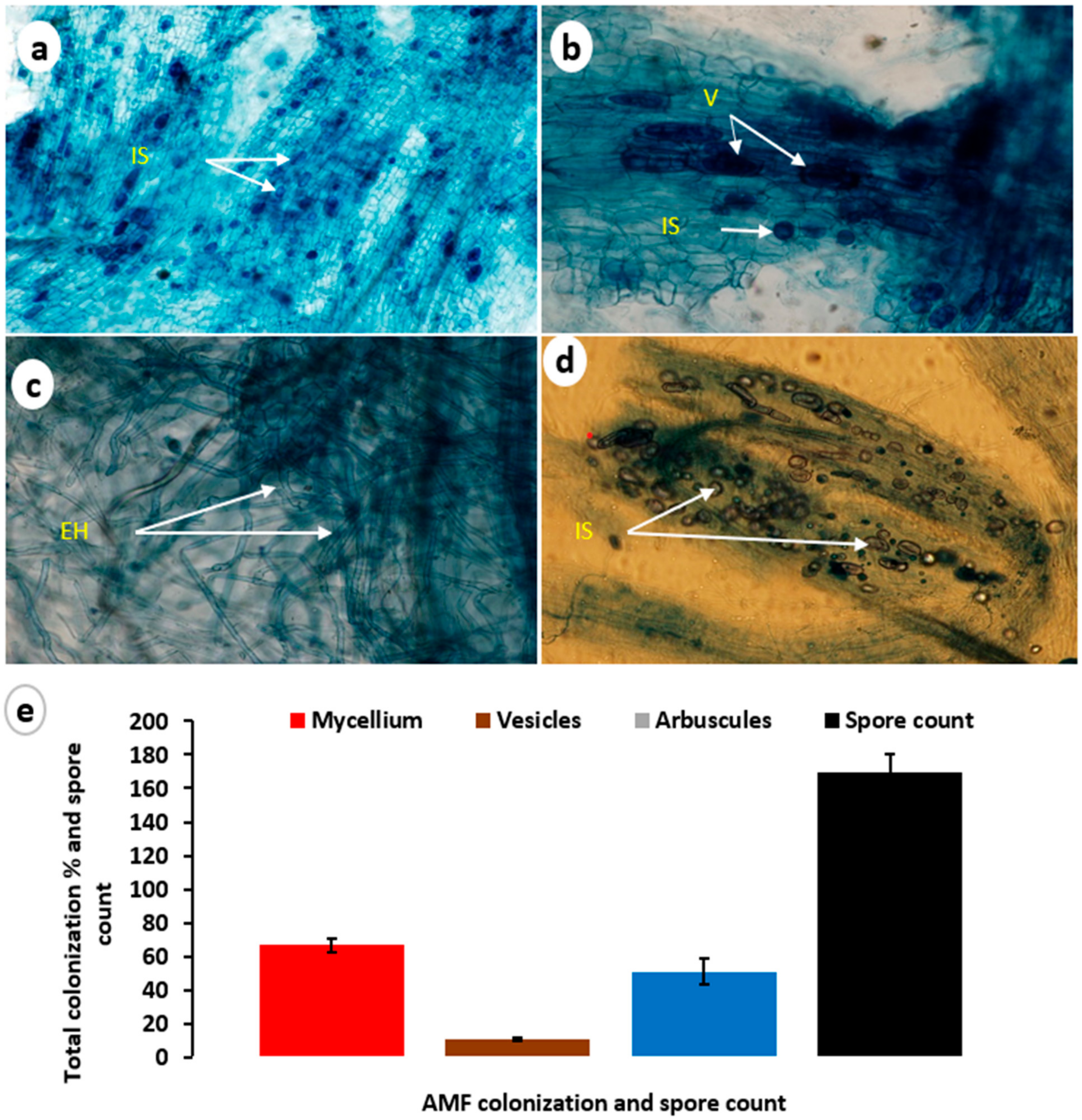
3.1. Mycorrhizal Colonization
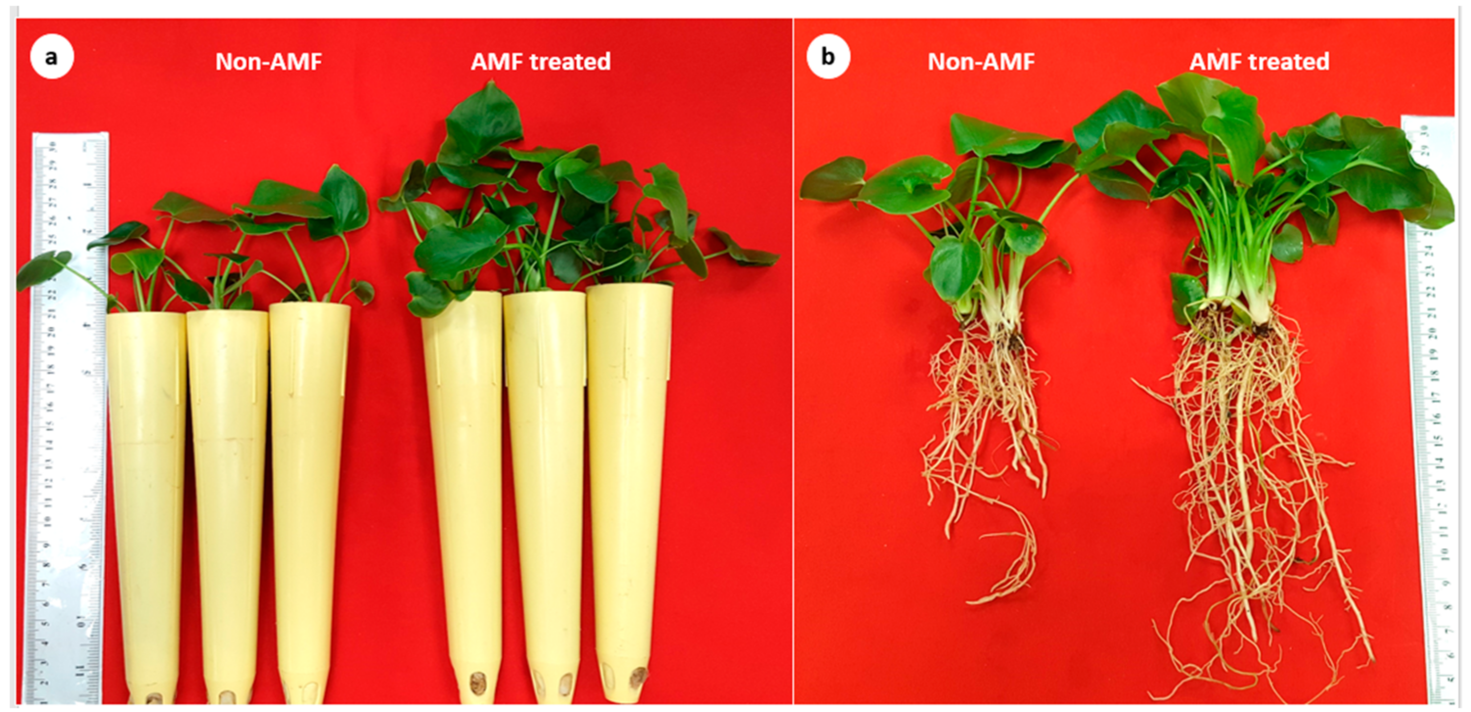
After eight weeks of cultivation, the roots of AMF-treated plants were harvested and examined for colonization with the host plants. The microscopic observation of the mycorrhizal status of P. bipinnatifidum plantlets indicated the presence of all predicted AMF structures (mycelium, vesicles, arbuscules, and spores) in the roots (Figure 2). The analysis of the mycorrhizal colonization showed the colonization percentage as mycelium 66.66%, vesicles 11.11%, and arbuscules 51.11%. The total spore count was also recorded as (169/100 g soil).

3.2. Plant Growth

Overall, plant growth was initially low. However, growth increased as the plantlets were adjusted to the ex vitro environment. Analysis of growth parameters such as the number of leaves per plant, leaf area, plant height, and shoot fresh and dry weight revealed that AMF plants had a significantly greater number of leaves (10.67 per plant), leaf area (75.63 cm2), plant height (14.17 cm), shoot fresh weight (3.30 g) and dry weight (0.315 g). These parameters were comparatively lower in non-AMF plants (Table 1). Figure 3a shows the promotive effect of vegetative growth AMF plants when compared to non-AMF plants. Root growth characteristics of AMF-treated plants were compared with non-AMF-treated plants and presented in Figure 3b and Figure 4. The length of the main root (23 cm), total root length/plantlet (485.73 cm), number of root tips/plantlet (236), average root diameter/plantlet (4.58 mm), total root surface area (697.76 cm2), total root volume/plantlet (79.98 cm3), root fresh weight (1.51 g) and root dry weight (0.16 g) were all significantly higher when compared to non-AMF plants (Figure 4).

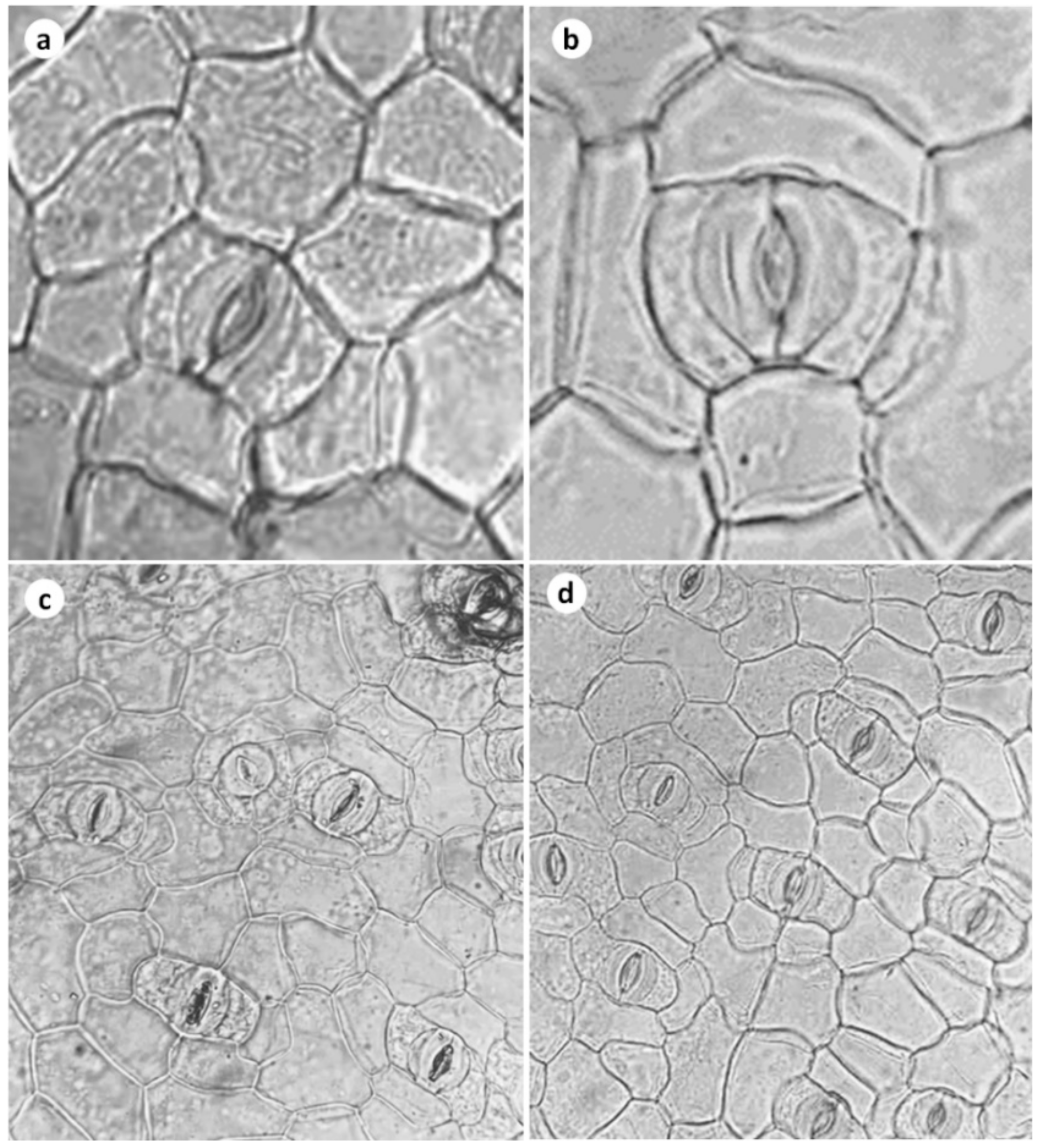
3.3. Stomatal Frequency, Stomatal Conductance, Leaf Gas Exchange, and Transpiration Rate

In the current study, the plants which were treated with AMF showed higher leaf gas exchange, net-CO2 assimilation, high transpiration rate, and stomatal conductance (Table 1) compared to non-AMF plants. The number of stomata per unit area was higher in the AMF-treated plants (136.68 mm2), whereas the aperture length and aperture width of the stomatal apparatus were almost similar in both AMF and non-AMF treated plants (Table 1; Figure 5). The net CO2 assimilation was 9.26 µmol CO2·m−2·s−1 in AMF plants, whereas CO2 assimilation was 7.10 µmol CO2·m−2·s−1 in non-AMF plants. Stomatal conductance was 0.049 mol H2O·m−2·s−1 and 0.023 mol H2O·m−2·s−1, respectively, in AMF and non-AMF plants. The transpiration rate was 1.6324 mol H2O·m−2·s−1 in AMF-treated plants and it was 0.8784 mol H2O·m−2·s−1 in non-AMF-treated plants (Table 1).

3.4. Chlorophyll and Carotenoid Content

The leaf chlorophyll and carotenoid content were estimated with AMF-treated and non-treated plants, and results are presented in Table 1. The chlorophyll a (1.512 mg·g−1 FW), chlorophyll b (0.598 mg·g−1 FW), chlorophyll a+b (2.529 mg·g−1 FW), chlorophyll a/b ratio (2.529) and carotenoid (0.533 mg·g−1 FW) were higher in AMF-treated plants compared to non-treated plants (Table 1).

4. Discussion

The acclimatization phase is one of the pivotal steps during the micropropagation of plants and it is the transition phase wherein plants are entering into autotrophic conditions (ex vitro) from heterotopic conditions (in vitro). Plants that have adjusted/habituated to heterotrophic nutrition and physical conditions (such as light, temperature, and humidity) may fail to overcome the unfavorable related ex vitro cultivation. During this transition, pivotal physiological processes for the plant’s survival must be enhanced. The acclimatization phase may affect the plant’s photosynthetic efficiency, reduce its defense against pathogens and hinder the proper development of the root and sap conditions systems [39]. Many researchers have used AMF at this crucial stage of the plant’s transition stage, as they provide several benefits to the plants with which they are associated. AMF provides protection against biotic and abiotic stresses as well as the improvement in plant growth and development [40]. AMF are obligatory biotrophs and the symbiosis basically involving of nutrients, in which the plant provides carbon through products of photosynthesis, and the AMF transfer nutrients from the soil to the plants [41]. In general, the AMF develops their hyphae rapidly through the soil for longer distances and adsorb nutrients efficiently to the plants. These features are useful to plants at the pivotal time of acclimatization. Due to these advantages, mycorrhization/AMF inoculation is widely recommended for the ex vitro establishment of horticultural plants [39,42]. In the present study we isolated two AMFs, namely Gigospora albida and G. marginata, and inoculated them to the plantlets in equal amounts of P. bipinnatifidum after transplantation to potting medium. After eight weeks of cultivation, the roots of treated plants were harvested and examined for colonization with the host plants. The microscopic observation of the mycorrhizal status of P. bipinnatifidum plantlets indicated the presence of all predicted AMF structures (mycelium, vesicles, arbuscules, and spores) in the roots (Figure 2). The analysis of the mycorrhizal colonization showed the colonization percentage as mycelium 66.66%, vesicles 11.11%, and arbuscules 51.11%. Similarly, Etilingera elatior microporpagated plants showed very good colonization of AMF fungi Gigospora albida and Claroideogloums etunicatum after the treatment with these fungi [10].
Varied reports suggest that colonization of micropropagated plants with AMF after ex vitro transplantation can enhance growth and plant performance. For example, AMF-inoculated plants have demonstrated an increase in plant height, shoot biomass, leaf number, and leaf area in Gerbera and Heliconia [43], Gloriosa superba [44], Musa acuminata [45], and Paeonia spp. [46]. The results of the present study also support this view and AMF plants had a significantly greater number of leaves (10.67 per plant), leaf area (75.63 cm2), plant height (14.17 cm), shoot fresh weight (3.30 g), and shoot dry weight (0.314 g) than control plants, according to an analysis of growth characteristics. In comparison, non-AMF plants had lower values for these metrics (Table 1; Figure 3a). It has been demonstrated very well that reduction in plant morphological characteristics, i.e., reduced stem length, number of leaves, leaf area (length and width), and overall vegetative growth of plants causes a severe reduction in photosynthesis, leaf water potential, and sap movement [47,48]. About these features, the use of beneficial AMF during acclimatization will help the plants to involve in proper growth and achieve higher biomass (stem length, number of leas, and leaf area) so that plants in the process of acclimatization will involve proper physiological functions including photosynthesis.
Figure 3b and Figure 4 provide data comparing the root growth traits of plants treated with AMF and without AMF. In comparison to non-AMF plants, the main root’s length (23 cm), total root length per plantlet (485.73 cm), number of root tips per plantlet (236), average root diameter per plantlet (4.58 mm), total root surface area (697.76 cm2), total root volume (79.98 cm3), root fresh weight (1.51 g), and root dry weight (0.16 g) were all significantly higher in AMF plants. A similar increase in root growth and biomass accumulation was reported in AMF-treated plants of Alpinia purpurata [15], Citrus limon [8], and Scutellaria integrifolia [12].
Tissue culture plants upon transplantation showed very low chlorophyll content and reduced net-CO2 assimilation, high transpiration rate, and stomatal conductance [7]. However, plants subjected to AMF treatments have restored these physiological functions several days after transplantation [2]. Furthermore, stomatal density and stomatal morphology are important characteristics that enable plants to involve net physiological functions such as CO2 assimilation, stomatal conductance, and transpiration [49]. Moreover, higher stomatal density observed in AMF plants is known to have a direct positive influence on stomatal conductance and enhanced photosynthetic rate [50]. The plants treated with AMF in the current study had a higher stomatal density and showed increased stomatal conductance, net CO2 assimilation, and leaf gas exchange, when compared to non-AMF plants (Table 1). In contrast to non-AMF plants, AMF plants had higher leaf stomatal densities, with more stomata per unit area (136.68 mm−2) in the AMF-treated plants (Table 1; Figure 5), while the aperture length and aperture width of the stomatal apparatus were nearly the same in AMF-treated plants and non-AMF-treated plants. The chlorophyll A (1.512 mg·g−1 FW) and carotenoid (0.533 mg·g−1 FW) were higher in AMF-treated plants compared to non-treated plants (Table 1). However, the quantity of chlorophyll b and chlorophyll a/b ratio were similar in AMF-treated and control plants (Table 1). An increment in chlorophyll was also reported in AMF-treated plants of Capsicum annuum [2], Glycyrrhiza glabra [13], and Vitis vinifera [47]. The higher stomatal frequency and stomatal conductance in AMF plants compared to non-AMF plants were attributed to the higher chlorophyll content, higher leaf area, and a greater number of leaves. Previous studies have also suggested a positive correlation between chlorophyll content and net photosynthetic rate [48,49,50,51]. Overall, the results of current experiments demonstrate the beneficial effects of AMF with the micropropagated plants enabling their physiological adjustments during acclimatization.

5. Conclusions

The current study offers new knowledge about the development and physiological response of control and AMF-treated P. bipinnatifidum plants grown in tissue culture. Plantlets of the P. bipinnatifidum benefited from the establishment, growth, and development of the arbuscular mycorrhizal fungi (Gigaspora albida and G. marginata). In terms of shoot and root growth, leaf gas exchange, chlorophyll content, and carotenoid content, the AMF-treated plants performed better. In addition, AMF-treated plants had more stomata, stomatal conductance, net CO2 assimilation, and transpiration rate as compared with non-AMF plants. Therefore, we conclude that AMF’s interaction with acclimatizing plants was the cause of superior growth, development, and physiological characteristics. Further, tissue culture plant mycorrhizal treatments are advantageous for the establishment during the acclimatization period. The use of additional AMF fungi and establishing consortium AMF fungi will be investigated further to aid in the acclimatization of tissue-cultured plants.

Author Contributions

Conceptualization, Y.H.D.; methodology, M.M.H., A.A.A. (AbdulAziz A. AlQarawi), T.S.A., A.A.A. (Ahmed Ali Alaizari) and J.A.M.; software, Y.H.D. and T.S.A.; validation, M.S.A. and H.N.M.; formal analysis Y.H.D., M.M.H., A.A.A. (AbdulAziz A. AlQarawi), T.S.A., J.A.M.; investigation, Y.H.D., M.M.H., A.A.A. (AbdulAziz A. AlQarawi), T.S.A., A.A.A. (Ahmed Ali Alaizari), J.A.M. and M.S.A.; resources, Y.H.D. and A.A.A. (AbdulAziz A. AlQarawi); data curation, Y.H.D., M.M.H., A.A.A. (AbdulAziz A. AlQarawi), T.S.A., J.A.M. and M.S.A.; writing—original draft preparation, Y.H.D., M.M.H. and H.N.M.; writing—review and editing, Y.H.D., M.M.H., A.A.A. (AbdulAziz A. AlQarawi), M.S.A. and H.N.M.; visualization, M.S.A. and H.N.M. All authors have read and agreed to the published version of the manuscript.

Funding

The authors acknowledge Researchers Supporting Project number (RSP2023R375), King Saud University, Riyadh, Saudi Arabia.

Data Availability Statement

All data are presented in the article.

Acknowledgments

The authors acknowledge the Researchers Supporting Project number (RSP2023R375), King Saud University, Riyadh, Saudi Arabia. Hosakatte Niranjana Murthy is thankful to the ‘Brain Pool (BP)’ program of the National Research Foundation of Korea (Grant No. 2022H1D3A2A02056665).

Conflicts of Interest

The authors declare no conflict of interest.

References

  1. Diaz-Perez, J.C.; Shackel, K.A.; Sutter, E.G. Effect of in vitro formed roots and acclimatization on water status and gas exchange of tissue-cultured apple shoots. J. Am. Soc. Hortic. Sci. 1995, 120, 435–440. [Google Scholar] [CrossRef]
  2. Estrada-Luna, A.; Davies, F.T., Jr. Arbuscular mycorrhizal fungi influence water relations, gas exchange, abscisic acid, and growth of micropropgated chile ancho pepper (Capsicum annuum) plantlets during acclimatization and post-acclimatization. J. Plant Physiol. 2003, 160, 1073–1083. [Google Scholar] [CrossRef] [PubMed]
  3. Wetzstein, H.Y.; Sommer, H.E. Leaf anatomy of tissue cultured Liquidambar styraciflua (Hemamelidacae) during acclimatization. Am. J. Bot. 1982, 69, 1579–1586. [Google Scholar] [CrossRef]
  4. Ziv, M.; Schwarz, A.; Fleminger, D. Malfunctioning stomata in vitreous leaves of carnation (Dianthus caryophyllus) plants propagated in vitro; implications for hardening. Plant Sci. 1987, 52, 127–134. [Google Scholar] [CrossRef]
  5. Bhusal, N.; Kim, H.S.; Han, S.G.; Yoon, T.M. Photosynthetic traits and plant-water relations of apple cultivars grown as bi-leader trees under long-term waterlogging conditions. Environ. Exp. Bot. 2020, 176, 104111. [Google Scholar] [CrossRef]
  6. Kozai, T. Acclimatization of micropropagated plants. In Biotechnology in Agriculture and Forestry, High-Tech and Micropropagation I; Bajaj, Y.P.S., Ed.; Springer: Berlin/Heidelberg, Germany, 1991; Volume 17, pp. 127–141. [Google Scholar]
  7. Preece, J.E.; Sutter, E.G. Acclimatization of micropropagated plants to the greenhouse and field. In Micropropagation Technology and Applications; Debergh, P.C., Zimmerman, R.H., Eds.; Kluwer Academic Publishers: Dordrecht, The Netherlands, 1991; pp. 71–93. [Google Scholar]
  8. Quatrini, P.; Gentile, M.; Carimi, F.; Pasquale, F.D.; Puglia, M. Effect of native arbuscular mycorrhizal fungi and Glomus mosseae on acclimatization and development of micropropgated Citrus limon (L.) Burm. J. Hortic. Sci. Biotechnol. 2003, 78, 39–45. [Google Scholar] [CrossRef]
  9. Hajiboland, R.; Joudmand, A.; Aliasgharzad, N.; Tolra, R.; Poschenrieder, C. Arbuscular mycorrhizal fungi alleviate low-temperature stress and increase freezing resistance as a substitute for acclimation treatment in barley. Crop Pasture Sci. 2019, 70, 218–233. [Google Scholar] [CrossRef]
  10. Yano-Melo, A.M.; Saggin, O.J., Jr.; Lima-Filho, J.M.; Melo, N.F.; Maia, L.C. Effect of arbuscular mycorrhizal fungi on the acclimatization of micropropagated banana plantlets. Mycorrhiza 1999, 9, 119–123. [Google Scholar] [CrossRef] [Green Version]
  11. Da Silva, A.R.; de Melo, N.F.; Yano-Melo, A.M. Acclimatization of micropropagated plants Etlingera elatior (Jack) R.M. Sm. Inoculated with arbuscular mycorrhizal fungi. S. Afr. J. Bot. 2017, 113, 164–169. [Google Scholar] [CrossRef]
  12. Joshee, N.; Mentreddy, S.R.; Yadav, A.K. Mycorrhizal fungi and growth and development of micropropagated Scutellaria integrifolia plants. Ind. Crop. Prod. 2007, 25, 169–177. [Google Scholar] [CrossRef]
  13. Yadav, K.; Aggarwal, A.; Singh, N. Arbuscular mycorrhizal fungi induced acclimatization and growth enhancement of Glycyrrhiza glabra L.: A potential medicinal plant. Agric. Res. 2013, 2, 43–47. [Google Scholar] [CrossRef] [Green Version]
  14. Ferrari, M.P.D.S.; Queiroz, M.D.S.; Andrade, M.M.D.; Alberton, O.; Goncalves, J.; Gazim, Z.C.; Magalhaes, H.M. Substrate-associated mycorrhizal fungi promote changes in terpene composition, antioxidant activity, and enzymes in Curcuma longa L. acclimatized plants. Rhizosphere 2020, 13, 100191. [Google Scholar] [CrossRef]
  15. Campos, M.A.D.S.; Silva, F.S.B.; Yano-Melo, A.M.; Melo, N.F.D.; Maia, L.C. Application of arbuscular mycorrhizal fungi during the acclimatization of Alpinia purpurata to induce tolerance to Meloidogyne arenaria. Plant Pathol. J. 2017, 33, 329–336. [Google Scholar] [CrossRef] [Green Version]
  16. Fortuna, P.; Citernesi, A.S.; Morini, S.; Viaglano, C.; Giovannetti, M. Influence of arbuscular mycorrhizae and phosphate fertilization on shoot apical growth of micropropagated apple and plum rootstocks. Tree Physiol. 1996, 16, 757–763. [Google Scholar] [CrossRef]
  17. Guillemin, J.P.; Gianinazzi, S.; Gianinazzi-Pearson, V.; Marchal, J. Contribution of arbuscular mycorrhizas of biological protection of micropropagated pineapple (Ananas comosus (L.) Merr) against Phytophthora cinnamomi Rands. Agric. Food Sci. 1994, 3, 241–251. [Google Scholar] [CrossRef]
  18. Varma, A.; Schuepp, H. Mycorrhization of the commercially important micropropagated plants. Crit. Rev. Biotechnol. 1995, 15, 313–328. [Google Scholar] [CrossRef]
  19. Chen, F.C.; Wang, C.Y.; Fang, J.Y. Micropropagation of self-heading Philodendron via direct shoot regeneration. Sci. Hortic. 2012, 141, 23–29. [Google Scholar] [CrossRef]
  20. Alawaadh, A.A.; Dewir, Y.H.; Alwihibi, M.S.; Aldubai, A.A.; El-Hendawy, S.; Naidoo, Y. Micropropagation of lacy tree philodendron (Philodendron bipinnatifidum Schott ex Endl.). HortScience 2020, 55, 294–299. [Google Scholar] [CrossRef] [Green Version]
  21. Grout, B.W.W.; Aston, M.J. Transplanting of cauliflower plants regenerated from meristem culture. II. Carbon dioxide fixation and development of photosynthetic ability. Hortic. Res. 1978, 17, 65–71. [Google Scholar]
  22. Grout, B.W.W.; Milan, S. Photosynthetic development of micropropagated strawberry plantlets following transplanting. Ann. Bot. 1985, 55, 129–131. [Google Scholar] [CrossRef]
  23. Dewir, Y.H.; Ibrahim, A.; Murthy, H.N.; Aldubai, A.A.; Al-Ali, A.M.; Al-Aizari, A.A.; Migdadi, H.; Alsadon, A.; Al-Suhaibani, N.A. Integration of salicylic acid in in vitro rooting medium improves photosynthesis and growth during acclimatization of Philodendron bipinnatifidum Schott ex endl. Plantlets. Propag. Ornam. Plants 2020, 20, 103–108. [Google Scholar]
  24. John, T.S. A list of naturally infected Brazilian tropical plant species with vesicular-arbuscular mycorrhiza. Acta Amaz. 1980, 10, 229–234. [Google Scholar] [CrossRef] [Green Version]
  25. Brundrett, M.C. Mycorrhizal associations and other means of nutrition of vascular plants: Understanding the global diversity of host plants by resolving conflicting information and developing reliable means of diagnosis. Plant Soil 2009, 320, 37–77. [Google Scholar] [CrossRef]
  26. Murashige, T.; Skoog, F. A revised medium for rapid growth and bioassays with tobacco tissue cultures. Physiol. Plant. 1962, 15, 473–497. [Google Scholar] [CrossRef]
  27. Gerdemann, J.; Nicolson, T.H. Spores of mycorrhizal Endogone species extracted from soil by wet sieving and decanting. Trans. Br. Mycol. Soc. 1963, 46, 235–244. [Google Scholar] [CrossRef]
  28. Schenck, N.C.; Perez, Y. Manual for Identification of VA Mycorrhizal Fungi; Synergistic Publication: Gainesville, FL, USA, 1990. [Google Scholar]
  29. Walker, C. Spore Extraction by Centrifugation-Sugar Flotation; Biological Research and Imaging Laboratory: Hampshire, UK, 1997. [Google Scholar]
  30. International Culture of Vesicular-Arbuscular Mycorrhizal Fungi (INAVAM). Available online: http://invam.caf.wvu.edu (accessed on 1 July 2022).
  31. Schüssler, A.; Walker, C. The Glomeromycota: A Species List with New Families and New Gener. In The Glomeromycota; CreateSpace Independent Publishing Platform: Scotts Valley, CA, USA, 2010. [Google Scholar]
  32. Redecker, D.; Schüßler, A.; Stockinger, H.; Stürmer, S.L.; Morton, J.B.; Walker, C. An evidence-based consensus for the classification of arbuscular mycorrhizal fungi (Glomeromycota). Mycorrhiza 2013, 23, 515–531. [Google Scholar] [CrossRef]
  33. Phillips, J.M.; Hayman, D. Improved procedures for clearing roots and staining parasitic and vesicular-arbuscular mycorrhizal fungi for rapid assessment of infection. Trans. Br. Mycol. Soc. 1970, 55, 158–161. [Google Scholar] [CrossRef]
  34. Trouvelot, A.; Kough, J.; Gianinazzi-Pearson, V. Evaluation of VA infection levels in root systems. Research for estimation methods having a functional significance. Physiol. Genet. Asp. Mycorrhizae 1986, 1, 217–221. [Google Scholar]
  35. Al-Qarawi, A.; Mridha, M.; Alghamdi, O. Diversity of structural colonization and spore population of arbuscular mycorrhizal fungi in some plants from Riyadh, Saudi Arabia. J. Pure Appl. Microbiol. 2012, 6, 1119–1125. [Google Scholar]
  36. Dewir, Y.H.; Alsadon, A.; Ibrahim, A.; El-Mahrouk, M. Effects of growing substrate, mode of nutrient supply, and Saffron corm size on flowering, growth, photosynthetic competence, and cormlet formation in hydroponics. HortTechnology 2022, 32, 234–240. [Google Scholar] [CrossRef]
  37. Lichtenethaler, H.K. Biosynthesis, accumulation and emission of carotenoids, α-tocopherol, plastoquinone and isoprene in leaves under high photosynthetic irradiance. Photosynth. Res. 2007, 92, 163–179. [Google Scholar] [CrossRef]
  38. Cotton, R. Cytotaxonomy of the Genus Vulpia; The University of Manchester: Manchester, UK, 1974. [Google Scholar]
  39. Kapoor, R.; Sharma, D.; Bhatnagar, A.K. Arbuscular mycorrhizae in micropropagation systems and their potential applications. Sci. Hortic. 2008, 116, 227–239. [Google Scholar] [CrossRef]
  40. Smith, S.E.; Read, D.J. Mycorrhizal Symbiosis. Mycorrhizal Symbiosis, 3rd ed.; Academic Press: New York, NY, USA, 2008. [Google Scholar]
  41. Smith, F.A.; Smith, S.E. What is the significance of the arbuscular mycorrhizal colonization of many economically important crop plants? Plant Soil 2011, 348, 63–79. [Google Scholar] [CrossRef]
  42. Azcon-Aguilar, C.; Barea, J.M. Applying mycorrhiza biotechnology to horticulture: Significance and potentials. Sci. Hortic. 1997, 68, 1–24. [Google Scholar] [CrossRef]
  43. Sato, A.Y.; Nannetti, D.D.C.; Pinto, J.E.B.P.; Siqueira, J.O.; Blank, M.D.F.A. Application of arbuscular mycorrhiza to micropropagated Heliconia and Gerbera plants during acclimatization period. Hortic. Bras. 1999, 17, 25–28. [Google Scholar] [CrossRef] [Green Version]
  44. Yadav, K.; Aggarwal, A.; Singh, N. Arbuscular mycorrhizal fungi (AMF) induced acclimatization, growth enhancement and colchicine content of micropropagated Gloriosa superba L. plantlets. Ind. Crop. Prod. 2013, 45, 88–93. [Google Scholar] [CrossRef]
  45. Koffi, M.C.; Declerck, S. In vitro mycorrhization of banana (Musa acuminata) plantlets improves their growth during acclimatization. Vitr. Cell. Dev. Biol.-Plant 2015, 51, 265–273. [Google Scholar] [CrossRef]
  46. Wen, S.S.; Cheng, F.Y.; Zhong, Y.; Wang, X.; Li, L.Z.; Zhang, Y.X.; Qiu, J.M. Efficient protocols for the micropropagation of tree peony (Paeonia suffruticosa “Jin Pao Hong”, P. suffruticosa “Wu Long Peng Sheng”, and P. × lemoninei “High Noon’) and application of arbuscular mycorrhizal fungi to improve plantlet establishment. Sci. Hortic. 2016, 201, 10–17. [Google Scholar] [CrossRef]
  47. Bavaresco, L.; Fogher, C. Lime-induced chlorosis of grape-vine as affected by rootstock and root infection with arbuscular mycorrhiza and Pseudomonas fluorescence. Vitis 1996, 35, 119–123. [Google Scholar]
  48. Bhusal, N.; Bhusal, S.J.; Yoon, T.M. Comparisons of physiological and anatomical characteristics between two cultivars in bi-leader apple trees (Malus × domestica Borkh.). Sci. Hortic. 2018, 231, 73–81. [Google Scholar] [CrossRef]
  49. Salomon, Y.; Dietrich, L.; Sevanto, S.; Holtta, T.; Dannoura, M.; Epron, D. Drought impacts on tree phloem: From cell-level responses to ecological significance. Tree Physiol. 2019, 39, 173–191. [Google Scholar] [CrossRef] [PubMed] [Green Version]
  50. Pardiso, R.; Arena, C.; De Micco, V.; Giordano, M.; Aronne, G.; De Pascale, S. Changes in leaf anatomical traits enhanced photosynthetic activity of soybean grown in hydroponics with plant growth promoting microorganisms. Front. Plant Sci. 2017, 8, 674. [Google Scholar] [CrossRef] [PubMed] [Green Version]
  51. Massonnet, C.; Costes, E.; Rambal, S.; Dreyer, E.; Regnard, J.L. Stomatal regulation of photosynthesis in apple leaves: Evidence for different water-use strategies between two cultivars. Ann. Bot. 2007, 100, 1347–1356. [Google Scholar] [CrossRef] [PubMed] [Green Version]
Figure 1. Arbuscular mycorrhizal fungi spores collected from the trap culture: crushed spore spores of Gigaspora albida (a,b); crushed spores of Gigaspora margarita (c,d).
Horticulturae 09 00276 g001
Figure 2. Photomicrographs indicating the root colonization structures and colonization status in the roots of Philodendron bipinnatifidum in response to arbuscular mycorrhizal fungi after 8 weeks acclimatization. ((ad) 400× magnification). The presence of extraradical hyphea (EH) indicated initiation of AMF colonization, which later propagated and developed various structural forms such as intraradical intact spores (IS); Vesicles (V). (e) AMF root colonization (Mycelium, Vesicles, and Arbuscules) and spore count of Philodendron plants.
Horticulturae 09 00276 g002
Figure 3. Photograph showing vegetative growth (a) and root growth (b) in non-AMF and AMF-treated Philodendron bipinnatifidum plants after 8 weeks of acclimatization.
Horticulturae 09 00276 g003
Figure 4. Root growth characteristics in non-AMF and AMF-treated Philodendron bipinnatifidum plants after 8 weeks of acclimatization. (a) Length of the main root per plantlet, (b) Total root length per plantlet, (c) Number of root tips per plantlet, (d) Root diameter per plantlet, (e) Total root surface area per plantlet, (f) Total root volume per plantlet, (g) Root fresh weight per plantlet and (h) Root dry weight per plantlet. * = Significant at p ≤ 0.05 according to unpaired t-test.
Horticulturae 09 00276 g004
Figure 5. Stomatal density in non-AMF (a,c) and AMF-treated (b,d) Philodendron after 8 weeks of acclimatization (40× magnification).
Horticulturae 09 00276 g005
Table 1. Stoma density, leaf gas exchange and pigments, and vegetative growth characteristics of Philodendron bipinnatifidum in response to arbuscular mycorrhizal fungi after 8 weeks acclimatization.
Growth ParametersNon-AMFAMF Treated
Number of stomata (mm2)78.93 ± 9.874136.68 ± 5.775 **
Aperture length (μm)11.00 ± 1.35411.5 ± 0.2886 NS
Aperture width (μm)8.25 ± 0.62929.5 ± 0.2887 NS
Net CO2 assimilation (μmol CO2·m−2·s−1)7.1905 ± 0.38659.2673 ± 0.4032 ***
Stomatal conductance (mol H2O·m−2·s−1)0.0239 ± 0.00070.0492 ± 0.0029 ***
Transpiration rate (mol H2O·m−2·s−1)0.8784 ± 0.02341.6324 ± 0.0901 ***
Chlorophyll a (mg·g−1 FW)1.379 ± 0.00241.512 ± 0.0130 **
Chlorophyll b (mg·g−1 FW)0.571 ± 0.00880.598 ± 0.0096 NS
Chlorophyll a+b (mg·g−1 FW)1.950 ± 0.01122.110 ± 0.0226 **
Chlorophyll a/b ratio2.414 ± 0.27512.529 ± 0.0188 NS
Carotenoids (mg·g−1 FW)0.423 ± 0.00040.533 ± 0.0070 ***
Number of leaves/plants7.00 ± 0.40810.67 ± 0.624 **
Leaf area/plant (cm2)31.11 ± 3.05875.63 ± 1.207 ***
Plant height (cm)12.13 ± 0.40914.17 ± 0.118 **
Shoot fresh weight/plant (g)1.776 ± 0.0813.307 ± 0.047 ***
Shoot dry weight/plant (g)0.148 ± 0.0130.315 ± 0.004 ***
NS = not significant, ** and *** = significant at p ≤ 0.01, and p ≤ 0.001, respectively, according to unpaired t-test.
Disclaimer/Publisher’s Note: The statements, opinions and data contained in all publications are solely those of the individual author(s) and contributor(s) and not of MDPI and/or the editor(s). MDPI and/or the editor(s) disclaim responsibility for any injury to people or property resulting from any ideas, methods, instructions or products referred to in the content.

