Article

Exogenous Melatonin Application Induced Morpho-Physiological and Biochemical Regulations Conferring Salt Tolerance in Ranunculus asiaticus L.

by
Eman Abdelhakim Eisa
1,2,
Péter Honfi
2,
Andrea Tilly-Mándy
2 and
Iman Mirmazloum
3,*
1
Department of Floriculture and Dendrology, the Hungarian University of Agriculture and Life Science (MATE), 1118 Budapest, Hungary
2
Botanical Gardens Research Department, Horticulture Research Institute, Agricultural Research Center (ARC), Giza 12619, Egypt
3
Department of Plant Physiology and Plant Ecology, Institute of Agronomy, Hungarian University of Agriculture and Life Sciences, 1118 Budapest, Hungary
*
Author to whom correspondence should be addressed.
Horticulturae 2023, 9(2), 228; https://doi.org/10.3390/horticulturae9020228
Submission received: 16 January 2023 / Revised: 6 February 2023 / Accepted: 7 February 2023 / Published: 8 February 2023

Abstract

:
This study presents the effects of exogenous melatonin application at different concentrations (50, 100, and 200 μM) on the morphological, physiological, and antioxidant defense systems of the buttercup plant under salinity stress (4.5 and 5.5 dS·m−1 EC). Expectedly, the salinity stress negatively affected the plant growth parameters, cell membrane stability, and POX enzyme activity of R. asiaticus compared to non-stressed plants. However, in a dose-dependent manner, exogenous melatonin foliar application decreased the salt stress-induced symptoms of retarded vegetative growth, physiological characteristics, and oxidative stress level. The results obtained, revealed the significant effectiveness of exogenous melatonin treatment at 200 μM concentration under salt stress conditions by enhancing the plant growth traits such as chlorophyll and carotenoids content, relative water content, proline content, peroxidase enzyme activity (POD), and by the decreased electrolyte leakage rate, and Na+ content, as well as delaying the emergence of flower buds under salinity stress. The salt tolerance index percentages (STI%) for all estimated characters are also calculated for all studied parameters. This study uncovered the beneficial effect of melatonin in reducing salt stress symptoms that can be used to reduce the salinity effect in ranunculus plant production.

1. Introduction

Salinity, particularly in arid and semi-arid regions, is the main environmental concern with detrimental effects on soil and consequent agricultural output [1]. The salt-affected land area has increased and will continue to expand due to persistently unsustainable agricultural practices and climate change [2], posing a serious threat to agricultural areas that appears to be worsening [3]. As a result, 30% of farmland will be affected in the next quarter-century, and as much as 50% will be impracticable by 2050 [4]. Therefore, it is now more crucial than ever to learn how plants adapt and cope with salt stress by understanding the potential inhibition and tolerance mechanisms. In many ways, osmotic and ionic stress caused by salt exposure alters the major biological activities of many plants, including sodium ion (Na+) toxicity, nutritional imbalance, physiological water shortage, metabolic disturbances, oxidative damage, and photo-inhibition [5,6]. Commonly, the cut flowers are not suggested as crops for saline soils or water recycling systems because they are considered intolerant to high salt concentration of irrigation water and soil [7]. Salinity, in particular, can affect the quality of the blooms, shorten plants stems, and lower the production in flora crops [7,8,9,10]. In light of this, it is crucial to test more floricultural varieties for salinity tolerance from environmental and economic points of view for sustainable development.
Ranunculus asiaticus, also known as the buttercup, a member of the Ranunculaceae family, is an indigenous species from Asia and the Mediterranean [11]. The buttercup has delightful and odorless flowers that have a diameter of around 5 cm and come in various colors, including yellow, orange, red, pink, and white. Ranunculus’s leaves are light green and grow to a maximum height of about 40 cm [12]. Due to its gorgeous inflorescences on strong and long stems, it is considered as an annual field-grown cut flower and is highly prized as a bedding or potted plant [13]. Depending on the variety, ranunculus can be propagated by tuberous roots or seeds [14,15]. Tuberous roots are more preferred since they often result in plants with earlier flowering time and more blooms per stem than seed-propagated plants [15]. A rosette of leaves with long petioles is produced from the tuberous roots of R. asiaticus in the fall after the first rain rehydrates the dried tissue in its natural Mediterranean habitat [16]. Six to eight fully developed leaves signal the onset of flowering, which continues through February and May. After shoot senescence, the tuberous roots are removed, stored over the summer, and sold for the following year’s plantings in gardens and flowerbeds [15]. Buttercups like partial exposure to the sun and prefer moderate low temperatures (night/day regime 5–10/12–25 °C; optimum day 16 °C) and medium to high light intensities [16]. According to Rauter et al. [17], ranunculus physiological characteristics and visual growth quality declined when the EC of irrigation water raised from 0.5 to 5.5 dS·m−1, resulting in a rise in root-zone salinity. Similar observations were reported by Valdez-Aguilar et al. [18] on the sensitivity of ranunculus to saline conditions when irrigation solution EC value was raised from 2.0 to 6.0 dS·m−1.
One of the most recent strategies that tried to reduce the impact of biotic and abiotic stressors on plants is the application of plant bio-stimulators or growth hormones to promote adaptation [19]. Melatonin (MT) is the common name for N-acetyl-5-methoxytryptamine, a derivative of the essential amino acid, tryptophan, which is also a multi-regulatory biological hormone that is vital for plant and animal lives [20]. MT is reported to be involved in the circadian rhythm [21], immunological stimulation [22], antioxidant systems [23], and the seasonal cycle of reproduction [24]. MT was first detected in plants in 1995 [25]. The involvement of MT in plant growth regulation and development, including chlorophyll production and photosynthesis, callus induction, flowering, rhizogenesis, and the ageing of the leaves, has been documented [26,27,28,29]. Additionally, melatonin is also famous for its frequent application to combat biotic and abiotic stresses in plants since its ROS scavenging ability was confirmed [30,31]. Recent studies showed that MT-treated plants were more tolerant to salinity stress, had lower levels of reactive oxygen species, electrolyte leakage, and cell damage, and had higher growth parameters than untreated plants [32,33]. The positive effect of MT application for plants exposed to stress was confirmed in different species such as in watermelon [34], cucumber [28,35], melon [36], basil [37], and maize [29]. However, no such trial (melatonin application) has been conducted on Ranunculus asiaticus grown under salinity stress. Therefore, the present work intends to exhibit the efficacy of MT in enhancing the salinity tress tolerance of buttercup plants in terms of morphological and physiological reactions associated with oxidative stress. In this regard, the salt tolerance index (STI%) for all the studied parameters is determined to be utilized as an indicant of salt tolerance.

2. Materials and Methods

2.1. Material, Cultivation Conditions, and Trial Layout

The pot trial was undertaken in a research greenhouse of the Floriculture and Dendrology Department at The Hungarian University of Agriculture and life science (MATE) (Budapest, Hungary). The planting process started on 1 October 2021, and the harvest occurred 150 days later. The average day and night temperature of the glasshouse was 20–15 °C, with 60% relative humidity. Healthy seedlings (30-days old) of R. asiaticus with four to five true leaves (size, 5–6 cm) were transplanted into ø (9 × 9 × 10) cm plastic pots; one plant per each pot and 135 plants per trial. The design of the experimental groups was a randomized complete block design (RCBD) in a split-plot arrangement; the main plots consisted of three different salinity levels: (1) the control plants typically watered with 80% of the field’s capacity; no MT; no salt treatment; (2) salinity treatment at EC 4.5 dS·m−1; (3) salinity treatment EC 5.5 dS·m−1, and the sub-plots included the different levels of MT treatments associated with salinity levels at EC 4.5 and 5.5 dS·m−1 (15 plants/subgroup). In each pot, the growing medium consisted of a uniform mixture of Klassmann TS3 Baltic peat (constituents are shown in Table S1), supplemented with 3 kg/m3 Osmocote Exact Potassium Dominant (Scotts, NSW, Australia) and 1 kg/m3 (soluble carbonate).

2.2. Salinity Treatment and MT Application

Prior to salinity treatments, the ranunculus seedlings were grown in separate pots for four weeks. The salt solution preparation involved adding NaCl and CaCl2 dihydrate at a molar ratio of 2:1 to attain electrical conductivity (EC) values of 4.5 and 5.5 dS·m−1 [17]; the electrical conductivity of the solution was determined using a handheld EC meter (Milwaukee EC 60 Inc, Szeged, Hungary). After six weeks of culture and salt treatments, different concentrations (0, 50, 100, and 200 μM in tap water (ethanol/water (v/v) = 1/10,000)) [38,39] of exogenous melatonin (Thermo Fischer, Geel, Belgium) were sprayed four times in two-week intervals using a handgun sprayer. The leaves were sprayed until complete wetness and dripping. Tap water was used for regular irrigation.

2.3. Morphological Characteristics

Plants (10 individual plants per treatment) were sampled two weeks after the fourth foliar application to estimate the growth parameters, such as the plant height (cm), (measured from the medium surface to the shoot apex using a meter rod), the number of leaves, (counted manually), fresh and dry weights (shoots and leaves in g). The total leaf area (Area Meter 350, ADC Bioscietific Ltd., Hoddesdon, UK) and the number of flower buds that appeared were also recorded for analysis.

2.4. Physiological and Biochemical Assesments

The five uppermost young fully expanded leaf samples were instantly frozen in liquid nitrogen and kept at −80 °C pending inspection. The photosynthetic pigments were measured for the acetone (80%) extract samples of each treatment and the absorbances (644, 663, and 480 nm) were recorded in a UV-VIS Spectrophotometer (Genesys 10S, Waltham, MA, USA). The carotenoid content was computed following the method of Lichtenthaler et al. [40], and the relative water content (RWC) was determined using the methods described by Turk and Erdal [41] and the formula RWC = (FW-DW)/(TW-DW) × 100.
The leaf’s proline content (PC) was determined by the methods of Bates et al. [42] at 520 nm. A standard curve was constructed to calculate the proline content in μmol g−1 leaf FW. The extent of electrolyte leakage (EL) was assessed via the methods of Reddy et al. [43]. The peroxidase enzyme activity (POD) was also determined spectrophotometrically (470 nm) using the standard guaiacol method [44].
Sodium content of the leaves was determined with a flame photometer according to Campbell’s method [45] and is expressed as grams per 100 g dry weight (g 100 g−1 DW). The Salt Tolerance Index (STI) was determined as a percentage (%) for each of the analyzed traits, as proposed by Sbei et al. [46]. STI = (T salt/T cont.) × 100, where T salt represents the characteristic’s average value recorded under the saline conditions induced by 5.5 EC, and T cont. represents the same traits’ average value under control conditions.

2.5. Statical Analysis

The experiment was set up in a completely randomized design. The Two-Way MANOVA followed by UNIANOVA for the variables with Bonferroni’s correction was run for all dependent variables, between factors at two levels: (1) treatments (Cont (no salinity and no MT), Salinity EC 4.5 and Salinity EC 5.5), (2) melatonin concentrations (0, 50, 100, 200 µM). Assumptions were as if the normality of the residuals for all dependent variables are accepted by Kolmogorov–Smirnov’s (p > 0.05) [47]. The homogeneity of variances by Leven’s F test was satisfied with all dependent variables p > 0.05 [48]. Tukey’s post hoc test was used for factor level comparisons [49,50], while Dunnett’s test was used to compare the control treatment with two salinity groups. Pairwise within-subject effects were compared via Bonferroni’s method. All statistics were gathered using the software IBM SPSS27 [51].

3. Results

Statistical analysis of the obtained data revealed highly significant differences amongst multivariate factors and for the interaction effect of treatment levels (Wilk’s lambda < 0.001) [52]. Thus we compared the two salinity levels with the control for the MT concentration effect, and compared the MT concentration levels with the applied salinity levels, then followed up with Univariate ANOVA for different variables using the Bonferroni’s correction to see significant differences in all individual variables p > 0.05 [53].

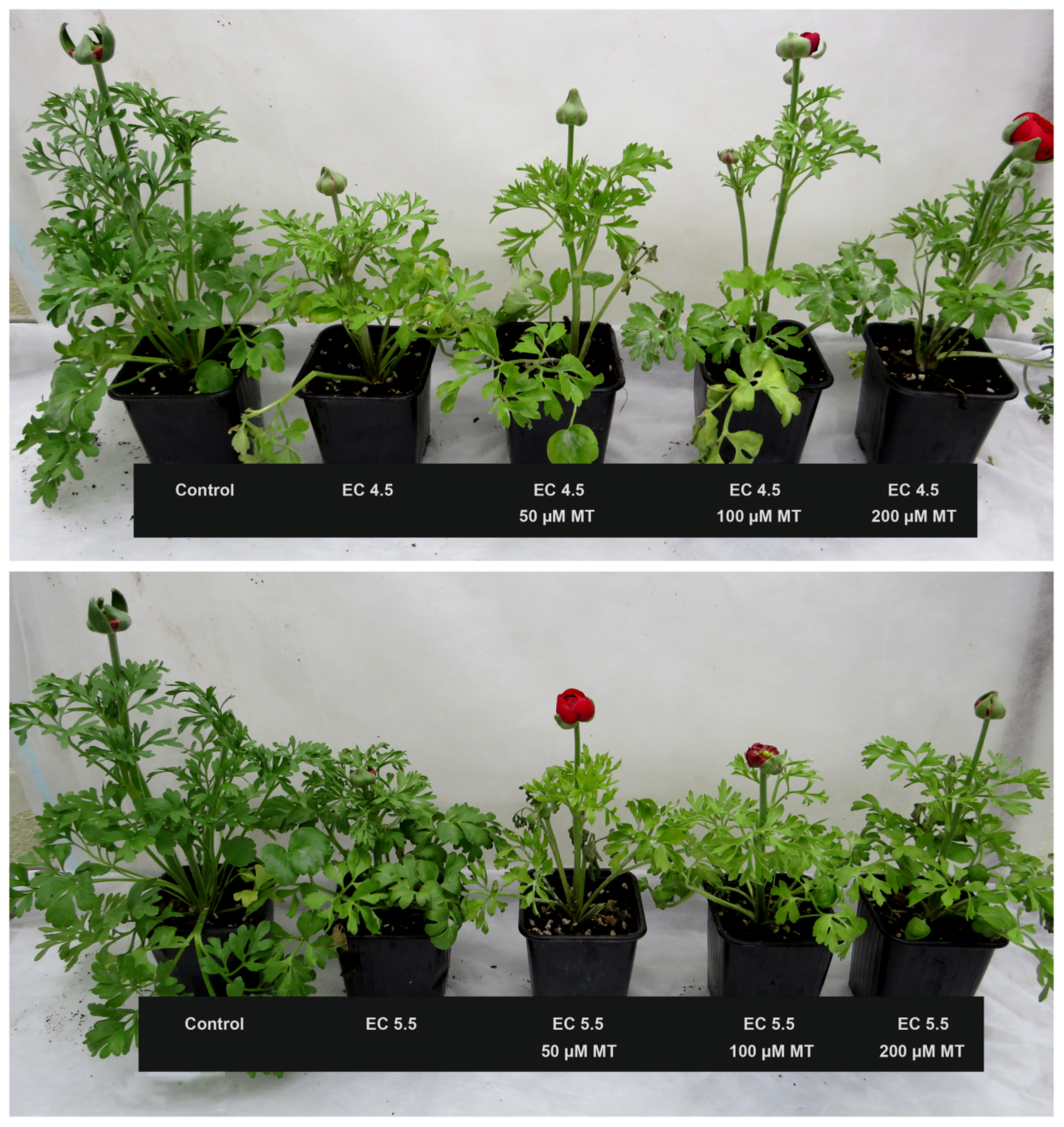
3.1. The Effect of Melatonin on Plant Morphology under Salinity Stress

The applied salinity stress at both levels had an obvious negative impact on the recorded growth parameters of buttercup plants (Table 1 and Figure 1). Comparing the control (non-stressed plants), salt-stressed plants at the EC 4.5 (0 MT) and EC 5.5 (0 MT) exhibited a substantial decrease in shoot length by (31.78 and 35.64%), leaf number by (32.23 and 34.03%), total leaf area by (58.34 and 60.98%), shoot FW by (29.32 and 36.61%) and shoot DW by (42.15 and 46.08%), respectively. However, foliar melatonin treatments (50, 100, and 200 µM) considerably enhanced the plant development and improved all vegetative parameters under salinity stress. Plants treated with 200 µM MT showed the best performance under both level of salinity treatments, manifested in the remarkable increase in shoot length (23.37 and 30.04%), leaf numbers (28.32 and 21.14%), total leaf area (58.01 and 58.79%), shoot FW (29.92 and 42.33%) and shoot DW (45.20 and 41.82%), compared with stressed plants of EC 4.5 MT0 and EC 5.5 MT0, respectively, when compared with the NO-MT treated plants (Table 2).

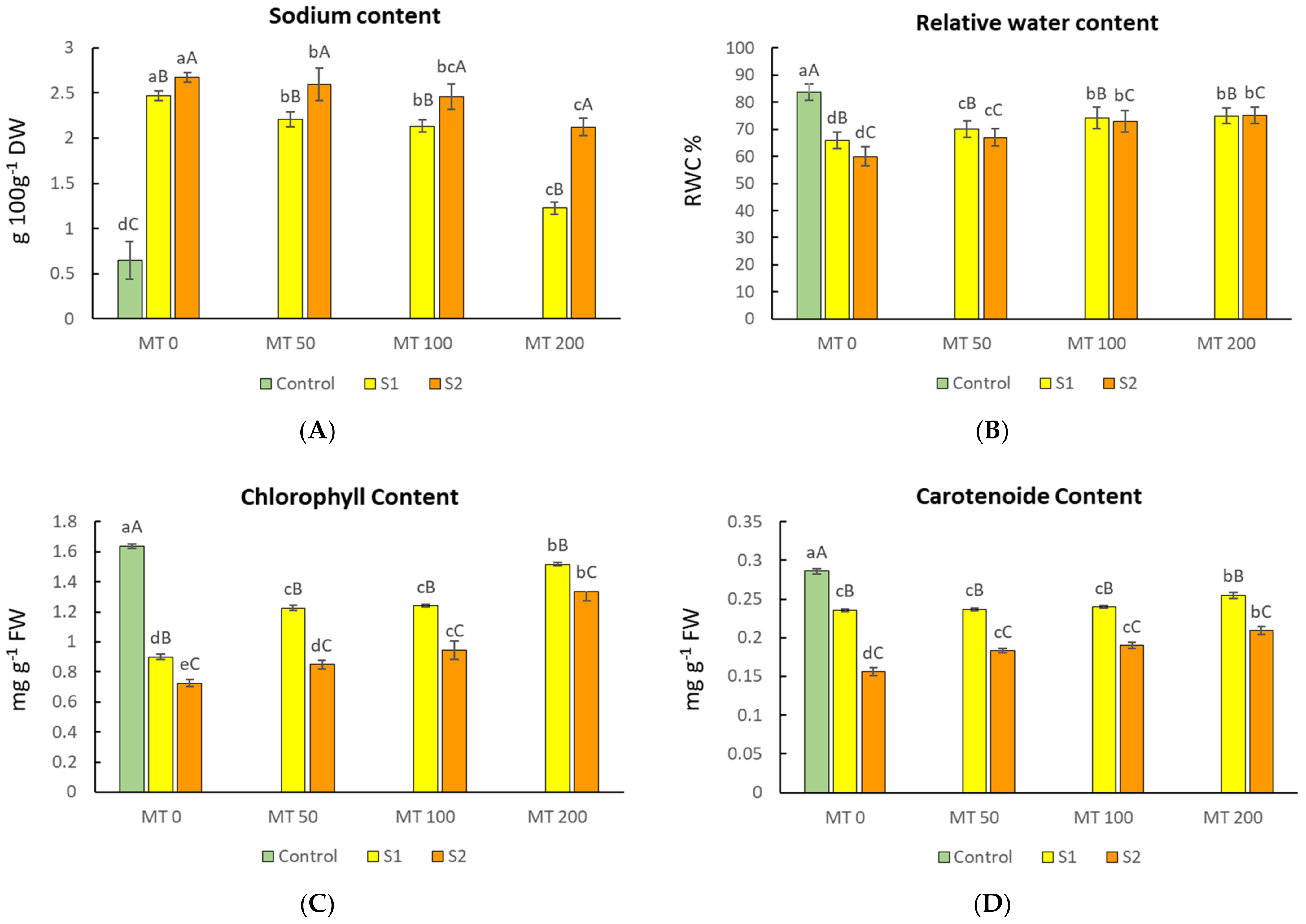
3.2. Changes in Sodium Ion Level and Relative Water Content (RWC) of the Leaves

Our results indicate the sensitivity of ranunculus to salinity with increased Na+. The Na+ concentration in dry leaves increased by 280 and 311% upon 4.5 (S1) and 5.5 (S2) dS·m−1 EC of irrigation water, respectively, when compared to control plants (Figure 1A). The application of exogenous MT at different concentrations (50, 100, and 150 μM) resulted in a marked reduction in Na+ content in leaf samples of R. asiaticus plants under both salinity levels (Figure 1A). The foliar application of 200 MT at both S1 and S2 levels revealed a significant effect and reduced the Na+ content by 50.4 and 20.5% in comparison to salinity-stressed plants without MT application (S10MT and S20MT, respectively).
The relative water content (RWC) of a plant tissue is one of the most reliable indicators of its water status and capacity for survival under stressful conditions. Our data revealed that saline conditions negatively affected the leaves’ RWC, particularly at EC 5.5 dS·m−1 which had the most severe effect (Figure 1B). Compared with plants under normal conditions (non-stressed plants), stressed plants, S1 0MT and S2 0MT exhibited a significant decrease (21.1 and 28.3%, respectively) in RWC.
On the other hand, the foliar melatonin treatment showed a slight improvement in RWC under the saline conditions. The increase in RWC was noted in the presence of 100 and 200 µM melatonin compared with non-MT-treated stressed plants which was still lower than that of control plants (Figure 1B). These outcomes imply that the foliar melatonin treatment has more favorable impacts on plant biomass and development upon salt stress, and that the 200 µM melatonin could better protect the plants under saline conditions than 100 and 50 µM melatonin.

3.3. Changes in Photosynthetic Pigments of Leaves

Saline conditions negatively affected the leaves’ total chlorophyll (Chl) and carotenoid (Car) content of buttercup plants, with the influence being most prominent at the higher NaCl concentration of EC 5.5 treatment (Figure 1C,D). The leaves’ Chl and Car content of non-MT-treated plants under both salinity levels (S1 0MT and S2 0MT) were estimated for a decrease of 45.12 and 55.49% in cases of chlorophyll and 17.24, and 44.82% in cases of carotenoids, respectively. However, the exogenous MT application at different doses (50, 100, and 150 μM) on R. asiaticus plants resulted in a significant increase in leaves’ photosynthetic pigments (total Chl and Car) in comparison to stressed plants without MT under both salinity levels (Figure 1C,D). The foliar application of 200 MT at both S1 and S2 levels revealed a significant effect, with the rise of leaves’ Chl and Car estimated at 68.89 and 83.56%, respectively, compared with stressed plants without MT application (S10MT and S20MT, respectively). These data indicate that 200 μM MT was able to inhibit the entrance of detrimental ions into the cells, safeguarding the cellular structure.

3.4. Changes in Electrolyte Leakage and Proline Content

The impacts of salinity stress and exogenous melatonin application on membrane integrity and electrolyte leakage (EL) are illustrated in Figure 2A. Saline conditions induced by NaCl 4.5 and 5.5 dS·m−1 significantly increased the electrolyte leakage by 64.01 and 67.74%, respectively, when compared to control plants. In contrast, in stressed plants subjected to foliar application of melatonin (50 µM, 100 µM, and 200 µM), there was a significant and concentration-dependent decrease in electrolyte leakage. The application of 200 µM MT resulted in the highest reduction of EL by 28.93 and 20.57% under both S1 and S2 salinity levels, when compared with salt-stressed plants without MT treatment (Figure 2A).
Proline, a compatible membrane protective solute with its role in maintaining water balance and enhancing cytoplasmic osmotic pressure is known to protect the cells during early dehydration. According to our findings, saline conditions showed a considerable rise in proline content in leaves, with its highest values being recorded at the S2 (EC 5.5) treatment. Stressed plants (S1 0MT and S2 0MT) exhibited a significant increase of about 50% in proline content in comparison with control plants. The foliar melatonin application at 50, 100, and 200 µM resulted in a gradual increase in proline content under the saline conditions when compared to non-MT-stressed plants (Figure 2B).

3.5. Changes in Peroxidase Enzyme Activity (POD)

A significant increase (p < 0.05) in POD activity was detected in buttercup plants under both salinity stress conditions (Figure 2C). The POD levels increased by 61.9 and 82.2% when plants were irrigated with saline water with EC of 4.5 and 5.5 dS·m−1, respectively, when compared to non-stressed plants. Under both S1 and S1 salinity conditions, all of the melatonin treatments (50 µM, 100 µM, and 200 µM) significantly increased (p < 0.05) the POD activity, with its highest level being detected after 200 µM MT treatment. A 48.8 and 46.7% increase in antioxidant enzyme activity was recorded in plants under S1 and S2 conditions, respectively, when treated with 200 µM MT compared with non-MT-stressed plants under both salinity levels. These findings suggest that the NaCl stress brought the POD activities to a certain level as expected and that the foliar MT application enhanced (in a linear concentration-dependent manner) the POD activity even more to protect the cells from ROS accumulation.

3.6. Changes in Flower Bud Emergence

Our results reveal that both salt stress conditions (S1 and S2) induced the flower buds’ appearance (Figure 3). Flowering time of salt stressed plants irrigated with saline water with EC of 4.5 and 5.5 dS·m−1 without melatonin treatment occurred 29 and 36 days sooner than in the control plant, respectively. This early flowering time was delayed after the application of melatonin for 22, 24, and 15 days in the case of S1 treatment and 17, 8, and 10 days in the case of S2 salinity treatment when 50, 100, and 200 µM of MT was applied, respectively, in comparison to control plants (unstressed plants without MT).

3.7. Salt Tolerance Index (STI)

Table 2 depicts the STI as a percentage of all examined variables between the non-melatonin-treated plants grown under unstressed conditions and stressed plants under S2 salinity level (EC 5.5 dS·m−1). The STI% demonstrated that Na+ content in leaves was the trait most sensitive to saline conditions, compared to STI% of other variables, which gave 437.9%, followed by POD activity and El value in the second place, which gave 321.3 and 309.9%, respectively. Proline content was in the third place for the salt response, which gave 202.5%. In addition, without significant differences, the remaining traits exhibited STI% values less than 100% and higher than 50%, except total Chl showed the lowest salinity-responsiveness, which gave 44.4%.

4. Discussion

Salinity stress is known to cause plant performance to deteriorate due to osmotic, ionic, or nutritional disorders, represented in the morphological characteristics of subjected plants. As an activator of antioxidant enzymes, melatonin is known to protect plants from oxidative stress [54]. The cut flower species and in particular the ranunculus is reported to be sensitive to salinity stress [17,18]. In the present work, exposing ranunculus plants to salinity stress induced by EC 4.5 and EC 5.5 caused a significant reduction in all the assessed vegetative growth characteristics when compared with unstressed plants (control) where the EC 5.5 was more harmful (Table 1 and Figure 1). Similarly, it has been reported that increasing EC from 2 to 3 dS·m−1 significantly stunted plant growth, dry weight, number of leaves, and diameter of flowering stems of white and pink R. asiaticus cultivars [18]. Poorter et al. [55] reported that the plant leaf-related parameters were significantly affected by salinity.
Melatonin (MT) is known as an endogenous regulator with an enhancement effect on both plant growth promotion [36,56,57] and also in protecting against abiotic stress [31,58,59,60,61] even when applied in small concentrations [62]. Many investigations have demonstrated MT’s efficacy as a dose-dependent stimulator of plant growth and development [35,63,64]. We evaluated the effects of melatonin on ranunculus plants under saline conditions and found that foliar spray with 200 μM MT was the most effective concentration to mitigate the adverse effects of stress in most of the traits studied. Our findings were in line with the recent investigation on cotton seedlings treated with 200 μM of melatonin [65]. Exogenous melatonin must be applied to plants at the appropriate concentration, which may be different for distinct plant species [66]. Melatonin. an indoleamine, shares IAA’s metabolic precursor, which may explain its influence on plant growth and development [67]. In addition, earlier research reported that environmental stress factors may induce flowering to be able to perpetuate the species’ survival [68,69]. Exogenous melatonin was involved in flowering bud growth, as reported by Kolář et al. [70]. Similar to our results (Figure 2D), in Arabidopsis, exogenous MT caused a delay in flowering time [71]. This may occur due to the involvement of exogenous MT in upregulation of flowering locus C (FLC), which suppresses the transcription of flowering locus T (FT) [56,72].
Excessive cytoplasmic NaCl levels interrupt ion balance and inhibit plant growth and development [73]. Accordingly, plants must be able to ingest and compartmentalize ions to increase their salt tolerance efficiently. Thus, plants redistribute cytoplasmic salt ions into vacuoles or store them in various tissues to cope with salinity stress [74,75]. Consequently, a more significant Na+ build-up will be observed in leaves. Likewise, in this experiment, Na+ content of the leaves dramatically increased at both saline conditions EC 4.5 and 5.5 dS·m−1 (Figure 1A). Similar outcomes were also noticed in the cotton leaves [65] and pepper cultivars ‘Granada’ and ‘Nobili’ [76]. However, exogenous MT treatment allowed ranunculus plants to retain lower Na+ in leaves than in non-MT-treated stressed plants (Figure 1A), and higher reduction was observed under S1 conditions by increasing MT levels. These findings agree with Castañares and Bouzo [36] and Wei et al. [77], reporting that MT treatment reduced ion toxicity in melons and rice, respectively when subjected to salt stress. This research also confirmed that 200 µM MT aids in preserving ion equilibrium while exposed to salt stress. Specifically, melatonin’s action in the presence of saline stress is to upregulate the transporter genes NHX1 and AKT1 [78], which are involved in preserving the ion homeostasis. Due to the extreme sensitivity of plants to salinity, water status is the primary factor in response to stress [79]. Using water relation properties in ranunculus leaves, we found a common reactivity to salinity, consistent with the findings of Stępień and Kłobus [80] on cucumber and on pepper cultivars [76]. Toxic ion accumulation, especially Na+ and Cl, may be to blame for the reduction in RWC by causing leaves to shrink and their stomata to close, thereby lowering the intracellular CO2 partial pressure [81]. According to Munns’ [82] study, higher sodium chloride concentrations led to a greater loss of water from the cells, which manifested as dehydration; thus, the cell growth rate was rapidly reduced in addition to a series of metabolic alterations like those imposed by water stress. MT application in our study, however, increased RWC gradually by increasing MT concentrations and the higher concentration was the most effective under both salinity levels S1 and S2 (Figure 1B) similar to those reported in borage plants [79], likely attributable to enhanced water absorption due to safeguarding the cell membrane [36,80]. It has been proven that melatonin can improve the thickness of a plant’s cuticle, which in turn helps to limit water loss. Melatonin treatment has been shown to improve stress tolerance in plants by keeping their turgor and water ratio stable [81].
Photosynthesis relies on chloroplast chlorophyll, which can capture and transmit light energy for plants and is a key indicator of plant physiological properties and performance [82]. In plants subjected to saline conditions, a decrease in total chlorophyll and carotenoids was observed, and a more severe effect was found under S2 conditions (Figure 1C,D). This is correlated with excessive quantities of Na+ that result in sluggish synthesis or rapid breakdown of photosynthetic pigments, which has an immediate impact on plant growth [83]. An application of melatonin in earlier studies enabled salt-stressed plants to keep their chlorophyll levels closer to normal such as in melon (Cucumis melo L.) [36], pistachio [60], maize seedlings [29], and in spearmint plants [84]. Similarly, in the current study, exogenous melatonin enhanced chlorophyll and carotenoid levels in ranunculus plants compared to non-MT-stressed plants. In general, S1 (4.5 EC) showed the higher pigment accumulation with MT application, and 200 μM MT was the most effective concentration applied under both salinity stress levels, supporting the hypothesis that MT pretreatment promotes biosynthesis and reduces chlorophyll breakdown [85]. This effect has been attributed to melatonin’s antioxidant properties and its influence on the genes for the chlorophyll-degrading enzymes CLH (chlorophyllase), PAO (pheophorbide an oxygenase), and the red chlorophyll catabolite reductase [56]. Additionally, high melatonin dosages improved chloroplast carotenoid content and lipid-soluble antioxidants [74]. Consequently, melatonin can stimulate carotenoid production and result in lowering the levels of photooxidative degradation [31].
Ion-specific salt degradation mainly occurs in the plasma membrane [86]. Electrolyte leakage can be attributed to a loss of membrane integrity, which reduces plants’ ability to hold K+ as a result of stress [87]. Consequently, plasma membrane electrolyte leakage is regarded as an essential indicator for selecting tolerant plants under salt stress [86]. From our data, both salinity levels increased EL rate, but S2 (EC 5.5) caused the highest EL rate. However, minimizing EL in plants treated with MT afforded a protective effect toward membrane damage under saline conditions (Figure 2A). Similar results were presented by Khan et al. [88] and Zhang et al. [89], who reported that MT treatment reduced EL in sugar beets and tomato under salinity stress, indicating that MT may alleviate the oxidative damage induced by salt conditions. Moreover, melatonin, is present on the hydrophilic side of lipid bilayers and can diffuse through lipid membranes and the cytoplasm [90,91,92]. Melatonin’s ability to organize itself in lipid membranes is concentration-dependent as at lower doses the molecules align themselves parallel to the lipid tail, while at higher doses they arrange themselves parallel to the bilayer [92].
Plants accumulate osmotic regulators as part of their adoptive stress-reduction strategy to preserve intracellular stability and shield their cells from the detrimental consequences of saline stress [64,93]. Ferchichi et al. [94] revealed that proline serves numerous functions, including stability of membranes and proteins, regulating gene expression in response to salinity conditions to maintain the redox balance. Previous investigations have shown that the melatonin-induced buildup of certain osmolytes diminishes cells’ osmotic potential, which enhances osmotic adjustment and hence increases plants’ water content under stressful environments possibly by upregulating the transcription of genes like BADH and P5CS [36,89]. Melatonin stimulates root development and aquaporin function, which in turn stimulate water uptake and distribution, and ultimately improves hydration status under salt conditions [95]. This mechanism is supported by the findings of the current study (Figure 2B), when MT enhanced proline accumulation under both salinity levels compared to non-stressed plants. Likewise, Sheikhalipour et al. [64] observed that MT-treated plants showed increased RWC and proline content under salinity conditions when compared with non-MT-treated plants under stress.
Additionally, ROS is a significant indicator of oxidative and salt stress [96]. The antioxidant machinery present in cells is responsible for maintaining ROS homeostasis, which is necessary for cells to avoid irreparable damage and maintain their integrity (antioxidant compounds and enzymes) [77,97]. The primary function of peroxidases as an ROS scavenger is to convert H2O2 into water molecules after SOD has converted O2•− to O2 and H2O2 [98]. Exogenous MT decreased the formation of ROS by scavenging ROS and promoting antioxidant activity [99]. In this investigation, MT treatments, especially those with a concentration of 200 µM MT (Figure 2D), enhanced the POD activity under stressful conditions and the highest activity was noticed under the S2 treatment (EC 5.5). Our results also corroborate the findings of Zhang [28] that MT can improve salt tolerance by upregulating antioxidant enzyme genes and lowering biological macromolecule breakdown to increase antioxidant enzyme activity under high salinity. Similar investigations into rice [77], naked oat [100], and rapeseed [101] showed the same trend.
Depending on the salinity tolerance index (STI%) values under severe stress of EC 5.5 (S1 treatment), the investigated characteristics in this study could be divided into three categories (Table 2). The first category consists of the variables having an STI% greater than 400%, as observed with Na content; the second category is the variables that exhibited STI% of more than 200%, as observed with POD activity, El, and proline. While the final category includes the variables with STI% values below 100%, as observed with flower bud emergence time, RWC, number of leaves, shoot length, shoot fresh weight, carotenoids, shoot dry weight, total chlorophyll, and leaf area, in descending order. Based on the high STI% values for Na content, followed by POD activity, El, and proline, it is conceivable to use these characteristics as obvious markers of ranunculus plants’ response to salt stress. Some scientific papers have debated the fractionated STI as a stress-response indicator and considered rather the biomass of the entire plant, for instance, as reported for chickpea [102] and Asian barley [46]. In this work, it can be stated that STI% was computed in a wide variety of ranunculus plant features, similar to an approach by Roshdy et al. in strawberry plants [103], that are more informative for future studies and plant breeding projects.

5. Conclusions

This study investigated the effect of exogenous melatonin treatment on R. asiaticus under two salinity levels (EC 4.5 and EC 5.5). The melatonin application enhanced RWC content and photosynthesis pigments, and reduced Na+ accumulation in leaves, resulting in strengthening the vegetative and growth parameters under saline conditions in a dose-dependent manner. Additionally, our data showed improvement in osmotic regulation capability by increasing osmolyte accumulation (Proline), as well as the protective evidence that exogenous melatonin application in buttercup seedlings enhanced the performance of the antioxidant defense system by diminishing the ROS generation as demonstrated by activation of POD and the decrease in EL. Considering STI% values under the most harmful salinity level (EC 5.5), sodium content in leaves, followed by enzyme activity, EL, and leaf proline content could indicate a Ranunculus plant’s salinity stress response. Overall, when applied at the optimal dose of 200 µM in this study, melatonin alleviates salinity stress on morpho-physiological characteristics, improving R. asiaticus’ tolerance, which can be considered as an effective practice for productions under stress conditions.

Supplementary Materials

The following supporting information can be downloaded at: https://www.mdpi.com/article/10.3390/horticulturae9020228/s1, Table S1: Klassmann TS3 Baltic peat chemical components.

Author Contributions

Conceptualization, A.T.-M. and P.H.; methodology, E.A.E., A.T.-M. and P.H.; validation, E.A.E., A.T.-M. and I.M.; formal analysis, E.A.E.; investigation, E.A.E.; resources, A.T.-M.; data curation, E.A.E. and I.M.; writing—original draft preparation, E.A.E.; writing—review and editing, I.M.; visualization, E.A.E. and I.M.; supervision, A.T.-M. and P.H. All authors have read and agreed to the published version of the manuscript.

Funding

This research received no funding. The APC was paid by the Hungarian University of Agriculture and Life Sciences (MATE).

Institutional Review Board Statement

Not applicable.

Informed Consent Statement

Not applicable.

Data Availability Statement

Not applicable.

Acknowledgments

E.A.E. is thankful to the Tempus Public Foundation of Hungary for the Stipendium Hungaricum scholarship.

Conflicts of Interest

The authors declare no conflict of interest.

References

  1. Porcel, R.; Aroca, R.; Ruiz-Lozano, J.M. Salinity Stress Alleviation Using Arbuscular Mycorrhizal Fungi: A Review. Agron. Sust. Dev. 2012, 32, 181–200. [Google Scholar] [CrossRef]
  2. Qadir, M.; Quillérou, E.; Nangia, V.; Murtaza, G.; Singh, M.; Thomas, R.; Drechsel, P.; Noble, A. Economics of Salt-Induced Land Degradation and Restoration. Nat. Resour. Forum 2014, 38, 282–295. [Google Scholar] [CrossRef]
  3. Bui, E. Soil Salinity: A Neglected Factor in Plant Ecology and Biogeography. J. Arid Env. 2013, 92, 14–25. [Google Scholar] [CrossRef]
  4. Chandrasekaran, M.; Boughattas, S.; Hu, S.; Oh, S.; Sa, T. A Meta-Analysis of Arbuscular Mycorrhizal Effects on Plants Grown under Salt Stress. Mycorrhiza 2014, 24, 611–625. [Google Scholar] [CrossRef]
  5. Shabala, S.; Cuin, T.A. Potassium Transport and Plant Salt Tolerance. Physiol. Plant 2007, 133, 651–669. [Google Scholar] [CrossRef]
  6. Pandolfi, C.; Mancuso, S.; Shabala, S. Physiology of Acclimation to Salinity Stress in Pea (Pisum sativum). Env. Exp. Bot. 2012, 84, 44–55. [Google Scholar] [CrossRef]
  7. Carter, C.T.; Grieve, C.M. Mineral Nutrition, Growth, and Germination of Antirrhinum majus L. (Snapdragon) When Produced Under Increasingly Saline Conditions. HortScience 2008, 43, 710–718. [Google Scholar] [CrossRef]
  8. Grieve, C.M. Salinity-Induced Enhancement of Horticultural Crop Quality. In Handbook of Plant and Crop Stress; Pessarakli, M., Ed.; Taylor and Francis: Boca Raton, FL, USA, 2011; pp. 1173–1194. [Google Scholar]
  9. Grieve, C.M.; Poss, J.A.; Shouse, P.J.; Carter, C.T. Modeling Growth of Matthiola Incana in Response to Saline Wastewaters Differing in Nitrogen Level. HortScience 2008, 43, 787–1793. [Google Scholar] [CrossRef]
  10. Shillo, R.; Ding, M.; Pasternak, D.; Zaccai, M. Cultivation of Cut Flower and Bulb Species with Saline Water. Sci. Hort. 2002, 92, 41–54. [Google Scholar] [CrossRef]
  11. De Hertogh, A.A. Holland Bulb Forcer’s Guide, 5th ed.; Alkemade Printing BV: Lisse, The Netherlands, 1996. [Google Scholar]
  12. Parkin, J. The Glossy Petals of Ranunculus. Ann. Bot. 1928, 4, 739–755. [Google Scholar] [CrossRef]
  13. Karlsson, M. Producing Ravishing Ranunculus; Greenhouse Product News: Sparta, MI, USA, 2003; pp. 44–48. Available online: https://gpnmag.com/article/producing-ravishing-ranunculus/ (accessed on 1 November 2022).
  14. Margherita, B.; Giampiero, C.; Pierre, D. Field Performance of Tissue-Cultured Plants of Ranunculus asiaticus L. Sci. Hortic. 1996, 66, 229–239. [Google Scholar] [CrossRef]
  15. Meynet, J. Ranunculus. In Physiology of Flower Bulbs: A Comprehensive Treatise on the Physiology and Utilization of Ornamental Flowering Bulbous and Tuberous Plants; De Hertogh, A.A., Le Nard, M., Eds.; Elsevier: Amsterdam, The Netherlands, 1993; pp. 603–610. [Google Scholar]
  16. Horovitz, A. Ranunculus. In Handbook of Flowering; Halevy, A., Ed.; CRC Press: Boca Raton, FL, USA, 1985; Volume 4, pp. 155–161. [Google Scholar]
  17. Rauter, S.; Sun, Y.; Stock, M. Visual Quality, Gas Exchange, and Yield of Anemone and Ranunculus Irrigated with Saline Water. Horttechnology 2021, 31, 763–770. [Google Scholar] [CrossRef]
  18. Valdez-Aguilar, L.A.; Grieve, C.M.; Poss, J.; Mellano, M.A. Hypersensitivity of Ranunculus asiaticus to Salinity and Alkaline PH in Irrigation Water in Sand Cultures. HortScience 2009, 44, 138–144. [Google Scholar] [CrossRef]
  19. Kamran, M.; Wennan, S.; Ahmad, I.; Xiangping, M.; Wenwen, C.; Xudong, Z.; Siwei, M.; Khan, A.; Qingfang, H.; Tiening, L. Application of Paclobutrazol Affect Maize Grain Yield by Regulating Root Morphological and Physiological Characteristics under a Semi-Arid Region. Sci. Rep. 2018, 1, 15. [Google Scholar]
  20. Qingbo, K.; Jun, Y.; Bomei, W.; Jianhong, R.; Lina, Y.; Xiping, D.; Shiwen, W. Melatonin Mitigates Salt Stress in Wheat Seedlings by Modulating Polyamine Metabolism. Front. Plant Sci. 2018, 9, 914. [Google Scholar]
  21. Mishima, K. Melatonin as a Regulator of Human Sleep and Circadian Systems. Nihon. Rinsho. Jpn. J. Clin. Med. 2012, 70, 1139–1144. [Google Scholar]
  22. Hardeland, R.; Madrid, J.; Tan, D.-X.; Reiter, R. Melatonin, the Circadian Multioscillator System and Health: The Need for Detailed Analyses of Peripheral Melatonin Signaling. J. Pineal Res. 2012, 52, 139–166. [Google Scholar] [CrossRef]
  23. Rodriguez, C.; Mayo, J.C.; Sainz, R.M.; Antolin, I.; Herrera, F.; Martin, V.; Reiter, R.J. Regulation of Antioxidant Enzymes: A Significant Role for Melatonin. J. Pineal Res. 2004, 36, 1–9. [Google Scholar] [CrossRef]
  24. Barrett, P.; Bolborea, M. Molecular Pathways Involved in Seasonal Body Weight and Reproductive Responses Governed by Melatonin. J. Pineal Res. 2012, 52, 376–388. [Google Scholar] [CrossRef]
  25. Hattori, A.; Migitaka, H.; Iigo, M.; Itoh, M.; Yamamoto, K.; Ohtani-Kaneko, R.; Hara, M.; Suzuki, T.; Reiter, R.J. Identification of Melatonin in Plants and Its Effects on Plasma Melatonin Levels and Binding to Melatonin Receptors in Vertebrates. Biochem. Mol. Boil. Int. 1995, 35, 627–634. [Google Scholar]
  26. Tan, D.; Manchester, L.; Korkmaz, A.; Ma, S.; Rosales-Corral, S.; Reiter, R.J. Fundamental Issues Related to the Origin of Melatonin and Melatonin Isomers during Evolution: Relation to Their Biological Functions. Int. J. Mol. Sci. 2012, 15, 15858–15890. [Google Scholar] [CrossRef] [PubMed]
  27. Wang, P.Y.L.; Liang, D.; Li, C.; Ma, F.; Yue, Z. Delayed Senescence of Apple Leaves by Exogenous Melatonin Treatment: Toward Regulating the Ascorbate–Glutathione Cycle. J. Pineal Res. 2012, 53, 11–20. [Google Scholar] [CrossRef] [PubMed]
  28. Zhang, H.J.; Yang, R.C.; Wang, L.; Sun, Q.Q.; Li, D.B.; Cao, Y.Y.; Weeda, S.; Zhao, B.; Ren, S.; Guo, Y.D. Melatonin Promotes Seed Germination under High Salinity by Regulating Antioxidant Systems, ABA and GA4 Interaction in Cucumber (Cucumis sativus L.). J. Pineal Res. 2014, 57, 269–279. [Google Scholar] [CrossRef]
  29. Ahmad, S.; Cui, W.; Kamran, M.; Ahmad, I.; Meng, X.; Wu, X.; Su, W.; Javed, T.; El Hamed, A.; Zhikuan, S.; et al. Exogenous Application of Melatonin Induces Tolerance to Salt Stress by Improving the Photosynthetic Efficiency and Antioxidant Defense System of Maize Seedling. J. Plant Growth Regul. 2021, 40, 1270–1283. [Google Scholar] [CrossRef]
  30. Yin, L.; Wang, P.; Li, M.; Ke, X.; Li, C.; Liang, D.; Wu, S.; Ma, X.; Li, C.; Zou, Y. Exogenous Melatonin Improves Malus Resistance to Marssonina Apple Blotch. J. Pineal Res. 2013, 54, 426–434. [Google Scholar] [CrossRef]
  31. Szafrańska, K.; Reiter, R.J.; Posmyk, M.M. Melatonin Application to Pisum sativum L. Seeds Positively Influences the Function of the Photosynthetic Apparatus in Growing Seedlings during Paraquat-Induced Oxidative Stress. Front. Plant Sci. 2016, 7, 1663. [Google Scholar] [CrossRef] [PubMed]
  32. Arnao, M.B.; Hernández-Ruiz, J. Functions of Melatonin in Plants: A Review. J. Pineal Res. 2015, 59, 133–150. [Google Scholar] [CrossRef]
  33. Shi, H.; Jiang, C.; Ye, T.; Tan, D.X.; Reiter, R.J.; Zhang, H.; Liu, R.; Chan, Z. Comparative Physiological, Metabolomic, and Transcriptomic Analyse Sreveal Mechanisms of Improved Abiotic Stress Resistance in Bermudagrass [Cynodon dactylon (L). Pers.] by Exogenous Melatonin. J. Exp. Bot. 2015, 66, 681–694. [Google Scholar] [CrossRef]
  34. Li, H.; Chang, J.; Chen, H.; Wang, Z.; Gu, X.; Wei, C.; Zhang, Y.; Ma, J.; Yang, J.; Zhang, X.; et al. Exogenous Melatonin Confers Salt Stress Tolerance to Watermelon by Improving Photosynthesis and Redox Homeostasis. Front. Plant Sci. 2017, 8, 295. [Google Scholar] [CrossRef]
  35. Wang, L.Y.; Liu, J.L.; Wang, W.X.; Sun, Y. Exogenous Melatonin Improves Growth and Photosynthetic Capacity of Cucumber under Salinity-Induced Stress. Photosynthetica 2016, 54, 19–27. [Google Scholar] [CrossRef]
  36. Castañares, J.L.; Bouzo, C.A. Effect of Exogenous Melatonin on Seed Germination and Seedling Growth in Melon (Cucumis melo L.) Under Salt Stress. Hortic. Plant J. 2019, 5, 79–87. [Google Scholar] [CrossRef]
  37. Bahcesular, B.; Yildirim, E.D.; Karaçocuk, M.; Kulak, M.; Karaman, S. Seed Priming with Melatonin Effects on Growth, Essential Oil Compounds and Antioxidant Activity of Basil (Ocimum basilicum L.) under Salinity Stress. Ind. Crops Prod. 2020, 146, 112165. [Google Scholar] [CrossRef]
  38. Zhang, Y.P.; Yang, S.J.; Chen, Y.Y. Effects of Melatonin on Photosynthetic Performance and Antioxidants in Melon during Cold and Recovery. Biol. Plant. 2017, 61, 571–578. [Google Scholar] [CrossRef]
  39. Bidabadi, S.S.; Vander, W.J.; Sabbatini, P. Exogenous Melatonin Improves Glutathione Content, Redox State and Increases Essential Oil Production in Two Salvia Species under Drought Stress. Sci. Rep. 2020, 10, 6883. [Google Scholar] [CrossRef]
  40. Lichtenthaler, H.K.; Wellburn, A.R. Determinations of Total Carotenoids and Chlorophylls a and b of Leaf Extracts in Different Solvents. Biochem. Soc. Trans. 1983, 11, 591–592. [Google Scholar] [CrossRef]
  41. Turk, H.; Erdal, S. Melatonin Alleviates Cold-Induced Oxidative Damage in Maize Seedlings by up-Regulating Mineral Elements and Enhancing Antioxidant Activity. J. Plant Nutr. Soil Sci. 2015, 178, 433–439. [Google Scholar] [CrossRef]
  42. Bates, L.; Waldren, R.; Teare, I. Rapid Determination of Free Proline for Water-Stress Studies. Plant Soil 1973, 39, 205–207. [Google Scholar] [CrossRef]
  43. Reddy, A.R.; Chaitanya, K.V.; Vivekanandan, M. Drought-Induced Responses of Photosynthesis and Antioxidant Metabolism in Higher Plants. J. Plant Physiol. 2004, 161, 1189–1202. [Google Scholar] [CrossRef]
  44. Zhang, J.; Kirkham, M.B. Drought-Stress-Induced Changes in Activities of Superoxide Dismutase, Catalase, and Peroxidase in Wheat Species. Plant Cell Physiol. 1994, 35, 785–791. [Google Scholar] [CrossRef]
  45. Korkmaz, D. Precipitation Titration: Determination of Chloride by the Mohr Method. Methods 2017, 4, 1–6. [Google Scholar]
  46. Sbei, H.; Shehzad, T.; Harrabi, M.; Okuno, K. Salinity Tolerance Evaluation of Asian Barley Accessions (Hordeum vulgare L.) at the Early Vegetative Stage. J. Arid L. Stud. 2014, 24, 183–186. [Google Scholar]
  47. West, S.G.; Finch, J.F.; Curran, P.J. Structural Equation Models with Nonnormal Variables: Problems and Remedies. In Structural Equation Modeling: Concepts, Issues, and Applications; Hoyle, R.H., Ed.; Sage Publications, Inc.: New York, NY, USA, 1995; pp. 56–75. [Google Scholar]
  48. Brown, M.B.; Forsythe, A.B. Robust Tests for the Equality of Variances. J. Am. Stat. Assoc. 1974, 69, 364–367. [Google Scholar] [CrossRef]
  49. Garson, G.D. Testing Statistical Assumptions; Statistical Associates Publishing: Asheboro, NC, USA, 2012. [Google Scholar]
  50. Tabachnick, B.; Fidell, L. IBM SPSS Statistics for Windows. In Using Multivar. Stat. Pearson, Boston. IBM Corp. Released 2020. IBM SPSS Stat. Wind. 27.0.; IBM Corp.: Armonk, NY, USA, 2013. [Google Scholar]
  51. IBM Corporation. Released 2020. IBM SPSS Statistics for Windows, Version 27.0.; IBM Corp.: Armonk, NY, USA, 2020. [Google Scholar]
  52. Olson, C.L. On Choosing a Test Statistic in Multivariate Analysis of Variance. Psychol. Bull. 1976, 83, 579. [Google Scholar] [CrossRef]
  53. Barbara, G.T.; Linda, S.F. Using Multivariate Statistics, 7th ed.; Pearson: New York, NY, USA, 2013; ISBN-13: 9780135350904; ISBN-10: 0134790545. [Google Scholar]
  54. Manchester, L.C.; Coto-Montes, A.; Boga, J.A.; Andersen, L.P.; Zhou, Z.; Galano, A.; Vriend, J.; Tan, D.X.; Reiter, R.J. Melatonin: An ancient molecule that makes oxygen metabolically tolerable. J. Pineal Res. 2015, 59, 403–419. [Google Scholar] [CrossRef] [PubMed]
  55. Poorter, H.; Niinemets, U.; Poorter, L.; Wright, I.; Villar, R. Causes and Consequences of Variation in Leaf Mass per Area (LMA): A Meta-Analysis. New Phytol. 2010, 182, 565–588. [Google Scholar] [CrossRef] [PubMed]
  56. Ruiz, M.B.A.J.H.; Arnao, M.B.; Hernández-Ruiz, J. Melatonin in Flowering, Fruit Set and Fruit Ripening. Plant Reprod. 2020, 33, 77–87. [Google Scholar] [CrossRef]
  57. Alyammahi, O.; Gururani, M.A. Chlorophyll-a Fluorescence Analysis Reveals Differential Response of Photosynthetic Machinery in Melatonin-Treated Oat Plants Exposed to Osmotic Stress. Agronomy 2020, 10, 1520. [Google Scholar] [CrossRef]
  58. Sadak, M.S.; Abdalla, A.M.; Abd Elhamid, E.M.; Ezzo, M.I. Role of Melatonin in Improving Growth, Yield Quantity and Quality of Moringa oleifera L. Plant under Drought Stress. Bull. Natl. Res. Cent. 2020, 44, 18. [Google Scholar] [CrossRef]
  59. Han, Q.; Huang, B.; Ding, C.; Zhang, Z.; Chen, Y.; Hu, C.; Zhou, L.; Huang, Y.; Liao, J.; Yuan, S.; et al. Osystem II in Cold-Stressed Rice Seedlings. Front. Plant Sci. 2017, 8, 785. [Google Scholar] [CrossRef]
  60. Kamiab, F. Exogenous Melatonin Mitigates the Salinity Damages and Improves the Growth of Pistachio under Salinity Stress. J. Plant Nutr. 2020, 43, 1468–1484. [Google Scholar] [CrossRef]
  61. Imran, M.; Latif Khan, A.; Shahzad, R.; Aaqil Khan, M.; Bilal, S.; Khan, A.; Kang, S.; Lee, I. Exogenous Melatonin Induces Drought Stress Tolerance by Promoting Plant Growth and Antioxidant Defence System of Soybean Plants. AoB Plants 2021, 13, plab026. [Google Scholar] [CrossRef]
  62. Zhang, N.; Sun, Q.; Zhang, H.; Cao, Y.; Weeda, S.; Ren, S.; Guo, Y.D. Roles of Melatonin in Abiotic Stress Resistance in Plants. J. Exp. Bot. 2015, 66, 647–656. [Google Scholar] [CrossRef]
  63. Wei, W.; Li, Q.T.; Chu, Y.N.; Reiter, R.J.; Yu, X.M.; Zhu, D.H.; Zhang, W.K.; Ma, B.; Lin, Q.; Zhang, J.S.; et al. Melatonin Enhances Plant Growth and Abiotic Stress Tolerance in Soybean Plants. J. Exp. Bot. 2015, 66, 695–707. [Google Scholar] [CrossRef]
  64. Sheikhalipour, M.; Mohammadi, S.A.; Esmaielpour, B.; Zareei, E.; Kulak, M.; Ali, S.; Nouraein, M.; Bahrami, M.K.; Gohari, G. Exogenous Melatonin Increases Salt Tolerance in Bitter Melon by Regulating Ionic Balance, Antioxidant System and Secondary Metabolism-Related Genes. BMC Plant Biol. 2022, 33, 380. [Google Scholar] [CrossRef]
  65. Jiang, D.; Lu, B.; Liu, L.; Duan, W.; Meng, Y.; Li, J.; Zhang, K.; Sun, H.; Zhang, Y.; Dong, H.; et al. Exogenous Melatonin Improves the Salt Tolerance of Cotton by Removing Active Oxygen and Protecting Photosynthetic Organs. BMC Plant Biol. 2021, 21, 331. [Google Scholar] [CrossRef]
  66. Bajwa, V.S.; Shukla, M.R.; Sherif, S.M.; Murch, S.J.; Saxena, P.K. Role of Melatonin in Alleviating Cold Stress in Arabidopsis thaliana. J. Pineal Res. 2014, 56, 238–245. [Google Scholar] [CrossRef]
  67. Hernández-Ruiz, J.; Cano, A.; Arnao, M.B. Melatonin: A Growth-Stimulating Compound Present in Lupin Tissues. Planta 2004, 220, 140–144. [Google Scholar] [CrossRef]
  68. Sharp, R.G.; Else, M.A.; Cameron, R.W.; Davies, W.J. Water Deficits Promote Flowering in Rhododendron via Regulation of Pre and Post Initiation Development. Sci. Hortic. 2009, 120, 511–517. [Google Scholar] [CrossRef]
  69. Wada, K.C.; Takeno, K. Stress-Induced Flowering. Plant Signal. Behav. 2010, 5, 944–947. [Google Scholar] [CrossRef]
  70. Kolář, J.; Johnson, C.H.; Macháčková, I. Exogenously Applied Melatonin (N -Acetyl-5-Methoxytryptamine) Affects Flowering of the Short-Day Plant Chenopodium rubrum. Physiol. Plant 2003, 118, 605–612. [Google Scholar] [CrossRef]
  71. Shi, H.; Wei, Y.; Wang, Q.; Reiter, R.; He, C. Melatonin Mediates the Stabilization of DELLA Proteins to Repress the Floral Transition in Arabidopsis. J. Pineal Res. 2016, 60, 373–379. [Google Scholar] [CrossRef] [PubMed]
  72. Zhang, Z.; Hu, Q.; Liu, Y.; Cheng, P.; Cheng, H.; Liu, W.; Xing, X.; Guan, Z.; Fang, W.; Chen, S.; et al. Strigolactone Represses the Synthesis of Melatonin, Thereby Inducing Floral Transition in Arabidopsis thaliana in an FLC-Dependent Manner. J. Pineal Res. 2019, 67, e12582. [Google Scholar] [CrossRef] [PubMed]
  73. Yuan, F.; Lyu, M.J.A.; Leng, B.Y.; Zhu, X.G.; Wang, B.S. The Transcriptome of NaCl-Treated Limonium Bicolor, Leaves Reveals the Genes Controlling Salt Secretion of Salt Gland. Plant. Mol. Biol. 2016, 91, 241–256. [Google Scholar] [CrossRef] [PubMed]
  74. Zhu, J.K. Regulation of Ion Homeostasis under Salt Stress. Curr. Opin. Plant Boil. 2003, 6, 441–445. [Google Scholar] [CrossRef] [PubMed]
  75. Yuan, F.; Lyu, M.J.; Leng, B.Y.; Zheng, G.Y.; Feng, Z.T.; Li, P.H.; Zhu, X.G.; Wang, B.S. Comparative Transcriptome Analysis of Developmental Stages of the Limonium Bicolor Leaf Generates Insights into Salt Gland Differentiation. Plant Cell Environ. 2015, 38, 1637–1657. [Google Scholar] [CrossRef]
  76. Hand, M.J.; Taffouo, V.D.; Nouck, A.E.; Nyemene, K.P.J.; Tonfack, B.; Meguekam, T.L.; Youmbi, E. Effects of Salt Stress on Plant Growth, Nutrient Partitioning, Chlorophyll Content, Leaf Relative Water Content, Accumulation of Osmolytes and Antioxidant Compounds in Pepper (Capsicum annuum L.) Cultivars. Not. Bot. Hortic. Agrobot. Cluj-Napoca 2017, 45, 481–490. [Google Scholar] [CrossRef]
  77. Wei, L.; Zhao, H.; Wang, B.; Wu, X.; Lan, R.; Huang, X.; Chen, B.; Chen, G.; Jiang, C.; Wang, J.; et al. Exogenous Melatonin Improves the Growth of Rice Seedlings by Regulating Redox Balance and Ion Homeostasis under Salt Stress. J. Plant Growth Regul. 2022, 41, 2108–2121. [Google Scholar] [CrossRef]
  78. Li, C.; Wang, P.; Wei, Z.; Liang, D.; Liu, C.; Yin, L.; Jia, D.; Fu, M.; Ma, F. The Mitigation Effects of Exogenous Melatonin on Salinity-Induced Stress in Malus hupehensis. J. Pineal Res. 2012, 53, 298–306. [Google Scholar] [CrossRef]
  79. Farouk, S.; AL-Huqail, A.A. Sustainable Biochar and/or Melatonin Improve Salinity Tolerance in Borage Plants by Modulating Osmotic Adjustment, Antioxidants, and Ion Homeostasis. Plants 2022, 11, 765. [Google Scholar] [CrossRef]
  80. Hu, W.; Zhang, J.; Wu, Z.; Loka, D.A.; Zhao, W.; Chen, B.; Wang, Y.; Meng, Y.; Zhou, Z.; Gao, L. Effects of Single and Combined Exogenous Application of Abscisic Acid and Melatonin on Cotton Carbohydrate Metabolism and Yield under Drought Stress. Ind. Crops Prod. 2022, 176, 114302. [Google Scholar] [CrossRef]
  81. Liu, J.; Wang, W.; Wang, L.; Sun, Y. Exogenous Melatonin Improves Seedling Health Index and Drought Tolerance in Tomato. Plant Growth Regul. 2015, 77, 317–326. [Google Scholar] [CrossRef]
  82. Liang, C.; Zheng, G.; Li, W.; Wang, Y.; Hu, B.; Wang, H.; Wu, H.; Qian, Y.; Zhu, X.-G.; Tan, D.-X.; et al. Melatonin Delays Leaf Senescence and Enhances Salt Stress Tolerance in Rice. J. Pineal Res. 2015, 59, 91–101. [Google Scholar] [CrossRef]
  83. Muller, O.; Cohu, C.M.; Stewart, J.J.; Protheroe, J.A.; Demmig-Adams, B.; Adams, W.W. Association between Photosynthesis and Contrasting Features of Minor Veins in Leaves of Summer Annuals Loading Phloem via Symplastic versus Apoplastic Routes. Physiol Plant. 2014, 152, 174–183. [Google Scholar] [CrossRef]
  84. Gohari, G.; Farhadi, H.; Panahirad, S.; Zareei, E.; Labib, P.; Jafari, H.; Mahdavinia, G.; Hassanpouraghdam, M.B.; Ioannou, A.; Kulak, M.; et al. Mitigation of Salinity Impact in Spearmint Plants through the Application of Engineered Chitosan-Melatonin Nanoparticles. Int. J. Biol. Macromol. 2023, 224, 893–907. [Google Scholar] [CrossRef]
  85. Liang, D.; Ni, Z.; Xia, H.; Xie, Y.; Lv, X.; Wang, J.; Lin, L.; Deng, Q.; Luo, X. Exogenous Melatonin Promotes Biomass Accumulation and Photosynthesis of Kiwifruit Seedlings under Drought Stress. Sci. Hortic. 2019, 246, 34–43. [Google Scholar] [CrossRef]
  86. Ashraf, M.; Ali, Q. Relative Membrane Permeability and Activities of Some Antioxidant Enzymes as the Key Determinants of Salt Tolerance in Canola (Brassica napus L.). Environ. Exp. Bot. 2008, 63, 266–273. [Google Scholar] [CrossRef]
  87. Parkash, V.; Singh, S. A Review on Potential Plant-Basedwater Stress Indicators for Vegetable Crops. Sustainability 2020, 12, 3945. [Google Scholar] [CrossRef]
  88. Khan, T.A.; Saleem, M.; Fariduddin, Q. Melatonin Influences Stomatal Behavior, Root Morphology, Cell Viability, Photosynthetic Responses, Fruit Yield, and Fruit Quality of Tomato Plants Exposed to Salt Stress. J. Plant Growth Regul. 2022, 1–25. [Google Scholar] [CrossRef]
  89. Zhang, P.; Liu, L.; Wang, X.; Wang, Z.; Zhang, H.; Chen, J.; Liu, X.; Wang, Y.; Li, C. Beneficial Effects of Exogenous Melatonin on Overcoming Salt Stress in Sugar Beets (Beta vulgaris L.). Plants 2021, 10, 886. [Google Scholar] [CrossRef]
  90. Saneoka, H.; Moghaieb, R.E.; Premachandra, G.S.; Fujita, K. Nitrogen Nutrition and Water Stress Effects on Cell Membrane Stability and Leaf Water Relations in Agrostis palustris Huds. Environ. Exp. Bot. 2004, 52, 131–138. [Google Scholar] [CrossRef]
  91. Catala, A. The Ability of Melatonin to Counteract Lipid Peroxidation in Biological Membranes. Curr. Mol. Med. 2007, 7, 638–649. [Google Scholar] [CrossRef] [PubMed]
  92. Huang, B.; Chen, Y.-E.; Zhao, Y.-Q.; Ding, C.-B.; Liao, J.-Q.; Hu, C.; Zhou, L.-J.; Zhang, Z.-W.; Yuan, S.; Yuan, M. Exogenous Melatonin Alleviates Oxidative Damages and Protects Photosystem II in Maize Seedlings under Drought Stress. Front. Plant Sci. 2019, 10, 677. [Google Scholar] [CrossRef] [PubMed]
  93. Zhu, J.K. Plant Salt Tolerance. Trends Plant Sci. 2001, 6, 66–71. [Google Scholar] [CrossRef] [PubMed]
  94. Ferchichi, S.; Hessini, K.; Dell’Aversana, E.; D’Amelia, L.; Woodrow, P.; Ciarmiello, L.; Fuggi, A.; Carillo, P. Hordeum Vulgare and Hordeum Maritimum Respond to Extended Salinity Stress Displaying Different Temporal Accumulation Pattern of Metabolites. Funct. Plant Biol. 2018, 45, 1096–1109. [Google Scholar] [CrossRef]
  95. Qiao, Y.; Ren, J.; Yin, L.; Liu, Y.; Deng, X.; Liu, P.; Wang, S. Exogenous Melatonin Alleviates PEG-Induced Short-Term Water Deficiency in Maize by Increasing Hydraulic Conductance. BMC Plant Biol. 2020, 20, 218. [Google Scholar] [CrossRef]
  96. Ahanger, M.A.; Aziz, U.; Alsahli, A.A.; Alyemeni, M.N.; Ahmad, P. Influence of Exogenous Salicylic Acid and Nitric Oxide on Growth, Photosynthesis, and Ascorbate-Glutathione Cycle in Salt Stressed Vigna angularis. Biomolecules 2020, 10, 42. [Google Scholar] [CrossRef]
  97. Liang, L.; Li, D.; Chen, Y.; Cheng, J.; Zhao, G.; Fahima, T.; Yan, J. Selenium Mitigates Salt-Induced Oxidative Stress in Durum Wheat (Triticum durum Desf.) Seedlings by Modulating Chlorophyll Fluorescence, Osmolyte Accumulation, and Antioxidant System. 3 Biotech 2020, 10, 368. [Google Scholar] [CrossRef]
  98. Hu, Z.; Fan, J.; Xie, Y.; Amombo, E.; Liu, A.; Gitau, M.M.; Khaldun, A.B.M.; Chen, L.; Fu, J. Comparative Photosynthetic and Metabolic Analyses Reveal Mechanism of Improved Cold Stress Tolerance in Bermudagrass by Exogenous Melatonin. Plant Physiol. Biochem. 2016, 100, 94–104. [Google Scholar] [CrossRef]
  99. Shi, H.; Chen, Y.; Tan, D.-X.; Reiter, R.J.; Chan, Z.; He, C. Melatonin Induces Nitric Oxide and the Potential Mechanisms Relate to Innate Immunity against Bacterial Pathogen Infection in Arabidopsis. J. Pineal Res. 2015, 59, 102–108. [Google Scholar] [CrossRef]
  100. Gao, W.; Feng, Z.; Bai, Q.; HeWang, J.Y. Melatonin-Mediated Regulation of Growth and Antioxidant Capacity in Salt-Tolerant Naked Oat under Salt Stress. Int. J. Mol. Sci. 2019, 20, 1176. [Google Scholar] [CrossRef]
  101. Zeng, L.; CaiJS, L.; Lu, G.; Li, C.; Fu, G.; Zhang, X.; Ma, H.; Liu, Q.; Zou, X.; Cheng, Y. Exogenous Application of a Low Concentration of Melatonin Enhances Salt Tolerance in Rapeseed (Brassica napus L.) Seedlings. J. Integr. Agric. 2018, 17, 328–335. [Google Scholar] [CrossRef]
  102. Mann, A.; Kaur, G.; Kumar, A.; Sanwal, S.K.; Singh, J.; Sharma, P.C. Physiological Response of Chickpea (Cicer arietinum L.) at Early Seedling Stage under Salt Stress Conditions. Legum. Res. 2019, 42, 625–632. [Google Scholar] [CrossRef]
  103. Roshdy, A.E.-D.; Alebidi, A.; Almutairi, K.; Al-Obeed, R.; Elsabagh, A. The Effect of Salicylic Acid on the Performances of Salt Stressed Strawberry Plants, Enzymes Activity, and Salt Tolerance Index. Agronomy 2021, 11, 775. [Google Scholar] [CrossRef]
Figure 1. The effects of exogenous melatonin application (0, 50, 100, and 200 µM) on Na+ level (A), RWC (B), chlorophyll (C), and carotenoids content (D) of R. asiaticus under saline conditions. Control: no-stress and no melatonin; S1: plants under salt stress EC 4.5 dS·m−1; S2: plants under salt stress EC 5.5 dS·m−1; different letters are for significantly different groups (Tukey/Dunnett p < 0.05). The lowercase letters are for significant differences amongst melatonin concentrations under fixed salinity treatments, and the uppercase letters for significant differences in the salinity treatments with the control group under fixed melatonin concentration levels (n ≥ 5).
Horticulturae 09 00228 g001
Figure 2. The effects of exogenous melatonin application (0, 50, 100, and 200 µM) on electrolyte leakage (A), proline content (B), peroxidase enzymes activity (C), and flower bud emergence time (D) of R. asiaticus under saline conditions. Control: no stress and no melatonin; S1: plants under salt stress EC 4.5 dS·m−1; S2: plants under salt stress EC 5.5 dS·m−1 different letters are for significantly different groups (Tukey/Dunnett p < 0.05). The lowercase letters are for significant differences amongst melatonin concentrations under fixed salinity treatments, while the uppercase letters are for significant differences in the salinity treatments with the control group under fixed melatonin concentration levels (n ≥ 5).
Horticulturae 09 00228 g002
Figure 3. Morphological changes in R. asiaticus plants being treated with different concentrations of exogenous melatonin under salinity stress.
Horticulturae 09 00228 g003
Table 1. The effects of exogenous melatonin application on growth parameters of R. asiaticus under salinity stress.
TreatmentsShoot Length (Cm)No of LeavesLeaf Area (Cm2)FW (g)DW (g)
Control (non-stressed)18.88 ± 0.2 aA6.67 ± 0.15 aA47.96 ± 0.2 aA18.11 ± 0.2 aA3.06 ± 0.02 aA
S1 Treatments
MT (0)12.88 ± 0.12 eB4.52 ± 0.12 eB19.98 ± 0.2 eB12.80 ± 0.1 eB1.77 ± 0.02 eB
MT (50)14.29 ± 0.22 dB5.07 ± 0.11 dB22.47 ± 0.2 dB13.90 ± 0.1 dB2.07 ± 0.01 dB
MT (100)15.62 ± 0.02 cB5.40 ± 0.18 cB27.99 ± 0.1 cB15.72 ± 0.2 cB2.25 ± 0.01 cB
MT (200)15.89 ± 1.3 bB5.80 ± 0.02 bB31.57 ± 0.2 bB16.63 ± 0.2 bB2.57 ± 0.02 bB
S2 Treatments
MT (0)12.15 ± 0.2 eC4.40 ± 0.02 eC18.71 ± 0.2 eC11.48 ± 0.2 eC1.65 ± 0.02 eC
MT (50)14.20 ± 0.1 dC4.87 ± 0.01 dC20.60 ± 0.3 dC13.64 ± 0.3 dC1.80 ± 0.01 dC
MT (100)15.55 ± 0.2 cC5.27 ± 0.01 cC26.25 ± 0.12 cC14.67 ± 0.1 cC2.24 ± 0.02 cC
MT (200)15.80 ± 0.1 bC5.33 ± 0.11 bC29.71 ± 0.4 bC16.34 ± 0.2 bC2.34 ± 0.02 bC
Control (no stress–no melatonin), S1: plants under salt stress EC 4.5 dS·m−1 were sprayed with MT (0 µM, 50 µM, 100 µM, and 200 µM); S2: plants under salt stress EC 5.5 dS·m−1 were sprayed with MT (0 µM, 50 µM, 100 µM, and 200 µM); different letters are for significantly different groups (Tukey/Dunnett p < 0.05). The lowercase letters are for significant differences amongst melatonin concentrations under fixed salinity treatments, and the uppercase letters for significant differences in the salinity treatments with the control group under fixed melatonin concentration levels (n ≥ 10).
Table 2. Salt tolerance index (STI)% of R. asiaticus traits.
TraitsSTI %
Shoot length64.4 ± 0.2
Number of leaves65.9 ± 0.2
Total leaf area39.0 ± 0.03
Shoot fresh weight (FW)63.4 ± 0.0
Shoot dry weight (DW)53.9 ± 0.3
Emergence of Flower buds74.3 ± 2.5
Total chlorophyll content44.4 ± 1.2
Total carotenoids content54.5 ± 2.1
Leaf Na+ content383.1 ± 23.7
Relative water content (RWC)71.7 ± 0.3
Proline content202.5 ± 2.1
Electrolyte leakage (El)309.9 ± 0.3
Peroxidase (POD)321.3 ± 28.5
The values represent the mean of at least five replicates for the comparison of tolerance index of all estimated traits under salt stress induced by EC 5.5 dS·m−1 without melatonin application.
Disclaimer/Publisher’s Note: The statements, opinions and data contained in all publications are solely those of the individual author(s) and contributor(s) and not of MDPI and/or the editor(s). MDPI and/or the editor(s) disclaim responsibility for any injury to people or property resulting from any ideas, methods, instructions or products referred to in the content.

