Review

In Vitro Micrografting of Horticultural Plants: Method Development and the Use for Micropropagation

1
State Key Laboratory of Crop Stress Biology for Arid Region, College of Horticulture, Northwest A&F University, Yangling 712100, China
2
The New Zealand Institute for Plant and Food Research Limited, Batchelar Road, Palmerston North 4410, New Zealand
*
Authors to whom correspondence should be addressed.
Horticulturae 2022, 8(7), 576; https://doi.org/10.3390/horticulturae8070576
Submission received: 11 June 2022 / Revised: 21 June 2022 / Accepted: 23 June 2022 / Published: 24 June 2022

Abstract

:
In vitro micrografting is an important technique supporting the micropropagation of a range of plant species, particularly woody plant species. Over the past several decades, in vitro micrografting has become a strategy to facilitate shoot recovery and acclimatization of in vitro-grown horticultural species. This review focuses on studies on horticultural crops over the past two decades that cover the establishment of in vitro micrografting, discusses factors affecting the success of in vitro micrografting, and provides commentary on the contribution of micrografting applications to the field of micropropagation. Considering the important roles of micrografting in the restoration of vigor and rooting competence, in promotion of shoot recovery following somatic embryogenesis and organogenesis, and in facilitation of shoot regrowth after cryopreservation, the potential use of this technique in facilitation of genetic engineering and safe conservation of horticultural species are specially highlighted.

1. Overall Developments and Characters of Micrografting

Plant grafting, a common practice for vegetative propagation of crops, refers to the natural or the deliberate connection of two discrete plant segments [1]. Grafting can be used to avoid juvenility of perennial woody species and can confer important agronomic traits to scions such as uniformity of plant architecture and tolerance to biotic and abiotic stresses [1,2,3,4]. In addition, the scion–rootstock combination can influence tree vigor, yield and fruit quality, and can extend the harvest season [5,6]. Following the advent of in vitro plant tissue culturing in the early 1900s [7], a grafting system using tissue culture (micrografting) was first demonstrated by Doorenbos [8] in ivy and then Holmes [9] in chrysanthemum in the 1950s, and was later developed and standardized for virus eradication from citrus species by Murashige et al. [10] and Navarro et al. [11]. To date, in vitro micrografting (IVM) has been widely applied (1) in pathogen management to facilitate the eradication, indexing and transmission of pathogens, as well as the assessments of graft incompatibility induced by pathogen infection [11,12,13,14,15]; (2) to facilitate in vitro rooting [16,17,18,19], to invigorate regenerating plant tissue cultures during micropropagation [19,20,21,22], and for the rapid assessment of graft compatibility [23,24,25,26]; and (3) in studies focusing on the molecular mechanism of graft compatibility, as well as the exchange and trafficking of macromolecules between scions and rootstocks [27,28,29,30].
IVM is an important technique that facilitates the micropropagation of horticultural crops and forest species because of the following characteristics. Firstly, IVM is often performed on seedlings to obtain rooted plants in species in which in vitro root induction is difficult [16,31]; secondly, it can reduce species-specific responses of scions to the culture medium, as the rootstock mediates the delivery of the hormonal and nutritional requirements necessary for the scion regrowth from the medium [18,32,33]; thirdly, IVM can be performed throughout the year using scions and rootstocks at the same physiological stage, while the success of in vivo grafting is season-dependent [17,21]. IVM is performed in an aseptic controlled environment with a high humidity. This stable in vitro environment and the probable pathogen-free status of micro-scion/rootstock may favor callus formation and rapid establishment of vascular reconnection between scions and rootstocks required for grafting success [34,35]. Micrografting protocols have been developed for many fruit crops including almond [18], apple [36], apricot [23], avocado [37], cacao [38], cashew [39], cherimoya [19], cherry [21], citrus [40,41], guava [4], grape [34], jujube [42], mulberry [43], hazelnut [44], kiwifruit [25], passion fruit [26], olive [45], peach [46], pear [47], pistachio [17], plum [48], walnut [49,50], and watermelon [51].
To highlight how IVM can be used to improve micropropagation in horticulture, this review presents findings from recent studies using IVM, with a focus on factors that affect the micrografting success.

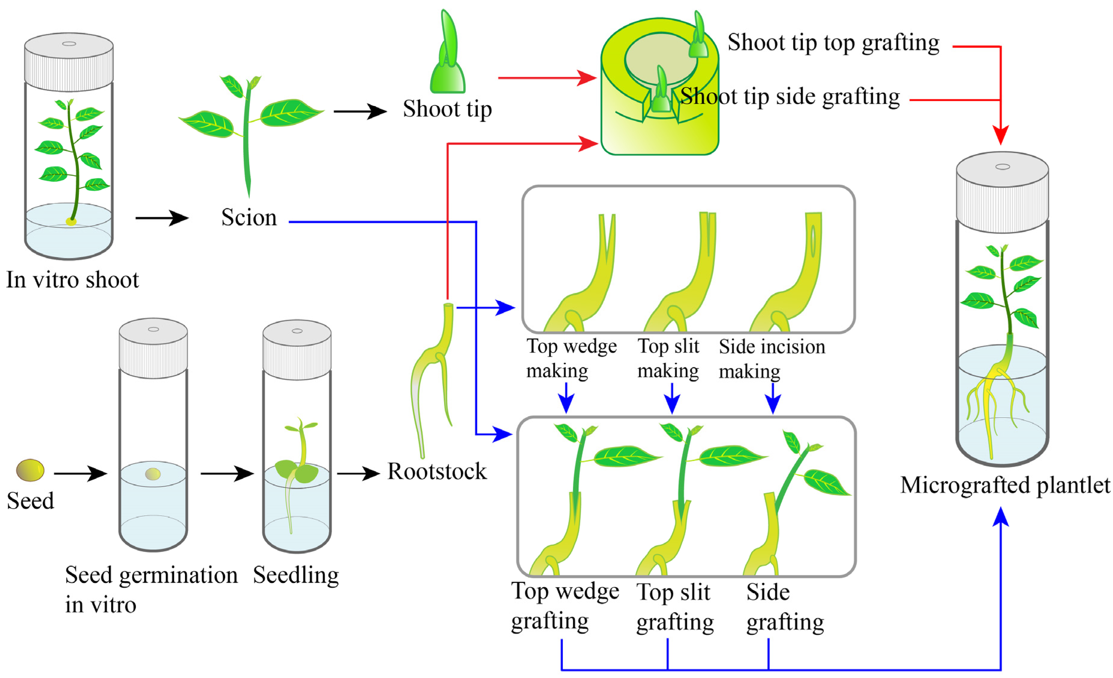
2. Establishment of Micrografting

2.1. Preparation of Scions

The origins and type of scion material are some of the determinants in successful micrografting [34,52,53,54]. The scions used in micrografting, usually shoots or shoot tips, can be obtained from in vitro or ex vitro grown plants. Scion material has traditionally been sourced from in vitro plants, having the advantage of being free from fungal and bacterial contaminations, the desired size, and being available year-around [35,53,55]. The use of ex vitro material, however, may introduce a seasonality component to the procedure, as the excised plant material may remain in a dormant stage [53]. However, shoot apices newly excised from actively growing trees in the field or in the greenhouse can be used in micrografting procedures [17,34]. Shoot apices sourced from in vivo plants are surface sterilized immediately prior to shoot/shoot tip preparation followed by micrografting. Usually, a short treatment of 70% ethyl alcohol is combined with a longer treatment of sodium hypochlorite or mercuric chloride for the surface sterilization [56].
Tissue browning is a common problem during the establishment of in vitro cultures [41,57]. Likewise, in IVM, wounding in scion/rootstock preparation may also cause browning and oxidation of plant tissues resulting in poor graft success [36,56,58]. The adverse effects of tissue browning may be minimized by presoaking scions in antioxidant solutions [39,59,60]. In cashew (Anacardium occidentale L.) and apple (Malus domestica), Thimmappaiah et al. [31] and Nunes et al. [61], respectively, reduced phenolic exudation by presoaking the cut edge of the scion with 0.01% ascorbic acid and 0.015% citric acid (1:1) prior to in vitro grafting. Reduced tissue browning in cashew can also be achieved by pre-conditioning in vitro stock shoots in culture media enriched with 0.1% polyvinylpyrrolidone before scion preparation [31]. In developing a protocol for micrografting of native and commercial roses, Davoudi Pahnekolayi et al. [59] showed that silver nitrate, as an antioxidant, played a key role in preventing production of phenolic compounds that could lead to micrografting failure. They found that a quick dip treatment (5–10 min) of wounded explants (scions and rootstocks) with silver nitrate (50 mg L−1) prior to micrografting could prevent tissue browning and consequently increase the survival of micrografts [59]. In contrast, Wu et al. [55] noted that micrografts of Protea cynaroides had reduced viability when scions were presoaked in ascorbic acid and citric acid solution; treatment with antioxidant solution induced more browning in scions than in those that were untreated. These results indicated that the fast operation of micrografting was more important in preventing tissue browning than pre-treatment, as suggested by Navarro [62]. Another possible reason could be the insufficient concentration of antioxidants which counterproductively promoted the spread of phenolic oxidation because of the improved wetness of the graft site [55]. Therefore, the response of antioxidants to the reduction/inhibition of phenolic browning may be species-dependent, and their concentrations and/or combinations are other critical points to be addressed.

2.2. Preparation of Rootstocks

In vitro germinated seedlings and segments of in vitro cultured shoots are the two major sources of rootstocks used in micrografting (Table 1). To prepare rootstock seedlings for micrografting, in vitro germination of seeds is the first step, with varied protocols needed to promote germination. Miguelez-Sierra et al. [38] found that cacao seeds did not require any pretreatment for the effective in vitro seed germination; in their protocol, seeds were taken from mature pods, were surface sterilized, inoculated in culture medium, and three-week-old seedlings were used as rootstocks. However, removal of the seed coat is necessary in many cases of in vitro germination. For example, for the preparation of in vitro rootstocks of almond, seeds were firstly removed from their endocarps (hard seed coat) before surface sterilization and in vitro germination [33,63]. Similarly, in pistachio, the mature kernels of seeds were surface sterilized after removing the outer pericarp and shells [17]. Likewise, the mature seeds of cashew [39] and jujube [42] were scarified in concentrated hydrochloric acid and sulfuric acid, respectively, before surface sterilization, to promote seed germination. In some cases, to achieve good germination, embryos have been removed from surface-sterilized seeds and grown in germination medium. In Protea cynaroides, a successful shoot tip micrografting technique was developed using 30-day-old in vitro-germinated embryos as rootstock [55]. Following seed germination, the duration of which varies between species, the root is shortened and the seedlings can be either decapitated above the cotyledons, leaving the epicotyls as the site for grafting [33,37], or cut below the cotyledons to make use of the hypocotyls as grafting sites [38,39,42,64].
In species with high levels of adventitious rooting, in vitro shoots can also be used as rootstocks [21,34,61,65]. Following grafting, the grafted shoots are cultured on a medium used for root induction of the rootstock genotype: this requires prior optimization of the rooting medium [65]. In cherry, Bourrain and Charlot [21] obtained a successful grafting rate of 79% when shoots (rootstock) were induced to root prior to grafting. In apple, Obeidy and Smith [66] achieved a graft success up to 45% when rooted in vitro shoots were used as rootstocks and apical 2-cm shoots as scions. When comparing the performance of Uapaca kirkiana (Muell. Arg) micrografts arising from in vitro rooted and unrooted rootstocks, Nkanaunena et al. [67] found that the three-month-old rooted rootstocks produced the highest graft success rate (at least 60%), with better development of grafted shoots. The positive response in the success of micrografting from the use of rooted rootstocks may be related to the species studied. Working on grapevine cultivar (cv.) ‘Superior’ micrografted onto different rootstocks, Sammona et al. [68] found no differences in grafting success between rooted and unrooted rootstocks.
Table 1. Applications of in vitro micrografting for restoration of scion vigor and establishment of plants with roots.
Plant Species (Scion) Scion Source and SizeRootstock Source and Age Grafting
Technique
Success Rate (%) and (No. Scions Tested) Reference
Amygdalus communis (Almond)Shoots of 1.5–2.0 cm in lengthAlmond seedlings of 2 weeks oldTop slit100 (1)[18]
Anacardium occidentale (Cashew)In vitro shoot apicesCashew seedlings, 5–8 cm in height (age not specified)Apical and side grafting45–73 (1)[69]
Shoots of 3–15 mm in lengthCashew seedlings of 20–25 days old Top slit and side grafting80 (top wedge) and 100 (side grafting) (1)[31]
Annona cherimola (Cherimoya)Nodal section of 2 cm in lengthCherimoya seedling of 42 days oldSide insertion 31–70 (3)[19]
Citrus deliciosa (Kinnow mandarin)Shoot tips less than 1.0 mm in sizeC. jambhiri, C. carrizo and C. reshnii seedlings of 15–20 days old Side reverse T insertionUp to 66.5 (1)[70]
Garcinia indicaApical shoots of 0.5–1.0 cm in length for the initial grafting and 1.0–1.5 cm for the subsequent graftingGarcinia indica seedlings of 2 months old Top slit95 (1)[71]
Malus domestica (Apple)Field grown shoots (size not specified)Apple shoots of 3 weeks oldVertical slit42–93 (3)[36]
Olea europea (Olive)Greenhouse-grown shoots of 1.0–1.5 cm in lengthOlive seedlings of 3 weeks oldTop slitUp to 83 (1)[45]
Opuntia ficus-indica (Cactus)Shoots of 0.5 cm in lengthShoots of O. strepacantha, O. robusta, O. cochinera, O. leucotricha and O. ficus-indica, 1.0 cm in length (age not specified)Top wedge and horizontal graft30 (top wedge) to 90 (horizontal) (1) [54]
Passiflora edulis (Passion fruit)Nodal segments of 1.5 cm in lengthPassion fruit shoots of 2 months old V-shaped joint with grafting devices73.3 (1)[26]
Pelecyphoraaselliformis (Cactus)Apical and subapical segments of 5 and 3 mm, respectivelyO. ficus-indica shoots, 10 mm in length (age not specified)Horizontal graft81 and 97 for the subapical and apical scions, respectively (1)[72]
Pistacia vera var. Siirt (Pistachio)Shoot tips of 0.5–10 mm in lengthPistachio seedlings of 10–14 days oldTop slit and top wedgeUp to 80 (1)[17]
Protea cynaroides (King Protea)Shoots of 5 mm in lengthKing Protea seedling of 30 days oldTop slit80 (1)[55]
Prunus dulcis (Almond)Apical shoots of 1.5–2.0 cm in lengthShoots of almond/peach hybrid rootstock of 3–7 weeks old Top slit50–70 (2)[65]
Shoots (size not specified)Almond seedlings of 2 weeks oldTop slit Up to 100 (1)[63]
Shoot tips of 4, 8 and 15 mm in lengthAlmond seedlings of 14 days old Top slit and top wedge90–100 (2)[33]
Prunus avium (Cherry)Shoot tips of
0.3–1.0 cm in length
P. avium × (P. canescens × P. tomentosa) shoots, 3–4 cm in length (age not specified)Top slitUp to 79 (1)[21]
Pyrus communis (‘Old Home’ x ‘Farmingdale 333′) (Pear)Shoots of 10 mm in length P. elaeagrifolia seedlings of 10–14 days old Cleft97.9 (1)[24]
Rosa hybrida cvs./(Rose)Shoots 10–15 mm in lengthR. canina and R. multiflora shoots, 20 mm in length (age not specified)With grafting devicesUp to 100 (2)[59]
Theobroma cacao (Cacao)Shoots of 4–6 mm in lengthCacao seedlings of 5–6 weeks old Not specified>50 (1)[73]
Bud sticks with apical or axillary buds (sourced from potted plants) of 1 cm in lengthCacao seedlings of 3 weeks oldTop slit and side grafting55–95 (1)[38]
Vitis vinifera (Grape)Shoot tips of 0.2–0.5 mm in lengthWhite to slightly coloured hypocotyls from white somatic embryosSide grafting18–30 (4)[74]
In vitro/in vivo derived shoot tips of 0.3–0.8 mm in length Shoots of V. vinifera × V. berlandieri, 1.0 cm in length (age not specified)Not specified40–61 (in vitro shoot tips) and 12–17
(in vivo shoot tips) (4)
[34]
Ziziphus mauritiana (Jujube)Shoots of 5–10 mm in lengthJujube seedlings (7 spp.) of 4 weeks old Top wedge28–100 (1)[42]

2.3. Grafting Techniques

The success of a micrografting procedure depends on the successful union of the rootstock and scion. The skill of the grafter is a key determinant of in vitro graft success. Various grafting techniques have been described, and the choice of which to use may depend on the type and size of the scion propagule and the purpose of the micrografting. The top-slit or top-wedge methods are the most frequently used in vitro grafting techniques and have been tested across a wide range of genera (Table 1). A slit or cleft is made onto the rootstock and wedge-shaped scions inserted into the cleft [17,33,55,75]. When rootstock thinner than scions are being used, micrografting can be performed by a reverse-cleft graft in which a slit is made at the bottom of the scions for the insertion of the rootstocks with a wedged top [76]. When small shoot tips were used as scions, their placement into the slit made at the top or directly over the rootstocks is usually referred as apical micrografting [69]. The insertion of small shoot tips into a slit on one side of the rootstock is termed side grafting (or side insertion) [69,74]. In Citrus, side grafting by inserting the shoot apices into inverted T-cuts of rootstocks has been successfully used [70]. Side insertion was also applied when longer shoots or nodal sections were used as scions [19,77,78]. The major methods applied in micrografting are illustrated in Figure 1.
In vitro propagules used in micrografting procedures are highly susceptible to moisture, whereby dehydration of the cut scion or rootstock surface can negatively affect the graft success. Therefore, in order to avoid dehydration, IVM should be performed promptly after the preparation of rootstock and scions to avoid dehydration [21]. In addition, ensuring a firm contact between the rootstock and scion is extremely important in developing a strong graft union [36,55].
Several devices have been used to enable fast and effective union between the rootstock and scion, such as the elastic electric-wire tube [44], aluminum foil [66,76], Parafilm® strip [42], silicon tube [13,79,80], paper bridge [59], silicone chip [30], plastic clamps [50] or alginate gel beads [44,75,81]. These grafting devices are used to support the graft and hold the scion and rootstock together during graft healing, particularly for the top-slit and top-wedge methods.
In addition to grafting devices, the practice of dipping the lower end of the scion in the culture medium before fitting it into the rootstock [12], or applying agar solution on the grafting zone as an adhesive material [36,82] to hold the graft, are also strategies to establish and fix the graft union, particularly when the scion does not fit properly into the rootstock. Pathirana and McKenzie [12] suggested that, in addition to delivering nutrients directly to the graft site, the strategy of dipping the lower end of the scion in the culture medium before grafting keeps the cut surfaces moist until the high relative humidity within the vessel is re-established after closure. This strategy resulted in a high success rate of 75–85% in micrografting of grapevine. Dobránszki et al. [82] described an effective method for IVM of apple using agar–agar solution to stick the scion to the vertical slit of the rootstock. Briefly they placed the V-shape cut scion base in an antioxidant solution (0.15 mg L−1, 0.1 mg L−1 ascorbic acid, and 0.1 mg L−1 gibberellic acid) to inhibit oxidative browning, followed by treatment with 1% agar–agar solution, and then two drops of agar solution were placed around the graft zone before attaching the scion to the rootstock. With this method, the graft success rate was 95% and all acclimatized plants survived [82]. In contrast, medium-supported grafting resulted in lower micrograft survival rates than the unsupported technique in Protea cynaroides [55]. Similarly, working on conifer micrografts, Ponsonby and Mantell [83] and Cortizo et al. [84] found either a reduction in graft union success or no response, respectively, when antioxidant additives or culture medium solution was applied to the micrograft union. Therefore, we suggest that medium-supported grafting be applied as a back-up strategy for IVM.

2.4. Culture Conditions

Various culture conditions have been tested to optimize the regrowth of micrografts with success dependent on the plant species and the source of plant material used. In rootstock seedlings, seeds are usually germinated under continuous darkness for 1 to 6 weeks [40,64,85,86], but successful protocols using seedlings germinated in light conditions have also been reported [27,51,72,87]. Working on grapefruit micrografted onto seedling sour orange, Ali et al. [40] showed that the grafting success was related to the light conditions during seedling development. The frequency of graft success increased from 5 to 50% when rootstock seedlings were obtained from seeds germinated under continuous darkness for two weeks compared with success using seeds germinated under continuous light [40]. Similarly, in Citrus cultivars Cadenera Fina and Pera (sweet oranges) micrografted onto different rootstocks, Navarro et al. [11] found a greater graft success rate when rootstock seedlings from seeds germinated and grown in darkness were used (37.5%) than seedlings germinated in light conditions (2.7%). In contrast, working on Tahitian lime and Valencia orange micrografted to seedlings from the mandarin (Cleopatra), Suárez et al. [87] found moderate rates of success that ranged from 14 to 28%, respectively, on rootstocks from seeds germinated under light conditions.
The conditions in which plants are grown following micrografting can influence graft success. Micrografted plants of jujube tree (Ziziphus mauritiana ‘Gola’) were first grown in darkness for ten days and then transferred to light conditions [42]. They found that a period in the dark before and after grafting was important to avoid the photo oxidation at the grafting point as well as to minimize the destruction of the auxins synthesized in the scion [42]. In almond, micrografted plants were cultured on rooting medium and incubated in the dark for 7 days, then transferred to the light of 35–40 μmol m−2 s−1 for two weeks, and finally to 60 μmol m−2 s−1 for one week before in vivo acclimatization [65]. Several studies have reported successful micrografts without a dark incubation period following the grafting procedure [13,24,72,82,86].
Different supporting systems have been used with growth media in micrografting procedures. Paper bridges, perlite and vermiculite have been used for supporting micrografted plants in liquid culture medium [63,88,89], as well as the solid and semi-solid culture media [21,25,34,72,86]. For example, liquid medium with perlite or a paper bridge was used as the supporting system in micrografting of almond [63] and lime shoots [90]. Liquid medium has also been used in micrografting cashew [39] and cut rose [59]. An adjustable paper bridge can be made to better support the micrografts cultured with liquid medium [66]. The advantages of using liquid medium lie in the better availability and absorption of nutrients, and the reduced damage to the root system when moving plants [39]. In micrografting of cherry, an agar-solidified medium with vermiculite was successfully used to obtain high-quality grafted plants [21]. In apple, the highest graft success was achieved when micrografted plants were cultured on agar-solidified medium [89].

2.5. Acclimatization of Micrografted Plants

Once micrografted plants are well rooted and showed clear scion regrowth, they can be transferred to potting mix following a gradual transition of light intensity and ventilation to achieve successful acclimatization [17,18,21,77]. Acclimatization is a critical phase within micrografting protocols: significant losses can occur when transplanting micrografted plants to ex vitro conditions [62]. Micrografts are removed from in vitro conditions and rinsed with tap water to remove any remaining medium from the roots, and finally grafted plants are transferred to pots containing substrate [21,22,33]. During the first few days, micrografted plants are maintained in high relative humidity and gradually transferred to ex vitro conditions [43,59,87,91]. Micrografted plants of jujube cultured for one month on growth medium and with the scion having grown to 5–10 cm in length (scions were initially with 5–10 mm) could be successfully acclimatized (83–87%) [42]. Miguelez-Sierra et al. [38] found that micrografted plants of cacao grown in vitro for two weeks could be transferred to ex vitro conditions. They observed that only plants with at least 1 cm of scion elongation and two expanded leaves survived acclimatization [38]. The presence of roots on the rootstock is essential for micrografted plants to survive acclimatization. Hieu et al. [26] found that the micrografted plants of passion fruit without roots did not survive acclimatization; plants with roots formed in vitro achieved the highest survival rate during the acclimatization phase. The survival rate during acclimatization of micrografted plants varies among species. For example, in apple, survival rate of grafted plants reached 100% [82], for almond it was 85–100% [92], it was 82% in cacao [36], and 75% in passionfruit [26], whereas in Tahitian lime and Valencia orange, survival rates ranged from 47 to 50%, respectively [87]. Contrasting results were also reported by Kobayashi et al. [93] in micrografting of sweet orange buds derived from organogenesis. In that instance, the fully developed in vitro micrografts grew slowly in the greenhouse, so the micrografted plants were re-grafted onto three-month-old seedlings of Rangpur lime for rapid acclimatization and normal development of the plants. In general, however, there was a higher acclimatization success rate of micrografted plants than for ungrafted plants when plants were difficult to manage in conventional tissue culture or to establish roots on [65].

3. Factors Affecting the Success of Micrografting

3.1. Scions

In micrografting of cacao, Miguelez-Sierra et al. [38] used two types of bud sticks with either apical or axillary buds as scions. While better graft survival was achieved with the apical shoot apices (85 to 100%), the latter showed a more rapid post-grafting regrowth, resulting in better acclimatization [38]. In micrografts of a cactus (Pelecyphora aselliformis Ehrenberg), the use of apical and sub-apical slices as scions produced successful rates of 97% and 81%, respectively [72]. For in vitro grafting of jujube, shoot tips excised from in vitro cultures as scions showed significantly higher survival (70%) and scion growth (3.3 cm) than those collected from mature trees (33% survival and scion growth of 1.3 cm) [42]. Working on micrografts of pistachio, Onay et al. [17] observed that there was a seasonal variation in success rate when micrografting ex vitro material as scions. They found that the month at which shoot tips were harvested from the field growing trees significantly affected graft success: 80% of success at June compared with 10 and 30% for February and December, respectively. In contrast, similar success rates (50 to 80%) were obtained year-round when using in vitro-sourced shoot tips as scions [17].
The size of scions can affect the success of micrografting. Thimmappaiah et al. [39] reported higher success of micrografting (80%) in cashew when scion size was 6–15 mm, while scions of less than 5 mm only had 0.5% of graft success. Similarly, in pistachio, a scion of 4–6 mm resulted in a significantly higher success rate (79%) than 2–4 mm and over 10 mm, which gave 57% and 17% micrografting success, respectively [17]. In micrografting of almond, Yıldırım et al. [33] noted no significant difference in the success rate with scion size ranging from 4 to 15 mm, but significantly improved shoot length and leaf number were observed when larger scion size was used (15 mm). By contrast, in micrografting of cherry, a scion size of 3–5 mm produced higher graft success (42–46%) than the 29% when 10-mm-long scions were used [21].
Channuntapipat et al. [65] compared the effects of various hardness of scions on micrografting survival in almond using wood-stem shoots as rootstocks. Results showed that the highest micrografting survival was found from ‘hard scion/hard stem’, while no successful graft was found from the combination of ‘soft scion/wood stem’ [65].

3.2. Rootstocks

Scions can be grafted onto either the hypocotyls or epicotyls of in vitro germinated seedlings. Hypocotyls are usually preferred because epicotyls may contain axillary meristems that compete with the scions and may grow following the grafting, thus adding extra steps of identifying and removing rootstock-derived shoots [78]. In micrografting of cashew, Thimmappaiah et al. [39] noted that side grafting of shoots onto the hypocotyl resulted in higher grafting success (100%) than top-wedge grafting on epicotyls (80%), attributing this to better cambial contact established when side-grafting to hypocotyls. Although comparable micrografting success was achieved when shoot apices of cashew were side micrografted onto the hypocotyl and epicotyl of seedlings, the hypocotyl grafts grew more vigorously than the epicotyl ones [69]. All these studies highlight the advantages of using of hypocotyls as the micrografting sites, when in vitro-germinated seedlings are used as the rootstocks.
Scions grafted onto the same species (homografts) had significantly higher success rates and subsequent growth than heterografts in cactus [54]. In this study, five different combinations of micrografts were tested, with the scion growth of homografts extending from 0.5 to 28.8 cm after 90 days of post-grafting cultures, significantly higher than with the four heterograft combinations [54]. Similarly, graft incompatibility was reported in micrografting in vitro Ziziphus mauritiana ‘Gola’ of west Africa onto an American jujube (Z. joazeiro), while achievable results were obtained using those originating from the Old World as rootstocks [42].
The age of rootstock seedlings can also affect the success of micrografting. Working with grapefruit grafted onto sour orange seedlings, Ali et al. [40] found that the highest percentage of successful micrografts was obtained when two- (60%) to three- (40%) week- old seedlings were used, while a further week of seedling growth resulted in a high percentage of unsuccessful grafts (70%). In addition, they observed that most (80%) of the shoot tips grafted onto one-week-old seedlings became quiescent and turned into calluses. Similarly, working on Kinnow mandarin grafted onto Carrizo citrange seedlings, Chand et al. [94] found the highest micrografting success (38%) when 12-day-old seedlings were used, whilst the success was reduced to 24% with 18-day-old rootstock seedlings. They suggested that the reduction in micrografting success with older rootstocks may be due to the harder stem, which made it more difficult to preparing the cut edges for the grafts as well as to insert the scions [94].

3.3. Micrografting Methods

The success of micrografting depends on numerous factors including the grafting method used [87]. Establishing good contact between the microscion and rootstock fosters the reconnection of the cambial tissue and is pivotal to formation of the micrograft union [55,65]. In micrografts of almond, Yıldırım et al. [33] found that the top-slit method resulted in better connection, leading to fusion between scion and rootstock with success rates of 90–100%, while significantly higher numbers of displaced micrografts were detected in top wedge micrografting, which produced only 30–40% of successful grafts. Similar results were produced in micrografting of pistachio by Onay et al. [17] where the top-slit method was compared with the top-wedge method, and 80% success was obtained from the top-slit compared with 60% from the top-wedge.
In Tahitian lime and Valencia orange micrografted onto mandarin seedlings, Suárez et al. [87] only found moderate rates of micrografting success, ranging from 14 to 28%, respectively, when scion shoot tips (<5 mm long) were placed in a slanted position on the decapitated surface of the rootstocks. Shoot tips either positioned at the top or on the side-chip buds of the rootstocks failed completely [87].
Estrada-Luna et al. [54] compared horizontal and wedge grafts for micrografting of Opuntia spp. They found the horizontal grafts more successful, owing the reduced scion displacement. In micrografting small Vitis shoot apices onto rootstocks of much greater diameter, Torres-vinals [74] observed that side grafting was more successful than top grafting (19% versus 14%). Similarly, in cashew, the side grafting of small shoot apices led to a significantly higher success rate (66%) than the apical grafting method (45%) [69].

3.4. Culture Conditions

The carbon source is a factor that may induce in vitro plant recalcitrance and affect micrografting success [21,95]. In cherry, the highest rates of successful micrografts (79%) were obtained when 30 g L−1 of glucose was used as a carbon source compared with sucrose (58%) [21]. In addition, they observed that the incremental increase of 30 to 75 g L−1 for both sucrose and glucose did not increase grafting success [21]. In contrast, in micrografts of cut roses, elevating sucrose concentration (50 g L−1) in the culture medium resulted in significantly higher micrografting success than 30 g L−1 [59]. Similarly, in Kinnow mandarin and Succari oranges micrografted on ‘Rough’ lemon seedlings, Naz et al. [96] found that increasing sucrose concentration in the culture medium from 30 to 50 g L−1 improved graft success rates from 21 to 33% in both cultivars. In addition, a further increment of graft success (to 38%) was found in Kinnow mandarin grown in culture medium supplemented with 70 g L−1 sucrose [96]. Similar improvements in successful micrografting for other Citrus species, by increasing sucrose concentration in the culture medium, have been reported by Navarro et al. [11,62], Ali et al. [40], and Singh et al. [97].
Almond micrografts cultured in liquid medium with perlite as a support system showed better survival and scion growth than those supported by paper bridges [63]. Thimmappaiah et al. [39] noted that liquid medium may be better for supporting growth of cashew micrografts as availability and absorption of nutrients was higher and there was less damage to the root system, compared with use of a solid medium. Liquid woody plant medium was successfully used to support Prunus micrografts [98]. In using agar-solidified medium to support micrografts of cherry, Bourrain and Charlot [21] found adding vermiculite to the medium increased the grafting success from 12 to 52%. Furthermore, they noticed that the aerated structure of the medium in the presence of vermiculite enhanced the root system and favored the development of secondary roots [21]. Successful micrograft unions in apple, cherry, and Citrus were achieved using either agar-solidified medium or vermiculite to hold the grafts during the healing period, with no difference in rate of success [66].
A range of mineral formulations with difference ionic content have been used in micrografting protocols [12,25,42,62,74,98,99]. There is no “best” or “standard” mineral formulation: the selection of a culture medium is influenced by plant species used. In addition, the phytohormones applied in the culture medium can also affect the development of micrografts. Cytokinin and auxin are the most frequently used phytohormones in growth media when micrografts are used [22,26,91]. In pistachio, Onay et al. [17] found that the micrografts cultured in a growth medium with 2.22 μM 6-benzylaminopurine (BAP) were significantly more successful (72%) than those cultured in medium containing 2.46 μM indole-3-butyric acid (IBA) (36%) or in phytohormone-free medium (47%). In contrast, in almond, the medium in which the grafted plants were cultured had no effect on micrografting success [18,33]. It is also worth noting that almond micrografts grown in medium containing 1.0 mg L−1 IBA had an increased number and length of roots, while grafts grown in medium with 1.0 mg L−1 BAP resulted in higher scion proliferation [18,33]. The different effects of auxin and cytokinin on the survival and growth of Kinnow mandarin micrografts were reported by Kumar et al. [70]. They found that the highest graft survival (66.9%) was observed in graft growth medium supplemented with 3.0 mg L−1 2,4-Dichlorophenoxyacetic acid (2, 4-D), while early bud sprouting and increased scion length were obtained in growth medium supplemented with 1.0 mg L−1 BAP [70].
There have been many successful reports of micrografting without the use of phytohormones in the growth medium [21,24,25,34,40,66,72,98]. This would minimize the need to test the genetic stability of regenerants, as high concentrations of plant growth regulators may cause somaclonal variation within in vitro tissue cultures [25,98,100,101,102].

4. Applications of Micrografting in Micropropagation

4.1. Root Promotion

In vitro rooting is an important stage of micropropagation protocols [56,103]. For some species, the main challenge of micropropagation has been the difficulty in inducing adventitious root formation [18,22,70,104]. IVM is an alternative means to provide roots and overcome rooting difficulties in the vegetative propagation of these species [18,22,55,71], Table 1. For example, IVM of microshoots onto rootstock seedlings was applied to solve the in vitro recalcitrance of Protea cynaroides, an important ornamental species endemic to South Africa [55]; Lens culinaris, an important pulse crop of Mediterranean area [16]; and some Prunus species [21,33,55,66]. The development of suitable micrografting techniques to overcome rooting difficulties of plant species has been highlighted in previous reviews [15,105,106].
Root induction is the limiting step in the in vitro propagation of Garcinia indica [71]. To overcome this difficulty, Chabukswar and Deodhar [71] proposed a micrograft protocol where shoot tips were repeatedly grafted on to in vitro juvenile seedlings to restore rooting competence. They conducted micrografting using the 2-month-old in vitro-grown seedlings as rootstocks to reinvigorate in vitro shoots established from 20-year-old trees. The elongated shoots (scions) about 0.5–1.0 cm in length were cut into a V-shape at the bottom and inserted into a vertical incision in the rootstock, after they had been decapitated at the lowest node. After the graft union formed (6–8 weeks), the apical region (1–1.5 cm in length) of the scion was cut, and it was re-grafted onto new in vitro seedling rootstocks. After five successive micrograftings, 75% of the grafts were rooted and successfully acclimatized [71]. In vitro rooting of Annona cherimola shoots was also difficult, but it could be achieved after 2–3 consecutive cycles of micrografting onto rootstock seedlings [19]. Similarly, three cycles of in vitro grafting improved the rooting ability in jujube [42]. While numerous studies made use of micrografting in order to promote rooting, in Juglans rejia (walnut), the in vitro adult clones did not produce adventitious roots, even after two consecutive cycles of micrografting onto rootstock seedlings [107]. However, an acceptable root induction rate could be obtained after 30 cycles of in vitro subcultures [107]. Further study is therefore still needed to improve the promotion of rooting in walnut. Micrografting for improved rooting often utilizes in vitro germinated seedlings as rootstocks; some examples can be found in Table 1.

4.2. Promotion of Shoot Proliferation

Long-established in vitro plants often demonstrate declined regenerative ability [108,109]. The reduced proliferation could be reversed in vitro following successive grafting onto vigorous rootstocks [19,33,106,110]. In order to improve the micropropagation process of three cherimoya cultivars, Padilla and Encina [19] micrografted its nodal segments onto in vitro germinated seedlings. It was found that proliferation from shoot segments was significantly improved in all micrografted plants compared with conventionally in vitro cultured segments [19]. The restoration of shoot proliferation was also achieved in the almond cultivars Ferragnes and Ferraduel micrografted onto in vitro germinated wild almond seedlings [33].
Farahani et al. [45] repeatedly micrografted mature olive segments (1–1.5 mm in length containing lateral meristem) onto three-week-old germinated seedlings for improved shoot proliferation of the cultivar Zard over a number of culture cycles. The micrografting success, shoot elongation and bud sprouting were improved, particularly after the third successive micrografting [45]. In Ziziphus mauritiana, repetitive micrografting (≥2 times) of in vitro shoots onto in vitro germinated seedlings improved the growth of the scions as well as the percentage of rooted micrografts [42]. Improved in vitro rooting and shoot proliferation were also achieved following micrografting in several plant species, such as cherimoya [19], mandarin and sweet orange [106,111]. The improved scion growth was a consequence of reinvigorated rooting either from direct support by the in vitro germinated seedlings [45] or through recovered adventitious rooting [19].

4.3. Embryo Rescue or the Promotion of Organogenesis-Derived Shoot Regrowth

Recovery of plants via de novo organogenesis and somatic embryogenesis can be important for obtaining genetically modified plants, and in vitro mutagenesis [112,113,114,115]. However, it can be problematic in some horticultural species owing the difficulties of rooting [116,117] or to inadequate callus maturation and tissue culture [37].
Micrografting has been applied to overcome the inability of many organogenesis-derived regenerants to readily produce roots [77,78,93,113]. The poor rooting ability observed in regenerated shoots of sunflower was resolved by micrografting shoots regenerated from leaves onto in vitro-germinated seedlings using a side insertion method [77]. In this method, best survival (75%) was obtained by inserting the 0.5–1.0-cm-long shoots with a wedge-shaped base into the longitudinal cut at the hypocotyl. The micrografted sunflowers were successfully acclimatized, and they flowered and produced seeds [77]. Kobayashi et al. [93] applied IVM to support the growth of sweet orange (Citrus sinensis) regenerated from thin sections of mature stem segments. Using the same regeneration system in four sweet orange cultivars, Almeida et al. [118] tested the use of micrografting with plantlets of Carrizo citrange as rootstocks to support shoot recovery after genetic transformation. IVM was also applied in pepper (Capsicum annuum) to obtain rooted transgenic plants regenerated from cotyledon-derived organogenesis [119].
Noticeably, ex vitro micrografting was used in legumes using in vitro-regenerated shoots as scions and ex vitro-germinated seedlings as rootstocks to facilitate grafting and acclimatization simultaneously in field pea (Pisum sativum L.) [120] and chickpea (Cicer arietinum L) [121]. Similar protocols were also used to obtain rooted plants from cotyledon-derived adventitious pear shoots [122].
IVM was first reported in 1992 to support the recovery of shoot regenerated from somatic embryos (SEs) of cocoa plants [123]. In avocado, Raharjo and Litz [37] proposed an effective micrografting procedure for SE shoot rescue. Briefly, in their study, SE-derived shoots of 5–10 mm in length were grafted (V-shaped cut) onto in vitro rootstock seedlings, and then grafted plants were grown on a phytohormone-free medium. Using this protocol, micrografted plants were established after 3–4 weeks, and 70.5% of the SE-derived shoots were rescued, whereas only 30.4% of nonmicrografted SE shoots survived and normal plantlets were never recovered [37]. In addition, the micrografting protocol was followed by ex vitro grafting, and this has served as a protocol for rescuing transformed avocado materials [37]. Palomo-Ríos et al. [124] also reported successful recovery of transgenic plants by IVM in avocado. In this study, globular somatic embryos established from immature zygotic embryos were transformed using Agrobacterium. After selection on kanamycin, the germinated somatic embryos were then elongated to 3–5 mm before micrografting onto in vitro-germinated seedlings to achieve better recovery [124]. Likewise, in seedless sweet orange, micrografting of ovary-derived somatic embryos onto in vitro seedlings was applicable to achieve full recovery or somatic organogenesis [117]. Some examples of applying IVM to promote the shoot recovery from de novo organogenesis and somatic embryogenesis are listed in Table 2.

4.4. Shoot Regrowth after Cryopreservation

Cryopreservation is currently considered an applicable strategy to facilitate long-term, cost-effective maintenance of plant genetic resources [125,126]. Shoot tip cryopreservation of many horticultural species has been established in cryobanks; a high level of post-thaw recovery is a requirement of successful cryopreservation [126,127]. Direct shoot tip recovery could not be obtained in some species, such as Citrus; thus, micrografting of cryopreserved shoot tips onto in vitro prepared seedlings was used to overcome this [128,129,130], Table 2. In successful recovery of citrus shoot tips after cryopreservation, Volk et al. [128] prepared six-week-old in vitro ’Carrizo’ citrange seedlings as rootstocks to support the shoot tips cryopreserved by a vitrification protocol. Briefly, in their study, rootstock seedlings with a height of at least 3 cm were decapitated 1 cm above the cotyledonary node with a 2-mm deep incision made into the cut surface, followed by horizontal cut through the seedling to create a “ledge” or “step” at the cut surface. Cryopreserved shoot tips were trimmed (0.2 mm of the basal portion) and placed on this rootstock ledge [128]. This post-thaw protocol resulted in 53% of regrowth on average for eight Citrus and Fortunella species [128]. Volk et al. [129] applied the same IVM procedure to support the post-thaw recovery of 150 pathogen-free citrus accessions representing 32 taxa after a droplet-vitrification cryopreservation. With this procedure, 24 taxa had mean regrowth levels of over 40% after cryopreservation [129]. There are ongoing efforts to use this successful procedure to recover plants following cryopreservation of a wide range of citrus species [86].
Table 2. Applications of in vitro micrografting in promoting of plants recovery after shoot organogenesis, somatic embryogenesis and shoot tip cryopreservation.
Plant Species (Scion)Scion Source and SizeRootstock Source and Age Grafting TechniqueSuccess Rate (%) and (No. Scions Tested) Reference
Citrus sinensis (Sweet orange)Shoots of 1–2 mm in length (sourced from greenhouse plants) Carrizo citrange seedlings of 2 weeks oldSide insertion90 (1)[93]
Shoots recovered from germinated somatic embryos (size not specified)Carrizo citrange seedlings (age not specified)Not specified90 (1)[117]
Citrus spp.Shoot tips (1–1.5 mm in length) cryopreserved by droplet-vitrificationCarrizo citrange seedlings up to 6 weeks oldSide grafting10–100 (32); average of 56%[129]
Helianthus annuus (Sunflower)Shoots (0.5–1 cm in length) from leaf explantsSunflower seedlings of 7–10 days oldSide insertion47–85 (7)[77]
Shoots (1 cm in length) from cotyledon explants Sunflower seedlings of 1–2 weeks old Side insertion69 (1)[78]
Lens culinaris (Lentil)Shoots (1–1.5 cm in length) from cotyledonary nodesLentil seedlings of 5–6 days old Top slit90–100 (3)[16]
Persea americana (Avocado)Shoots regenerated from somatic embryos (SEs)Avocado seedlings of 7–12 days oldTop slit70.5 (1)[37]
Shoots derived from previously micrografted SE shoots100 (1)
Shoots of 5–10 mm in length59 (1)
Shoots (size not specified) regenerated from genetically transformed SEs Avocado seedlings of 3 weeks old Top slit83.6 (1)[112]
Shoots of 3–5 mm in length from genetically transformed SEsAvocado seedlings of 4 weeks old Top slit60–80 (1)[124]
Solanum lycopersicum
F1 hybrids (Tomato)
Shoots (size not specified) regenerated from cotyledon explantsTomato seedlings of 3 weeks oldTop slit75–83 (3)[131]
Ziziphus jujuba (Chinese jujube)Shoot tips (0.2–1 mm in length) cryopreserved by droplet-vitrificationZ.spinosa seedling of 4 weeks oldSide grafting5–75 (1)[132]
Micrografting was necessary to support the recovery of cryopreserved Chinese jujube (Ziziphus jujuba) shoot tips [132]. Cryopreserved shoot tips cultured on recovery medium, without micrografting, developed only leaves without shoot regrowth. In contrast, a high shoot recovery rate of 75% was obtained when shoot tips were micrografted onto sour jujube (Ziziphus spinosa) rootstock seedlings. This procedure was also effective to produce plants free of jujube witches’ broom phytoplasmas. Therefore, micrografting provided technical support in cryopreservation and production of phytoplasma-free plants [132].
While micrografting in Citrus to support cryopreservation protocols has been moving from research [128] to full-scale implementation [86,129], it has not been well explored in other species. Micrografting has potential to improve the regrowth of cryoprocessed shoot tips of woody plants that are still recalcitrant to cryopreservation. Plants that might benefit from this approach include Pistacia species, in which shoot tip cryopreservation resulted in low recovery levels ranging from 5.0 to 17.6% [133]. In avocado, although SEs have been shown to be amenable to cryopreservation, studies focusing on shoot tip cryopreservation are still needed for the safe conservation of elite avocado cultivars [134]. Micrografting could therefore be considered as a tool to support shoot tip regrowth after cryopreservation in the cases of pistachio, avocado, and other recalcitrant plant species.

5. Conclusions and Future Prospects

This review focuses on studies highlighting the use of micrografting in micropropagation of horticultural species in the 21st century. As an important technique supporting basic and applied research, IVM consists of preparation of rootstocks and scions, the micrografting process and post-grafting cultures, steps that have all been developed from in vitro tissue culture techniques.
IVM protocols have been developed for many plant species with differing degrees of success. The successful recovery of plants following micrografting depends on numerous factors, such as the origin and preparation of rootstocks and scions, grafting methods, graft growth conditions, as well acclimatization, with these factors being genotype- and species-specific, just as in in vitro tissue culture. Therefore, further development and optimization of micrografting techniques, especially for recalcitrant species, are needed, to expand the use of micrografting in micropropagation. While successful micrografting protocols have been established in most horticultural species, they rely on technically difficult protocols, performed by well-equipped, skilled and well-trained technicians. Progress is thus still needed to simplify micrografting procedures. In addition, the programs applying IVM for micropropagation always include the transfer of the micrografted plants to ex vitro conditions, and thus, the effects of acclimatization on the survival of micrograft plantlet as well as long-term survival in case of partial graft incompatibility should also be evaluated.
Although progress has been made over the years on in vitro plant tissue cultures, in some plant species, IVM is still applied as a necessary step to provide roots for in vitro grown propagules, thus enabling further acclimatization (Figure 2). In species with problematic in vitro rooting, seedlings produced from in vitro germinated seeds are often used as rootstocks in micrografting procedures. For species that showed decreased rooting and shoot proliferation following prolonged in vitro cultures, consecutive micrografting of shoots onto in vitro germinated seedlings has proved effective in restoration of vigor and rooting competence in some instances (Figure 2).
Moreover, IVM has assisted the recovery of shoots generated from de novo organogenesis and somatic embryogenesis (Figure 2), which have been widely used as sources of explants for genetic transformation. Therefore, IVM has been implemented as a step in supporting the genetic transformation of horticultural species such as citrus and avocado. Likewise, the cryopreservation of citrus shoot tips also relies on IVM to sustain post-thaw recovery and shoot regrowth after cryopreservation (Figure 2). The large-scale cryopreservation of citrus has been established with the assistance of micrografting, ensuring the safe and long-term preservation of its valuable genetic resources. The successes observed in citrus cryopreservation would encourage the use of micrografting in the recovery of cryopreserved species that are still recalcitrant to cryopreservation procedures.

Author Contributions

M.-R.W. and J.C.B.: data collection, analysis, manuscript writing and revisions; A.-L.Z. and X.L.: data collection and analysis, table and figure preparation; D.Z. and Q.-C.W.: manuscript proposal and manuscript revision. All authors have read and agreed to the published version of the manuscript.

Funding

We acknowledge funding from the National Key R&D Programs of China (project no. 2019YFD1001803) for M.-R.W., X.L., D.Z. and Q.-C.W. and from The New Zealand Institute for Plant and Food Research Limited (project no. 0/601000/01) for J.C.B.

Institutional Review Board Statement

Not applicable.

Informed Consent Statement

Not applicable.

Data Availability Statement

Not applicable.

Conflicts of Interest

The authors declare no conflict of interest.

References

  1. Mudge, K.; Janick, J.; Scofield, S.; Goldschmidt, E.E. A History of Grafting. In Horticultural Reviews; Janick, J., Ed.; John Wiley & Sons, Inc.: Hoboken, NJ, USA, 2009; Volume 35, pp. 437–493. [Google Scholar]
  2. King, S.R.; Davis, A.R.; Liu, W.; Levi, A. Grafting for Disease Resistance. Hortscience 2008, 43, 1673–1676. [Google Scholar] [CrossRef] [Green Version]
  3. Goldschmidt, E.E. Plant grafting: New mechanisms, evolutionary implications. Front. Plant Sci. 2014, 5, 727. [Google Scholar] [CrossRef] [PubMed] [Green Version]
  4. Macan, G.P.F.; Cardoso, J.C. In vitro grafting of Psidium guajava in Psidium cattleianum for the Management of the Meloidogyne enterolobii. Int. J. Fruit Sci. 2020, 20, 106–116. [Google Scholar] [CrossRef]
  5. Fazio, G. Genetics, breeding, and genomics of apple rootstocks. In The Apple Genome, Compendium of Plant Genomes; Korban, S.S., Ed.; Springer: Cham, Switzerland, 2021; pp. 105–130. [Google Scholar]
  6. Martins, V.; Silva, V.; Pereira, S.; Afonso, S.; Oliveira, I.; Santos, M.; Ribeiro, C.; Vilela, A.; Bacelar, E.; Silva, A.P.; et al. Rootstock affects the fruit quality of ‘Early Bigi’ sweet cherries. Foods 2021, 10, 2317. [Google Scholar] [CrossRef] [PubMed]
  7. Thorpe, T.A. History of Plant Tissue Culture. In Methods in Molecular Biology, 2nd ed.; Loyola-Vargas, V.M., Vázquez-Flota, F., Eds.; Humana Press Inc.: Totowa, NJ, USA, 2006; Volume 318, pp. 9–32. [Google Scholar]
  8. Doorenbos, J. Rejuvenation of Hedera helix in graft combinations. Koninklijke Nederlandse Akademie van Wetenschappen: Series C. Biol. Med. Sci. 1953, 57, 99–102. [Google Scholar]
  9. Holmes, F.O. Elimination of spermivirus from the nightingale chrysanthemum. Phytopathology 1956, 46, 599–600. [Google Scholar]
  10. Murashige, T.; Bitters, W.P.; Rangan, E.M.; Nauer, E.M.; Roistacher, C.N.; Holliday, P.B. A technique of shoot apex grafting and its utilization towards recovering virus-free citrus clones. HortScience 1972, 7, 118–119. [Google Scholar] [CrossRef]
  11. Navarro, L.; Roistacher, C.N.; Murashige, T. Improvement of shoot-tip grafting in vitro for virus-free citrus. J. Am. Soc. Hortic. Sci. 1975, 100, 471–479. [Google Scholar]
  12. Pathirana, R.; McKenzie, M.J. Early detection of grapevine leafroll virus in Vitis vinifera using in vitro micrografting. Plant Cell Tiss. Organ Cult. 2005, 81, 11–18. [Google Scholar] [CrossRef]
  13. Cui, Z.-H.; Agüero, C.B.; Wang, Q.-C.; Walker, M.A. Validation of micrografting to identify incompatible interactions of rootstocks with virus-infected scions of Cabernet Franc. Aust. J. Grape Wine Res. 2019, 25, 268–275. [Google Scholar] [CrossRef]
  14. Hao, X.-Y.; Jiao, B.-L.; Wang, M.-R.; Wang, Y.-L.; Shang, B.-X.; Wang, J.-Y.; Wang, Q.-C.; Xu, Y. In vitro biological indexing of grapevine leafroll-associated virus 3 in red- and white-berried grapevines (Vitis vinifera). Aust. J. Grape Wine Res. 2021, 27, 483–490. [Google Scholar] [CrossRef]
  15. Chilukamarri, L.; Ashrafzadeh, S.; Leung, D.W.M. In Vitro grafting—Current applications and future prospects. Sci. Hortic. 2021, 280, 109899. [Google Scholar] [CrossRef]
  16. Gulati, A.; Schryer, P.; McHughen, A. Regeneration and micrografting of lentil shoots. In Vitro Cell. Dev. Biol.-Plant 2001, 37, 798–802. [Google Scholar] [CrossRef]
  17. Onay, A.; Pirinç, V.; Yıldırım, H.; Basaran, D. In vitro Micrografting of Mature Pistachio (Pistacia vera var. Siirt). Plant Cell Tiss. Organ Cult. 2004, 77, 215–219. [Google Scholar] [CrossRef]
  18. Işıkalan, Ç.; Namli, S.; Akbas, F.; Ak, B.E. Micrografting of almond (Amygdalus communis) cultivar ‘Nonpareil’. Aust. J. Crop. Sci. 2011, 5, 61–65. [Google Scholar]
  19. Padilla, I.M.G.; Encina, C.L. The use of consecutive micrografting improves micropropagation of cherimoya (Annona cherimola Mill.) cultivars. Sci. Hortic. 2011, 129, 167–169. [Google Scholar] [CrossRef]
  20. Luo, J.; Gould, J.H. In Vitro shoot-tip grafting improves recovery of cotton plants from culture. Plant Cell Tiss. Organ Cult. 1999, 57, 211–213. [Google Scholar] [CrossRef]
  21. Bourrain, L.; Charlot, G. In vitro micrografting of cherry (Prunus avium L.‘Regina’) onto ‘Piku® 1’rootstock [P. avium × (P. canescens × P. tomentosa)]. J. Hortic. Sci. Biotechnol. 2014, 89, 47–52. [Google Scholar] [CrossRef]
  22. Koufan, M.; Mazri, M.A.; Essatte, A.; Moussafir, S.; Belkoura, I.; El Rhaffari, L.; Toufik, I. A novel regeneration system through micrografting for Argania spinosa (L.) Skeels, and confirmation of successful rootstock-scion union by histological analysis. Plant Cell Tiss. Organ Cult. 2020, 142, 369–378. [Google Scholar] [CrossRef]
  23. Errea, P.; Garay, L.; Marín, J.A. Early detection of graft incompatibility in apricot (Prunus armeniaca) using in vitro techniques. Physiol. Plant 2001, 112, 135–141. [Google Scholar] [CrossRef]
  24. Dumanoğlu, H.; Çelik, A.; Büyükkartal, H.N.; Dousti, S. Morphological and anatomical investigations on in vitro micrografts of OHxF 333 / Pyrus elaeagrifolia interstock / rootstock combination in pears. J. Agric. Sci. 2014, 20, 269–279. [Google Scholar]
  25. Bao, W.W.; Zhang, X.C.; Zhang, A.L.; Zhao, L.; Wang, Q.C.; Liu, Z.D. Validation of micrografting to analyze compatibility, shoot growth, and root formation in micrografts of kiwifruit (Actinidia spp.). Plant Cell Tiss. Organ Cult. 2020, 140, 209–214. [Google Scholar] [CrossRef]
  26. Hieu, T.; Phong, T.H.; Khai, H.D.; Mai, N.T.N.; Cuong, D.M.; Luan, V.Q.; Tung, H.T.; Nam, N.B.; Nhut, D.T. Efficient production of vigorous passion fruit rootstock for in vitro grafting. Plant Cell Tiss. Organ Cult. 2022, 148, 635–648. [Google Scholar] [CrossRef]
  27. Fragoso, V.; Goddard, H.; Baldwin, I.T.; Kim, S.-G. A simple and efficient micrografting method for stably transformed Nicotiana attenuata plants to examine shoot-root signaling. Plant Meth. 2011, 7, 1–8. [Google Scholar] [CrossRef] [Green Version]
  28. Paultre, D.S.G.; Gustin, M.P.; Molnar, A.; Oparka, K.J. Lost in transit: Long-distance trafficking and phloem unloading of protein signals in Arabidopsis homografts. Plant Cell 2016, 28, 2016–2025. [Google Scholar] [CrossRef] [Green Version]
  29. Hao, L.; Zhang, Y.; Wang, S.; Zhang, W.; Wang, S.; Xu, C.; Yu, Y.; Li, T.; Jiang, F.; Li, W. A constitutive and drought-responsive mRNA undergoes long-distance transport in pear (Pyrus betulaefolia) phloem. Plant Sci. 2020, 293, 110419. [Google Scholar] [CrossRef]
  30. Tsutsui, H.; Yanagisawa, N.; Kawakatsu, Y.; Ikematsu, S.; Sawai, Y.; Tabata, R.; Arata, H.; Higashiyama, T.; Notaguchi, M. Micrografting device for testing systemic signaling in Arabidopsis. Plant J. 2020, 103, 918–929. [Google Scholar] [CrossRef]
  31. Thimmappaiah; Shirly, R.A.; Sadhana, P.H. In vitro propagation of cashew from young trees. In Vitro Cell. Dev. Biol. -Plant 2002, 38, 152–156. [Google Scholar] [CrossRef]
  32. Perez-Tornero, O.; Burgos, L. Different media requirements for micropropagation of apricot cultivars. Plant Cell Tiss. Organ Cult. 2000, 63, 133–141. [Google Scholar] [CrossRef]
  33. Yıldırım, H.; Onay, A.; Süzerer, V.; Tilkat, E.; Ozden-Tokatli, Y.; Akdemir, H. Micrografting of almond (Prunus dulcis Mill.) cultivars “Ferragnes” and “Ferraduel”. Sci. Hortic. 2010, 125, 361–367. [Google Scholar] [CrossRef]
  34. Aazami, M.A.; Hassanpouraghdam, M.B. In Vitro Micro-Grafting of Some Iranian Grapevine Cultivars. Rom. Biotech. Lett. 2010, 15, 5576–5580. [Google Scholar]
  35. Ashrafzadeh, S. In Vitro grafting–twenty-first century’s technique for fruit tree propagation. Acta Agric. Scand. B Soil Plant Sci. 2020, 74, 584–587. [Google Scholar] [CrossRef]
  36. Dobránszki, J.; Jámbor-Benczúr, E.; Hudák, I.; Magyar-Tábori, K. Model experiments for establishment of in vitro culture by micrografting in apple. Int. J. Hortic. Sci. 2005, 11, 47–49. [Google Scholar] [CrossRef]
  37. Raharjo, S.H.T.; Litz, R.E. Micrografting and ex vitro grafting for somatic embryo rescue and plant recovery in avocado (Persea americana). Plant Cell Tiss. Organ Cult. 2005, 82, 1–9. [Google Scholar] [CrossRef]
  38. Miguelez-Sierra, Y.; Hernández-Rodríguez, A.; Acebo-Guerrero, Y.; Baucher, M.; El Jaziri, M. In vitro micrografting of apical and axillary buds of cacao. J. Hort. Sci. Biotech. 2017, 92, 25–30. [Google Scholar] [CrossRef]
  39. Thimmappaiah; Puthra, G.T.; Anil, S.R. In vitro grafting of cashew (Anacardium occidentale). Sci. Hortic. 2002, 92, 177–182. [Google Scholar] [CrossRef]
  40. Ali, H.M.A.; Elamin, O.M.; Ali, M.A. Propagation of grapefruit (Citrus paradisi macf) by shoot tip micrografting. Gezira J. Agric. 2004, 2, 37–49. [Google Scholar]
  41. Singh, P.; Patel, R.M. Factors affecting in vitro degree of browning and culture establishment of pomegranate. Afr. J. Plant Sci. 2016, 10, 43–49. [Google Scholar] [CrossRef] [Green Version]
  42. Danthu, P.; Touré, M.; Soloviev, P.; Sagna, P. Vegetative propagation of Ziziphus mauritiana var. Gola by micrografting and its potential for dissemination in the Sahelian Zone. Agrofor. Syst. 2004, 60, 247–253. [Google Scholar] [CrossRef]
  43. Ma, F.; Guo, C.; Liu, Y.; Li, M.; Ma, T.; Mei, L.; Hsiao, A.I. In vitro shoot-apex grafting of mulberry (Morus alba L.). Hortscience 1996, 31, 460–462. [Google Scholar] [CrossRef]
  44. Nas, M.N.; Read, P.E. Simultaneous micrografting, rooting and acclimatization of micropropagated American chestnut, grapevine and hybrid hazelnut. Eur. J. Hortic. Sci. 2003, 68, 234–237. [Google Scholar]
  45. Farahani, F.; Razeghi, S.; Peyvandi, M.; Attaii, S.; Mazinani, M.H. Micrografting and micropropagation of olive (Olea europea L.) Iranian cultivar: Zard. Afri. J. Plant Sci. 2011, 5, 671–675. [Google Scholar]
  46. Edriss, M.H.; Baghdady, G.A.; Abdrabboh, G.A.; Abd El-Razik, A.M.; Abdel Aziz, H.F. Micro-grafting of Florida prince peach cultivar. Nat. Sci. 2015, 13, 54–60. [Google Scholar]
  47. Hassanen, S.A. In vitro grafting of pear (Pyrus spp.). World Appl. Sci. J. 2013, 24, 705–709. [Google Scholar] [CrossRef]
  48. Vozárová, Z.; Nagyová, A.; Nováková, S. In vitro micrografting of different Prunus species by cherry-adapted Plum pox virus isolate. Acta Virol. 2018, 62, 109–111. [Google Scholar] [CrossRef]
  49. Wang, G.; Li, X.; Chen, Q.; Tian, J. Studies on factors affecting the microshoot grafting survival of walnut. Acta Hortic. 2010, 861, 327–332. [Google Scholar] [CrossRef]
  50. Ribeiro, H.; Ribeiro, A.; Pires, R.; Cruz, J.; Cardoso, H.; Barrroso, J.M.; Peixe, A. Ex vitro rooting and simultaneous micrografting of the walnut hybrid rootstock ‘Paradox’ (Juglans hindsi × Juglans regia) cl. ‘Vlach’. Agronomy 2022, 12, 595. [Google Scholar] [CrossRef]
  51. Zhang, N.; Zeng, H.X.; Shi, X.F.; Yang, Y.X.; Cheng, W.S.; Ren, J.; Li, Y.H.; Li, A.C.; Tang, M.; Sun, Y.H.; et al. An efficient in vitro micrografting technology of watermelon. Acta Hortic. 2015, 1086, 65–70. [Google Scholar] [CrossRef]
  52. Edriss, M.H.; Burger, D.W. Micro-grafting shoot-tip culture of citrus on three trifoliolate rootstocks. Sci. Hort. 1984, 23, 255–259. [Google Scholar] [CrossRef]
  53. Deogratias, J.M.; Castellani, V.; Dosba, F.; Juarez, J.; Arregui, J.M.; Ortega, C.; Ortega, V.; Llacer, G.; Navarro, L. Study of growth parameters on apricot shoot-tip grafting in vitro (STG). Acta Hortic. 1991, 293, 363–372. [Google Scholar] [CrossRef]
  54. Estrada-Luna, A.A.; López-Peralta, C.; Cárdenas-Soriano, E. In vitro micrografting and the histology of graft union formation of selected species of prickly pear cactus (Opuntia spp.). Sci. Hortic. 2002, 92, 317–327. [Google Scholar] [CrossRef]
  55. Wu, H.C.; du Toit, E.S.; Reinhardt, C.F. Micrografting of Protea cynaroides. Plant Cell Tiss. Organ Cult. 2007, 89, 23–28. [Google Scholar] [CrossRef] [Green Version]
  56. Teixeira da Silva, J.A.; Gulyás, A.; Magyar-Tábori, K.; Wang, M.-R.; Wang, Q.-C.; Dobránszki, J. In vitro tissue culture of apple and other Malus species: Recent advances and applications. Planta 2019, 249, 975–1006. [Google Scholar] [CrossRef] [PubMed] [Green Version]
  57. Jones, A.M.; Saxena, P.K. Inhibition of phenylpropanoid biosynthesis in Artemisia annua L.: A novel approach to reduce oxidative browning in plant tissue culture. PLoS ONE 2013, 8, e76802. [Google Scholar] [CrossRef] [Green Version]
  58. Jonard, R.; Hugard, J.; Macheix, J.J.; Martinez, J.; Mosella-Chancel, L.; Poessel, J.L.; Villemur, P. In Vitro micrografting and its applications to fruit science. Sci. Hortic-Amst. 1983, 20, 147–159. [Google Scholar] [CrossRef]
  59. Davoudi Pahnekolayi, M.; Tehranifar, A.; Samiei, L.; Shoor, M. Optimizing culture medium ingredients and micrografting devices can promote in vitro micrografting of cut roses on different rootstocks. Plant Cell Tiss. Organ Cult. 2019, 137, 265–274. [Google Scholar] [CrossRef]
  60. Naddaf, M.E.; Rabiei, G.; Moghadam, E.G.; Mohammadkhani, A. In vitro production of PPV-free Sweet cherry (Prunus avium cv. Siahe-Mashhad) by meristem culture and micro-grafting. J. Plant Bioinform. Biotechnol. 2021, 1, 51–59. [Google Scholar] [CrossRef]
  61. Nunes, J.C.O.; Abreu, M.F.; Dantas, A.C.M.; Pereira, A.J.; Pedrotti, E.L. Caracterização morfológica de microenxertia em macieira. Rev. Bras. Frutic. 2005, 27, 80–83. [Google Scholar] [CrossRef]
  62. Navarro, L. Application of shoot-tip grafting in vitro to woody species. Acta Hortic. 1988, 227, 43–55. [Google Scholar] [CrossRef]
  63. Asadi Zargh Abad, M.; Shekafandeh, A. In vitro grafting of ‘Sahand’ cultivar on two wild almond rootstocks and evaluation of its some physiological and biochemical traits vis-a-vis different rootstocks. Plant Cell Tiss. Organ Cult. 2021, 145, 507–516. [Google Scholar] [CrossRef]
  64. Juárez, J.; Aleza, P.; Navarro, L. Applications of citrus shoot-tip grafting in vitro. Acta Hort. 2015, 1065, 635–642. [Google Scholar] [CrossRef]
  65. Channuntapipat, C.; Sedgley, M.; Collins, G. Micropropagation of almond cultivars Nonpareil and Ne Plus Ultra and the hybrid rootstock Titan x Nemaguard. Sci. Hortic. 2003, 98, 473–484. [Google Scholar] [CrossRef]
  66. Obeidy, A.A.; Smith, M.A.L. A versatile new tactic for fruit tree micrografting. Hortic. Sci. 1991, 26, 776. [Google Scholar] [CrossRef] [Green Version]
  67. Nkanaunena, G.A.; Kwapata, M.B.; Bokosi, J.M.; Maliro, M.F.A. The effect of age and type of rootstock, method of scion placement and light intensity levels on the performance of Uapaca kirkiana (Muell. Arg) micrografts cultured in vitro. UNISWA J. Agric. 2001, 10, 22–29. [Google Scholar] [CrossRef]
  68. Sammona, O.S.; Abde Elhamid, N.A.; Samaan, M.S.F. Effects of some factors on the micropropagation and micrografting of some grape rootstocks in vitro. J. Agric. Sci. 2018, 26, 133–146. [Google Scholar] [CrossRef]
  69. Mneney, E.E.; Mantell, S.H. In Vitro micrografting of cashew. Plant Cell Tiss. Organ Cult. 2001, 66, 49–58. [Google Scholar] [CrossRef]
  70. Kumar, R.A.J.; Kaul, M.K.; Saxena, S.N.; Singh, A.K.; Khadda, B.S. Standardization of protocol for in Vitro shoot tip grafting in Kinnow mandarin (Citrus deliciosa). Indian J. Agric. Sci. 2014, 84, 1376–1381. [Google Scholar]
  71. Chabukswar, M.M.; Deodhar, M.A. Restoration of rooting competence in a mature plant of Garcinia indica through serial shoot tip grafting in vitro. Sci. Hortic. 2006, 108, 194–199. [Google Scholar] [CrossRef]
  72. Badalamenti, O.; Carra, A.; Oddo, E.; Carimi, F.; Sajeva, M. Is in vitro micrografting a possible valid alternative to traditional micropropagation in Cactaceae? Pelecyphora aselliformis as a case study. SpringerPlus 2016, 5, 1–4. [Google Scholar] [CrossRef] [Green Version]
  73. Jenderek, M.M.; Holman, G.E.; Irish, B.M.; Souza Vidigal, F. In Vitro Seed Germination and Rootstock Establishing for Micrografting of Theobroma cacao L. In Proceedings of the ASHS 2015 Annual Conference, New Orleans, LA, USA, 4–7 August 2015. [Google Scholar]
  74. Torres-Viñals, M.; Sabaté-Casaseca, S.; Aktouche, N.; Grenan, S.; Lopez, G.; Porta-Falguera, M.; Torregrosa, L. Large-scale production of somatic embryos as a source of hypocotyl explants for Vitis vinifera micrografting. Vitis 2004, 43, 163–168. [Google Scholar] [CrossRef]
  75. Mhatre, M.; Bapat, V.A. Micrografting in grapevine (Vitis spp.). In Protocols for Micropropagation of Woody Trees and Fruits; Jain, S.M., Häggman, H., Eds.; Springer: Dordrecht, The Netherlands, 2007; pp. 249–258. [Google Scholar]
  76. Liu, X.; Liu, M.; Ning, Q.; Liu, G. Reverse-cleft in vitro micrografting of Ziziphus jujuba Mill. Infected with jujube witches’ broom (JWB). Plant Cell Tiss. Organ Cult. 2012, 108, 339–344. [Google Scholar] [CrossRef]
  77. Zhang, Z.; Finer, J.J. Sunflower (Helianthus annuus L.) organogenesis from primary leaves of young seedlings preconditioned by cytokinin. Plant Cell Tiss. Organ Cult. 2015, 123, 645–655. [Google Scholar] [CrossRef]
  78. Zhang, Z.; Finer, J.J. Use of cytokinin pulse treatments and micrografting to improve sunflower (Helianthus annuus L.) plant recovery from cotyledonary tissues of mature seeds. In Vitro Cell. Dev. Biol. Plant 2016, 52, 391–399. [Google Scholar] [CrossRef]
  79. Gebhardt, K.; Goldbach, H. Establishment, graft union characteristics and growth of Prunus micrografts. Physiol. Plant. 1988, 72, 153–159. [Google Scholar] [CrossRef]
  80. Hao, X.-Y.; BI, W.-L.; Cui, Z.-H.; Pan, C.; Xu, Y.; Wang, Q.-C. Development, histological observations and Grapevine leafroll-associated virus-3 localisation in in vitro grapevine micrografts. Ann. Appl. Biol. 2017, 170, 379–390. [Google Scholar] [CrossRef]
  81. Saleh, S.S.; AbdAllah, M.N.; El Botaty, E.S.M. Using new micro grafting techniques in grape. J. Plant Prod. 2019, 10, 1015–1022. [Google Scholar] [CrossRef]
  82. Dobránszky, J.; Magyar-Tábori, K.; Jámbor-Benczúr, E.; Lazányi, J. New in vitro micrografting method for apple by sticking. Int. J. Hort. Sci. 2000, 6, 79–83. [Google Scholar] [CrossRef]
  83. Ponsonby, D.J.; Mantell, S.H. In vitro establishment of Picea pungens f. glauca and P. sitchensis seedling rootstocks with an assessment of their suitabilities for micrografting with scions of various Picea species. J. Hortic. Sci. 1993, 68, 463–475. [Google Scholar] [CrossRef]
  84. Cortizo, M.; Afonso, P.; Fernández, B.; Rodríguez, A.; Centeno, M.; Ordás, R. Micrografting of mature stone pine (Pinus pinea L.) trees. Ann. For. Sci. 2004, 61, 843–845. [Google Scholar] [CrossRef] [Green Version]
  85. Sertkaya, G. Effects of different rootstocks in micrografting on growing of Washington Navel orange plants obtained by shoot tip grafting. Biotechnol. Biotechnol. Equip. 2004, 18, 82–88. [Google Scholar] [CrossRef] [Green Version]
  86. Volk, G.M.; Jenderek, M.M.; Walters, C.; Bonnart, R.; Shepherd, A.; Skogerboe, D.; Hall, B.D.; Moreland, B.; Krueger, R.; Polek, M. Implementation of Citrus shoot tip cryopreservation in the USDA-ARS National Plant Germplasm System. Acta Hortic. 2019, 1234, 329–334. [Google Scholar] [CrossRef]
  87. Suárez, I.E.; Álvarez, C.; Díaz, C.L. Micrografting of Valencia orange and Tahiti lime. Temas Agrar. 2021, 26, 26–35. [Google Scholar] [CrossRef]
  88. Lahoty, P.; Singh, J.; Bhatnagar, P.; Raipurohit, D.; Singh, B. In vitro multiplication of Nagpur mandarin (Citrus reticulata Blanco) through STG. Vegetos 2013, 26, 318–324. [Google Scholar] [CrossRef]
  89. Bhatt, K.M.; Banday, F.A.; Mir, M.A.; Rather, Z.A.; Hussain, G. In vitro grafting in apple (Malus domestica Borkh) cv. Lal Ambri. Karnataka J. Agric. Sci. 2013, 26, 399–402. [Google Scholar]
  90. Prasaei, Z.; Hedayat, M.; Rastgoo, S.; Bayat, F. Evaluation rootstock, sucrose concentration and culture medium supporting systems in the micrografting and acclimatization of lime (Citrus aurantifulia). Agric. Biotechnol. J. 2018, 10, 1–17. [Google Scholar]
  91. Zhu, B.; Cao, H.-N.; Zong, C.-W.; Piao, R.-Z.; Chen, L.; Zhou, L. Micrografting technology in grapevine (Vitis vinifera L.). Fruit Veg. Cereal Sci. Biotechnol. 2007, 1, 60–63. [Google Scholar]
  92. Yıldırım, H.; Akdemir, H.; Süzerer, V.; Ozden, Y.; Onay, A. In vitro micrografting of the almond cultivars “Texas”, “Ferrastar” and “Nonpareil”. Biotechnol. Biotechnol. Equip. 2013, 27, 3493–3501. [Google Scholar] [CrossRef]
  93. Kobayashi, A.K.; Bespalhok, J.C.; Pereira, L.F.P.; Vieira, L.G.E. Plant regeneration of sweet orange (Citrus sinensis) from thin section of mature stem segments. Plant Cell Tiss. Organ Cult. 2003, 74, 99–102. [Google Scholar] [CrossRef]
  94. Chand, L.; Sharma, S.; Kajla, S. Effect of rootstock and age of seedling on success of in vitro shoot tip grafting in Suárez. Indian J. Hort. 2016, 73, 8–12. [Google Scholar] [CrossRef]
  95. Benson, E.E. In vitro plant recalcitrance in vitro plant recalcitrance: An introduction. In Vitro Cell. Dev. Biol.-Plant 2000, 36, 141–148. [Google Scholar] [CrossRef]
  96. Naz, A.A.; Jaskani, M.J.; Abbas, H.; Qasim, M. In vitro studies on micrografting technique in two cultivars of citrus to produce virus free plants. Pak. J. Bot. 2007, 39, 1773–1778. [Google Scholar]
  97. Singh, A.D.; Meetei, N.T.; Kundu, S.; Salma, U.; Mandal, N. In vitro micrografting using three diverse indigenous rootstocks for the production of Citrus tristeza virus-free plants of Khasi mandarin. In Vitro Cell. Dev. Biol.-Plant 2019, 55, 180–189. [Google Scholar] [CrossRef]
  98. Conejero, A.; Romero, C.; Cunill, M.; Mestre, M.A.; Martínez-Calvo, J.; Badenes, M.L.; Llácer, G. In vitro shoot-tip grafting for safe Prunus budwood exchange. Sci. Hortic. 2013, 150, 265–370. [Google Scholar] [CrossRef]
  99. Revilla, M.A.; Pacheco, J.; Casares, A.; Rodríguez, R. In vitro reinvigoration of mature olive trees (Olea europaea L.) through micrografting. In Vitro Cell. Dev. Biol. Plant 1996, 32, 257–261. [Google Scholar] [CrossRef]
  100. Gao, X.; Yang, D.; Cao, D.; Ao, M.; Sui, X.; Wang, Q.; Kimatu, J.N.; Wang, L. In vitro micropropagation of Freesia hybrid and the assessment of genetic and epigenetic stability in regenerated plantlets. J. Plant Growth Regul. 2010, 29, 257–267. [Google Scholar] [CrossRef]
  101. Sales, E.K.; Butardo, N.G. Molecular analysis of somaclonal variation in tissue culture derived bananas using MSAP and SSR markers. Int. J. Biol. Vet. Agric. Food Eng. 2014, 8, 572–579. [Google Scholar] [CrossRef]
  102. Krishna, H.; Alizadeh, M.; Singh, D.; Singh, U.; Chauhan, N.; Eftekhari, M.; Sadah, R.K. Somaclonal variation and their applications in horticultural crops improvement. 3 Biotech 2016, 6, 54. [Google Scholar] [CrossRef] [Green Version]
  103. Souza, J.A.; Bettoni, J.C.; Dalla Costa, M.; Baldissera, T.C.; dos Passos, J.M.; Primieri, S. In vitro rooting and acclimatization of ‘Marubakaido’ apple rootstock using indole-3-acetic acid from rhizobacteria. Commun. Plant Sci. 2022, 12, 16–23. [Google Scholar] [CrossRef]
  104. Amghar, I.; Ibriz, M.; Ibrahimi, M.; Boudra, A.; Gaboun, F.; Meziani, R.; Iraqi, D.; Mazri, M.A.; Diria, G.; Abdelwahd, R. In vitro root induction from Argan (Argania spinosa (L.) Skeels) adventitious shoots: Influence of ammonium nitrate, auxins, silver nitrate and putrescine, and evaluation of plantlet acclimatization. Plants 2021, 10, 1062. [Google Scholar] [CrossRef]
  105. Hussain, G.; Wani, M.S.; Mir, M.A.; Rather, Z.A.; Bhat, K.M. Micrografting for fruit crop improvement. Afr. J. Biotechnol. 2014, 13, 2474–2483. [Google Scholar] [CrossRef] [Green Version]
  106. Vidoy-Mercado, I.; Narváez, I.; Palomo-Ríos, E.; Litz, R.E.; Barceló-Muñoz, A.; Pliego-Alfaro, F. Reinvigoration/rejuvenation induced through micrografting of tree species: Signaling through graft union. Plants 2021, 10, 1197. [Google Scholar] [CrossRef] [PubMed]
  107. Dolcet-Sanjuan, R.; Claveria, E.; Gruselle, R.; Meier-Dinkel, A.; Jay-Allemand, C.; Gaspar, T. Practical factors controlling in vitro adventitious root formation from walnut shoot microcuttings. J. Am. Soc. Hort. Sci. 2004, 129, 198–203. [Google Scholar] [CrossRef]
  108. Hackett, W.P. Juvenility, maturation, and rejuvenation in woody plants. Hortic. Rev. 1985, 7, 109–155. [Google Scholar] [CrossRef]
  109. Chakraborty, T.; Chaitanya, K.V.; Akhtar, N. Analysis of regeneration protocols for micropropagation of Pterocarpus santalinus. Plant Biotechnol. Rep. 2022, 16, 1–15. [Google Scholar] [CrossRef]
  110. Abousalim, A.; Mantell, S.H. Micrografting of pistachio (Pistacia vera L. cv. Mateur). Plant Cell Tiss. Organ Cult. 1992, 29, 231–234. [Google Scholar] [CrossRef]
  111. Huang, L.-C.; Hsiao, C.-K.; Lee, S.-H.; Huang, B.-L.; Murashige, T. Restoration of vigor and rooting competence in stem tissues of mature citrus by repeated grafting of their shoot apices onto freshly germinated seedlings in vitro. In Vitro Cell. Dev. Biol. Plant 1992, 28, 30–32. [Google Scholar] [CrossRef]
  112. Raharjo, S.H.T.; Witjaksono, N.F.N.; Gomez-Lim, M.A.; Padilla, G.; Litz, R.E. Recovery of avocado (Persea americana Mill.) plants transformed with the antifungal plant defensin gene PDF1.2. In Vitro Cell. Dev. Biol.-Plant 2008, 44, 254–262. [Google Scholar] [CrossRef]
  113. Duclercq, J.; Sangwan-Norreel, B.; Catterou, M.; Sangwan, R.S. De novo shoot organogenesis: From art to science. Trends Plant Sci. 2011, 16, 597–606. [Google Scholar] [CrossRef]
  114. Tiwari, V.; Chaturvedi, A.K.; Mishra, A.; Jha, B. An efficient method of Agrobacterium-mediated genetic transformation and regeneration in local Indian cultivar of groundnut (Arachis hypogaea) using grafting. Appl. Biochem. Biotechnol. 2015, 175, 436–453. [Google Scholar] [CrossRef]
  115. Liu, Y.; Yang, X.; Zhao, Y.; Yang, Y.; Liu, Z. An effective method for Agrobacterium tumefaciens-mediated transformation of Jatropha curcas L. using cotyledon explants. Bioengineered 2020, 11, 1146–1158. [Google Scholar] [CrossRef]
  116. De Pasquale, F.; Giuffrida, S.; Carimi, F. Minigrafting of shoots, roots, inverted roots, and somatic embryos for rescue of in vitro Citrus regenerants. J. Am. Soc. Hort. Sci. 1999, 124, 152–157. [Google Scholar] [CrossRef] [Green Version]
  117. Cardoso, J.C.; Curtolo, M.; Latado, R.R.; Martinelli, A.P. Somatic embryogenesis of a seedless sweet orange (Citrus sinensis (L.) Osbeck). In Vitro Cell. Dev. Biol.-Plant 2017, 53, 619–623. [Google Scholar] [CrossRef]
  118. Almeida, W.A.B.; Mourão Filho, F.A.A.; Pino, L.E.; Boscariol, R.L.; Rodriguez, A.P.M.; Mendes, B.M.J. Genetic transformation and plant recovery from mature tissues of Citrus sinensis L. Osbeck. Plant Sci. 2003, 164, 203–211. [Google Scholar] [CrossRef]
  119. Balázs, E.; Bukovinszki, Á.; Csányi, M.; Csilléry, G.; Divéki, Z.; Nagy, I.; Mitykó, J.; Salánki, K.; Mihálka, V. Evaluation of a wide range of pepper genotypes for regeneration and transformation with an Agrobacterium tumefaciens shooter strain. S. Afr. J. Bot. 2008, 74, 720–725. [Google Scholar] [CrossRef] [Green Version]
  120. Das, A.; Kumar, S.; Nandeesha, P.; Yadav, I.S.; Saini, J.; Chaturvedi, S.K.; Datta, S. An efficient in vitro regeneration system of fieldpea (Pisum sativum L.) via shoot organogenesis. J. Plant Biochem. Biotechnol. 2014, 23, 184–189. [Google Scholar] [CrossRef]
  121. Roy, P.K.; Lodha, M.L.; Mehta, S.L. In vitro regeneration from internodal explants and somaclonal variation in chickpea (Cicer arietinum L). J. Plant Biochem. Biotechnol. 2001, 10, 107–112. [Google Scholar] [CrossRef]
  122. Nakajima, I.; Ito, A.; Moriya, S.; Saito, T.; Moriguchi, T.; Yamamoto, T. Adventitious shoot regeneration in cotyledons from Japanese pear and related species. In Vitro Cell. Dev. Biol.-Plant 2012, 48, 396–402. [Google Scholar] [CrossRef]
  123. Aguilar, M.E.; Villalobos, V.M.; Vasquez, N. Production of cocoa plants (Theobroma cacao L.) via micrografting of somatic embryos. In Vitro Cell. Dev. Biol.-Plant 1992, 28, 15–19. [Google Scholar] [CrossRef]
  124. Palomo-Ríos, E.; Cerezo, S.; Mercado, J.A.; Pliego-Alfaro, F. Agrobacterium-mediated transformation of avocado (Persea americana Mill.) somatic embryos with fluorescent marker genes and optimization of transgenic plant recovery. Plant Cell Tissue Organ Cult. 2017, 128, 447–455. [Google Scholar] [CrossRef]
  125. Bettoni, J.C.; Bonnart, R.; Volk, G.M. Challenges in implementing plant shoot tip cryopreservation technologies. Plant Cell Tissue Organ Cult. 2021, 144, 21–34. [Google Scholar] [CrossRef]
  126. Wang, M.-R.; Lambardi, M.; Engelmann, F.; Pathirana, R.; Panis, B.; Volk, G.M.; Wang, Q.-C. Advances in cryopreservation of in vitro-derived propagules: Technologies and explant sources. Plant Cell Tissue Organ Cult. 2021, 144, 7–20. [Google Scholar] [CrossRef]
  127. Panis, B. Sixty years of plant cryopreservation: From freezing hardy mulberry twigs to establishing reference crop collections for future generations. Acta Hortic. 2019, 1234, 1–7. [Google Scholar] [CrossRef] [Green Version]
  128. Volk, G.M.; Bonnart, R.; Krueger, R.; Lee, R. Cryopreservation of citrus shoot tips using micrografting for recovery. Cryoletters 2012, 33, 418–426. [Google Scholar] [PubMed]
  129. Volk, G.M.; Bonnart, R.; Shepherd, A.; Yin, Z.; Lee, R.; Polek, M.; Krueger, R. Citrus cryopreservation: Viability of diverse taxa and histological observations. Plant Cell Tissue Organ Cult. 2017, 128, 327–334. [Google Scholar] [CrossRef]
  130. Yi, J.-Y.; Balaraju, K.; Baek, H.-J.; Yoon, M.-S.; Kim, H.-H.; Lee, Y.-Y. Cryopreservation of Citrus limon (L.) Burm. F Shoot Tips. Using a Droplet-vitrification Method. Korean J. Plant Res. 2018, 31, 684–694. [Google Scholar]
  131. Grigoriadis, I.; Nianiou-Obeidat, I.; Tsaftaris, A.S. Shoot regeneration and micrografting of micropropagated hybrid tomatoes. J. Hortic. Sci. Biotechnol. 2005, 80, 183–186. [Google Scholar] [CrossRef]
  132. Wang, R.R.; Mou, H.Q.; Gao, X.X.; Chen, L.; Li, C.; Li, M.F.; Wang, Q.C. Cryopreservation for eradication of Jujube witches’ broom phytoplasma from Chinese jujube (Ziziphus jujuba). Ann. Appl. Biol. 2015, 166, 218–228. [Google Scholar] [CrossRef]
  133. Akdemir, H.; Onay, A. Biotechnological Approaches for Conservation of the Genus Pistacia. In Sustainable Development and Biodiversity-Biodiversity and Conservation of Woody Plants; Ahuja, M.R., Mohan Jain, S., Eds.; Springer Nature: Cham, Switzerland, 2017; Volume 17, pp. 221–244. [Google Scholar]
  134. O’Brien, C.; Hiti-Bandaralage, J.; Folgado, R.; Hayward, A.; Lahmeyer, S.; Folsom, J.; Mitter, N. Cryopreservation of woody crops: The avocado case. Plants 2021, 10, 934. [Google Scholar] [CrossRef]
Figure 1. Schematic illustrations of in vitro micrografting methods. An in vitro-germinated seedling decapitated at the epicotyl is demonstrated as the rootstock. Black arrows indicate the preparation of scion and rootstock before the grafting process. Red arrows indicate the micrografting of shoot tips onto the rootstock using the top grafting and side grafting methods. Blue arrows indicate the use of in vitro shoots as scions in micrografting, and the top-wedge grafting, the top-slit grafting and the side grafting are illustrated.
Horticulturae 08 00576 g001
Figure 2. A summary of the applications of in vitro micrografting (IVM) for improved micropropagation. A (black arrows), the use of IVM for in vitro rooting and reinvigoration. A1 indicates the use of consecutive micrografting for reinvigoration of in vitro adventitious rooting and vegetative growth; A2 illustrates the one-step use of micrografting for in vitro rooting. B (red arrows), the use of IVM to assist in the recovery and regrowth of shoots derived from somatic embryogenesis/shoot organogenesis. C (blue arrows), the use of IVM to support the regrowth of cryopreserved shoot tips. The green arrow indicates rootstock preparation.
Horticulturae 08 00576 g002
Publisher’s Note: MDPI stays neutral with regard to jurisdictional claims in published maps and institutional affiliations.

