Article

Electrolytic Disinfection of Irrigation Water for Intensive Crop Production in Greenhouses as Demonstrated on Tomatoes (Solanum lycopersicum Mill)

1
Division Phytomedicine, Faculty of Life Science, Humboldt-Universität zu Berlin, 14195 Berlin, Germany
2
Agricultural Sciences Faculty, Francisco de Paula Santander University, Cúcuta 540003, Colombia
3
Division Biosystem Engineering, Faculty of Life Science, Humboldt-Universität zu Berlin, 14195 Berlin, Germany
*
Author to whom correspondence should be addressed.
Horticulturae 2022, 8(5), 414; https://doi.org/10.3390/horticulturae8050414
Submission received: 13 April 2022 / Revised: 29 April 2022 / Accepted: 4 May 2022 / Published: 6 May 2022
(This article belongs to the Special Issue Innovative System for Disinfection in Greenhouses)

Abstract

:
Shortage of water availability and awareness of the need for sustainable resource management have generated a significant increase in the use of recycled water for irrigation and processing of crops and harvest products, respectively. As a result, irrigation systems face the challenge of neutralizing plant pathogens to reduce the risk of their dispersal and the subsequent occurrence of diseases with potentially high economic impacts. We evaluated the efficacy of an innovative electrolytic disinfection system based on potassium hypochlorite (KCLO) to inactivate major pathogens in hydroponically grown tomatoes: Fusarium oxysporum (Synder and Hans), Rizocthonia solani (Kühn), Tobacco mosaic virus (TMV) and Pepino mosaic virus (PepMV). The electrolytically derived disinfectant was prepared on-site and added to the recirculating fertigation solution once a week for 60 min in an automated manner using sensor technology at a dosage of 0.5 mg of free chlorine/L (fertigation solution at pH 6.0 ± 0.3 and ORP 780 ± 31 mV). Tomato fruit yield and pathogen dispersal were determined for 16 weeks. At the applied dosage, the disinfectant has been shown to inhibit the spread of plant pathogenic fungi and, remarkably, plant viruses in recirculating fertigation solutions. Phytotoxic effects did not occur.

1. Introduction

Water is a fundamental element in greenhouse crop production, but its availability is increasingly limited mainly due to climate change, overexploitation of aquatic ecosystems and contamination of water sources [1]. Irrigation systems in protected agriculture face the challenge of ensuring water supply and at the same time adapting to the needs of new legislative and social restrictions by governments that increasingly limit the amount of fresh water available for crop production [2,3,4]. A conscious handling and use of the resource water, especially its reuse, poses a challenge for farmers and gardeners to meet this demand. However, the water/nutrient recirculation technology increases the risk of plant disease epidemics both in nurseries and in commercial crop production. It facilitates the dispersion of important plant pathogens (virus, fungi and bacteria) and nematodes if suitable and efficient decontamination treatment is dispensed with [5,6,7]. Plant pathogens causing serious economic losses have been detected in different water sources and irrigation systems [8,9,10]. The tomato crop, with an annual global production of over 180 million tons [11], is a host plant susceptible to several water transmissible viral and fungal pathogens. These include Tobacco mosaic virus (TMV) and Pepino mosaic virus (PepMV) and the fungi Fusarium oxysporum and Rhizoctonia solani, which have all been detected in surface and irrigation water [9,12,13].
The fungus F. oxysporum Schlechtend. Fr. f. sp. lycopersici (Sacc.) W.C. Snyder & H.N. Hansen is the pathogenic agent that causes the vascular wilt disease in tomato [14]. At present, three physiological races (1, 2 and 3) are distinguished by their virulence in tomato cultivars. In particular, races 1 and 2 are considered to be of concern due to their worldwide distribution in tomato cultivation areas [15,16]. The pathogen infects tomato plants through root penetration, mainly by wounds and natural openings. Subsequently, mycelium invades new cells and spreads through the vascular system, leading to its obstruction and functional collapse. Chlamydospores, macroconidia and microconidia are produced in the infected tissues [17]. Infected plants wilt and die, or continue to grow in a weakened state [18]. Fruit production is drastically reduced and yield loss may vary between 10 and 80% [18,19,20]. R. solani Kühn (teleomorph Thanatephorus cucumeris [A. B. Frank] Donk) is another economically important fungal pathogen in tomato that is found worldwide [21,22]. The species complex is classified by various anastomosis groups (AGs) based on the hyphal fusion reaction. In tomatoes, those grouped to AG-3 and AG-4 are reported to be highly aggressive and responsible for economic losses in nurseries or greenhouses [23,24]. Seeds infected with R. solani show an inhibition of germination. After emergence, seedlings develop a root rot with blackening and narrowing of the stem base, called damping-off. Likewise, leaf spot and rot on stems and fruit are also described [25].
Pepino mosaic virus (PepMV, family Alphaflexiviridae, genus Potexvirus) was first identified in 1974 on vegetal material of pepino (Solanum muricatum) from Peru [26]. A quarter of a century later, the virus was isolated from diseased tomatoes in the Netherlands and Great Britain [27,28] and spread across Europe (Germany, Austria, Belgium, France, Italy, Canary Islands, Portugal and Spain) [29]. PepMV is transmitted mechanically with a high efficiency [26]. Thus, in tomato crops it is easily spread, either by contact with infected plants or with contaminated tools, clothes, irrigation water, or in fertigation solution in closed circulation systems [27,30,31,32]. Symptoms of PepMV may vary depending on the genotype, environmental conditions, plant age and variety of tomato [33]. Typical foliar symptoms ranging from patterns of light green mosaic to light yellowing, angular leaf spots, deformations, blistering leaves, nettlehead of young apical leaves and, in some cases, necrosis of leaves and stems [28,31,33,34,35]. However, the most devastating symptoms of PepMV infection are those on fruits as they diminish the economic value of the crop. These include fruit marbling, flaming, blotchy ripening and ‘open fruits’ [31,36]. Production losses have been estimated between 15 and 80% [33]. Tobacco mosaic virus (TMV, family Virgaviridae, genus Tobamovirus), which affects important crops such as tobacco, tomato and other members of the family Solanaceae, is also easily transmissible by mechanical means and also has a high longevity in vitro. Since its description by Adolf Mayer in 1886 [37], it has gained great importance both as model organism in virus research and cause of economical relevant crop losses. Although symptoms of TMV infection also vary with virus strain, tomato variety and environmental conditions, mosaic, mottling, stunting and leaf curling are most often described [38]. Economic losses result from poor fruit quality, such as nonuniform fruit color, distorted fruits and delayed fruit ripening [39].
Different cultural, physical, chemical and biological methods are applied to eliminate pathogens in recirculating fertigation solutions. These technologies include filtration, sedimentation, pasteurization, photocatalytic processes, ultraviolet light, ionization, surfactants and chlorination [40]. The latter is among the most commonly used, as this disinfectant is a highly effective and inexpensive germicide [7].
Chlorination can be carried out in gaseous form (Cl2) or as hypochlorite (OCl) in liquid or granular form. It results in variable distribution and stability of the free chlorine species, which have varying efficacy in inactivating pathogens. Thus, dissolving hypochlorite in water will result in the formation of hypochlorous acid (HOCl), a stronger oxidizer. With increasing alkalinity of irrigation water, the free chlorine species HOCl converts to OCl, which is a much weaker oxidizer. Optimal treatment conditions for achieving the highest microbicidal efficacy are at pH 6, at which the active form of undissociated hypochlorous acid is most prevalent [41,42]. Being small and non-polar, it is able to penetrate cell walls, causing damage to proteins and membranes and disruption of metabolic processes [42]. One particular challenge in the field of agriculture is the dependence of the disinfection effect on water quality, in particular, here, on pH value, dissolved nutrient elements and organic material.
Today, new electrochemical technologies can efficiently generate potassium hypochlorite (KClO), which contains free chlorine. The solution is generated in situ and may be used in the decontamination of fertigation solutions in hydroponic systems. In the present study the efficacy of a disinfection system using KClO as an inactivator of tomato plant pathogens in Nutrient Film Technique (NFT) was evaluated. Here, the pathogens F. oxysporum, R. solani, PepMV and TMV were used as test organisms due to their economic importance in hydroponic tomato production.

2. Materials and Methods

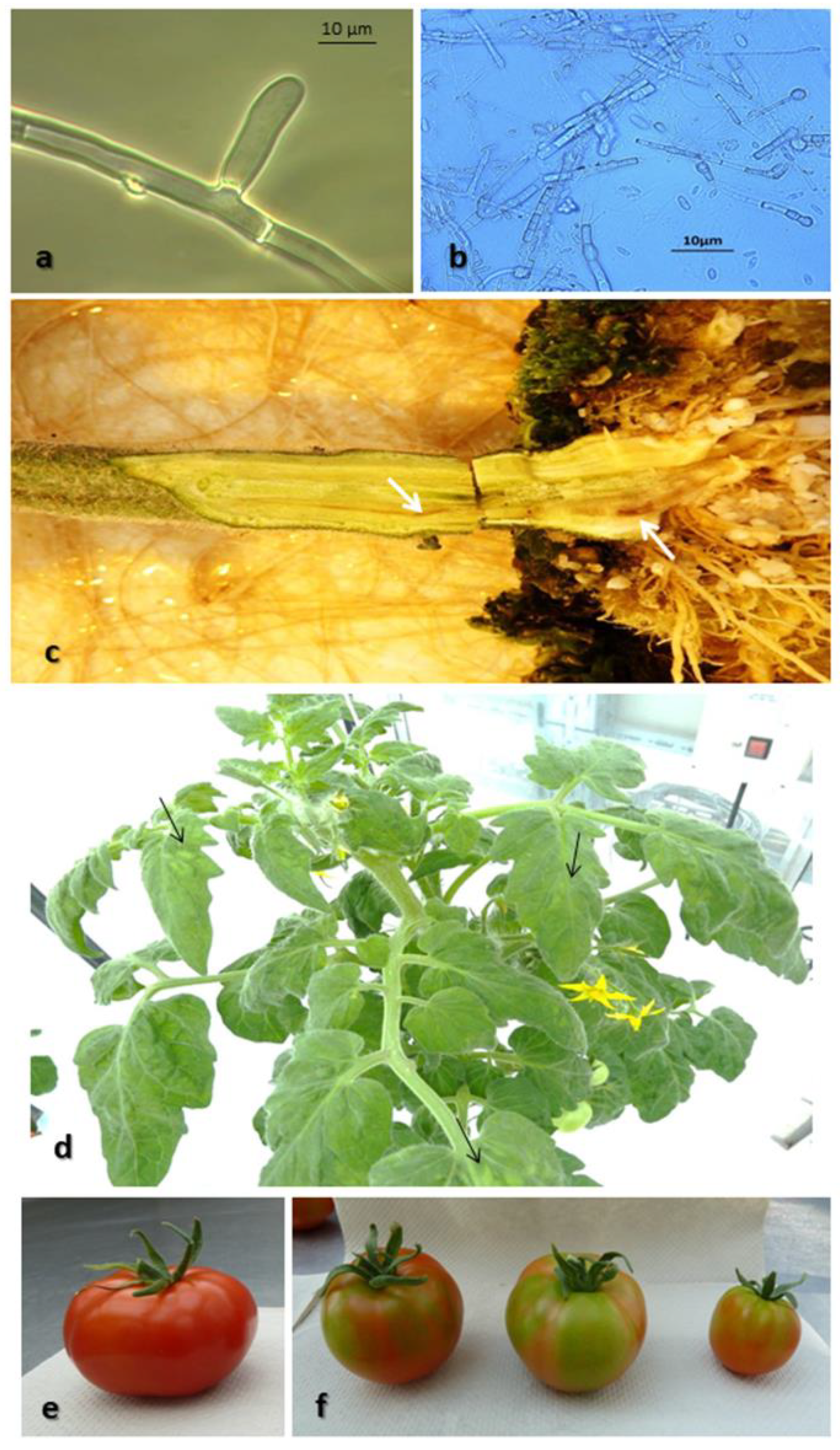
2.1. Plant Material, Plant Pathogens and Inoculum

Seeds of the small bush tomato cv. Hoffmanns Rentita were sown in perlite, grown in the greenhouse (22 °C, 16h photoperiod and relative humidity 30–60%), and transferred to rockwool cubes (10 × 10 × 7 cm3) 15 days after sowing (das).
The culture of conidia and mycelial fragments of F. oxysporum f. sp. lycopersicum (DSM-62059, DSMZ, 38124 Braunschweig, Germany) was carried out using “Spezieller nährstoffarmer Agrar” (SNA) [43] at 22 °C with an 8 h light: dark regime, for 10 days. Following the flooding of the plates with high-purity water, a sterile rod was used to gently scrape the surface to detach spores. The resulting spore suspension was then cultured further in liquid medium at 30 °C for 10 days, with continuous shaking at 120 rpm. Large mycelial fragments were filtered from the spore suspension through two layers of cheesecloth. R. solani PM-5 (collection of the Division Phytomedicine, Humboldt-Universität zu Berlin, 14195 Berlin, Germany) was cultured on “Potato Dextrose Agar” (PDA) at 22 °C with an 8 h light: dark regime for 12 days. To produce an inoculum, colonized PDA plugs (5 × 5 mm2) were excised and added to 100 mL of Potato dextrose broth (PDB). The broth was incubated at 30 °C under darkness for 12 days on an orbital shaker. Mycelium was collected and homogenized in a blender (Clatronic, model SM2452) for 30 s with high-purity water. A hemocytometer was used to quantify all fungal propagule suspensions, which were then diluted with sterile deionized water to obtain 106 propagules/mL prior to the chlorine treatment. Propagules viability was monitored on PDA by determining the colony forming units (CFU)/mL.
Tomato plants were infected with F. oxysporum f. sp. lycopersicum and R. solani by immersion of the roots in the respective fungal solution (1 × 10⁶ propagules/mL) for 2 min prior transfer to rockwool cubes. Infection was verified microscopically 2 weeks later by evaluating root tissue.
Virus-infected tomato plants were achieved by mechanical inoculation of tomato seedlings at the two-leaf stage 30 das with PepMV isolate PV-0554 (DSMZ, Braunschweig, Germany) or TMV isolate PV-1701 (DSMZ, 38124 Braunschweig, Germany), respectively. Infection was verified 2 weeks later by DAS-ELISA.

2.2. Detection of Plant Pathogens in Plant Material

An infection with F. oxysporum and/or R. solani was determined using 24 small root sections approx. 2 mm in length, of each tomato plant. Twelve sections each were placed on SNA [43], and PDA as previously described. To facilitate microscopic evaluation, antibiotics (100 mg Penicillin G, 10 mg Chlortetracycline and 50 mg Streptomycin sulphate per litre of nutrient media) were added to PDA. Surface disinfection was also conducted using 2% commercial bleach solution (6.15% NaOCl) for 2 min. Plates were incubated at 22 °C under an 8 h light: a dark regime for 10 days. Morphological characteristics were used to identify F. oxysporum and R. solani isolates under the microscope. Stem sections of each tomato plant were tested at the end of the in vivo trial in a similar manner. All trials were carried out with three replicates.
DAS-ELISA to detect PepMV and TMV was performed using commercially available assays (RT-1022 and RT-1065, respectively) according to the suppliers’ instructions (DSMZ, 38124 Braunschweig, Germany). Each sample was tested with at least two replicates. The optical density (OD) of the samples at 405 nm was rated after 60 and 120 min substrate incubation. The cut-off value was defined as three times the mean value of three homogenates of different healthy (negative) samples. All samples with values above the cut-off were regarded as PepMV and TMV positive, respectively.

2.3. Detection of Plant Pathogens in Nutrient Solution

Both treated and untreated samples with fertigation solution were obtained every week to determine whether contamination with fungal and viral pathogens had occurred.
The fungal pathogens F. oxysporum and R. solani were measured from 100 mL samples. A haemocytometer was used to count characteristic conidia using a microscope for 20 subsamples (each 10 µL). Subsequently, ten 2 mL subsamples were transferred to Petri dishes with SNA and PDA (see Section 2.1), incubated at 22 °C under an 8 h light: a dark regime for 10 days and evaluated by counting CFU.
Detection of the viral pathogens PepMV and TMV in fertigation solution requires concentration of the viruses prior to testing by ELISA [44]. Therefore, samples of 10 l each were concentrated by tangential flow filtration (TFF) with subsequent further concentration of the retentate using ultracentrifugation, as described by Bandte et al. [45]. The pellets were re-suspended and pooled in 300 μL high-purity water and finally tested by ELISA for plant material (see Section 2.1).

2.4. Electrolytic Disinfection of Fertigation Solution

A single chamber brine electrolysis plant was used to produce hypochlorite on-site (nt-BlueBox mini; newtec Umwelttechnik GmbH; 13507 Berlin, Germany), as previously described by Schuch et al. [46]. Chlorine (Cl2) was formed with a 10 A direct current and a 13 V voltage applied with titanium electrodes to a brine solution containing potassium chloride (KCl) and fresh water. In turn, Cl2 was disproportionated to hypochlorous acid (HClO) and Cl in the presence of hydroxyl ions (OH) in an aqueous solution. The device produced a potassium hypochlorite (KClO) solution containing 36.6 mg of free chlorine/L. A manually handheld apparatus (Pocket Colorimeter II, Hach Lange GmbH, 40549 Düsseldorf, Germany) was used to check the free chlorine content in this electrolytically generated stock solution and in the working solutions, as per the manufacturer’s instructions.
The stock solution was automatically injected into the recirculating fertigation solution using a dosing system (Dosatronic GmbH, 88213 Ravensburg, Germany), as described by Rodriguez et al. [47]. The target concentration to be set by the dosing system and maintained for a period of 60 min weekly was 0.5 mg free chlorine per L.

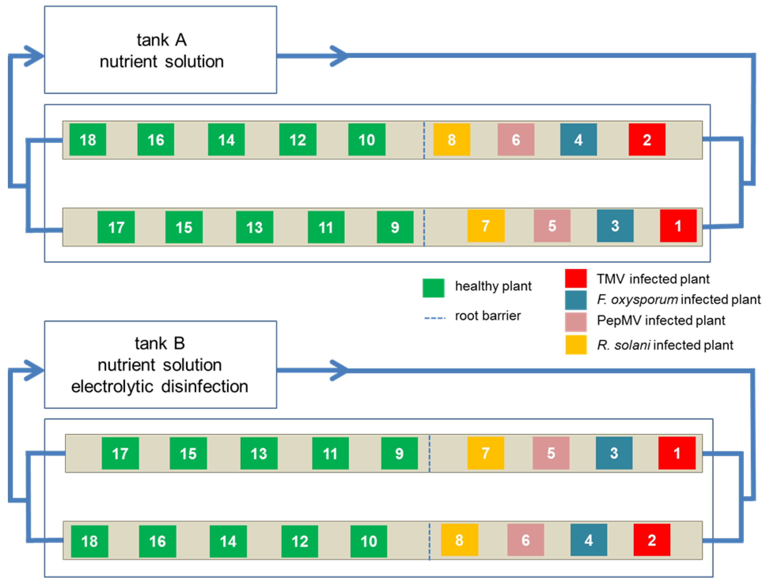
2.5. Experimental Design

The arrangement of the experimental plants to evaluate the efficacy of the fertigation solution treatment is shown schematically in Figure 1. In each set-up, 18 plants distributed on two channels (6 m × 0.25 m × 0.10 m), with an inclination of 1% and fed with fertigation solution via a storage tank of 500 L. The flow rate of the fertigation solution through the channels was continuously 400 L h−1. One storage tank each was subjected to the disinfection treatment; the other tank contained only fertigation solution and served as the untreated control. An additional pump per tank with a flow rate of 400 L h−1 was installed to generate turbulence and homogenize the solution within the respective system.
Pruning and harvesting was carried out using disposable rubber gloves that were changed after handling each plant in order to prevent mechanical spread of the pathogens. A root barrier (1626 mesh/cm2) prevented root contact between healthy (test) and infected (control) plants. The 16-week cultivation period consisted of the following commercial practices: 22 °C, 16 h photoperiod and relative humidity 30–60%. Tap water and a stock solution of nutrients (ammonium nitrate 0.4 mmol/L, potassium nitrate 3.3 mmol/L, monopotassium phosphate 0.4 mmol/l, magnesium sulfate 2.6 mmol/L, calcium nitrate 1.7 mmol/L, and 10 mg/L Fe EDTA 13%). It also included micronutrients, according to Göhler and Molitor [48]. An adjusted pH value of 6.0 and electrical conductivity (EC) value of 1.8 were used. Weekly measurements of the composition and pH of the fertigation solution were carried out in the laboratory and corrected as needed. The experiment was carried out twice.

2.6. Data Collection and Analysis

The efficacy of the sanitation treatment on the dispersal of fungal and viral plant pathogens was measured as well as the effect on yield and fruit quality of tomato.
Tomato plants were evaluated individually on a weekly basis for both fungal and viral infections by isolation from plant tissue or DAS-ELISA, respectively. Harvesting of tomato fruits at skin color gauge 9 [49] was carried out weekly, starting in the eighth week of cultivation. Thereby, the number and fresh weight of the fruits were determined per plant. Small fruits with a diameter below 40 mm, or with severe discoloration or growth cracks were defined unmarketable. Data on tomato fruits were subjected to two-way analysis of variance. Means were compared by Fisher’s F-test followed by Tukey’s t-test at significance level α = 0.05. Significant differences are represented by different letters.

3. Results

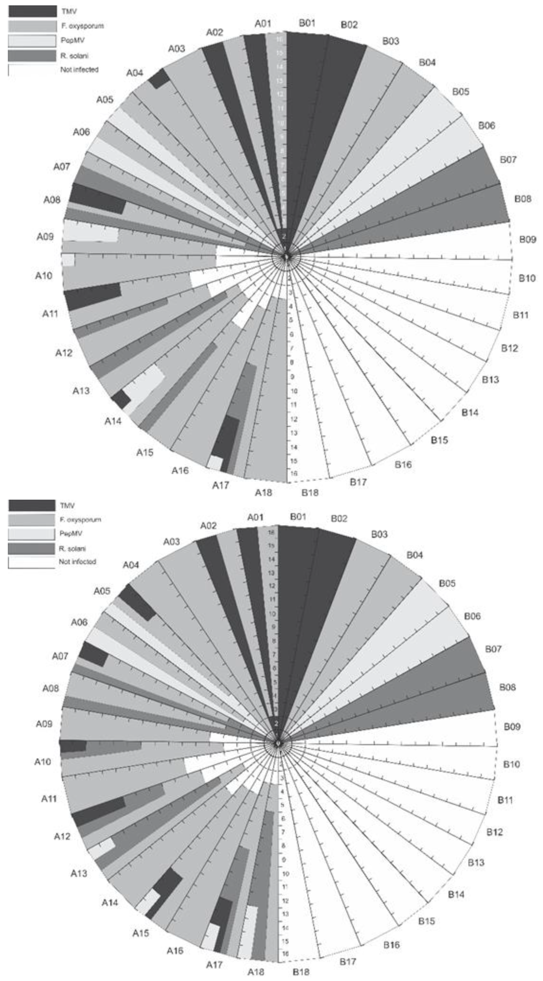
3.1. Detection of Plant Pathogens in Fertigation Solution

Fungal and viral pathogens were only detected in untreated fertigation solution. Thereby, F. oxysporum and R. solani could already be isolated in the fifth and tenth week after experimental set up, respectively. On average, four CFU were formed corresponding to four propagules in 20 mL fertigation solution. Propagules were first observed by light microscopy, microconidia in the case of F. oxysporum and mycellium for R. solani. They were quantified 14 weeks after experimental set-up by using a haemocytometer. TMV and PepMV were first detected by DAS-ELISA 12 weeks after starting the experiment.

3.2. Effect of Fertigation Solution Treatment on Pathogen Dispersal

The fertigation solution of both tanks was continuously contaminated naturally by the infected control plants (source plants). As expected, treatment with the disinfectant did not cure those plants. They remained infected throughout the experiment. However, the treatment led to sanitation as the dispersal of both fungal and viral pathogens was inhibited in all experimental runs. None of the tomato plants supplied with fertigation solution treated weekly with 0.5 mg free chlorine/L for 60 min (pH 6.0 ± 0.3 and ORP 780 ± 31 mV) were infected with F. oxysporum, R. solani, TMV or PepMV (Figure 2). By contrast, the untreated fertigation solution enabled the spread of the fungal and viral pathogens and the infection of previously healthy test plants. Thus, all 10 tomato plants (A09 to A18) were infected with F. oxysporum in both the first and second survey. In each survey, four out of ten formerly healthy tomato plants became infected with PepMV (A09, A10, A14, A17 and A13, A15, A17, A18, respectively). Four out of ten (A12, A13, A15 and A17) in the first survey and five out of ten (A10, A12, A13, A17 and A18) previously healthy tomato plants in the second survey were infected with R. solani. Additionally, three out of ten (A11, A14 and A17) tomato plants in the first trial and four out of ten (A10, A12, A15 and A17) tomato plants in the second trial were also infected with TMV. The first newly infected plants were detected 3 and 4 weeks after experimental set-up, respectively. The infection of these plants occurred by chance and was not determined in relation to the position of the infected source plants. For example, in the second survey, plant A11 was infected exclusively with F. oxysporum, but remained free of R. solani, TMV and PepMV, despite being closest to the plants infected with the latter three pathogens.
With the exception of one plant (A03) in the first survey and two plants (A03 and A04) in the second one, infected source plants cultivated without sanitizing treatment acquired mixed infections with at least one other pathogen (Figure 2). The weekly disinfection treatment prevented further infection of the infected source plants with the other pathogens. None of those plants contracted any other of the four pathogens.

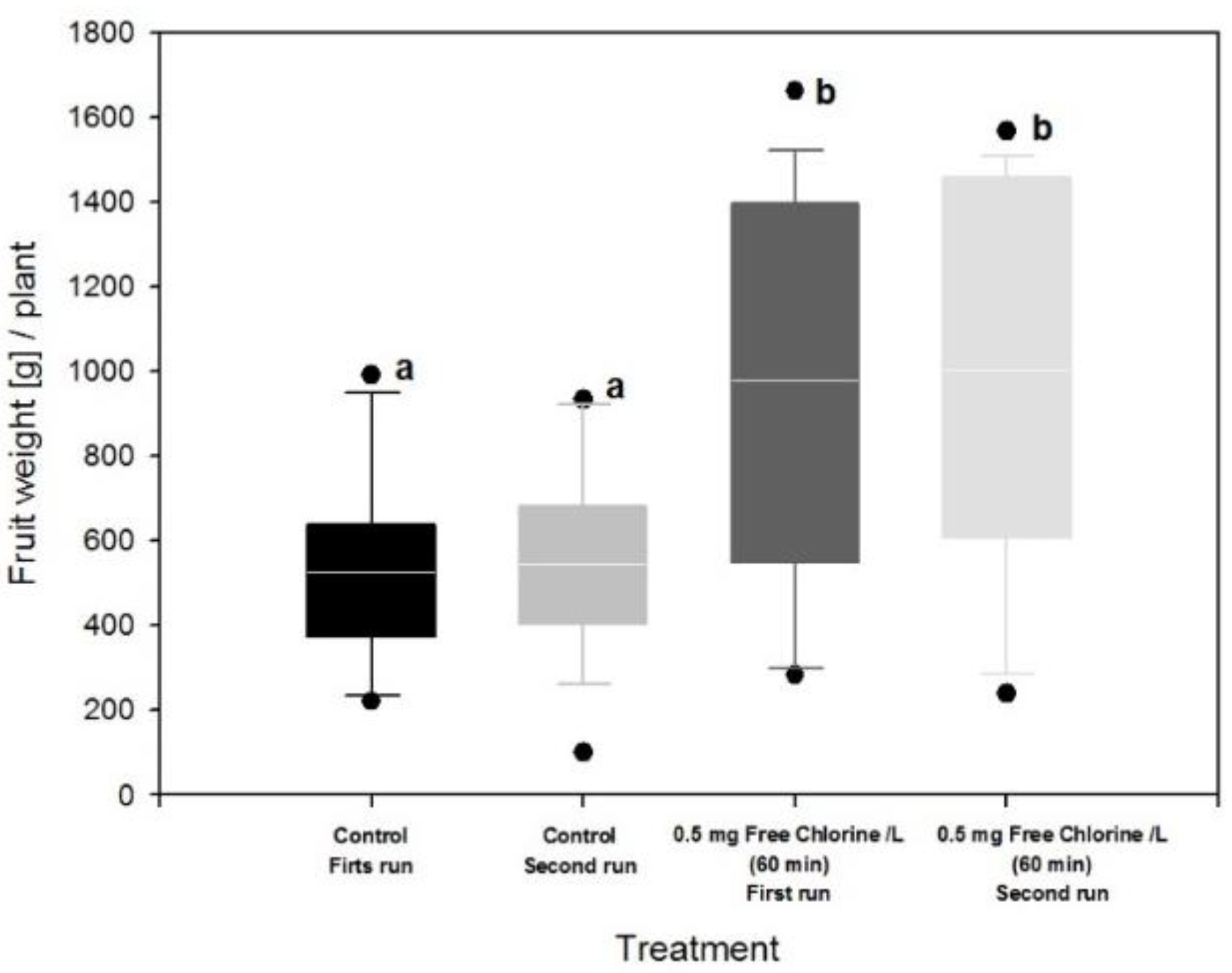
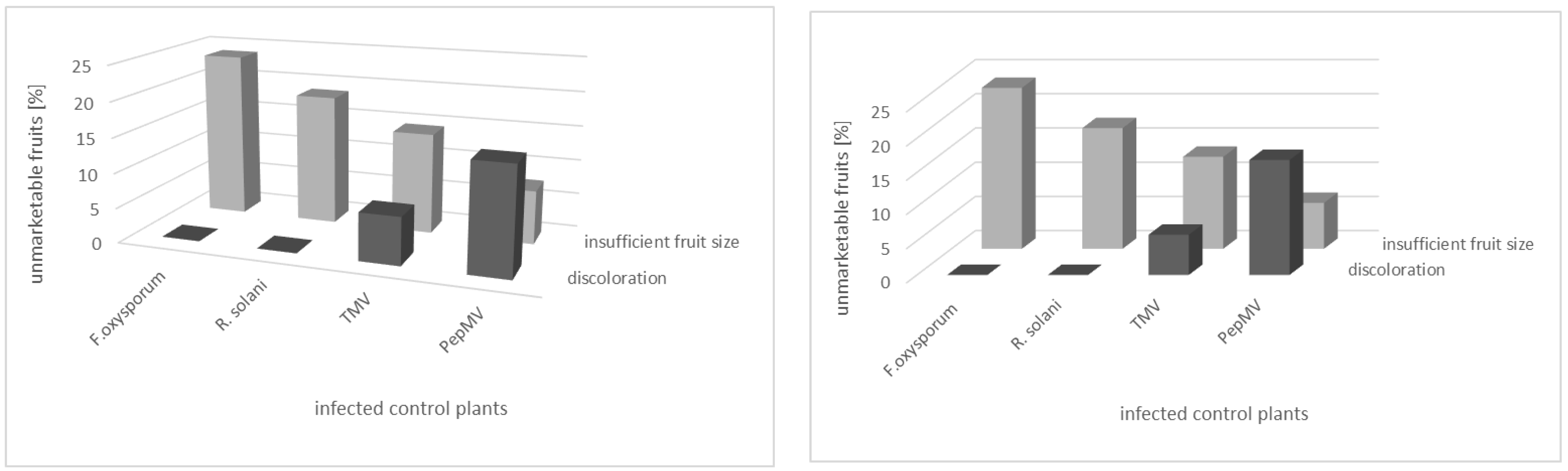
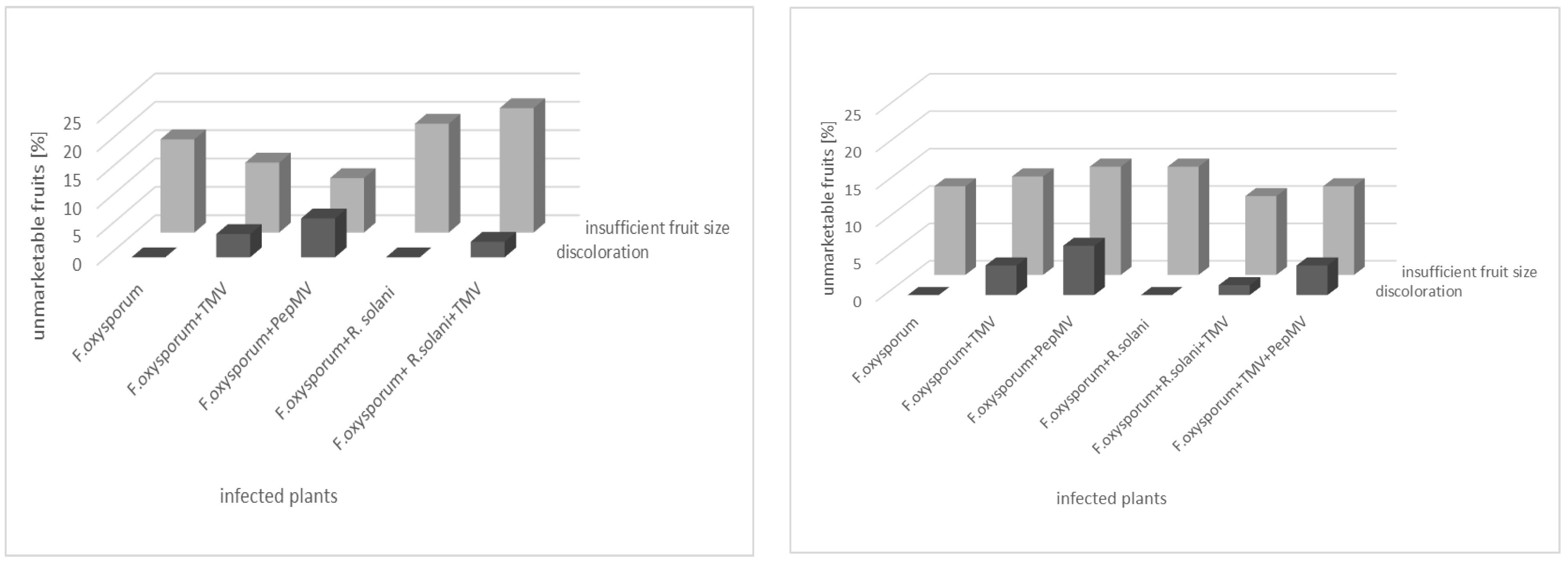
3.3. Effect of Fertigation Solution Treatment on Tomato Plant and Fruit

Infected tomato plants, both source as well as test plants, developed characteristic symptoms of the particular disease (Figure 3). In the case of F. oxysporum, plants showed a delay in growth, asymmetric chlorosis of leaflets and wilting. Leaf symptoms progressed from the base to apex; the vascular tissue of root and stem exhibited a reddish color. Plants infected with R. solani were characterized by a reduced size and roots that became necrotic and rotten. Virus infection caused symptoms on tomato plants only in the case of TMV: delay in growth, leaf curling, mosaic of young and necrosis of older leaves.
Treatment of the nutrient solution with the disinfectant did not cause leaf damage in any of the plants, nor did the plants show differences in growth compared to the plants supplied with untreated fertigation solution. In both trials, the tomato plants supplied with sanitized fertigation solution indicated a significantly higher yield per plant (Figure 4). However, when fungal and viral pathogens leading to disease were considered, differences in yield were observed. The plants supplied with untreated fertigation solution yielded lower than the corresponding plants fed with sanitized fertigation solution, regardless of whether they were the control plants or the test plants (Table 1). Thus, the yield of the test plants was about twice as high than that of the infected control plants and slightly, but not significantly, higher than that of the untreated fertigation solution. Potassium hypochlorite treatment significantly increased the number of fruits per plant in both surveys (Table 2). However, for plants grown without sanitizing treatment, the proportion of unmarketable fruit was high, about 90% in infected control plants and about 60% in test plants. The corresponding test plants supplied with treated fertigation solution yielded only about 5% of unmarketable tomatoes. Interestingly, fruits of plants infected by TMV or PepMV never reached the required quality for marketability.
The number of tomatoes not marketable due to color changes or insufficient fruit size was dependent on the infecting pathogen. The fruit size of all infected plants was reduced (Figure 5 and Figure 6). As is clearly evident from the single infected control plants, almost one-fourth and one-fifth of the unmarketable fruits came from F. oxysporum- and R. solani-infected plants, respectively. All of these fruits were below the minimum size and thus unmarketable (Figure 5). A singular fungal infection never led to market-relevant discoloration of fruits. However, about 15% of the fruits of PepMV-infected plants and about 6% of those of TMV-infected plants showed such severe discoloration. All mixed-infected plants yielded fruits of insufficient size, with the proportion of non-marketable fruits between 10 and 20% in each case (Figure 6). Discoloration of the fruits occurred again exclusively with mixed infections in which either the virus TMV or PepMV was involved. Mixed infections with the two fungal pathogens F. oxysporum and R. solani resulted exclusively in smaller fruits.

4. Discussion

Infected plants may harbor and disseminate large numbers of virulent pathogens into the drain, which are then delivered to water storage tanks and, when the water is recycled for irrigation, subsequently redistributed to susceptible crop plants. Different studies indicate that the water used in irrigation and as a component in horticultural fertigation solutions poses a potential risk in facilitating the dispersal of plant pathogens, particularly in nurseries and companies using recirculating systems [50,51,52,53].
The pathogens TMV, PepMV, F. oxysporum, and R. solani are four of the most important diseases in tomato production. All of these pathogens can be dispersed with the fertigation solution. Dissemination of fungal pathogens via fertigation solution is usually faster than that of viruses. Thus, infection of tomato plants grown in the same cycle of fertilizer solution occurred after 3 and 5 weeks for F. oxysporum and R. solani, respectively [47], but only after 10 weeks for PepMV [45]. F. oxysporum is able to invade both the (i) young apical tissue and (ii) mature tissue of the tomato root, creating entry points for other pathogens: for example, for plant viruses that cannot actively enter the plant themselves, but rely on wounds or the help of vectors. Interestingly, infection of tomato plants infected with F. oxysporum via the fertigation solution and subsequently infected with PepMV occurred at 12 weeks at the earliest, which was 2 weeks later than single infection. Likewise, pre-infection of tomato with the pathogen Pythium aphanidermatum significantly delayed infection with PepMV via fertigation solution [32]. Initial fungal infection may lead to reduced vitality making plants less susceptible to viral pathogens.
We evaluated the use of KCLO as a disinfection treatment method in recirculating fertigation solutions for its efficacy in the inactivation of the tomato pathogens F. oxysporum, R. solani, TMV and PepMV. The experiments simulated production under semi-commercial conditions. Here, the recirculating fertigation solution was gradually contaminated by tomato plants that had previously been infected with the selected pathogens. Not only does the transmission of plant pathogens occurs naturally in the field or bed by water but also artificially, as has been demonstrated in many other studies of disinfection strategies in agriculture [54,55,56,57], whereby the fertigation solution or irrigation water is contaminated by adding a default concentration of propagules. Although a higher pathogen concentration is usually employed, it does not guarantee transmission of the pathogen and spread of the disease in the crop. For example, Jenkins and Averre [58] failed to infect tomato plants by using a recirculating solution contaminated artificially with a solution containing propagules of Erwinia spp. and F. oxysporum sp. radicis-lycopersici. Spores of F. oxysporurn f. sp. cyclaminis introduced into the fertigation solution settled down on the bottom of the containers and were not transported during irrigation cycles [59]. Therefore, we installed an additional pump in the storage tank to ensure homogeneous distribution of fungal propagules.
The efficacy of the disinfectant KCLO was evaluated using a naturally infected fertigation solution. In this way, it can be practically tested whether concentration and exposure time are sufficient to safely inactivate the various pathogens in the recirculating fertigation solution and to prevent further spread in the crop. Studies by Cayanan et al. [55] show, as might be expected, a dependence of the required dosage on the pathogen species. For this reason, we included different pathogens from different taxonomic groups and with different physical and biological properties in the studies, and we also considered mixed infections. Especially in the case of spore-forming fungi, an accumulation of a few surviving reproductive units must be prevented in order to prevent infection via fertigation solution and disease of the plants in the medium term. In contrast to physical methods, chlorination has a certain residual disinfectant activity. This can counteract reinfection by plants remaining in the stand and, in the case of discontinuous disinfection, increase the frequency between treatments. However, chlorine also produces long-lived by-products such as chloramines, which have potential environmental and human health impacts [7]. Therefore, the respective dosage must be adjusted so that the permissible limit value of undesirable byproducts is not exceeded in the harvested products.
Most studies report pathogen mortality as a function of chlorine concentration in in vitro experiments using NaOCl or Cl2 without considering the phytotoxic effects caused by these concentrations in plants. Although F. oxysporum and R. solani can be killed at 14 mg L−1 for 6 min and 12 mg L−1/L for 10 min contact time, respectively, phytotoxicity occurs [55]. Lévesque et al. [2] demonstrated the inactivation efficacy of electrochemical disinfection in hydroponic irrigation water contaminated with mycelial suspensions of R. solani. Thereby, 1.14 mA cm−2 for a contact time of 6 min in the presence of fertilizer resulted in a notable but not complete inactivation of R. solani. A further increase in current density up to the highest achievable (9.09 mA cm−2) did improve inactivation, but did not lead to complete inactivation. For this, 20 mg L−1 chloride needs to be present in the fertigation solution [60].
In our studies, electrolytically derived potassium hypochlorite in a weekly application prevented the spread of pathogens and infection of plants. However, this did not lead to phytotoxic effects such as chlorosis, deformation, leaf drop, leaf necrosis and discoloration or yield depression, in contrast to Raudales et al. [61] and van Haute et al. [62]. The results obtained on simultaneous contamination with two fungal and two viral pathogens confirm the studies conducted by Rodríguez et al. [47] with the same two fungal pathogens. For complete inactivation of only PepMV a weekly application of 0.2 mg free chlorine/L for 1 h is sufficient [45]. Our previous experiments with the very stable TMV at this dose were not successful (data not shown). Surprisingly, 0.5 mg free chlorine/L supplied for 1 h weekly is sufficient for complete inactivation, as is the case for the two fungal pathogens F. oxysporum and R. solani.
Electrolytic disinfection of water is mostly used with sodium chloride as salt. The use of potassium chloride has the advantage that potassium is a macro element that is needed by plants in large quantities. Sodium, on the other hand, accumulates on the surface of the root system and in the irrigation system and can form crystals. This increases the electrical conductivity EC of the solution and can cause a phytotoxic reaction. Magnesium chloride-based disinfectant solutions are less compatible with plants than those made from potassium and are therefore impractical [63].
Chlorate and perchlorate residues were found in various vegetables, such as lettuce, spinach, tomato and cucumber, and herbs [64]. Such contamination can occur at various stages of the food production chain: already during cultivation, harvesting or processing, for example through the use of chlorinated water or chlorinated disinfectants. Ref. [65] showed a highly significant correlation between the application of hypochlorite and chlorate accumulation in tomatoes. Fortunately, the accumulation of both chlorate and perchlorate was not found in tomato fruits following electrolytic disinfection of fertigation solution. The maxim must consequently be: as little as possible, as much as necessary. Ref. [46] showed that ammonia rapidly binds the free chlorine and thus markedly increases the usage of disinfectant or chlorate input up to the chlorine loss limit. These changes in chemical processes in irrigation and fertigation solutions (e.g., chlorine losses) can be monitored and considered with the use of a chlorine sensor, administered as needed; over- and under-dosing are thus avoided. With the preferred use of nitrate fertilizers, the amount of disinfectant can be further minimized.
The use of disinfection technology with electrolytically produced KLCO and its automatized injection into the fertigation solution in horticulture is innovative. However, it is essential to record different variables, such as ORP, pH, EC, temperature, to (i) correctly inject the required amount of chlorine, (ii) achieve the desired disinfection effect and (iii) not pollute the environment unnecessarily. If this is considered, the threshold for the risk of accumulation of chlorates and perchlorates in vegetables and fruits (0.05 mg/kg) established by the European Food Safety Authority (EFSA) in 2020 should not be reached, either [66,67].

5. Conclusions

The potassium hypochlorite (KCLO)-based disinfection system provided efficient inactivation of pathogens (F. oxysporum, R. solani, PepMV and TMV) in fertigation solutions under semi-commercial tomato production conditions at a weekly dose of 0.5 mg free chlorine/L for 60 min.
PH value and the oxidation-reduction potential (ORP) should be considered as an active tool for monitoring the disinfection process and should remain within the thresholds of pH 6.0 ± 0.3 or ORP 780 ± 31 mV during the treatment with the disinfectant to achieve the highest disinfection effect. In the fertigation solution, the ORP depends on the concentration ratio between the disinfectant and the organic impurities. When the disinfectant is used up and not sufficiently replenished, the ORP shifts to lower values, while high values occur when the disinfectant is overdosed or there is little organic matter in the nutrient solution. Thus, the sensor-based dosing allows overdosing problems that can lead to phytotoxicity and the accumulation of chlorates and perchlorates in plant tissues and reductions in the quality of fruit for marketing to be avoided.
Finally, electrolytic disinfection with KCLO is an innovative, reliable and affordable technology that allows high quality vegetables to be produced and reduces economic losses caused by plant pathogens in hydroponic systems with recirculation of the fertigation solution. Even a few infected plants provide enough inoculum for pathogens to replicate and spread through the crop. Regular treatment of irrigation water is therefore essential to protect farmers from diversifiable risks and resulting economic losses, while meeting the requirement and demand for water and nutrient conservation.

Author Contributions

Conceptualization M.B.; methodology M.B. and C.B.; validation M.B.; formal analysis, M.H.R.; investigation M.H.R. and M.B.; resources, U.S. and C.B.; data curation, M.H.R.; writing—original draft preparation M.H.R.; writing—review and editing, M.H.R., M.B., C.B. and U.S.; visualization M.H.R. and M.B.; supervision M.B. and C.B.; project administration, U.S. and C.B.; funding acquisition, U.S. and C.B. All authors have read and agreed to the published version of the manuscript.

Funding

This research was funded by the German Federal Ministry of Food and Agriculture (BMEL). On behalf of the BMEL, The Federal Office of Agriculture and Food (BLE) managed the project technically and in terms of organization (project no. 28RZ5IP035).

Institutional Review Board Statement

Not applicable.

Informed Consent Statement

Not applicable.

Data Availability Statement

Not applicable.

Acknowledgments

We thank Yuan Gao (newtec Umwelttechnik GmbH, 13507 Berlin, Germany) for providing the brine electrolysis plant and Thomas Gaskin for English proofreading.

Conflicts of Interest

The authors declare no conflict of interest. The funders had no role in the design of the study; in the collection, analyses or interpretation of data; in the writing of the manuscript or in the decision to publish the results.

References

  1. FAO. The State of the World’s Land and Water Resources for Food and Agriculture—Systems at Breaking Point; Earthscan: London, UK, 2021; p. 82. [Google Scholar]
  2. Lévesque, S.; Graham, T.; Bejan, D.; Lawson, J.; Zhang, P.; Dixon, M. An electrochemical advanced oxidation process (EAOP) for the inactivation of Rhizoctonia solani in fertigation solutions. Can. J. Plant Sci. 2020, 100, 415–424. [Google Scholar] [CrossRef] [Green Version]
  3. FAO. Towards a Water and Food Secure Future; FAO & WWC: Marseille, France; Rome, Italy, 2015; p. 76. [Google Scholar]
  4. FAO. Water. 2022. Available online: https://www.fao.org/water/en/ (accessed on 5 January 2022).
  5. Stanghellini, M.; Rasmussen, S.; Kim, D.; Rorabaugh, P. Efficacy of nonionic surfactants in the control of zoospore spread of Pythium aphanidermatum in a recirculating hydroponic system. Plant Dis. 1996, 80, 422–428. [Google Scholar] [CrossRef]
  6. Schnitzler, W.H. Pest and disease management of soilless culture. Acta Hortic. 2004, 648, 191–203. [Google Scholar] [CrossRef]
  7. Stewart-Wade, S.M. Plant pathogens in recycled irrigation water in commercial plant nurseries and greenhouses: Their detection and management. Irrig. Sci. 2011, 29, 267–297. [Google Scholar] [CrossRef]
  8. Hong, C.; Richardson, P.; Kong, P.; Bush, E. Efficacy of chlorine on multiple species of Phytophthora in recycled nursery irrigation water. Plant Dis. 2003, 87, 1183–1189. [Google Scholar] [CrossRef]
  9. Hong, C.; Moorman, G. Plant pathogens in irrigation water: Challenges and opportunities. Crit. Rev. Plant Sci. 2005, 24, 189–208. [Google Scholar] [CrossRef]
  10. Mehle, N.; Ravnikar, M. Plant viruses in aqueous environment–survival, water mediated transmission and detection. Water Res. 2012, 46, 4902–4917. [Google Scholar] [CrossRef]
  11. FAO. Crops and Livestock Products; The Food and Agriculture Organization FAOstat: Roma, Italy, 2022. [Google Scholar]
  12. Büttner, C.; Koenig, R. Plant viruses in irrigation water. In Biology, Detection, and Management of Plant Pathogens in Irrigation Wate; Hong, C., Moorman, G.W., Wohanka, W., Büttner, C., Eds.; APS Press, The American Phytopathological Society: St. Paul, MN, USA, 2014; pp. 97–110. [Google Scholar]
  13. Wick, R.L. Fungal plant pathogens in irrigation water. In Biology, Detection and Management of Plant Pathogens in Irrigation Water; Hong, C., Moorman, G.W., Wohanka, W., Büttner, C., Eds.; APS Press, The American Phytopathological Society: St. Paul, MN, USA, 2014; pp. 77–82. [Google Scholar]
  14. Walker, J.C. Fusarium Wilt of Tomato; APS, Ed.; APS Press, The American Phytopathological Society: St. Paul, MN, USA, 1971; Volume 6, p. 56. [Google Scholar]
  15. Reis, A.; Costa, H.; Boiteux, L.S.; Lopes, C.A. First report of Fusarium oxysporum f. sp. lycopersici race 3 on tomato in Brazil. Fitopatol. Bras. 2005, 30, 426–428. [Google Scholar] [CrossRef] [Green Version]
  16. Đorđević, M.; Vatchev, T.; Girek, Z.; Šević, M.; Zečević, B.; Zdravković, J.; Ivanović, M. Reaction of different tomato cultivars toward race 1 of Fusarium oxysporum f. sp. lycopersici. Genetika 2012, 44, 109–118. [Google Scholar]
  17. Nelson, P.E. Life cycle and epidemiology of Fusarium oxysporum. In Fungal Wilt Diseases of Plants; Mace, M., Bell, A.A., Beckman, C.H., Eds.; Academic Press: New York, NY, USA, 1981; pp. 51–80. [Google Scholar] [CrossRef]
  18. Jarvis, W.; Shoemaker, R. Taxonomic status of Fusarium oxysporum causing foot and root rot of tomato. Phytopathology 1978, 68, 1679–1680. [Google Scholar] [CrossRef]
  19. Kapoor, I. Fungi involved in tomato wilt syndrome in Delhi, Maharashtra and Tamil Nadu. Indian Phytopathol. 1988, 41, 208–213. [Google Scholar]
  20. Gupta, S.K.; Thind, T. Disease Problems in Vegetable Production; Scientific Publishers Journals Department: Jodhpur, India, 2018; p. 576. [Google Scholar]
  21. Domsch, K.H.; Gams, W.; Anderson, T.H. Compendium of Soil Fungi; Academic Press: London, UK; New York, NY, USA; Toronto, ON, Canada; Sydney, Australia; San Francisco, CA, USA, 1980; p. 859. [Google Scholar]
  22. Ogoshi, A. Introduction—the genus Rhizoctonia. In Rhizoctonia species: Taxonomy, Molecular Biology, Ecology, Pathology and Disease Control; Sneh, B., Jabaji-Hare, S., Neate, S., Dijst, G., Eds.; Kluwer Academic Publishers: Dordrecht, The Netherlands, 1996; pp. 1–9. [Google Scholar] [CrossRef]
  23. Sneh, B.; Burpee, L.; Ogoshi, A. Identification of Rhizoctonia Species; APS Press, The American Phytopathological Society: St. Paul, MN, USA, 1991; p. 133. [Google Scholar]
  24. Paulitz, T.C.; Bélanger, R.R. Biological control in greenhouse systems. Annu. Rev. Phytopathol. 2001, 39, 103–133. [Google Scholar] [CrossRef]
  25. González García, M. Reseña de “Aspectos de sistemática y biologíca del complejo Rhizoctonia”. Fitosanidad 2008, 12, 147–159. [Google Scholar]
  26. Jones, R.A.C.; Koenig, R.; Lesemann, D. Pepino mosaic virus, a new potexvirus from pepino (Solanum muricatum). Ann. Appl. Biol. 1980, 94, 61–68. [Google Scholar] [CrossRef]
  27. Wright, D.; Mumford, R. Pepino mosaic Potexvirus (PepMV): First records in tomato in the United Kingdom. Plant Dis. Not. 1999, 89, 400. [Google Scholar]
  28. van der Vlugt, R.A.; Stijger, C.I.; Verhoeven, J.T.J.; Lesemann, D.E. First Report of Pepino mosaic virus on Tomato. Plant Dis. 2000, 84, 103. [Google Scholar] [CrossRef]
  29. Cordoba-Selles, M.d.C.; García-Rández, A.; Alfaro-Fernández, A.; Jordá-Gutiérrez, C. Seed transmission of Pepino mosaic virus and efficacy of tomato seed disinfection treatments. Plant Dis. 2007, 91, 1250–1254. [Google Scholar] [CrossRef] [Green Version]
  30. Lacasa, A.; Guerrero, M.; Hita, I.; Martínez, M.; Hernández, M. La diseminación del virus del mosaico del pepino dulce (Pepino mosaic Virus) en las labores de entutorado y desbrotado de las plantas de tomate. Bol. San. Veg. Plagas 2001, 21, 489–501. [Google Scholar]
  31. Spence, N.; Basham, J.; Mumford, R.; Hayman, G.; Edmondson, R.; Jones, D. Effect of Pepino mosaic virus on the yield and quality of glasshouse-grown tomatoes in the UK. Plant Pathol. 2006, 55, 595–606. [Google Scholar] [CrossRef]
  32. Schwarz, D.; Beuch, U.; Bandte, M.; Fakhro, A.; Büttner, C.; Obermeier, C. Spread and interaction of Pepino mosaic virus (PepMV) and Pythium aphanidermatum in a closed nutrient solution recirculation system: Effects on tomato growth and yield. Plant Pathol. 2010, 59, 443–452. [Google Scholar] [CrossRef]
  33. Jordá, C.; Pérez, A.L.; Martínez-Culebras, P.; Abad, P.; Lacasa, A.; Guerrero, M. First report of Pepino mosaic virus on tomato in Spain. Plant Dis. 2001, 85, 1292. [Google Scholar] [CrossRef]
  34. Hanssen, I.; Paeleman, A.; Vandewoestijne, E.; Van Bergen, L.; Bragard, C.; Lievens, B.; Vanachter, A.; Thomma, B. Pepino mosaic virus isolates and differential symptomatology in tomato. Plant Pathol. 2009, 58, 450–460. [Google Scholar] [CrossRef]
  35. Roggero, P.; Masenga, V.; Lenzi, R.; Coghe, F.; Ena, S.; Winter, S. First report of Pepino mosaic virus in tomato in Italy. Plant Pathol. 2001, 50, 798. [Google Scholar] [CrossRef]
  36. Hanssen, I.M.; Paeleman, A.; Wittemans, L.; Goen, K.; Lievens, B.; Bragard, C.; Vanachter, A.C.; Thomma, B.P. Genetic characterization of Pepino mosaic virus isolates from Belgian greenhouse tomatoes reveals genetic recombination. Eur. J. Plant Pathol. 2008, 121, 131–146. [Google Scholar] [CrossRef]
  37. Mayer, A. Concerning the mosaic disease of tobacco. In Phytopathological Classics; Johnson, J., Ed.; APS, The American Phytopathological Society: St. Paul, MN, USA, 1886; Volume 7, pp. 11–24. [Google Scholar]
  38. Jones, J.; Jones, J.; Stall, R.; Zitter, T. Compendium of Tomato Diseases, 1st ed.; APS, The American Phytopathological Society: St. Paul, MN, USA, 1991; p. 73. [Google Scholar]
  39. Scholthof, K. Tobacco mosaic virus. In The Plant Health Instructor; APS, The American Phytopathological Society: St. Paul, MN, USA, 2005. [Google Scholar] [CrossRef]
  40. Hong, C. Component analyses of irrigation water in plant disease epidemiology. In Biology, Detection and Management of Plant Pathogens in Irrigation Water; Hong, C., Moorman, G.W., Wohanka, W., Büttner, C., Eds.; APS Press, The American Phytopathological Society: St. Paul, MN, USA, 2014. [Google Scholar] [CrossRef]
  41. De Hayr, R.; Bodman, K.; Forsberg, L. Bromine and chlorine disinfestation of nursery water supplies. Comb. Proc. Int. Plant Propag. Soc. 1994, 44, 60–66. [Google Scholar]
  42. Fisher, P.R.; Huang, J.; Raudales, R.E.; Meador, D.P. Chlorine for irrigation water treatment. In Biology, Detection, and Management of Plant Pathogens in Irrigation Water; Hong, C., Moorman, G.W., Wohanka, W., Büttner, C., Eds.; APS Press, The American Phytopathological Society: St. Paul, MN, USA, 2014; pp. 235–249. [Google Scholar] [CrossRef]
  43. Nirenberg, H. Studies on the morphological and biological differentiation in the Fusarium-section Liseola. In Mitteilungen der Biologischen Bundeanstalt für Land-und Forstwirtschaft; P. Parey: Berlin, Germany, 1976; Volume 169, pp. 1–117. [Google Scholar]
  44. Büttner, C.; Bandte, M.; Pettitt, T.R. Filtration and centrifugation for detection of plant pathogens in irrigation water. In Biology, Detection and Management of Plant Pathogens in Irrigation Water; Hong, C., Moorman, G.W., Wohanka, W., Büttner, C., Eds.; APS Press, The American Phytopathological Society: St. Paul, MN, USA, 2014; pp. 139–148. [Google Scholar] [CrossRef]
  45. Bandte, M.; Rodriguez, M.H.; Schuch, I.; Schmidt, U.; Buettner, C. Plant viruses in irrigation water: Reduced dispersal of viruses using sensor-based disinfection. Irrig. Sci. 2016, 34, 221–229. [Google Scholar] [CrossRef]
  46. Schuch, I.; Dannehl, D.; Bandte, M.; Suhl, J.; Gao, Y.; Schmidt, U. Minimising chlorate in the electrolytic disinfection of irrigation water. Landtechnik 2016, 71, 25–33. [Google Scholar]
  47. Rodriguez, M.H.; Bandte, M.; Gaskin, T.; Fischer, G.; Büttner, C. Efficacy of electrolytically-derived disinfectant against dispersal of Fusarium oxysporum and Rhizoctonia solani in hydroponic tomatoes. Sci. Hortic. 2018, 234, 116–125. [Google Scholar] [CrossRef]
  48. Göhler, F.; Molitor, H.D. Erdelose Kulturverfahren im Gartenbau; Eugen Ulmer Stuttgart: Stuttgart, Germany, 2002; p. 268. [Google Scholar]
  49. OECD. Tomatoes. In International Standards for Fruit and Vegetables; OECD Publishing: Paris, France, 2019. [Google Scholar] [CrossRef]
  50. Pettitt, T.R. Irrigation water and the health of nursery crops. In Biology, Detection, and Management of Plant Pathogens in Irrigation Water; Hong, C., Moorman, G.W., Wohanka, W., Büttner, C., Eds.; APS Press, The American Phytopathological Society: St. Paul, MN, USA, 2014; pp. 13–22. [Google Scholar] [CrossRef]
  51. Moorman, G.W.; Gevens, A.J.; Granke, L.L.; Hausbeck, M.K.; Hendricks, K.; Roberts, P.D.; Pettitt, T.R. Sources and distribution systems of irrigation water and their potential risks for crop health. In Biology, Detection, and Management of Plant Pathogens in Irrigation Water; Hong, C., Moorman, G.W., Wohanka, W., Büttner, C., Eds.; APS Press, The American Phytopathological Society: St. Paul, MN, USA, 2014; pp. 3–12. [Google Scholar] [CrossRef]
  52. Moorman, G.W. Irrigation water and the health of greenhouse crops. In Biology, Detection, and Management of Plant Pathogens in Irrigation Water; Hong, C., Moorman, G.W., Wohanka, W., Büttner, C., Eds.; APS Press, The American Phytopathological Society: St. Paul, MN, USA, 2014; pp. 23–29. [Google Scholar] [CrossRef]
  53. Marčiulynas, A.; Marčiulynienė, D.; Lynikienė, J.; Gedminas, A.; Vaičiukynė, M.; Menkis, A. Fungi and oomycetes in the irrigation water of forest nurseries. Forests 2020, 11, 459. [Google Scholar] [CrossRef] [Green Version]
  54. Younis, B.A.; Mahoney, L.; Schweigkofler, W.; Suslow, K. Inactivation of plant pathogens in irrigation water runoff using a novel UV disinfection system. Eur. J. Plant Pathol. 2019, 153, 907–914. [Google Scholar] [CrossRef] [Green Version]
  55. Cayanan, D.F.; Zhang, P.; Liu, W.; Dixon, M.; Zheng, Y. Efficacy of chlorine in controlling five common plant pathogens. HortScience 2009, 44, 157–163. [Google Scholar] [CrossRef] [Green Version]
  56. Machado, P.d.S.; Alfenas, A.C.; Coutinho, M.M.; Silva, C.M.; Mounteer, A.H.; Maffia, L.A.; de Freitas, R.G.; Freitas, C.d.S. Eradication of plant pathogens in forest nursery irrigation water. Plant Dis. 2013, 97, 780–788. [Google Scholar] [CrossRef] [PubMed] [Green Version]
  57. Mehle, N.; Gutiérrez-Aguirre, I.; Prezelj, N.; Delić, D.; Vidic, U.; Ravnikar, M. Survival and transmission of potato virus Y, Pepino mosaic virus, and potato spindle tuber viroid in water. Appl. Environ. Microbiol. 2014, 80, 1455–1462. [Google Scholar] [CrossRef] [PubMed] [Green Version]
  58. Jenkins Jr, S.; Averre, C. Root diseases of vegetables in hydroponic culture systems in North Carolina greenhouses. Plant Dis. 1983, 67, 968–970. [Google Scholar] [CrossRef]
  59. Rattink, H. Epidemiology of Fusarium wilt in cyclamen in an ebb and flow system. Neth. J. Plant Pathol. 1990, 96, 171–177. [Google Scholar] [CrossRef]
  60. Lévesque, S.; Graham, T.; Bejan, D.; Lawson, J.; Zhang, P.; Dixon, M. Inactivation of Rhizoctonia solani in fertigation water using regenerative in situ electrochemical hypochlorination. Sci. Rep. 2019, 9, 1–16. [Google Scholar] [CrossRef]
  61. Raudales, R.E.; Parke, J.L.; Guy, C.L.; Fisher, P.R. Control of waterborne microbes in irrigation: A review. Agric. Water Manag. 2014, 143, 9–28. [Google Scholar] [CrossRef]
  62. Haute, S.v.; Sampers, I.; Jacxsens, L.; Uyttendaele, M. Selection criteria for water disinfection techniques in agricultural practices. Crit. Rev. Food Sci. Nutr. 2015, 55, 1529–1551. [Google Scholar] [CrossRef]
  63. Buck, J.; Van Iersel, M.; Oetting, R.; Hung, Y.-C. Evaluation of acidic electrolyzed water for phytotoxic symptoms on foliage and flowers of bedding plants. Crop Prot. 2003, 22, 73–77. [Google Scholar] [CrossRef]
  64. Hakme, E.; Herrmann, S.S.; Poulsen, M.E. Chlorate and perchlorate residues in food products on the Danish market. Food Addit. Contam. Part A Chem. Anal. Control Expo. Risk Assess 2022, 39, 551–559. [Google Scholar] [CrossRef]
  65. Dannehl, D.; Schuch, I.; Gao, Y.; Cordiner, S.; Schmidt, U. Effects of hypochlorite as a disinfectant for hydroponic systems on accumulations of chlorate and phytochemical compounds in tomatoes. Eur. Food Res. Technol. 2016, 242, 345–353. [Google Scholar] [CrossRef]
  66. Commission Regulation (EU) 2020/685 of 20 May 2020 Amending Regulation (EC) No 1881/2006 as Regards Maximum Levels of Perchlorate in Certain Foods (Text with EEA Relevance); European Comission: Brussels, Belgium, 2020; pp. 3–5.
  67. Commission Regulation (EU) 2020/749 of 4 June 2020 Amending Annex III to Regulation (EC) No 396/2005 of the European Parliament and of the Council as Regards Maximum Residue Levels for Chlorate in or on Certain Products (Text with EEA Relevance); European Comission: Brussels, Belgium, 2020; pp. 7–20.
Figure 1. Schematic view of experimental set-up. Fertigation solution was continuously recirculated in the individual NFT-system by means of a pump. Each tank supplied the fertigation solution to the 18 tomato plants (8 infected and 10 healthy plants), each arranged in two rows. A root barrier (mesh size of 1626/cm2) avoided root contact between infected donator plants and healthy plants. In addition to the bush growth of the tomato variety “Hoffmanns Rentita”, pruning management and spacing prevented the transmission of the pathogens from plant to plant. Only the solution in tank B was treated with the electrolytically generated disinfectant. The experiment was performed twice.
Horticulturae 08 00414 g001
Figure 2. Dispersal of F. oxysporum, R. solani, TMV, PepMV and infection of tomato plants in an NFT system within a 16-week survey dependent on a sanitizing treatment of the recirculating fertigation solution. Non-infected plants (A09 to A18 and B09 to B18) and those initially infected with either F. oxysporum (A03, A04, B03 and B04), R. solani (A07, A08, B07 and B08), TMV (A01, A02, B01 and B02), or PepMV- (plant A05, A06, B05 and B06) are cultivated in flat channels with recirculating fertigation solution flow. Plants with the letter A are provided with untreated fertigation solution, those with the letter B with treated (0.5 mg KClO/L for 60 min a week at pH 6.0 and ORP 780 mV). All plants were tested weekly for an infection with the various pathogens. A thin black line marks the time when the respective pathogen was first detected in a particular plant. Survey 1 (top) and Survey 2 (below).
Horticulturae 08 00414 g002
Figure 3. Fungal and viral pathogens causing characteristic symptoms in tomato plants. (a) R. solani mycelium; (b) F. oxysporum chlamydospores, macroconidia and microconidia; (c) vascular discoloration by F. oxysporum (white arrows) and root rot by R. solani; (d) TMV-induced mottling, curling (black arrows) and stunting leaves; (e) fruit of a healthy tomato plant; (f) PepMV-induced discoloration of fruits and reduction of tomato size.
Horticulturae 08 00414 g003
Figure 4. Box and whisker plot showing the maximum and minimum values (whiskers), the upper and lower quartiles (boxes) and the median (middle horizontal line) of the mean total yield of individual tomato plants during 10 harvest weeks dependent on a sanitizing treatment of the fertigation solution. Circles denote outliers beyond the whiskers. Significant differences are represented by different letters n = 18.
Horticulturae 08 00414 g004
Figure 5. Influence of a single-pathogen infection (control plants B1 to B8) on color and size of tomato fruit harvested over a 10-week period. Tomato plants were grown in a hydroponic NFT system with recirculating fertigation solution that was disinfected weekly to prevent pathogen spread. Fruits with discoloration, diameter less than 40 mm or cracks were considered unmarketable. (Left): first survey, (right): second survey.
Horticulturae 08 00414 g005
Figure 6. Influence of pathogens on the amount of unmarketable tomato fruits harvested from infected control plants (A1 to A8). During the cultivation period of 16 weeks, natural dispersal of the primarily single infected plants led to mixed infections. Tomato plants were grown in an NFT hydroponic system with recirculating fertigation solution. Harvest period was 10 weeks. Fruits with discoloration and diameter less than 40 mm or cracks were considered unmarketable. (Left): first survey, (right): second survey.
Horticulturae 08 00414 g006
Table 1. Total tomato yield of plants grown in an NFT hydroponic system with recirculating fertigation solution according to sanitizing treatment during a harvest period of 10 weeks. Mean values are shown for infected control plants (n = 8) and test plants (n = 10). Control: no sanitation, sanitation: 0.5 mg free chlorine/L for 60 min, weekly. Significance of comparisons calculated using Tukey-test, with values differing significantly from each other (p < 0.05) being annotated by different letters (a, b). Values prefixed with ± represent the standard deviation.
Total Yield [Kg]Yield by Plant [Kg]
SurveyTomato PlantsNo Sanitation0.5 mg Cl/L No Sanitation0.5 mg Cl/L
1F. oxysporum, R. solani, TMV and PepMV-infected control plants3.344.170.42 ± 0.13 b0.52 ± 0.16 b
Test plants6.1513.460.61 ± 0.28 b1.35 ± 0.17 a
2F. oxysporum, R. solani, TMV and PepMV-infected control plants3.554.270.44 ± 0.18 b0.53 ± 0.18 b
Test plants6.3213.810.63 ± 0.23 b1.38 ± 0.14 a
Table 2. Number of tomato fruits and non-marketable fruits of tomato plants grown in an NFT hydroponic system with recirculating fertigation solution dependent on a sanitizing treatment during a harvest period of 10 weeks. Mean values are shown for infected control plants (n = 8) and test plants (n = 10). Control: no sanitation, sanitation: 0.5 mg free chlorine/L for 60 min, weekly. Fruits with discoloration, of diameter less than 40 mm or cracks, were considered unmarketable. Significance of comparisons were calculated using Tukey-test with values differing significantly from each other (p < 0.05) being annotated by different letters (a, b). Values prefixed with ± represent the standard deviation.
Mean Fruit/Plant [No.]Unmarketable Fruit [%]
SurveyTomato PlantsNo Sanitation0.5 mg Cl/L No Sanitation0.5 mg Cl/L
1F. oxysporum, R. solani, TMV and PepMV—infected control plants9.13 ± 1.55 b14.75 ± 1.69 ab93.1586.44
Test plants10.90 ± 3.04 ab27.40 ± 2.22 a63.314.38
2F. oxysporum, R. solani, TMV and PepMV—infected control plants9.50 ± 1.60 b14.88 ± 2.02 ab92.1184.04
Test plants11 ± 2.81 ab28.70 ± 2.00 a58.194.88
Publisher’s Note: MDPI stays neutral with regard to jurisdictional claims in published maps and institutional affiliations.

