Article

Determination of Phylloplane Associated Bacteria of Lettuce from a Small-Scale Aquaponic System via 16S rRNA Gene Amplicon Sequence Analysis

1
Department of Biochemistry and Microbiology, Rhodes University, P.O. Box 94, Grahamstown 6140, South Africa
2
Animal Resources Research Programme, Abi Zonal Agricultural Research and Development Institute, National Agricultural Research Organisation, Arua P.O. Box 219, Uganda
3
Department of Ichthyology and Fisheries Science, Rhodes University, P.O. Box 94, Grahamstown 6140, South Africa
*
Author to whom correspondence should be addressed.
Horticulturae 2022, 8(2), 151; https://doi.org/10.3390/horticulturae8020151
Submission received: 1 December 2021 / Revised: 26 January 2022 / Accepted: 6 February 2022 / Published: 10 February 2022
(This article belongs to the Collection Recent Advances in Hydroponic and Aquaponic Research)

Abstract

:
Fresh vegetables harbour diverse bacterial populations on their surfaces which are important for plant health and growth. Information on epiphytic bacteria is limited to only a few types of vegetables and it is unknown how the lettuce epiphytic bacterial community structure may respond when a probiotic product is added to an aquaponic system. In this study, we evaluated lettuce growth and analysed epiphytic bacterial communities of lettuce based on metabarcoding analysis of the V3-V4 region of the 16S rRNA gene obtained from paired-end Illumina MiSeq reads. The addition of Bacillus probiotics resulted in a significant increase of nitrate and phosphate in the deep-water culture solution, as well as increased vegetative growth of lettuce. Metabarcoding analysis revealed that the most abundant phyla on lettuce leaf surfaces were Proteobacteria, Bacteroidetes, Firmicutes, and Actinobacteria. The in-depth bacterial composition analysis indicated that genera Chryseobacterium, Bacillus, Pantoea, Pseudoduganella, Flavobacterium, Paludibacter, and Cloacibacterium were dominant in leaf samples obtained from Bacillus-treated systems. Analysis of lettuce epiphytic bacterial communities of the fresh lettuce leaf surfaces also indicated the presence of food-borne pathogens belonging to the Shigella and Aeromonas genera, which were less abundant in the probiotic treated systems. This study provides the first characterization of the epiphytic bacterial community structure and how it can be modulated by the addition of a probiotic mixture to the nutrient solution of aquaponic systems.

1. Introduction

The above-ground surfaces of plants are colonized by microbial communities that may affect their growth and survival through enhancing nutrient acquisition and tolerance to environmental stress [1,2]. The surface of plant leaves, the phylloplane, provides a habitat for a wide diversity of microorganisms [1,2,3]. The phylloplane is inhabited by bacteria, archaea, filamentous fungi, and yeasts [3]. These microorganisms may either be associated with plant surfaces as epiphytes or within plant tissue as endophytes [4]. In soil-grown plants, epiphytic bacterial communities differ in composition among and within plants of the same species as well as over growing seasons which may be caused by changes in solar radiation, temperature, humidity, and nutrient availability [4]. Although leaf surfaces host a variety of microorganisms, bacteria are the most abundant with densities averaging 106 to 107 cells per cm2 [5]. Epiphytic bacteria may provide specific benefits to plants which include increased stress tolerance [6], nitrogen fixation [7], protection of plants against pathogen infections [8,9], and stimulation of plant growth [10].
Characterization of the epiphytic bacterial communities of plants grown in aquaponic systems is important within the context of food safety. Human pathogens such as Escherichia coli O157:H7, have been reported on leaf surfaces and internally on soil-grown lettuce and spinach which persist for several weeks [11]. Similarly, Shiga toxin-producing E. coli have been reported on the leaf surfaces of lettuce, basil, and tomatoes grown in indoor aquaponic and hydroponic systems [12], although these were not internalized. Weller et al. [13] reported detectable but low levels of generic E. coli in three out of five water samples collected from hydroponic systems. Of the leafy greens, lettuce is the most frequently grown in aquaponics systems because of its low nutrient requirements and economic value [14]. While many studies have investigated interactions between agricultural plants and microorganisms in the soil or rhizosphere [15], only a few studies have investigated the bacterial diversity on lettuce cultivated in aquaponic systems [12,13,16,17,18]. These studies focused on E. coli contamination while the phylloplane microbiome was not studied. Plants do not provide a homogeneous microbial habitat; knowledge of the distribution of bacteria across morphological regions of plants may contribute to understanding the role of plant-associated bacterial communities [12]. While the need to conduct such studies has been recognized for microbiological analyses of leaves of cultured terrestrial plants, more research on plants grown in aquaponics or hydroponic systems is needed [17].
The use of a mixture of Bacillus subtilis and B. licheniformis is an environmentally safe method to improve fish growth and water quality, however, their effects on the microbiome of soilless plants require further investigation. Bacillus is a major plant growth-promoting probiotic bacteria that colonizes different plant parts, including the phylloplane, and exerts beneficial effects on plant health [19,20]. In an attempt to influence microbial diversity in aquaponic systems, the use of probiotics has been tested to determine its influence on water quality [21,22] in order to enhance immunity against diseases in farmed fish species [23], to promote plant growth [21,24], and to study the microbial diversity of root-associated bacteria in aquaponic systems [25]. However, these studies did not report on the effect of probiotics on the microbiome inhabiting the plant surface. Because only a proportion of the viable plant microbiota is culturable, culture-independent methods have been developed to characterize the bacterial composition associated with different plants grown in aquaponic systems. Next-generation sequencing provides a culture-independent molecular technique that greatly expands the ability to obtain comprehensive information on the complexity of microbial communities associated with plants from aquaponic systems [25]. In this study, Illumina-based 16S rRNA gene sequencing was used to determine and compare the epiphytic bacterial communities of lettuce plants grown in aquaponic systems treated with a probiotic (Sanolife®PRO-W, INVE Aquaculture, Dendermonde, Belgium) containing a mixture of Bacillus subtilis and B. licheniformis to untreated systems under indoor conditions. In addition, as lettuce growth may be influenced by nutrient availability, the effect of the addition of the commercial mixture of Bacillus on water quality and lettuce growth was also monitored.

2. Materials and Methods

2.1. System Design

Small-scale aquaponics systems were established in a constant environment growth room [25] to grow Mozambique tilapia (Oreochromis mossambicus) and lettuce (Lactuca sativa, Locarno cultivar). Four randomly assigned aquaponic units were used. Each system was one experimental unit comprising a 100 L fish tank, a 50 L sump with a submersible pump (SOBO 6000, 85 W, Zhongshan SOBO Electric Co., Ltd., Zhongshan, China), a 125 L flood-and-drain gravel media bed fitted with a bell siphon, and a 125 L deep-water hydroponic culture unit. The dissolved oxygen (DO) of the systems’ water was maintained above 6 mg L−1. The water flow rate through each system was 180 L h−1. Rainwater was first filtered through a Milli-Q Millipore ultrapure water purification system (Milli-Q®, Merck KGaA, Darmstadt, Germany) fixed with 0.22 µm membrane filters before either filling the systems or replacing evaporated water. Aquatic thermostat heaters positioned in the fish tanks maintained the water temperature between 25 and 26.5 °C [25].

2.2. Lettuce Growth Conditions

Locarno-variety lettuce seedlings at the four-leaf stage were transferred into the aquaponic systems that had been operating for 12 months. Seedling roots were first submerged in sterilized water for 1 min and rinsed prior to planting. After cleaning, the seedlings were weighed and then placed in a polystyrene floating raft with their roots submerged in the water. The lettuce plants were spaced 15 cm apart, with 16 lettuce plants per raft. Lighting (Sylvania F58W/GRO Gro-Lux fluorescent tubes, Feilo Sylvania, Erlangen, Germany) was controlled with a digital timer (13 h light and 11 h dark phase). The growth room temperatures were 18 °C and 22 °C for night and day temperatures, respectively [25].

2.3. Addition of Probiotics

Aquaponics units were treated with 5.31 g of a Bacillus mixture (Sanolife®PRO-W; 5.0 × 10¹⁰ CFU g1), twice a week for the duration of the trial. The probiotic was dissolved in water prior to being added to the sump of the aquaponics system. Control aquaponics units were not treated with the probiotic mixture. Fe was supplemented into the aquaponics systems in the chelated form, i.e., 6% Fe-EDDHA (2 mg L−1 every two weeks). When the pH dropped below pH 6.5, calcium hydroxide, 2 g Ca(OH)2, was added to the system’s sump.

2.4. Sampling for Water Quality Parameters

Water temperature, pH, total dissolved solids (TDS), and electrical conductivity (EC) were measured daily using a water quality multi-meter (PHT-027, Zhejiang, China). Dissolved oxygen (DO) was measured using a DO meter (Pen–850045, Sper Scientific Ltd., Scottsdale, AZ, USA). Nitrate (NO3) and phosphate (PO43−) were assayed three times per week using Spectroquant test kits (Merck Pty Ltd., products; 1.14752.0001 and 1.14848.0001, respectively) following the manufacturer’s instructions, and absorbances were read using a spectrophotometer (Merck Spectroquant® Pharo 300 spectrophotometer, Merck, Darmstadt, Germany) [25].

2.5. Chlorophyll Fluorescence and Vegetative Growth of Lettuce

Prior to harvesting, the leaf chlorophyll content index (CCI) was assessed using a portable Apogee MC-100 chlorophyll concentration meter, (Apogee Instruments, Inc., Logan, UT, USA). Chlorophyll fluorescence was measured using a PEA+ chlorophyll fluorescence analyser (Hansatech Instruments Ltd., Norfolk, UK).
The 16 plants from each hydroponic unit were harvested and absolute growth rate (AGR) and height gain (HG, cm plant−1) were calculated using the equations:
AGR = (M2 − M1)/T
HG = FH − IH
where: M2 = average final plant mass (g plant−1), M1 = average initial plant mass (g plant−1), T = number of days of the growth period, FH = average final height (cm plant−1), and IH = average initial height (cm plant−1).
The plant biomass was determined by measuring the plant height (base to growing tip, cm), leaves per plant, shoot fresh weight (g), and root length (cm). Shoots were detached from the roots using a sterilized scalpel and the weight of each fresh shoot and root was recorded using a tared electronic scale (Adam Equipment, PGL-2002, accuracy 0.01 g). The lettuce shoots and roots were separately dried at 72 °C for 48 h using a benchtop Laboratory oven (Labcon laboratory equipment (Pty) Ltd., Krugersdorp, South Africa) for dry mass determination.

2.6. Sample Collection and Isolation of Leaf Epiphytic Bacteria

Leaf samples from lettuce plants with no obvious damage were collected from the Bacillus-treated and control systems 30 d after planting. To obtain representative samples of the bacterial communities on the lettuce leaf surfaces, leaves were not surface-sterilized or prewashed. Two leaf samples from each lettuce plant were cut using sterile scissors and six plants in each experimental unit were sampled, i.e., there were twelve leaf samples from each experimental unit. The scissors were flame-sterilized between samples. The leaf samples were stored in sterile zip-lock plastic bags. The samples from each system were pooled to be treated as one sample for further analyses. This was done to remove pseudoreplication bias. Epiphytic bacteria were isolated through a leaf washing technique described by Sánchez-López et al. [6] and Sare et al. [26]. Analyses commenced within an hour after the collection of samples and sterile procedures were followed. Each sample was suspended in an Erlenmeyer flask containing 150 mL sterile distilled water and kept in a rotary shaker at 150 rpm for 2 h at 20 °C. Two washing steps were conducted for each sample. A 300 μL aliquot of leaf wash solution was then sterile filtered (0.2 µm Supor® Membrane disc filters, PALL Life Sciences, Ann Arbor, MI, USA) by vacuum filtration for DNA extraction.

2.7. DNA Extraction, PCR Amplification of 16SrRNA Gene and Sequencing

Filter paper (0.2 µm pore size) containing the bacteria was added to bead-beating tubes and processed with a ZymoBIOMICSTM DNA Miniprep Kit (Zymo Research, Tustin, CA, USA) following the manufacturer’s protocol. The extracted DNA concentration was determined using a NanoDrop™ 2000 spectrophotometer (Thermo Fisher Scientific, Waltham, MA, USA). PCR amplification and electrophoresis were performed as described by Kasozi et al. [27]. A primer pair with a forward primer 16Sa-F (5′–TCG TCG GCA GCG TCA GAT GTG TAT AAG AGA CAG CAG CAG CCG CGG TAA- 3′) and reverse primer 16Sa-R (5′–GTC TCG TGG GCT CGG AGA TGT GTA TAA GAG ACA GGT AAG GTT CYT CGC GT-3′) were used for the PCR amplification. Next generation sequencing (NGS) was performed on the Illumina MiSeq platform (Illumina®, San Diego, CA, USA) with the reagent kit v3 at the Aquatic Genomics Research Platform of the South African Institute of Aquatic Biodiversity (SAIAB).

2.8. Sequence Analysis

The paired-end Illumina amplicon sequences in FASTQ format were processed with MOTHUR platform version 1.41.3 release [27,28]. Sequence reads shorter than 100 bases, reads longer than 550 bases, and those with ambiguous nucleotides were removed. Furthermore, chimeric sequences were removed using the VSEARCH [29] in the MOTHUR program. Unique reads were checked for chimeric sequences, which were removed from the datasets. Sequence reads were classified using a Naïve Bayesian classifier against the SILVA bacterial database (release version 132). Chloroplast, mitochondria, eukaryote, or unknown kingdom sequences were removed prior to further analysis. The OTUs were clustered at a 0.03 cut-off. The taxonomical classification was done to genus level and α-diversity metrics, including observed species richness (Sobs), Chao1, Shannon, and InvSimpson, were calculated using MOTHUR. Common and unique OTUs generated from the lettuce epiphytic DNA sequences were determined by constructing a Venn diagram of two replicates per treatment. Species identification for the dominant 35 OTUs was performed using the NCBI Basic Local Alignment Search Tool (BLAST). A heatmap was constructed to visualize the distribution of the dominant OTUs using the Morpheus application (https://software.broadinstitute.org/morpheus, accessed on 21 October 2021).

2.9. Analysis of Lettuce Growth and Water Quality

Lettuce growth data were expressed as mean ± standard error of the mean (SE), and a student’s t-test compared means for lettuce growth between treatments. The student’s t-test is more suitable for small sample sizes [30] than the non-parametric alternative tests. To test whether there was a significant difference between treatments in the change in water quality over the culture period, repeated measures analysis of variance (RM-ANOVA) was used with treatment and time as the independent variables and water quality measurements as the dependent variables. Differences between means or interactions between main effects were considered significant when the Type 1 error probability was less than 5%, i.e., p < 0.05. All analyses were performed using the Statistica 12® software package (TIBCO Software, Palo Alto, CA, USA, version 13.5.0).

3. Results

3.1. Vegetative Growth of Lettuce and Chlorophyll Fluorescence

Lettuce shoot fresh weight was 15.3% more in the Bacillus probiotic treated systems when compared to the lettuce harvested from the control systems (shoot weight, 45.03 ± 0.06 g plant−1 and 39.07 ± 0.91 g plant−1, respectively) (Table 1). Root fresh weight in the Bacillus probiotic treatment plants (2.42 ± 0.005 g plant−1) was 21.0% higher than in the control plants (2.00 ± 0.04 g plant−1). Mean shoot and root dry weights from the Bacillus probiotic treatment were significantly higher than those of the control group (p < 0.05). Mean chlorophyll content index (CCI) values did not differ between the Bacillus probiotic treatment and the control (p = 0.26). The chlorophyll fluorescence parameter increased in the Bacillus probiotic treatment compared to the control (Table 1). The absolute growth rate and height gain were also significantly increased in the lettuce grown in the Bacillus probiotic treated aquaponic systems (Table 1).

3.2. Water Quality Parameters in the Deep-Water Growth Beds

The water temperature in the deep-water control grow beds ranged between 25.0 °C and 26.5 °C and between 24.9 °C and 26.5 °C for the Bacillus probiotic treated deep-water growth beds (Table 1). The average temperature did not differ between treatments (RM-ANOVA, p = 0.82). DO remained above 6 mg L−1 and the probiotic treatment did not affect the DO concentration. The pH of the systems was also not affected by the probiotic (RM-ANOVA, p = 0.15). TDS in the control systems ranged between 734 and 931 mg L−1, and between 661 and 839 mg L−1 in the Bacillus-treated systems (Table 2). Average concentrations of TDS and EC were not significantly affected by the Bacillus probiotic mixture (RM ANOVA, p = 0.96; p = 0.97, respectively). Nitrate and phosphate concentrations increased significantly in the Bacillus probiotic treated systems relative to the controls (RM-ANOVA, p = 0.009, p < 0.0001, respectively, Table 2).

3.3. Sequencing Summary and Diversity of Leaf Epiphytic Bacteria

Leaf samples obtained from the probiotic treated systems had 96,579 sequence reads, and the control system samples yielded 104,664 raw sequences. After removal of poor-quality reads, 71,364 sequences for the probiotic treated and 76,103 control sequences remained. In addition, 35,965 unique sequences were generated for both the probiotic treated and control samples. Chimera removal resulted in 68,658 and 74,830 sequence reads for probiotic treated and control samples, respectively. Chimeric sequences were 2.7%. The non-target sequences accounted for 41,719 and 46,428 in probiotic treated and control samples, respectively, implying that microorganisms, in addition to bacteria, also inhabited the leaf surfaces. After quality filtering, removal of chimeras, and single reads, 6971 and 1868 reads remained for the probiotic treated and control samples, respectively (Table 3).
The results shown in Table 4 show high variability between treatments, however, there was no significant effect of treatment with the Bacillus probiotic on InvSimpson, Sobs, Chao1, and Shannon (p > 0.05; Table 4). The diversity of the epiphytic bacterial communities varied widely within the Bacillus treatment. Sequence analyses for leaf samples from both systems showed a low estimated coverage ranging from 87.62 to 92.44% and 87.29 to 90.51% for Bacillus-treated and control systems, respectively (Table 4).

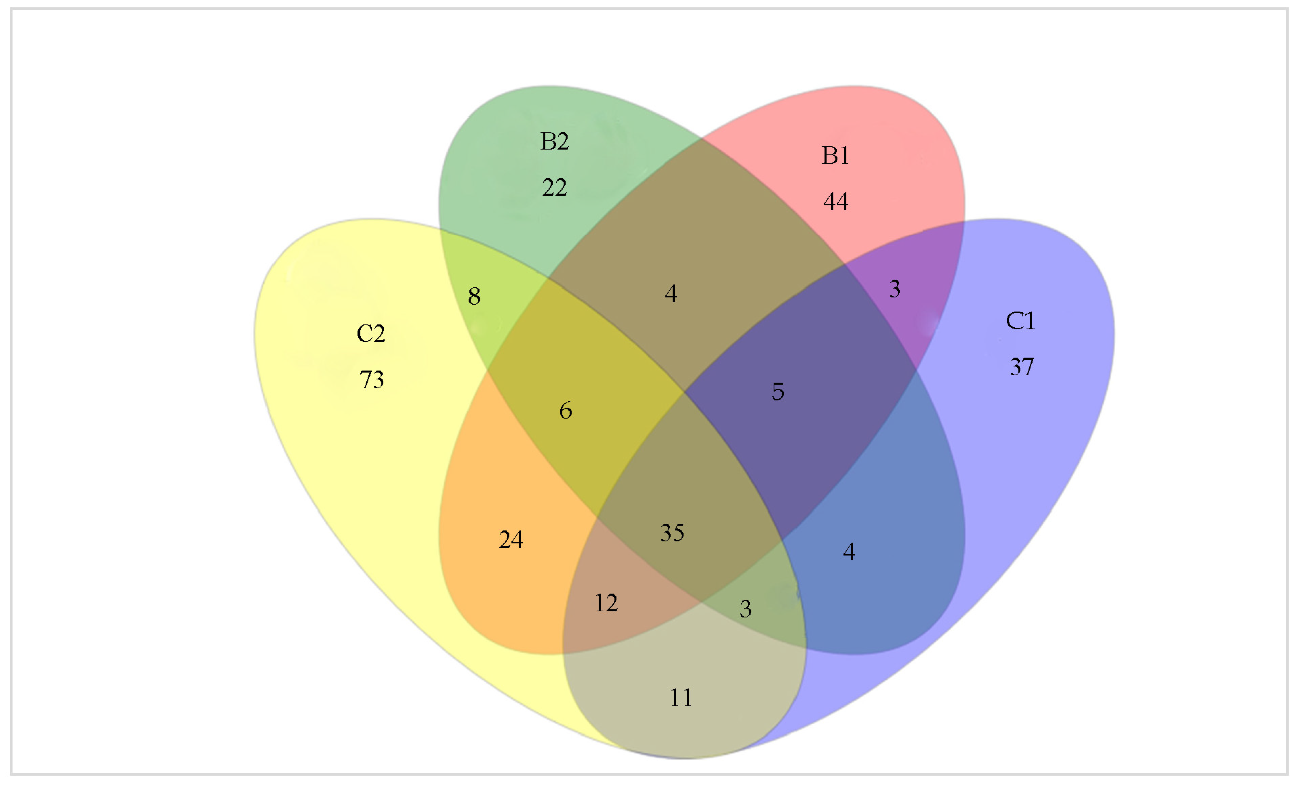
3.4. Shared and Unique Operational Taxonomic Units

In the control systems a greater number of unique OTUs, relative to those in Bacillus-treated systems, were observed, i.e., 37 and 73 were found on leaf surfaces from the control systems compared to 22 and 44 in leaf samples obtained from Bacillus probiotic treated systems (Figure 1). A total of 133 and 87 OTUs were found in leaf samples obtained from Bacillus-treated systems B1 and B2, respectively, while 110 and 172 were found in leaf samples collected from control systems C1 and C2, respectively. The number of OTUs shared between samples from Bacillus probiotic treated systems (B1 and B2) was 50, while 61 OTUs were shared between samples from the control systems. The control samples generated the most unique OTUs of this dataset.

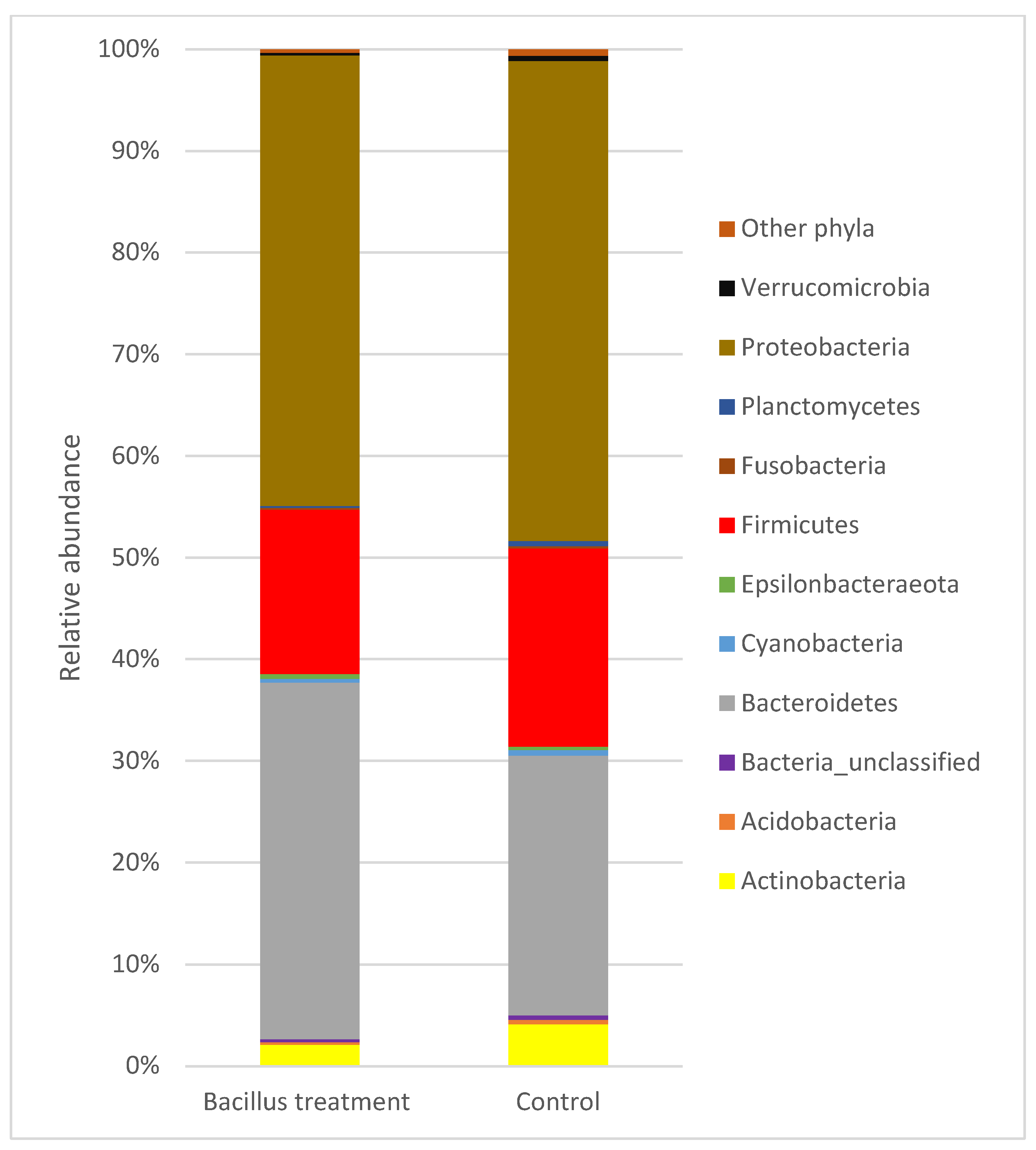
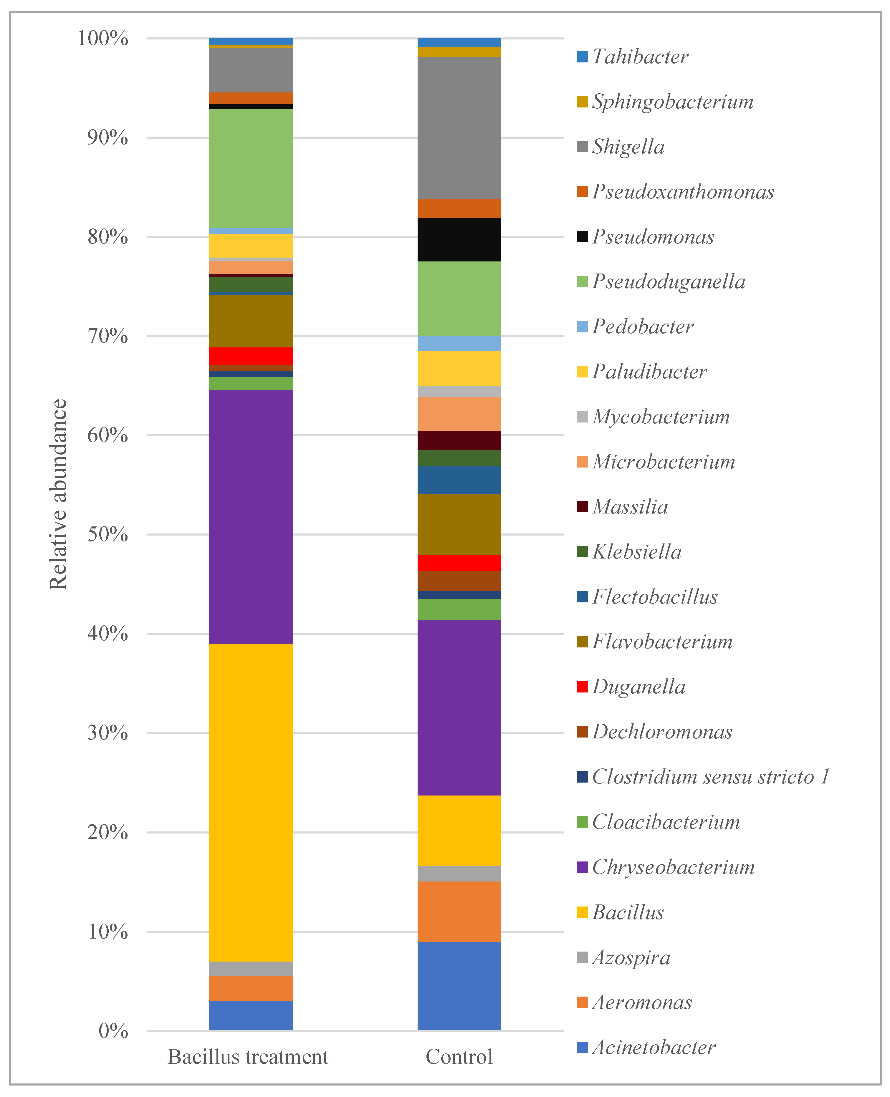
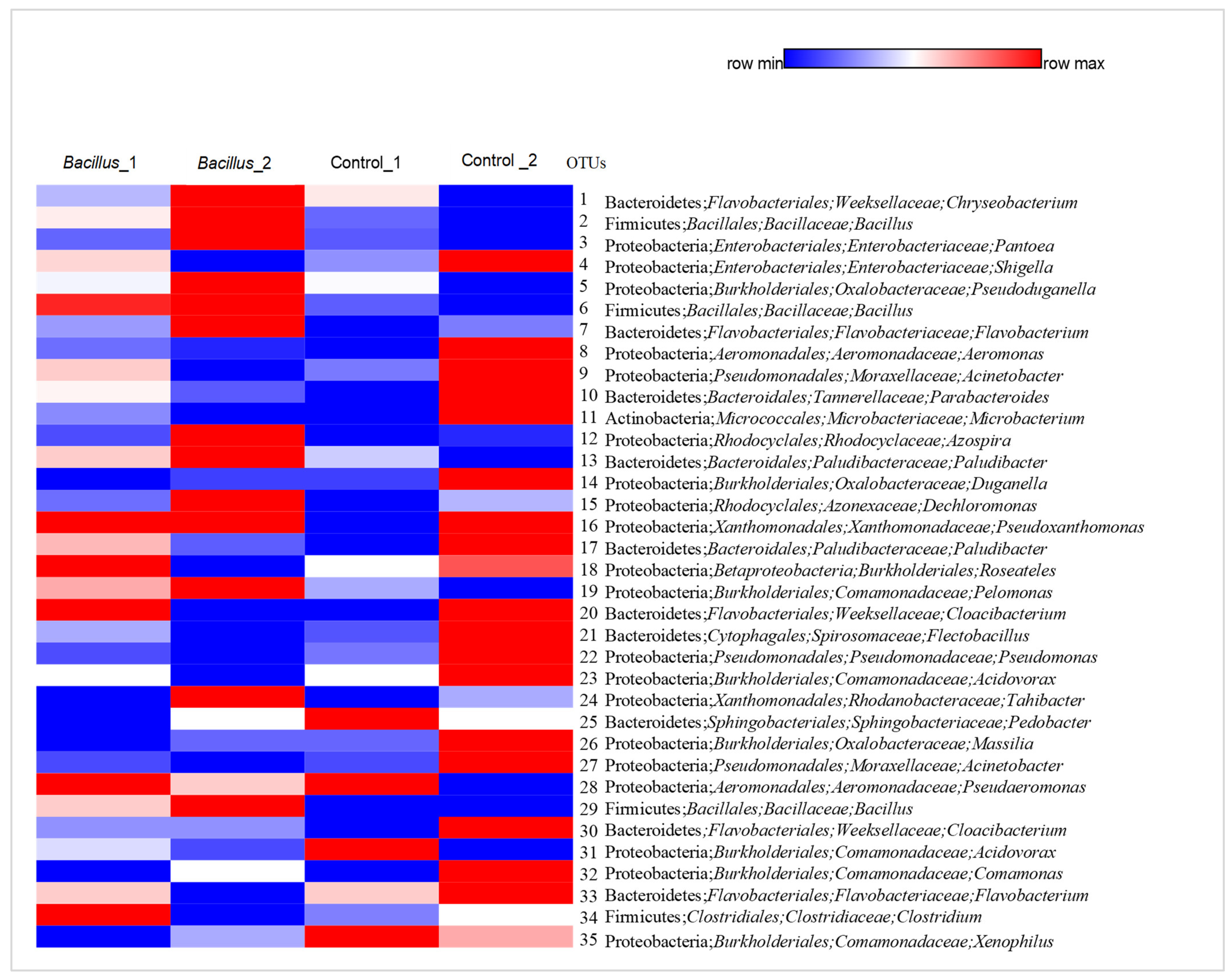
3.5. Overview of Bacterial Taxa Associated with Lettuce Leaf Samples

A total of 17 phyla were identified, with 10 having a relative abundance of more than 0.2% in the control and the Bacillus probiotic treated systems. The bacterial taxa were mostly members of the Proteobacteria, Bacteroidetes, Firmicutes, and Actinobacteria phyla (Figure 2). In the Bacillus probiotic treated systems, Proteobacteria and Bacteroidetes were the common inhabitants of lettuce epiphytes with average relative abundances of 44.33% and 35.06% of the total bacterial community, respectively. Similarly, in the control systems, the Proteobacteria and Bacteroidetes constituted 47.24% and 25.50% of the total bacterial community, respectively. Other major dominant phyla from the Bacillus-treated systems included Firmicutes (16.20%) and Actinobacteria (2.11%) while these phyla in the control systems constituted 19.49% and 4.13% of the total bacterial community, respectively. A fraction of the total sequences (0.28–0.46%) could not be classified into any known phyla. Other phyla including Acidobacteria, Epsilonbacteraeota, Planctomycetes, Fusobacteria, Verrucomicrobia, and Cyanobacteria were present in lower abundances. Gammaproteobacteria was the dominant class within the phylum Proteobacteria in all samples (Figure 2).
Twenty-three genera had a relative abundance greater than 0.5% of total bacteria reads (Figure 3). The lettuce leaf surfaces from Bacillus probiotic treated systems had a high relative abundance of Bacillus (27.20%), Chryseobacterium (21.90%), Pseudoduganella (10.26%), and Flavobacterium (4.49%). Other genera including Shigella (3.86%), Aeromonas (2.14%), Paludibacter (2.02%), and Cloacibacterium (1.14%) also inhabited the lettuce epiphytes. In the control systems, the dominant genera included Chryseobacterium (9.13%), Shigella (7.38%), Acinetobacter (4.63%), Pseudoduganella (3.90%), Bacillus (3.67%), Aeromonas (3.14%), Flavobacterium (3.16%), and Paludibacter (1.80%).
The distribution of the top 35 dominant OTUs are exhibited in a heatmap (Figure 4). The red illustrates the highest abundance, and the blue the lowest abundance of species. The 35 abundant OTUs (≥10 sequences) belonged to four phyla, including Proteobacteria, Bacteroidetes, Firmicutes, and Actinobacteria (Figure 4). The differences in the distribution of dominant OTUs were observed in lettuce leaf samples obtained from aquaponic systems receiving similar treatment. The corresponding OTUs were identified to species level (sequence similarity between 97–100%) based on 16S rRNA gene sequences in the NCBI database (Table 5).

4. Discussion

4.1. Water Quality Management and Lettuce Growth

Water temperature, pH, DO, EC, and TDS were maintained at similar levels in the deep-water growth beds between Bacillus probiotic treated and control aquaponic systems. These water parameters were maintained within levels recommended for aquaponics [31,32]. An oxygen concentration above 4 mg L−1 of the root environment in the hydroponics component is required to prevent root rot [31]. In our study, the DO was above 6 mg L−1 which is a sufficient concentration for the growth of plants and fish. EC and TDS remained within the recommended range for plant growth, with both increasing over time.
Nitrate and phosphate are dissolved forms of nitrogen and phosphorus, respectively, which are essential nutrients for plant growth. In our study, the aquaponics systems treated with the Bacillus probiotic mixture had significant increases in both nitrate and phosphate concentrations in the hydroponic water. The increase in nitrate and phosphate is corroborated by previous studies [25]. Because Bacillus mineralizes different forms of phosphorus [19], the addition of the probiotic may have led to the conversion of complex forms of essential nutrients, such as phosphorus and ammonium nitrogen, to simple bioavailable forms that can be used by plant roots to improve lettuce growth.
Probiotic supplementation increased the fresh and dry biomass of the lettuce with the fresh weight of the shoots and roots significantly higher in the Bacillus probiotic systems compared to the controls. The increase in shoot weight was most likely because of the increased nitrate and phosphate concentrations in the probiotic-treated aquaponic systems [25]. The dark-adapted value of Fv/Fm is generally used to assess the plant photosynthetic performance, with the value for unstressed leaves being 0.81–0.83 [25]. In this study, the chlorophyll fluorescence parameter averaged 0.82 for leaf samples from Bacillus probiotic treated aquaponic systems.

4.2. Bacterial Community Composition and Diversity

The composition of leaf epiphytic bacteria is determined by various factors, including environmental conditions, plant health, plant age, and plant genotype as well as the nutritional characteristics of the phyllosphere or grow media [3,15]. The α-diversity of the epiphytic bacterial community of the leaf samples in our study was evaluated by four indices, namely Sobs, Chao1, Shannon, and InvSimpson. Sobs and Chao1 are abundance-based estimators of species richness; Shannon and InvSimpson describe species richness and species evenness [33]. There were no significant differences in α-diversity indices between leaf samples obtained from Bacillus-treated and control systems, implying that leaf samples had similar species richness. The similarity between treatments was possibly due to the high variability of alpha diversity metric values within a treatment. Mercier and Lindow [34] reported that the epiphytic bacterial populations vary from one leaf to another, even on leaves of the same plant, due to changes in the utilizable carbon and nitrogen sources on leaf surfaces. Schlechter et al. [35] reviewed studies on factors that influence epiphytic bacterial communities and reported that the heterogenic nutrient conditions and fluctuating water availability of leaves are among key properties influencing bacterial colonization. The variability of the epiphytic bacterial communities within a treatment might be related to physicochemical properties. These factors relevant to aquaponics may include water quality, plant metabolism, and heterogenic nutrient conditions [35].
The Good’s coverage, which is an index of gene sequencing depth, was relatively low with an average coverage range of 88.9 to 90.0% for the control and Bacillus-treated samples. This implies that lettuce leaf surfaces were inhabited by other microbes as revealed by the sequences assigned to eukaryotes, chloroplasts, and mitochondria observed during sequence analysis. Bringel and Couée [4] reported that sequencing depth is a limitation for the detection of phyllosphere-specific bacteria.
The leaf epiphytic bacterial communities in the present study were dominated by the phylum Proteobacteria, followed by Bacteroidetes, Firmicutes, and Actinobacteria. These phyla are major colonizers of leaf surfaces in soil-based plants [3]. The dominance of Proteobacteria has been reported in other studies where representatives of this phylum play important roles in nitrification, nitrogen fixation, methylotrophy, or anoxygenic photosynthesis [15]. The genera Chryseobacterium (OTU1), Bacillus (OTU2), Pantoea (OTU3), Pseudoduganella (OTU5), Flavobacterium (OTU7) Paludibacter (OTU17), and Cloacibacterium (OTU30) were dominant in leaf samples obtained from Bacillus-treated systems. The source of epiphytic bacteria includes air and/or water which are enriched with microorganisms [36]. Endophytic bacteria from the roots could possibly transfer to the above-water parts of plants. These factors might explain the dominance of OTUs (1, 2, 3, 5, 7, 17, and 30) on lettuce leaf surfaces observed in our study.
Chryseobacterium was the most abundant genus that colonized the lettuce leaf surfaces from Bacillus probiotic treated systems. Members of this genus are reported to exhibit plant-growth-promoting activities [19,37]. They can produce siderophore, protease, cellulase, amylase, and xylanase and have antifungal activity. Kumar et al. [38] reported that rice leaf-associated Chryseobacterium showed anti-fungal activity against rice blast disease when inoculated in rice seedlings. Given that leaf Chryseobacterium is capable of secreting chemicals that suppress other pathogenic microbes, its ecological role in aquaponics may be of particular importance.
Other dominant OTUs assigned to Pantoea, Pseudoduganella, and Flavobacterium were detected on the leaf samples from Bacillus probiotic treated systems. Bacteria from the genus Pantoea have been isolated from a range of ecological niches and their biological roles have been reported as plant epiphytes, as biological control agents, or as plant-growth promoters [39]. They can establish quorum sensing systems on leaves, which may lead to the suppression of pathogens [40]. Species in the genus Pseudoduganella are adapted to oligotrophic conditions and they are considered nitrogen-fixing organisms [41]. Members of the genus Flavobacterium are associated with the ability to degrade complex organic compounds, often representing a significant fraction of leaf-associated microbiomes in different plant species [42,43]. Other dominant OTUs enriched to leaf samples from Bacillus probiotic treated systems were assigned to Paludibacter and Cloacibacterium. The genus Paludibacter includes species often associated with cellulose and chitin degradation [44]. Members of Cloacibacterium are capable of producing extracellular polymeric substances linked to the phytoremediation of toxic pollutants [45].

4.3. Occurrence of Shigella and Aeromonas Species on Leaf Surfaces of Lettuce

In our study, irrespective of the system, the fresh lettuce leaf surface was inhabited by members of the genus Shigella. The presence of the genus Shigella, a food-borne pathogen from leaf samples of the control systems (7.38% of the total bacterial community in the control systems compared to 3.86% in the Bacillus probiotic treated systems), is of concern because infections caused by Shigella species continue to be an important cause of diarrheal diseases [46]. An infective dose of Shigella of 10–100 bacterial cells can be sufficient to produce disease [47]. The comparative 16S rRNA gene sequence based on the NCBI BLAST search results indicated that this OTU had the highest sequence similarity to the Shigella sonnei strain CECT 4887 (98.28% sequence identity). Although little information is available to this specific strain, S. sonnei has been shown to multiply in lettuce and lettuce extract [46] and can grow within a pH range between 4.5 and 9.0 [48].
Another potential food-borne pathogen assigned to genus Aeromonas was identified from lettuce leaves from the control and Bacillus probiotic treated systems (3.14% of the total bacterial community in the control compared to 2.14% in the probiotic treated systems). Aeromonas species have also been isolated from lettuce sourced from retail facilities [49,50]. The NCBI BLAST search indicated the bacterial epiphyte OTU8 from our study had a 99.36% sequence identity with the Aeromonas hydrophila strain DSM 30187. Related studies in aquaponics have reported the presence of Aeromonas. Chitmanat et al. [51] reported the presence of Aeromonas hydrophila in aquaponics systems containing hybrid catfish (Clarias macrocephalus × Clarias gariepinus). Schmautz et al. [52] reported the presence of Aeromonas (0.25% of the total bacterial community) in the faecal samples of tilapia reared in an aquaponic system. A. hydrophila is amongst the most frequently reported opportunistic pathogenic bacteria in fish [53,54,55] with zoonotic potential [53]. In studies from retail facilities, Aeromonas spp. has been identified in organic vegetables at various occurrences at 7.3% [56] and 34% [49]. The transfer of this pathogen to aquaponic systems onto lettuce surfaces can be through water, equipment, fish feed, and media grow bed aggregates [50,55]. Our results indicate that lettuce leaf samples obtained from the probiotic treated systems had lower relative abundances of Shigella and Aeromonas than control systems, suggesting that application of the commercial probiotic product containing Bacillus subtilis and B. licheniformis may decrease pathogen colonization on leaf surfaces. B. subtilis has been suggested to inhibit pathogenic bacterial growth in plant tissue as well as decrease the harmful effects of plant pathogens by altering the expression of stress-responsive genes, proteins, phytohormones, and related metabolites [19]. The lower relative abundances of Shigella and Aeromonas on the lettuce leaf samples from the Bacillus probiotic treatments might also be due to compounds with antimicrobial properties produced by B. subtilis. Blake et al. [57] reported that B. subtilis produces surfactin, a compound that disrupts the cell membranes of other organisms and forms the basis of biocontrol against various plant pathogens. The relative decrease of these pathogens may also be a result of competition for nutrients between the bacterial species. However, further studies are required to investigate the mechanisms of how the commercial product containing a mixture of B. subtilis and B. licheniformis inhibit plant pathogens in aquaponic systems.

5. Conclusions

In conclusion, this study provides the characterization of the epiphytic bacterial community structure, and the results show how it can be modulated by the addition of a commercial probiotic to an aquaponic system. Our study has demonstrated a potential risk of pathogen contamination in lettuce grown in an indoor aquaponic system, however, there was a relatively lower abundance of Shigella and Aeromonas on lettuce leaf surfaces in systems supplemented with a Bacillus probiotic. Further investigation is needed to establish how the Bacillus probiotic influences epiphytic bacterial communities. The risk of pathogenic bacteria also requires further examination to ensure the risk of contamination of fresh fruit and vegetables grown in aquaponic systems is minimized through appropriate handling, cleaning, sanitizing, and possibly the supplementation with Bacillus probiotics.

Author Contributions

Conceptualisation, N.K.; H.K. and B.W.; methodology, N.K., H.K. and B.W.; data curation, N.K. and H.K.; writing of original draft, N.K.; and reviewing and editing, H.K. and B.W. All authors have read and agreed to the published version of the manuscript.

Funding

This study was financial supported by Rhodes University and the DAAD scholarship Programme for In-Region Rhodes University of South Africa, 2018 under the Grant number: 57408782.

Institutional Review Board Statement

The study was conducted in accordance with the ethical guidelines for the use of animals in research and was approved by the Animal Research Ethics Committee (AREC) of Rhodes University, South Africa (RU-AREC references: 29102018, 2019-1145-2120, and 2020-2824-4869).

Informed Consent Statement

Not applicable.

Data Availability Statement

Raw sequences from 16S RNA gene profiling were deposited into the National Center for Biotechnology Information (NCBI) under project accession number PRJNA623517 through the Sequence Read Archive (SRA) study accession number: SRR14373968.

Acknowledgments

We acknowledge the Aquatic Genomics Research Platform at the South African Institute of Aquatic Biodiversity (SAIAB) and Gwynneth F. Matcher for her assistance in the high-throughput sequencing application.

Conflicts of Interest

The authors declare no conflict of interest.

References

  1. Lindow, S.E.; Brandl, M.T. Microbiology of the Phyllosphere. Appl. Environ. Microbiol. 2003, 69, 1875–1883. [Google Scholar] [CrossRef] [PubMed] [Green Version]
  2. Williams, T.R.; Marco, M.L. Phyllosphere Microbiota Composition and Microbial Community Transplantation on Lettuce Plants Grown Indoors. mBio 2014, 5, 01564-14. [Google Scholar] [CrossRef] [PubMed] [Green Version]
  3. Vorholt, J.A. Microbial life in the phyllosphere. Nat. Rev. Genet. 2012, 10, 828–840. [Google Scholar] [CrossRef] [PubMed]
  4. Bringel, F.; Couée, I. Pivotal roles of phyllosphere microorganisms at the interface between plant functioning and atmospheric trace gas dynamics. Front. Microbiol. 2015, 6, 486. [Google Scholar] [CrossRef] [PubMed] [Green Version]
  5. Leveau, J.H.J. Microbial communities in the phyllosphere. In Biology of the Plant Cuticle; Riederer, M., Mueller, C., Eds.; Blackwell Publishing: Oxford, UK, 2006; pp. 334–367. [Google Scholar]
  6. Sánchez-López, A.S.; González-Chávez, M.D.C.A.; Solís-Domínguez, F.A.; Carrillo-González, R.; Rosas-Saito, G.H. Leaf Epiphytic Bacteria of Plants Colonizing Mine Residues: Possible Exploitation for Remediation of Air Pollutants. Front. Microbiol. 2018, 9, 3028. [Google Scholar] [CrossRef]
  7. Fürnkranz, M.; Wanek, W.; Richter, A.; Abell, G.; Rasche, F.; Sessitsch, A. Nitrogen fixation by phyllosphere bacteria associated with higher plants and their colonizing epiphytes of a tropical lowland rainforest of Costa Rica. ISME J. 2008, 2, 561–570. [Google Scholar] [CrossRef]
  8. Lindow, S.E.; Leveau, J.H. Phyllosphere microbiology. Curr. Opin. Biotechnol. 2002, 13, 238–243. [Google Scholar] [CrossRef]
  9. Rastogi, G.; Coaker, G.L.; Leveau, J.H. New insights into the structure and function of phyllosphere microbiota through high-throughput molecular approaches. FEMS Microbiol. Lett. 2013, 348, 1–10. [Google Scholar] [CrossRef] [Green Version]
  10. Berlec, A. Novel techniques and findings in the study of plant microbiota: Search for plant probiotics. Plant Sci. 2012, 193–194, 96–102. [Google Scholar] [CrossRef]
  11. Erickson, M.C.; Webb, C.C.; Diaz-Perez, J.C.; Phatak, S.C.; Silvoy, J.J.; Davey, L.; Payton, A.S.; Liao, J.; Ma, L.; Doyle, M.P. Surface and Internalized Escherichia coli O157:H7 on Field-Grown Spinach and Lettuce Treated with Spray-Contaminated Irrigation Water. J. Food Prot. 2010, 73, 1023–1029. [Google Scholar] [CrossRef]
  12. Wang, Y.-J.; Deering, A.J.; Kim, H.-J. The Occurrence of Shiga Toxin-Producing E. coli in Aquaponic and Hydroponic Systems. Horticulturae 2020, 6, 1. [Google Scholar] [CrossRef] [Green Version]
  13. Weller, D.; Saylor, L.; Turkon, P. Total Coliform and Generic E. coli Levels, and Salmonella Presence in Eight Experimental Aquaponics and Hydroponics Systems: A Brief Report Highlighting Exploratory Data. Horticulturae 2020, 6, 42. [Google Scholar] [CrossRef] [PubMed]
  14. Yep, B.; Zheng, Y. Aquaponic trends and challenges—A review. J. Clean. Prod. 2019, 228, 1586–1599. [Google Scholar] [CrossRef]
  15. Stone, B.W.G.; Weingarten, E.A.; Jackson, C.R. The Role of the Phyllosphere Microbiome in Plant Health and Function. Annu. Plant Rev. Online 2018, 1, 533–556. [Google Scholar] [CrossRef]
  16. Riggio, G.; Jones, S.; Gibson, K. Risk of Human Pathogen Internalization in Leafy Vegetables during Lab-Scale Hydroponic Cultivation. Horticulturae 2019, 5, 25. [Google Scholar] [CrossRef] [Green Version]
  17. Wall, G.L.; Clements, D.P.; Fisk, C.L.; Stoeckel, D.M.; Woods, K.L.; Bihn, E.A. Meeting Report: Key Outcomes from a Collaborative Summit on Agricultural Water Standards for Fresh Produce. Compr. Rev. Food Sci. Food Saf. 2019, 18, 723–737. [Google Scholar] [CrossRef] [Green Version]
  18. Fox, B.K.; Tamaru, C.S.; Hollyer, J.; Castro, L.F.; Fonseca, J.M.; Jay-russell, M.; Low, T. A preliminary study of microbial water quality related to food safety in recirculating aquaponic fish and vegetable production systems. Food Saf. Technol. 2012, 51, 1–11. [Google Scholar]
  19. Radhakrishnan, R.; Hashem, A.; Abd_Allah, E.F. Bacillus: A Biological Tool for Crop Improvement through Bio-Molecular Changes in Adverse Environments. Front. Physiol. 2017, 8, 667. [Google Scholar] [CrossRef]
  20. Afzal, I.; Shinwari, Z.K.; Sikandar, S.; Shahzad, S. Plant beneficial endophytic bacteria: Mechanisms, diversity, host range and genetic determinants. Microbiol. Res. 2019, 221, 36–49. [Google Scholar] [CrossRef]
  21. da Silva Cerozi, B.; Fitzsimmons, K. Use of Bacillus spp. to enhance phosphorus availability and serve as a plant growth promoter in aquaponics systems. Sci. Hortic. 2016, 211, 277–282. [Google Scholar] [CrossRef] [Green Version]
  22. Hlordzi, V.; Kuebutornye, F.K.; Afriyie, G.; Abarike, E.D.; Lu, Y.; Chi, S.; Anokyewaa, M.A. The use of Bacillus species in maintenance of water quality in aquaculture: A review. Aquac. Rep. 2020, 18, 100503. [Google Scholar] [CrossRef]
  23. Hai, N.V. The use of probiotics in aquaculture. J. Appl. Microbiol. 2015, 119, 917–935. [Google Scholar] [CrossRef] [PubMed]
  24. Ahmad, F.; Ahmad, I.; Khan, M. Screening of free-living rhizospheric bacteria for their multiple plant growth promoting activities. Microbiol. Res. 2008, 163, 173–181. [Google Scholar] [CrossRef] [PubMed]
  25. Kasozi, N.; Kaiser, H.; Wilhelmi, B. Effect of Bacillus spp. on Lettuce Growth and Root Associated Bacterial Community in a Small-Scale Aquaponics System. Agronomy 2021, 11, 947. [Google Scholar] [CrossRef]
  26. Sare, A.R.; Stouvenakers, G.; Eck, M.; Lampens, A.; Goormachtig, S.; Jijakli, M.H.; Massart, S. Standardization of Plant Microbiome Studies: Which Proportion of the Microbiota is Really Harvested? Microorganisms 2020, 8, 342. [Google Scholar] [CrossRef] [Green Version]
  27. Kasozi, N.; Kaiser, H.; Wilhelmi, B. Metabarcoding Analysis of Bacterial Communities Associated with Media Grow Bed Zones in an Aquaponic System. Int. J. Microbiol. 2020, 2020, 1–10. [Google Scholar] [CrossRef]
  28. Schloss, P.D.; Westcott, S.L.; Ryabin, T.; Hall, J.R.; Hartmann, M.; Hollister, E.B.; Lesniewski, R.A.; Oakley, B.B.; Parks, D.H.; Robinson, C.J.; et al. Introducing mothur: Open-Source, Platform-Independent, Community-Supported Software for Describing and Comparing Microbial Communities. Appl. Environ. Microbiol. 2009, 75, 7537–7541. [Google Scholar] [CrossRef] [Green Version]
  29. Rognes, T.; Flouri, T.; Nichols, B.; Quince, C.; Mahé, F. VSEARCH: A versatile open source tool for metagenomics. PeerJ 2016, 4, e2584. [Google Scholar] [CrossRef]
  30. De Winter, J.C.F. Using the Student’s t-test with extremely small sample sizes. Pract. Asses. Res. Eval. 2013, 18, 1–12. [Google Scholar] [CrossRef]
  31. Somerville, C.; Cohen, M.; Pantanella, E.; Stankus, A.; Lovatelli, A. Small-Scale Aquaponic Food Production. In Integrated Fish and Plant Farming; Technical Paper No. 589; FAO Fisheries and Aquaculture: Rome, Italy, 2014; pp. 90–91. [Google Scholar]
  32. Yanes, A.R.; Martinez, P.; Ahmad, R. Towards automated aquaponics: A review on monitoring, IoT, and smart systems. J. Clean. Prod. 2020, 263, 121571. [Google Scholar] [CrossRef]
  33. Kim, B.-R.; Shin, J.; Guevarra, R.B.; Lee, J.H.; Kim, D.W.; Seol, K.-H.; Lee, J.-H.; Kim, H.B.; Isaacson, R.E. Deciphering Diversity Indices for a Better Understanding of Microbial Communities. J. Microbiol. Biotechnol. 2017, 27, 2089–2093. [Google Scholar] [CrossRef] [PubMed] [Green Version]
  34. Mercier, J.; Lindow, S.E. Role of Leaf Surface Sugars in Colonization of Plants by Bacterial Epiphytes. Appl. Environ. Microbiol. 2000, 66, 369–374. [Google Scholar] [CrossRef] [PubMed] [Green Version]
  35. Schlechter, R.O.; Miebach, M.; Remus-Emsermann, M.N. Driving factors of epiphytic bacterial communities: A review. J. Adv. Res. 2019, 19, 57–65. [Google Scholar] [CrossRef] [PubMed]
  36. Dong, C.J.; Wang, L.L.; Li, Q.; Shang, Q.M. Bacterial communities in the rhizosphere, phyllosphere and endosphere of tomato plants. PLoS ONE 2019, 14, e0223847. [Google Scholar] [CrossRef] [PubMed]
  37. Montero-Calasanz, M.C.; Göker, M.; Rohde, M.; Spröer, C.; Schumann, P.; Busse, H.J.; Schmid, M.; Klenk, H.P.; Tindall, B.J.; Camacho, M. Chryseobacterium oleae sp. nov., an efficient plant growth promoting bacterium in the rooting induction of olive tree (Olea europaea L.) cuttings and emended descriptions of the genus Chryseobacterium, C. daecheongense, C. gambrini, C. gleum, C. joostei, C. jejuense, C. luteum, C. shigense, C. taiwanense, C. ureilyticum and C. vrystaatense. Syst. Appl. Microbiol. 2014, 37, 342–350. [Google Scholar] [CrossRef]
  38. Kumar, M.; Charishma, K.; Sahu, K.; Sheoran, N.; Patel, A.; Kundu, A.; Kumar, A. Rice leaf associated Chryseobacterium species: An untapped antagonistic flavobacterium displays volatile mediated suppression of rice blast disease. Biol. Control 2021, 161, 104703. [Google Scholar] [CrossRef]
  39. De Maayer, P.; Chan, W.-Y.; Blom, J.; Venter, S.N.; Duffy, B.; Smits, T.H.M.; Coutinho, T.A. The large universal Pantoea plasmid LPP-1 plays a major role in biological and ecological diversification. BMC Genom. 2012, 13, 625. [Google Scholar] [CrossRef] [Green Version]
  40. Pusey, P.L.; Stockwell, V.O.; Reardon, C.L.; Smits, T.H.; Duffy, B. Antibiosis Activity of Pantoea agglomerans Biocontrol Strain E325 Against Erwinia amylovora on Apple Flower Stigmas. Phytopathology 2011, 101, 1234–1241. [Google Scholar] [CrossRef] [Green Version]
  41. Baldani, J.I.; Rouws, L.; Cruz, L.M.; Olivares, F.L.; Schmid, M.; Hartmann, A. The Family Oxalobacteraceae. In The Prokaryotes; Rosenberg, E., DeLong, E.F., Lory, S., Stackebrandt, E., Thompson, F., Eds.; Springer: Berlin/Heidelberg, Germany, 2014; pp. 943–946. [Google Scholar]
  42. Kolton, M.; Erlacher, A.; Berg, G.; Cytryn, E. The Flavobacterium Genus in the Plant Holobiont: Ecological, Physiological, and Applicative Insights. In Microbial Models: From Environmental to Industrial Sustainability; Microorganisms for, Sustainability; Castro-Sowinski, S., Ed.; Springer: Singapore, 2016; pp. 189–207. [Google Scholar]
  43. Kolton, M.; Frenkel, O.; Elad, Y.; Cytryn, E. Potential Role of Flavobacterial Gliding-Motility and Type IX Secretion System Complex in Root Colonization and Plant Defense. Mol. Plant-Microbe Interact. 2014, 27, 1005–1013. [Google Scholar] [CrossRef] [Green Version]
  44. Dai, Y.; Yan, Z.; Jia, L.; Zhang, S.; Gao, L.; Wei, X.; Mei, Z.; Liu, X. The composition, localization and function of low-temperature-adapted microbial communities involved in methanogenic degradations of cellulose and chitin from Qinghai-Tibetan Plateau wetland soils. J. Appl. Microbiol. 2016, 121, 163–176. [Google Scholar] [CrossRef]
  45. Wang, X.; Hua, Z.; Mao, H. Influential factors for metal ions removal using extracellular polymeric substances produced by Cloacibacterium normanense. Water Environ. J. 2018, 32, 650–656. [Google Scholar] [CrossRef]
  46. Kapperud, G.; Rørvik, L.M.; Hasseltvedt, V.; Høiby, E.A.; Iversen, B.G.; Staveland, K.; Johnsen, G.; Leitao, J.; Herikstad, H.; Andersson, Y.; et al. Outbreak of Shigella sonnei infection traced to imported iceberg lettuce. J. Clin. Microbiol. 1995, 33, 609–614. [Google Scholar] [CrossRef] [PubMed] [Green Version]
  47. Zaidi, M.B.; Estrada-García, T. Shigella: A Highly Virulent and Elusive Pathogen. Curr. Trop. Med. Rep. 2014, 1, 81–87. [Google Scholar] [CrossRef] [PubMed] [Green Version]
  48. Bagamboula, C.F.; Uyttendaele, M.; Debevere, J. Acid tolerance of Shigella sonnei and Shigella flexneri. J. Appl. Microbiol. 2002, 93, 479–486. [Google Scholar] [CrossRef] [Green Version]
  49. McMahon, M.A.; Wilson, I.G. The occurrence of enteric pathogens and Aeromonas species in organic vegetables. Int J. Food Microbiol. 2001, 70, 155–162. [Google Scholar] [CrossRef]
  50. Umutoni, N.; Jakobsen, A.N.; Mukhatov, K.; Thomassen, G.M.B.; Karlsen, H.; Mehli, L. Occurrence, diversity and temperature-dependent growth kinetics of Aeromonas spp. in lettuce. Int. J. Food Microbiol. 2020, 335, 108852. [Google Scholar] [CrossRef] [PubMed]
  51. Chitmanat, C.; Pimpimol, T.; Chaibu, P. Investigation of Bacteria and Fish Pathogenic Bacteria Found in Freshwater Aquaponic System. J. Agric. Sci. 2015, 7, 254–259. [Google Scholar] [CrossRef] [Green Version]
  52. Schmautz, Z.; Graber, A.; Jaenicke, S.; Goesmann, A.; Junge, R.; Smits, T.H.M. Microbial diversity in different compartments of an aquaponics system. Arch. Microbiol. 2017, 199, 613–620. [Google Scholar] [CrossRef] [PubMed]
  53. Cipriano, R.L. Aeromonas Hydrophila and Motile Aeromonad Septicemias of Fish; US Fish & Wildlife Publications: Washington, DC, USA, 1984; Volume 68, pp. 1–23. [Google Scholar]
  54. Lehane, L.; Rawlin, G.T. Topically acquired bacterial zoonoses from fish: A review. Med. J. Aust. 2000, 173, 256–259. [Google Scholar] [CrossRef]
  55. Tekedar, H.C.; Karsi, A.; Akgul, A.; Kalindamar, S.; Waldbieser, G.C.; Sonstegard, T.; Schroeder, S.G.; Lawrence, M.L. Complete Genome Sequence of Fish Pathogen Aeromonas hydrophila AL06-06. Genome Announc. 2015, 3, e00368-15. [Google Scholar] [CrossRef] [Green Version]
  56. Santos, M.I.; Cavaco, A.; Gouveia, J.; Novais, M.; Nogueira, P.; Pedroso, L.; Ferreira, M.A.S.S. Evaluation of minimally processed salads commercialized in Portugal. Food Control 2012, 23, 275–281. [Google Scholar] [CrossRef]
  57. Blake, C.; Christensen, M.N.; Kovács, Á.T. Molecular Aspects of Plant Growth Promotion and Protection by Bacillus subtilis. Mol. Plant-Microbe Interact. 2021, 34, 15–25. [Google Scholar] [CrossRef] [PubMed]
Figure 1. A Venn diagram of shared and unique OTUs of epiphytic bacterial communities of lettuce plants grown in Bacillus probiotic treated and control systems. B1 and B2 represent leaf samples obtained from Bacillus probiotic supplemented systems and C1 and C2 represent leaf samples from the control systems.
Horticulturae 08 00151 g001
Figure 2. The relative abundances of taxa (at the phylum level) of leaf epiphytic bacteria from lettuce plants obtained from Bacillus probiotic treated and control aquaponic systems. Phyla less than 0.2% of the total reads are displayed under “other phyla” and include the phyla Armatimonadetes, Chloroflexi, Chlamydiae, Dependentiae, Nitrospirae, Patescibacteria, and Spirochaetes.
Horticulturae 08 00151 g002
Figure 3. The average relative abundance of bacterial genera of leaf epiphytic bacterial communities of lettuce plants obtained from Bacillus probiotic treated and control systems. Genera representing greater than 0.5% of the total reads in at least one sample are presented.
Horticulturae 08 00151 g003
Figure 4. A probiotic heatmap showing the distribution of the 35 dominant OTUs based on the abundance of 16S rRNA gene sequences (≥10 sequences) from leaf samples of lettuce grown in Bacillus treated and control aquaponic systems. Each column in the heatmap represents a sample and each row represents an OTU. The relative colour code refers to sequence abundance, with high abundances (red colours) and low abundances (blue colours). Taxonomy (right column) indicates the phylum, order, family, and genus of each OTU.
Horticulturae 08 00151 g004
Table 1. Vegetative growth and chlorophyll fluorescence of ‘Locarno’ lettuce cultivar grown in aquaponic systems treated with a Bacillus probiotic. Significant differences between Bacillus treated and control systems (p < 0.05) are marked by an asterisk. Data are means ± standard error (SE).
VariablesBacillus Treatment Controlp-Value
Initial plant mass (g plant−1)6.18 ± 0.026.21 ± 0.040.619
Shoot fresh weight (g plant−1)45.03 ± 0.0639.07 ± 0.910.022 *
Root fresh weight (g plant−1)2.42 ± 0.0052.00 ± 0.040.011 *
Height gain (cm plant−1)26.66 ± 0.4821.60 ± 0.380.014 *
Leaf number (plant−1)19.60 ± 0.2215.0 ± 0.190.004 *
Absolute growth rate (g day−1)1.38 ± 0.0051.16 ± 0.030.019 *
Fv/Fm0.82 ± 0.050.72 ± 0.010.011 *
CCI2.36 ± 0.052.25 ± 0.050.260
Shoot dry weight (g plant−1)2.07 ± 0.071.72 ± 0.010.043 *
Root dry weight (g plant−1)0.27 ± 0.0050.17 ± 0.010.016 *
Table 2. Water quality parameters in control and Bacillus probiotic treated systems in the deep-water growth beds. Mean value ranges are given in parentheses.
VariablesBacillus
Treatment
Control
Temperature
(°C)
25.94
(24.96–26.50)
25.94
(25.04–26.50)
DO
(mg L−1)
6.72
(6.53–6.83)
6.79
(6.67–6.90)
TDS
(mg L−1)
741
(661–839)
835
(734–931)
pH6.87
(6.74–6.97)
6.79
(6.68–6.90)
EC
(ms cm−1)
1.05
(1.05–1.19)
1.20
(1.03–1.35)
Nitrate (mg L−1)33.25
(25.27–43.60)
19.73
(14.50–27.37)
Phosphate (mg L−1)2.92
(2.34–4.43)
1.60
(1.14–2.39)
Table 3. Summary of sequencing data.
TreatmentNumber of Raw Sequences Number of Sequences before ChimerasNumber of Sequences after ChimerasEukaryotes and Unknown ReadsNumber of Reads after Screening and Filtering
Bacillus treatment
Sample B148,78835,34834,87424,544622
Sample B247,79136,01633,78417,1756349
Control
Sample C163,09841,06140,33616,5441119
Sample C241,56635,04234,494 29,884749
Table 4. Coverage and alpha diversity of epiphytic bacterial communities from lettuce leaf samples grown in Bacillus-treated and control systems. Mean values are followed by the standard error of the mean and the range is given in parentheses. No significant effect of Bacillus probiotic treatment on alpha diversity and coverage was observed (t-test, p > 0.05).
Coverage and IndicesTreatmentp-Value
BacillusControl
InvSimpson13.53
(7.95–19.12)
23.25
(10.34–36.15)
0.561
Observed richness110
(87–133)
141
(110–172)
0.506
Good’s coverage (%)90.0
(87.62–92.44)
88.9
(87.29–90.51)
0.735
Chao1218.65
(177.08–260.22)
255.41
(195.55–255.27)
0.907
Shannon3.32
(2.94–3.70)
3.81
(3.23–4.39)
0.554
Table 5. The 35 dominant bacterial OTUs and their closest phylogenetic neighbours from lettuce leaf samples obtained from a small-scale coupled aquaponic system through BLAST searches.
Bacterial OTUsRelated Species (NCBI-Blast)Similarity (%)Accession
OTU1Chryseobacterium rhizoplanae strain JM-53498.27%NR_134711.1
OTU2Bacillus australimaris strain MCCC 1A0578798.72%NR_148787.1
OTU3Pantoea rwandensis strain LMG 2627599.14%NR_118121.1
OTU4Shigella sonnei strain CECT 488798.28%NR_104826.1
OTU5Pseudoduganella eburnea strain 10R 5-2198.28%NR_159256.1
OTU6Bacillus haynesii strain NRRL B-4132798.50%NR_157609.1
OTU7Flavobacterium lindanitolerans strain IP-1098.26%NR_044208.1
OTU8Aeromonas hydrophila strain DSM 3018799.36%NR_119190.1
OTU9Acinetobacter vivianii strain NIPH 216898.71%NR_148847.1
OTU10Parabacteroides chartae strain NS31-398.92%NR_109439.1
OTU11Microbacterium laevaniformans strain DSM 2014098.72%NR_044935.1
OTU12Azospira oryzae PS98.72%NR_074103.1
OTU13Paludibacter propionicigenes WB497.41%NR_074577.1
OTU14Duganella sacchari strain Sac-22 98.28%NR_108216.1
OTU15Dechloromonas hortensis strain MA-198.72%NR_042819.1
OTU16Pseudoxanthomonas mexicana strain NBRC 10103497.43%NR_113973.1
OTU17Paludibacter propionicigenes WB497.41%NR_074577.1
OTU18Roseateles depolymerans strain 61A96.56%NR_111995.1
OTU19Pelomonas saccharophila strain NBRC 10303798.71%NR_114189.1
OTU20Cloacibacterium normanense strain NRS198.92%NR_042187.1
OTU21Flectobacillus roseus strain GFA-1198.70%NR_116312.1
OTU22Pseudomonas plecoglossicida strain NBRC 10316299.36%NR_114226.1
OTU23Acidovorax temperans strain PHL99.35%NR_028715.1
OTU24Tahibacter aquaticus strain PYM5-1197.22%NR_115098.1
OTU25Pedobacter glacialis strain 8-2498.70%NR_134125.1
OTU26Massilia alkalitolerans DSM 17462 strain YIM 3177598.92%NR_043094.1
OTU27Acinetobacter lwoffii strain JCM 684099.14%NR_113346.1
OTU28Pseudaeromonas sharmana strain GPTSA-698.28%NR_043470.1
OTU29Bacillus haynesii strain NRRL B-4132797.64%NR_157609.1
OTU30Cloacibacterium rupense strain NBRC 10493198.92%NR_114274.1
OTU31Acidovorax monticola strain K-4-1697.63%NR_164911.1
OTU32Comamonas composti strain YY28799.57%NR_044039.1
OTU33Flavobacterium notoginsengisoli strain SYP-B54098.92%NR_145940.1
OTU34Clostridium beijerinckii strain JCM 139099.57%NR_113388.1
OTU35Xenophilus aerolatus strain 5516S-299.35%NR_116099.1
Publisher’s Note: MDPI stays neutral with regard to jurisdictional claims in published maps and institutional affiliations.

