Article

Phytochemical Profiling, Mineral Elements, and Biological Activities of Artemisia campestris L. Grown in Algeria

1
Laboratoire de Valorisation et Promotion des Ressources Sahariennes (VPRS), Département de Chimie, Faculté des Mathématiques et des Sciences de la Matière, Université Kasdi Merabah, Ouargla 30000, Algeria
2
Centre de Recherche en Biotechnologie (C.R.B.T.) Constantine, Constantine 25000, Algeria
3
Nuclear Research Centre of Birine, Ain Oussera, Djelfa 17200, Algeria
4
Chemistry Department, University of Hamma Lakhdar El-Oued, B.P.789, El-Oued 39000, Algeria
5
Agronomy Department, University of Mohamed Khider Biskra, Biskra 07000, Algeria
6
School of Natural and Applied Sciences, Kampala International University, Kampala P.O. Box 20000, Uganda
7
Department of Plant Production Technology and Commodities Science, University of Life Science in Lublin, Akademicka 15 Str., 20-950 Lublin, Poland
8
Nutrition and Bromatology Group, Department of Analytical Chemistry and Food Science, Faculty of Food Science and Technology, University of Vigo—Ourense Campus, E32004 Ourense, Spain
*
Author to whom correspondence should be addressed.
Horticulturae 2022, 8(10), 914; https://doi.org/10.3390/horticulturae8100914
Submission received: 14 September 2022 / Revised: 26 September 2022 / Accepted: 1 October 2022 / Published: 6 October 2022

Abstract

:
Artemisia campestris L. is commonly used in folk medicine due to its antioxidant, antidiabetic, nutritional, and culinary properties. Our study assessed the total phenolics contents, antioxidant, and pharmacological activities of various organic extracts prepared from the aerial parts of Artemisia campestris, and its mineral elements and chemical profile were analyzed. ICP-OES was used to analyze the mineral profile and the LC-MS/MS analysis was used to characterize the phytochemical profiling. A series of antioxidant tests were carried out using DPPH, ABTS, beta-carotene, GOR, RP, CUPRAC, and O-Phenanthroline assays. In vitro potent inhibitory actions of A. campestris extracts were investigated to evaluate their anti-cholinesterase, anti-lipase and anti-diabetic activities. The photoprotective effect of the plant was measured by the sun protection factor. The most powerful inhibitor of α-amylase was AcPEE (IC50 = 11.79 ± 0.14 μg/mL), which also showed a significant butyrylcholinesterase inhibitory effect (IC50 = 93.50 ± 1.60 μg/mL). At IC50 = 23.16 ± 0.19 μg/mL, AcEAE showed the most powerful inhibitory effects on acetylcholinesterase. A. campestris was found to have a strong photoprotective ability, absorbing UV radiations with SPF values ranging from 26.07 ± 0.22 to 40.76 ± 0.11. The results showed that A. campestris extract has strong antioxidant activity in all the test samples except for the carotene bleaching assay. The LC/MS-MS results showed that AcDE, AcEAE, and AcBE identified 11 compounds belonging to Polyphenols Compounds. Our result also showed that A. campestris contains a high concentration of essential minerals, including macro-and micro-elements with their values close to the FAO’s recommended concentration. A. campestris has the capacity to improve pharmaceutical formulations, health, and medical research, due to its compositions and potent biological properties.

1. Introduction

Medicinal plants have long been regarded for their medicinal properties and nutritional importance, both of which help in treating various diseases and for drug development, according to a survey conducted by the World Health Organization [1]. In light of the antioxidant properties that they have, natural chemicals that plants produce have the ability to reduce oxidative stress [2]. As a result, research into bioactive substances and natural antioxidants, with the aim of validating traditional medicines, develop drugs, and apply them in several illnesses that affect humans have attracted significant interest [3]. Algeria has a rich plant flora, with 3139 species spread over 150 botanical groups, 653 of which are endemic [4]. This study focuses on Artemisia campestris, an endemic plant of the desert Sahara. It is known as tedjok in Tamahaq, the indigenous language of the Touareg inhabitants in the South of Algeria, and as oum nafsa in Arabic [5]. Within the genus Artemisa, which contains roughly three hundred different species overall, Artemisia campestris belongs to varieties which also include a variety of flowering plants [6]. In the Algerian flora, there are eleven different species of Artemisia, including A. herba-alba, A. campestris ssp. eu-campestris, A. campestris ssp. gIutinosa, A. absinthium, A. atlantica, A. ar-borescens L., A. judaica L., A. atlantica, A. Verlotorum, and A. vulgaris L., A. alba [7]. These species consist of small shrubs that are perennial, biennial, or annual, and grow to around 1 m in height [8]. The aerial parts and the roots of Artemisia campestris are utilized in treating chronic diseases, particularly diabetes, diseases of the skin, and hypertension, by local inhabitants’ folk medicinal treatments in the Hoggar (Tamanrasset-South of Algeria) [9,10]. Additionally, there is proof that Artemisia campestris species are utilized in common medications to treat obesity [11]. Moreover, Artemisia campestris has culinary uses, specifically as a food preservative [12]. Several engaging pharmacological activities of the Artemisia campestris species have been investigated. Such activities involve antifungal [13], antihypertensive, hypotensive, and vasorelaxant effects [14], as well as antidiabetic [15], antioxidant, antibacterial, and anti-inflammatory [16], and neuroprotective activity [17]. In several of these cases, phenolic compounds, which are abundant, may be accountable for these effects. A study performed on the phytochemical profiling of Artemisia species reported the presence of tannins, polyphenols, flavonoids, saponins, and essential oils [15,16,17,18]. Additionally, the isolation of coumarins from Artemisia campestris and chloroacetophenones from Artemisia caerulescens has additionally been reported [19]. Artemisinin, an efficient antimalarial sesquiterpene lactone containing an endoperoxide, was isolated from Artemisia annua. This study aims to evaluate the biological effects of A. campestris, along with characterizing its chemical constitution, such as phenolic compounds, and mineral compositions using different extracts of A. campestris. LC-MS/MS was used to analyze the phytochemical profile of A. campestris extracts. The pharmacological activities evaluated and reported in this study include photoprotective, anti-Alzheimer, anti-lipase, alpha-amylase and antioxidant activities. Furthermore, A. campestris’ mineral profile was examined using inductively coupled plasma optical emission spectrometry. However, little or no attention has been directed to analyzing the chemical composition, particularly the mineral content, of A. campestris. The novelty of our study largely lies in the extent to which we evaluated and validated the anti-lipase, photoprotective properties, and mineral profile of A. campestris, all of which have not been studied previously.

2. Materials and Methods

2.1. Plant Material

During the flowering season in March 2018, A. campestris plants were harvested from their natural habitats in the Sahara Desert (Tamanrasset–Algeria (22°47′13″ N, 5°31′38″ E), Oued Tifouguine region). The plant material was identified taxonomically based on the identification methods described by Quezel and Santa [5], Dr. Reggani Adelmalek from the University of Tamanrasset, and Dr. Halis Yousef, Centre for Scientific and Technical Research on Arid Regions. A standard sample was stored in the departmental Herbarium of the Saharan Resources Valorization and Promotion Laboratory under the code (Ac.TMT: 03/18). The aerial parts were cleaned and dried. Dry aerial parts were pounded with an electric blender and kept wrapped in paper until use.

2.2. Extraction of Secondary Metabolites

The plant materials were macerated in a hydro-methanolic solution (80:20) (methanol/Water) (v/v) at room temperature for 24 h with steady stirring. The process of extraction was repeated three times with the solvent being reconditioned. The resulting suspension was filtered through Whatman filter paper and condensed in a vacuum evaporator. The filtrate was evaporated at 40 °C using a rotary evaporator (BUCHI, R-100, flail, Switzerland). The residue was dissolved in water and extracted with a series of solvents with increasing polarity. The organic fractions were concentrated to obtain the following dry extracts; methanol (AcME: 0.23%), petroleum ether (AcPEE: 0.05%), dichloromethane (AcDE: 0.35%), ethyl acetate (AcEAE: 0.79%), butanol (AcBE: 1.45%), and aqueous (AcAE: 7.95%).

2.3. Inductively Coupled Plasma ICP-OES

The analysis of the mineral contents was performed using ICP-OES (Inductively coupled plasma-optical emission spectrometry) [20,21,22,23]. A 500 mg powdered sample of A. campestris was placed in a burning cup. The powder was placed in a flask (200 mL capacity) and mixed with 3 mL of HNO3, which has a concentration of 65% (Sigma Aldrich-Germany), along with 3 mL of hydrochloric acid at a 37% concentration (Sigma Aldrich-Germany). Subsequent agitation was performed for 90 min followed by heating the mixture to 105 °C to ensure that all of the powder was transformed into liquid. The sample was then diluted with 50 mL distilled water and transferred to a new tube, where it was allowed to settle and homogenize. Finally, the supernatant was subjected to an ICP-OES analysis to determine the minerals present in the sample; each sample was examined in triplicate.

2.4. Liquid Chromatography-Mass Spectrometry (LC-MS-MS)

A qualitative analysis of constituents present in different fractions of A. campestris was performed using UPLC-ESI-MS-MS Shimadzu 8040 Ultra-High sensitivity with UFMS technology equipped with a binary bump Nexera XR LC-20AD. Separation was achieved with an Ultra-force C18 column (I.D. 2.5 mm × 100 mm, 1.8 μm particle size; Restek) at 25 °C oven temperature. The chromatographic separation was carried out using a mixture of 30% (water, 0.1% formic acid) as mobile phase A and 70% methanol as mobile phase B. The duration of each gradient elution was as follows: B; 0.10 min 5% B; 10 min 15% B; 30 min 95% B; 45 min 15% B; 50 min 5% B; 60 min 100%. The flow rate was established at 0.3 mL/min, while the injection volume was 6 μL, passed through a Millex-LCR (PTFE) filter with 0.22 μm pore sizes. The separation was performed at room temperature, while the run lasted for 60 min.
The ESI conditions employed in the MS/MS are as follows: 230 KPs CID gas; −6.00 Kv conversion dynode; 350 °C interface temperature; 250 °C DL temperature; 3.00 L/min nebulizing gas flow, 400 °C heat block; and 15.00 L/min drying gas flow. The ion trap mass spectrometer was used in both negative and positive ions in the MRM mode (multiple reaction monitoring). Accurate identification was made according to their typical fragments by comparing the mass spectra with the published literature.

2.5. Assessment of Total Phenolics, Flavonoids Compounds

2.5.1. Total Phenolics Content

Spectrophotometrically, the phenolic content of the extracts was determined in a microplate using the Folin–Ciocalteu method with some modifications [24]. First, 100 μL of Folin–Ciocalteu reagent (1:10) was mixed with 20 μL of each extract or varying concentrations of the gallic acid (standard); 75 μL of sodium carbonate (7.5%) was then added to the mixture. The mixture was kept at ambient temperature for 2 h, and kept in darkness while it was incubated. A 96-well microplate reader (Perkin Elmer EnSpire, Singapore) was used to record the reading of the absorbance at 765 nm. The total phenol levels were defined as μg GAE/mg extract, using a calibration graph of gallic acid.

2.5.2. Total Flavonoids Content

The flavonoid content of the extracts was determined using a slight modification method of the microplate assay reported by Topcu et al. [25]. The method depends on forming a complex between Al3+ and the flavonoids. A total of 50 μL of the test sample was placed in a 96-well microplate containing 130 μL (MeOH). This was followed by the addition of 10 μL of 1 M potassium acetate and 10 μL of 10% aluminium nitrate. The mixture was incubated at room temperature for 40 min. The absorbance of the sample was measured at 415 nm using quercetin as a positive control. The data were reported in μg quercetin equivalent per mg of extract (μg QE/mg) depending on the quercetin calibration curve.

2.6. Antioxidant Activity

2.6.1. DPPH Scavenging Activity

The ability of the different extracts of A. campestris to inhibit the DPPH free radical using the 2,2-diphenyl-1–picrylhydrazyl free radical was investigated using the procedure described by Blois [26]. A DPPH solution (0.1 M) newly prepared in methanol was used in combination with 40 μL of plant extract at various concentrations for the reaction. After incubation of the mixture in the dark for 30 min, the absorbances were measured at 517 nm using a microplate reader (Perkin Elmer EnSpire, Singapore). The synthetic antioxidants, BHA and BHT, were used as standard. Percentage (%) DPPH free radical scavenging activity was calculated using the Formula (1):
%   Inhibition = ( A b s 517   b l a n k A b s 517   s a m p l e ) A b s 517   b l a n k × 100
Abs517blank: absorbance of control reaction. Abs517sample: absorbance of test sample.
The percentage inhibition curve at various concentrations was used in order to calculate the IC50 value, which corresponds to the extract concentration and is expressed as (μg/mL).

2.6.2. ABTS Scavenging Activity

According to the procedure described by Re et al. [27], the extracts of A. campestris were conducted in order to determine their ability to scavenge free radicals in the presence of ABTS (2,2′-azinobis (3-ethylbenzothiazoline)-6-sulfonic. After reacting a 7 mM aqueous solution of ABTS with 2.45 mM of potassium persulfate (K2S2O8) for 16 h, while storing the mixture in the dark at room temperature, the cation ABTS+ was generated. After dilution, the ABTS solution had an absorbance of 0.700 ± 0.020. In a 96-well microplate reader (PerkinElmer Multi-mode Plate Reader EnSpire, USA), 40 μL of each sample prepared in methanol at different concentrations was mixed with 160 μL of the ABTS mixture and stored in the dark for 10 min. The absorbance was measured at a wavelength of 734 nm. The antioxidant standards were BHA and BHT, respectively. Formula (1) was used to calculate the inhibition percentage.

2.6.3. β-Carotene-Linoleic Acid Bleaching Activity

The capacity of A. campestris extracts to inhibit β-carotene by the model system (β-carotene/linoleic acid) was determined using the method described by Marco [28]. The method consists of preparing β-carotene/linoleic acid emulsion by dissolving 0.5 mg of beta-carotene in 1 mL of chloroform. This mixture was transferred to a flask containing 20 μL linoleic acid and 200 μL Tween 40. The chloroform was removed using a vacuum evaporator, and 50 μL of hydrogen peroxide was added, followed by vigorously agitation. At 470 nm, the absorbance of the mixture was adjusted to between 0.8 and 0.9. 160 μL; the emulsion was mixed with 40 μL of the plant extracts or synthetic antioxidants (BHA and BHA) at various concentrations. Then, the mixture places in each well of the 96-well microplate. After incubating the microplate at 50 °C, the absorbance was measured at a wavelength of 470 nm at different times ranging from t = 0 min to t = 120 min, with each having a 30 min interval. The following equation was used to express the percentage inhibition:
I% =1 − [(AH0 − AHt)/(AC0 − ACt)] × 100
  • I (%): percentage of inhibition
  • AH0: Absorbance of β-carotene in extract at t = 0.
  • AC0: Absorbance of β-carotene in negative control at t = 0.
  • AHt: Absorbance of β-carotene in extract at 120 min.
  • ACt: Absorbance of β-carotene in negative control at 120 min.

2.6.4. Galvinoxyl (GOR) Scavenging Activity

The Galvinoxyl free radical (GOR) antioxidant test was performed using the Shi et al. method [29]. For this experiment, 40 μL of different extract concentrations in methanol was mixed with 160 μL of Galvinoxyl methanolic solution at 0.1 mM. Using a spectrophotometer at 428 nm, the absorbance of the resulting solution was measured after 120 min of incubation at room temperature in darkness. The standards were BHT and BHA. Galvinoxyl’s methanolic solution was employed as a control for this test. The inhibition percentage I (%) was expressed as follows:
Inhibition (%) = [(A (Control) − A (Sample)/A (Control)] × 100

2.6.5. Reducing Power Activity

The capacity of the samples to reduce Fe3+ contained in the complex K3Fe(CN)6 to Fe2+ was assessed by reducing iron ions. The reducing power was evaluated using the approach outlined by Oyaizu [30]. In brief, 10 μL of each A. campestris extract or standard at various concentrations was mixed with 40 μL of phosphate buffer solution (0.2 M, pH 6.6) and 50 μL 1% potassium ferricyanide. Afterwards, the mixture was incubated for 20 min at 50 °C. Then, 50 μL of 10% trichloroacetic acid, 40 μL of distilled water, and 10 μL of a 0.1% ferric chloride solution were added. A microplate reader was used to record the 700 nm absorbance reading (Perkin Elmer, EnSpire, Singapore). BHA and ascorbic acid were used as reference points in the assay.

2.6.6. Cupric Reducing Antioxidant Capacity (CUPRAC) Activity

In the CUPRAC procedure, the reduction in copper ions Cu2+ is the basis for measurement [31]. Various concentrations of the sample (40 μL) were added to 50 μL each of 10 Mm CuCl2 and 7.5 mM neocuprine; 60 μL CH3COONH4 buffer (1 M, pH = 7). The resulting mixture was incubated at room temperature for 1 h under a dark environment. The absorbance was measured at 450 nm using the microplate reader (Perkin Elmer Enspire, Singapore). BHA and BHT were used as the antioxidant standards. The concentration, given an absorbance of 0.5 (A0.5), was calculated from the absorbance curve at different concentrations.

2.6.7. O-Phenanthroline Chelating Activity

The O-phenanthroline chelating activities of the extracts were determined using the method of Szydłowska-Czerniak et al. [32]. We added 50 μL FeCl3 (0.2%), 30 μL O-Phenanthroline methanol solution (0.5%), and 110 M methanol each to 10 μL of various concentrations of the extract and incubated them at room temperature for 20 min before the analysis. An orange-red solution absorbance was detected at 510 nm using a 96-well microplate reader. BHT and BHA were used as the standards.

2.7. Enzymes Inhibitory Activity

2.7.1. Cholinesterase Inhibitory Activity

The inhibitory effect of the aerial parts of A. campestris on butyrylcholinesterase (BChE) and acetylcholinesterase (AChE) was evaluated by employing the method of Ellman [33]; S-Butyrylthiocholine iodide (BuCI) and acetylcholine iodide (ACI) were used as substrates for the reaction. A solution of 150 μL of 100 mM sodium phosphate buffer (pH 8.0) with 10 μL of the sample at different concentrations and 20 μL AChE (5.32 × 10⁻³ U) or BChE (6.85 × 10⁻³ U) was mixed in a 96-well microplate. The solution was incubated at 25 °C for 15 min. A 10 μL of 0.5 mM, DTNB [5,5-Dithio-bis(2-nitrobenzoic acid)] was added along with 10 μL of acetylthiocholine iodide (0.71 mM) or 10 μL of butyrylthiocholine chloride (0.2 mM), at a wavelength of 412 nm. The absorbance of AChE or BChE enzymes was recorded every 5 min for 15 min. The inhibitory activity was measured by comparing the reaction rates of samples relative to the blank samples using the following formula:
Inhibition% = [(E − S)/E] × 100
E: activity of the enzyme without sample; S: activity of the enzyme with the sample. Galantamine was used as a reference compound (positive control).

2.7.2. α-Amylase Inhibitory Activity

The alpha-amylase inhibitory activity was investigated according to Zengin et al. [34], using the iodine/potassium iodide method, with slight modifications. The reaction mixture was prepared in a 96-well microplate by adding 25 μL of the sample at various concentrations with amylase solution in 1 U of sodium phosphate buffer (pH = 6.9 with 6 Mm NaCl). After incubating the resultant solution at 37 °C for 10 min, the reaction was initiated by adding 50 μL of 1% starch solution. A control was simultaneously prepared without the enzyme solution. Re-incubation for 20 min at 37 °C was performed, followed by the addition of 25 μL 1 M HCl and 100 μL of iodine-potassium iodide solution to stop the reaction. The absorbance was measured at 630 nm, and the % inhibition of α-amylase was estimated as follows:
I% = 1 − [(Absc − Abse) − (Abss − Absb)/(Absc − Abse)].
Abss = Absorbance (Extract, Starch, Enzyme, IKI, HCl); Absb = Absorbance (Extract, sodium phosphate buffer, IKI); Abse = Absorbance (solvent vol Extract, Enzyme, Starch, HCl, IKI); Absc = Absorbance (solvent vol Extract, sodium phosphate buffer, Starch, HCl, IKI).

2.7.3. Pancreatic Lipase Inhibitory Activity

The assay for evaluating the inhibitory potential against pancreatic lipase was performed using the method of Souza et al. [35], with minor modifications; where P-nitrophenyl butyrate (p-NPB) was utilized as a substrate to assess the reaction. An aliquot containing 100 μL of enzyme solution (1 ppm in Tris-HCl (50 mM, pH 8.0) was mixed to obtain 50 μL of each sample at various concentrations. The resultant solution was then incubated at 37 °C for 20 min. Following that, the reaction was initiated by adding 50 μL of p-NPP (p-Nitrophenol Palmitate) after 120 min of incubation at 37 °C. Orlistat was used as a positive control. A 96-well microplate reader measured the solution’s absorbance at 630 nm (Perkin Elmer, EnSpire) at t = 0 min and t = 120 min. In order to calculate the percentage of pancreatic lipase inhibition, we used the following equations [36]:
I (%) = [(AbsA − Absa) − (AbsB − Absb)/(AbsA − Absa)] × 100
where:
  • AbsA: the activity in the absence of an inhibitor;
  • Absa: the negative control in the absence of an inhibitor;
  • AbsB: the activity in the presence of an inhibitor;
  • Absb: the negative control in the presence of an inhibitor.

2.8. Photoprotective Activity

As described by Mansur et al. [37], the sun protection factor of A. campestris extracts was determined in vitro to investigate its ability to protect against UV damage (SPF). First, the samples were diluted to a 2000 ppm concentration in absolute methanol. The absorbance was then measured at seven wavelengths, each with a 5 nm gap, ranging from 290 to 320 nm. All measurements were carried out in triplicate, and the following equation was used to calculate the SPF:
SPF   spectrophotometric = CF   ×   290 320   EE ( λ ) ×   I ( λ ) ×   Abs   ( λ )
CF: correction factor (=10); EE: erythemal effect spectrum; I: solar intensity spectrum; Abs: absorbance of sunscreen product, EE × I: is a constant calculated by Sayre et al. and is displayed in Table 1 [38].

2.9. Statistical Data Analysis

All tests were carried out in triplicate. The obtained results were statistically analyzed using SPSS descriptive statistics (IBM SPSS Statistics, version 21 PL, IBM, United States) and one-way ANOVA (GraphPad Prism 5 program). To answer the question of how individual features differ from each other, an additional test was obtained, called the multiple comparison test (the so-called post-hoc); the Tukey test was used, and the differences in values were considered at p < 0.05.

3. Results

3.1. Mineral Analysis

Table 2 shows the main concentrations of mineral elements found in A. campestris. The results obtained were arranged in values as the mean and standard error (±SD), measured on the basis of triplicate analyses. Sixteen elements (Ca, Fe, Mg, Fe, Na, Co, Cd, Cr, Li, Mo, Ni, Pb, Sr, Ti, Zn and Cu) were found in our samples as reported. The coefficient of variation is a unitless measure (most often expressed as a percentage) and this property makes it possible to compare the diversity of statistical features with each other, regardless of the scale of the units. It should be noted that similar comparisons were not made using absolute measures of dispersion. It can also be used to figure out the arithmetic mean. The magnesium content in the aerial parts of A. campestris turned out to be the most stable (V = 0.32%), while molybdenum showed the greatest variability (V = 23.45%) (Table 2).

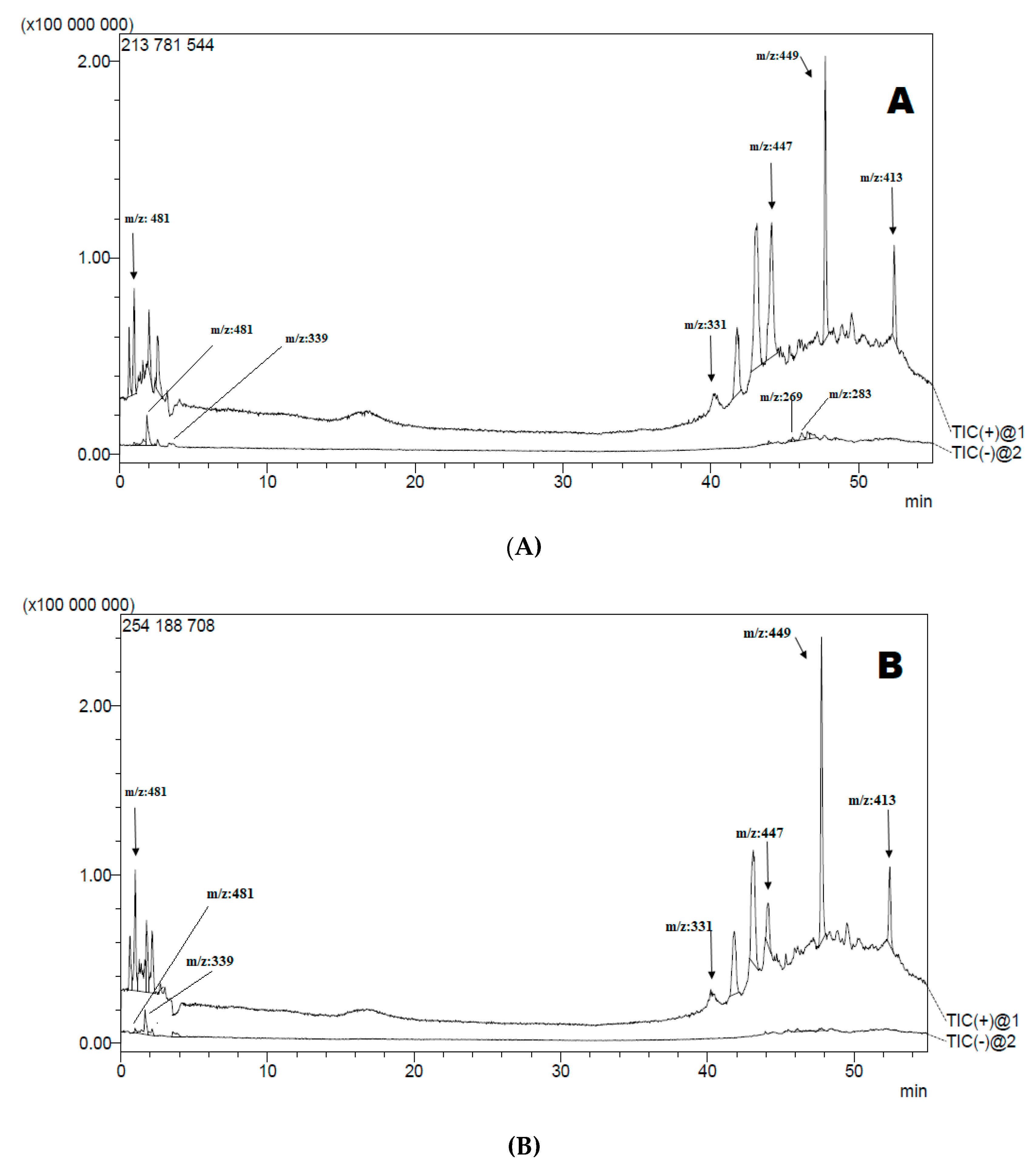
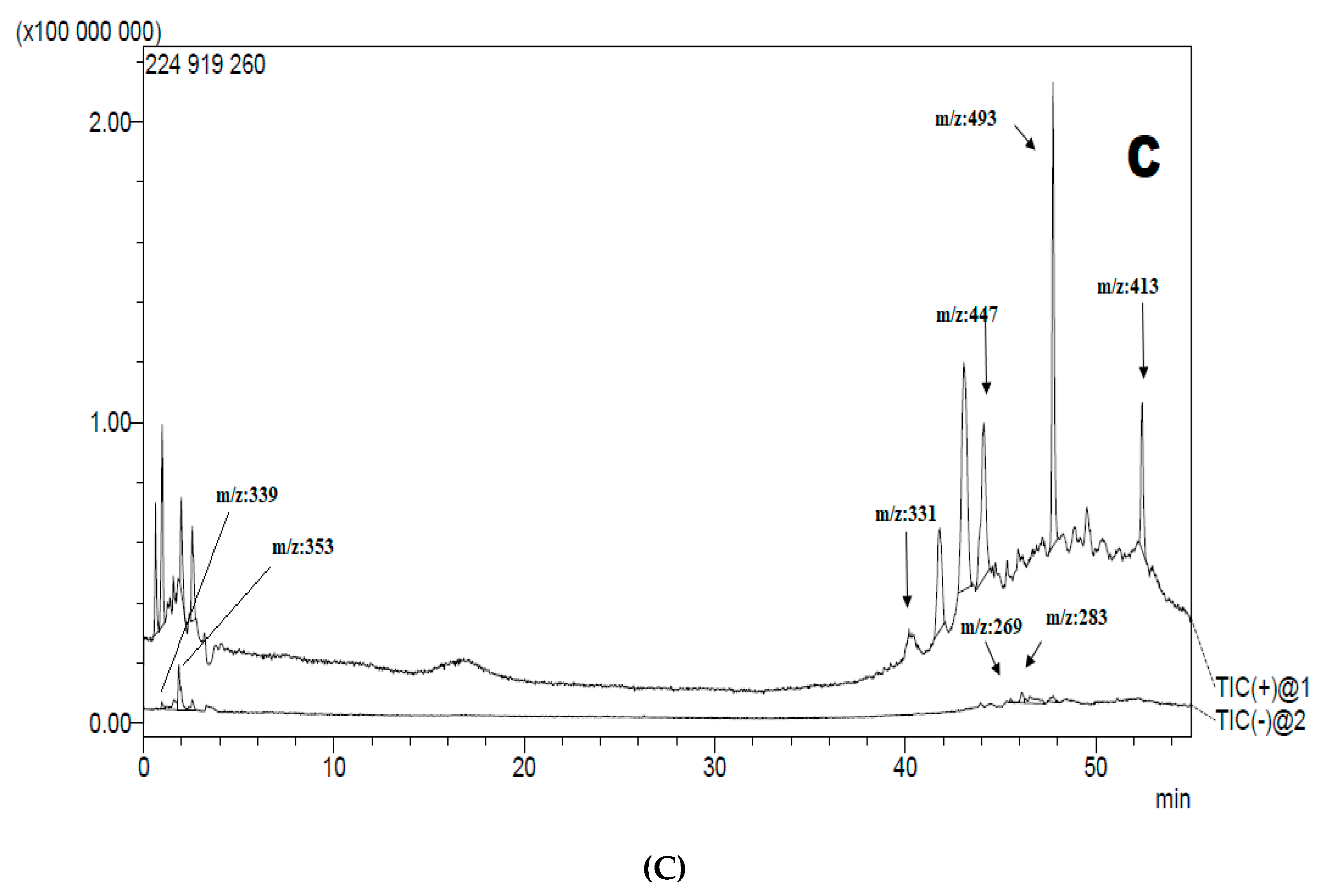
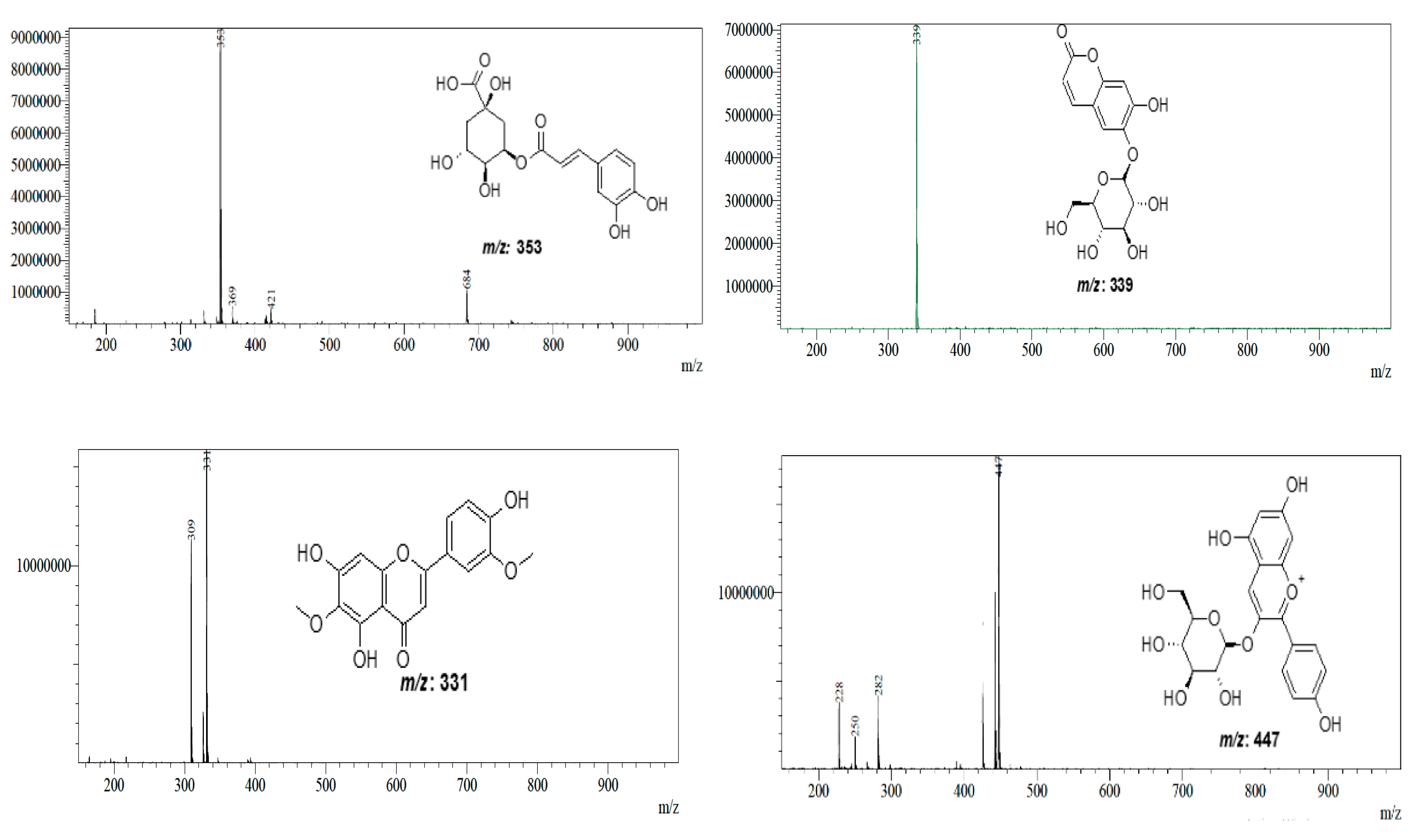
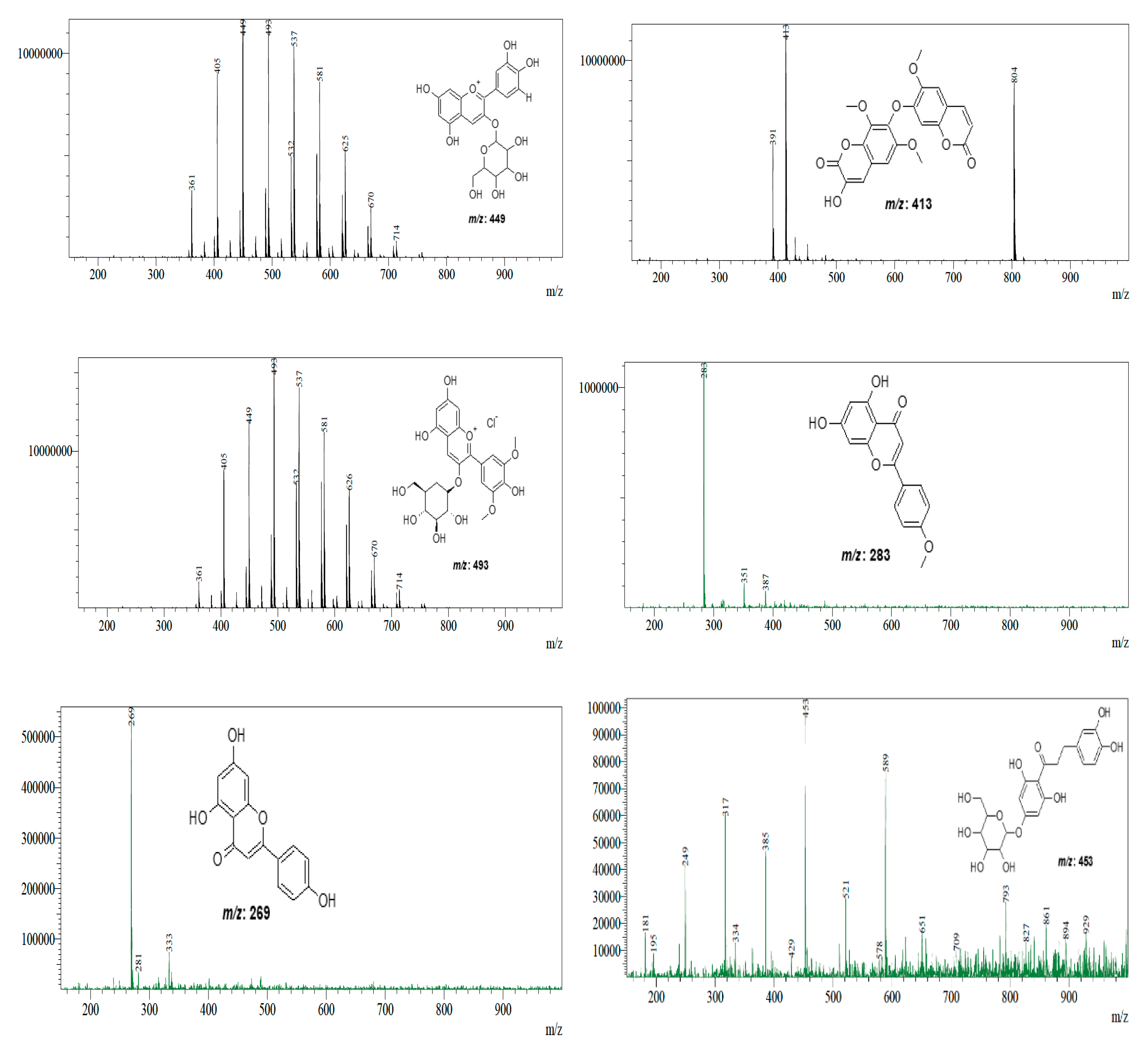
3.2. Liquid Chromatography-Mass Spectrometry Analysis LC-MS-MS

After establishing the optimal UPLC-ESI-MS-MS conditions, the AcDE, AcEAE, and AcBE were analyzed utilizing the method’s full scan with negative and positive ions mode. Figure 1 and Figure 2 illustrate the UPLC-ESI-MS-MS and mass spectra of eleven chemicals identified from A. campestris extracts. Table 3 outlines retention time (Rt), m/z, and the formula of compounds proposed or deduced based on data reported in previously identified in Artemisia genus.

3.3. Assessment of Total Bioactive Compounds

Table 4 summarizes the spectrophotometric results of the bioactive components in A. campestris extracts. Through employing a calibration curve (Phenol: y = 0.0034x + 0.1044, R2 = 0.9972; Flavonoid: y = 0.0048x, R2 = 0.997), the total phenol and flavonoid contents of the extract were, respectively, expressed in µg GAE/mg and µg QE/mg. The highest successive contents of AcEAE for phenolic compounds and flavonoid were 527.333 ± 0.61 µg GAE/mg and 203.4194 ± 0.14 µg QE/mg. In contrast, when compared to the other extracts, AcEAE demonstrated that it contained a high concentration of phenolic and flavonoid components. It was followed by the AcDE, which showed a high content of phenolic compounds and flavonoids at 203.607 ± 0.67 µg GAE/mg and 69.444 ± 0.147 µg QE/mg, respectively.

3.4. Antioxidant Activity

Due to the complexity of phytochemicals, it is expected that the antioxidant capabilities of plant extracts need to be investigated using more than one method. The tests of DPPH, ABTS, β-Carotene, Reducing Power, CUPRAC, GOR, and Phenanthroline were used to identify the antioxidant activities of A. campestris extracts. The results of these assays are listed in Table 5, which shows comprehensive results that are easier to consider, more basic to interpret, and correlate to other compounds. The indices of A0.5 and IC50 were used to measure the antioxidant activity of the extracts under consideration in this study, which were calculated using linear regression analysis. The A0.5 and IC50 levels have an inverse dependence with the activity’s efficiency.

3.4.1. DPPH Scavenging Activity

In the case of the hydrogen proton transfer method, against stable radicals the DPPH assay measures the antioxidant capability of substances according to their ability to scavenge free radicals [47]. A. campestris extracts exhibited a significant capacity of scavenging the radical DPPH. Both standards and plant extracts decreased in the order of BHA > AcEAE > BHT > AcDE > AcAE > AcME > AcBE. According to the results presented in Table 5, AcEAE (IC50: 10.45 ± 0.19 µg/mL) showed strong antioxidant activity in the DPPH method, in which the IC50 was relatively close to the IC50 stated by the BHA (IC50: 5.73 ± 0.41 µg/mL) and more efficient compared with the standard (BHT) (IC50: 22.32 ± 0.19 µg/mL).

3.4.2. ABTS Scavenging Activity

The ABTS radicals represent another approach for assessing the antiradical activity of A. campestris extracts, which operate on the same principle as DPPH and evaluate electron transfer in the media; the ABTS+ scavenging capacities among the various extracts were in the following order: BHT > BHA > AcEAE > AcDE > AcME > AcAE > AcBE. The result obtained from studying the highest concentration (IC50: 9.52 ± 0.12 µg/mL) (Table 5). AcEAE showed an excellent high capacity to scavenge the radical ABTS+.

3.4.3. β-Carotene-Linoleic Acid Bleaching Activity

To evaluate the capability of A. campestris fractions to inhibit lipid peroxidation, we performed the beta-carotene bleaching method. The results in Table 5 showed that AcBE (IC50: 183.87 ± 1.30 µg/mL) had a moderate effect compared with BHT and BHA (IC50: 1.05 ± 0.01;0.90 ± 0.02 µg/mL, respectively). Moreover, AcME, AcPEE, AcDE, AcEAE and AcAE were not active against β-carotene bleaching.

3.4.4. Galvinoxyl (GOR) Scavenging Activity

For the GOR assay, Table 5 showed that AcEAE had high antioxydant activity where the IC50 (IC50: 2.45 ± 0.03 µg/mL) is more effective compared to the values presented by the standards, followed by BHT and BHA (IC50: 3.32 ± 0.18; 5.38 ± 0.06 µg/mL, respectively), with others in the order AcDE (IC50: 16.11 ± 0.02 µg/mL) > AcBE (IC50: 62.37 ± 0.16 µg/mL) > AcME (IC50: 68.21 ± 0.13 µg/mL) > AcAE (IC50: 152.18 ± 0.47 µg/mL).

3.4.5. Reducing Power Activity

The mechanism of reducing power assay is based on the transformation of Fe3+ into Fe2+ [48]. AcEAE was the most effective in reducing iron ions, with the lowest A0.5 value (A0.5: 16.05 ± 0.16 µg/mL). In decreasing order, the effective concentrations at which the absorbance was 0.5 were as follows: BHA > Ascorbic acid > AcEAE > AcDE > AcME > AcAE.

3.4.6. Cupric Reducing Antioxidant Capacity Activity

For CUPRAC, the results in the Table 5 showed that AcEAE had a strong antioxidant activity, with its A0.5: 9.94 ± 0.21 µg/mL value near to the value presented by BHT (A0.5: 9.62 ± 0.87 µg/mL).

3.4.7. O-Phenanthroline Activity

Table 5 shows the ability of A. campestris to decrease the Fe3+ ion using the o-phenanthroline method [32]. Similarly, AcEAE also exhibited the highest chelating activity (A0.5: 7.12 ± 0.15 µg/mL). The antioxidant activity determined by the o-phenanthroline assay follows the decreasing order of BHA > BHT > AcEAE > AcDE > AcBE > AcAE.

3.5. Enzymes Inhibition Activity

3.5.1. Cholinesterase Inhibitory Activity

Additionally, the efficacy of each extract to inhibit enzyme activity was examined using the enzymes acetylcholinesterase (AChE) and butyrylcholinesterase (BChE) (Table 6), with the aim of increasing patients’ cognitive capacity and reestablishing cholinergic transmission [49]. AcEAE showed a significant inhibitory effect for ACHE; it was more effective than AcPEE, whilst AcDE showed the weakest inhibitory effectiveness (IC50 > 200 μg/mL). Although a substantial inhibition was shown towards BChE where the AcPEE displayed the most heightened inhibitory effect, both AcBE and AcAE had no effect on AchE and BchE.

3.5.2. α-Amylase Inhibitory Activity

In order to evaluate the antidiabetic efficient of A. campestris extracts was determined by estimating the potential of the different extracts to inhibit the α-amylase enzyme. From the results obtained (Table 6), the reaction of Artemisa extracts offered the optimum response with a substantially lower IC50 value than that of acarbose, except that the AcAEwas not active against α–amylase. The highest α–amylase inhibitory activity was found in AcPEE (IC50 = 11.97 ± 0.14 μg/mL). This activity is three 333 times higher than acarbose (IC50 = 3650.93 ± 10.70 µg/mL).

3.5.3. Pancreatic Lipase Inhibitory Activity

Table 6 shows the data on the Pancreatic Lipase inhibitory activity of extracts from A. campestris aerial parts. Anti-lipase activity was significantly inhibited by A. campestris extracts, with the AcPEE being the most effective compared to the reference (IC50 = 0.06 ± 0.001 μg/mL). The inhibitory activity follows the decreasing order of orlistat > AcPEE > AcDE > AcEAE, respectively, while AcME, AcBE and AcAE did not affect pancreatic lipase.

3.6. Photoprotective Activity

The Sun Protection Factor (SPF) was determined as an indicator of the photoprotective efficiency A. campetris, as presented in Table 7. SPF values of the extracts of A. campestris ranged from 24.79 ± 0.07 to 40.76 ± 0.1. These findings revealed that all of the extracts had evidence of photoprotective activity. In addition, the extracts of AcME, AcDE, AcEAE, and AcBE were found to have high photoprotective effects with SPF 42.07 ± 0,17; 40.76 ± 0.11; 39.51 ± 0.09; 38 ± 0.05, respectively.
The coefficients of variation of the SPF values of the photoprotective activity turned out to be very low (V = 0.13–1.00), which indicates a very high accuracy in their determination (Table 7).

4. Discussion

A wide range of illnesses can be treated or at least reduced by using plants and/or plant materials; in addition, plants are essential sources for food applications [50]. According to the WHO (World Health Organization), there is abundant proof that minerals have biochemical nutritional and structural activities that are important for overall physical and mental health [51]. The mineral composition of A. campestris has not been investigated in previous studies; hence, this is the first time such a study has been undertaken. The present purpose is to serve the lacuna in research by utilizing the ICP-OES method in order to provide empirical proof of the mineral make-up of A. campetris, along with the specific concentrations. The results of this study can be used as a database for the healthcare industry, and the mineral analysis reveals that A. campestris is a valuable Ca, Fe, Mg, and Na source. In human nutrition, these mineral components are essential. Calcium (Ca) is the most prevalent element in aerial parts of A. campetris, with a concentration of 10,538.1 ± 69.9 mg/Kg. Calcium is an important structural mineral that is commonly present in bones and teeth, and it regulates nerve and muscle function. It plays a critical function in enzyme activation [52]. A. campestris is an excellent source of calcium. A. campestris contains 1181.8 ± 82.37 mg/Kg of iron (Fe), which is the second most common element as shown in this study. Iron helps in the formation of haemoglobin in the transfer of oxygen; it is also a key component of some enzymes that performs biological oxidation in cellular respiration [52,53]. Magnesium and Sodium were both abundant with concentrations of 990.7 ± 3.14 mg/kg and 289.41 ± 1.319 mg/kg, respectively. Additionally, magnesium is a component of many enzyme systems and is also found in bones and teeth [54]. Sodium is the most abundant cation in extracellular fluids, and also plays a role in controlling the volume of plasma and the acid-base equilibrium of cells [55]. The experimental data revealed that the other mineral concentrations in A. campestris decreased in the following order: Cu > Zn > Li > Mn > Sr > Ti > Ni > Co > Mo with a concentration of 70.22 ± 0.94 mg/Kg. A. campestris had a high Cu content above the WHO allowable limits. As per FAO/WHO, the permitted limit of Cu content for fruit samples is 4.5 mg/kg [56]. It is therefore advisable for consumers of A. campestris to take note of this to avoid exposure to Cu in excessive doses; although, the plant is usually consumed in smaller quantities, thus reducing the chance of excess exposure. Other essential elements such as zinc and manganese were detected at significant levels. Consequently, A. campestris can be considered an excellent source of zinc and manganese. Pb, Cr, and Cd were also detected, and have been shown to be of toxicological concern in other studies. According to the World Health Organization’s ADI for lead, the allowed limit for lead concentration in medicinal plants is 10 mg/kg of the plant matter [57]. At 0.68 ± 0.305 mg/Kg, the A. campestris lead content is within the acceptable range [58]. In addition, our results are lower than the maximum permissible limits (MPL) set by the WHO for cadmium, which is 0.3 mg/kg. Interestingly, A. campestris contains large concentrations of macroelements such as Ca, Mg, and Na. This analysis demonstrated the enormous potential of plants in producing a wide range of minerals to benefit the treatment of various diseases.
The findings of the phytochemical study performed on the aerial parts of AcDE, AcEAE, and AcBE utilizing LC-MS-MS analysis (Table 3) led to the tentative identification of eleven chemicals, the majority of which belong to the flavonoid family. Flavonoids were present as flavones, including Acacetin [46], Apigenin [45] and 5, 7, 4′-trihydroxy-3, 6-dimethoxyflavone (Jaceosidin) [41]. Recently, Carazzone et al. [42] reported Pelargonidin-3-O-glucuronide, Cyanidin 3-O-galactoside, and Malvidin 3-O-glucoside in Cichorium intybus, a species from the family Asteraceae [42], which were identified based on their m/z values. These three anthocyanidins have m/z values of 447 (AcDE, AcEAE, AcBE), 449 (AcDE, AcBE), and 493 (AcBE), respectively (Table 3). AcDE, AcEAE, and AcBE were all confirmed to contain derivative coumarins according to the results of their full scan, and MS spectra, 3-hydroxy-6′-dimethoxy-7-(6′-methoxy-7′-coumarinyloxy) coumarin (Arteminorin B) produced protonated molecular ions at 413 m/z; the identity of this compound was confirmed to be bicoumarin by He et al., in Artemisia minor [43], in addition to a [M − H] ion at 339 m/z, which was assigned as Esculetin-6-O-glucoside [44]. Furthermore, the chromatograms of AcDE, AcEAE, and AcBE demonstrated accuracy at m/z 353 in the negative ion mode, which led to the tentative identification of the quinic acid derivative 5-O-caffeoylquinic acid [40]. Dihydrochalcone derivative, which was found in AcEAE, responded to positive ionization at m/z 453; the compound was therefore identified as 3-hydroxyphloretin 6′-O-hexoside [41].
AcDE, AcEAE contained sesquiterpene lactone glucosides with the molecular ion at m/z 481, which were identified as 15-O-β-D-glucopyranosyl-11β, and 13-dihydro urospemal A [39]. The majority of tentatively identified compounds are already known from the Artemisia genus.
In plants, phenolic chemicals constitute a major category of secondary metabolites with important pharmacological effects on the human body; these compounds provide protection against stressors via various mechanisms [59]. The number of polyphenols in A. campestris extracts was determined by examining the extracts with solvents at different polarities. Variable levels of polyphenols and flavonoids were found in this quantitative study. The AcEAE and AcDE yielded the highest total phenolics and flavonoids. Our findings were much greater than those obtained by Megdiche et al. [60] and Djeridane et al. [61]. According to our findings, polar organic solvents proved to be the most efficient in the process of extracting phenolic and flavonoid components. Several factors, including the solvents (degrees of polarity) utilized, ambient and ecological conditions, extraction and quantification processes, and geographic location, have been documented to influence phytochemical content.
Antioxidants protect against free-radical induced tissue damage by inhibiting radical production, scavenging them or promoting their breakdown [59]. Since a given antioxidant’s reaction varies in different testing systems, this led to the assessment of the antioxidant activity of plant extracts using a diversity of approaches, including direct, indirect, competitive procedures, reduction, chelation, and/or inhibition principles [62]. Due to the elevated levels of phenolics and flavonoids in the AcEAE, the above methods show that this extract has a potent antioxidant capacity, which gives AcEAE its distinctive character. Indeed, the strongest antioxidants are phenolic chemicals, which include hydroxyl groups in their structure that allow hydrogen or electrons to be transferred to a reactive molecule.
Synthetic-free radicals termed DPPH and ABTS are commonly used to assess plant extracts and pure compounds for their potential anti-radical ability [63]. The AcEAE had potent antioxidant power against the radical DPPH and the radical ABTS. The change from purple (DPPH) to yellow (DPPHH) was due to the reception of electrons measurable at 515 nm. The ABTS decolorization method is used to quantify the loss of color due to the addition of an antioxidant to the blue-green chromophore 2.2′azinobis-3-ethylbenzothiazoline-6-sulfonic acid (ABTS_+). The obtained results appear to be better correlated with the finding of Akrout et al. [64] using infusion and ethanol 50%. On the other hand, the obtained results of DPPH are similar to those reported by Megdich et al. [60]. The antioxidant properties of AcEAE were low and comparable to those of standard antioxidants saved from the bleaching of carotene. However, it was found that AcBE was the only one that moderately prevented the oxidation of β-carotene brought about by the free radicals produced as a by-product of the oxidation of linoleic acid. It is an important property that the ability to operate on the products of lipid peroxidation provides. Comparing our results with those reported previously, AcBE is incredibly effective in β-carotene [65].
The AcEAE of iron and copper ions was reduced by A. campestris with great efficiency, which may be due to their ability to rebuild oxidized molecules and, therefore, break the hydrogen subtract. Our research shows the evaluation of antioxidant activities for the first time using CUPRAC and O-Phenanthroline. Table 5 presents the ferric reducing power A0.5 of A. campestris extracts which represents the speed of reducing Fe3+(CN)6 to Fe2+(CN)6 by direct electron donation. Then, the complexes of ferric ferrous from the reaction mixture experienced high absorption at 700 nm [66]. Comparable to the results reported by Boulanouar et al., A. campestris oil has been previously reported to exhibit a strong reducing power activity [67], with an IC50 = 0.305 ± 0.006 mg·mL−1.
Another approach conducted for the first time in the genus of A. campestris is the use of the Galvinoxyl free radical scavenging assay. Similarly, AcEAE demonstrates a strong antioxidant activity which is better than the standard. Consequently, when hydrogen donors interact with the free radical Gox it is reduced to GoxH [29]. This reduction was observed spectrophotometrically as a disappearance of color when they were quenched.
The neurodegenerative disorder known clinically as Alzheimer’s disease, is a deadly neurodegenerative disease characterized by increasing brain damage and mental illness [68]. Using cholinesterase inhibitors as a key treatment strategy for AD, acetylcholinesterase and butyrylcholinesterase inhibitors extend the availability of the neurotransmitter acetylcholine to the brain receptors by inhibiting the enzymes that are affected within the hydrolysis of acetylcholine [69]. As demonstrated in Table 6, A. campestris exhibited good inhibitory efficacy for the AcEAE against AChE (IC50 = 23.16 ± 0.19 μg/mL) afterwards AcPEE (IC50 = 59.03 ± 0.58 μg/mL). The AcPEE, nonetheless, moderately inhibited BChE activity (IC50 =93.50 ± 1.60 μg/mL) compared with the Galantamine standard. AcBE and AcAE were reported to be inactive. Compared to the results found for the essential oil of A. campestris reported by Cheraif et al. [70], a significant enzyme inhibitory effect was shown by the AcPEE towards both the AChE and BChE enzymes. For this purpose, we can reach the conclusion that the chemical constituents are capable of behaving as antagonists of AChE and BChE; studies have already demonstrated that particular flavonoids and phenolic acids, such as acacetin and apigenin, possess a significant effect on inhibiting activity towards AChE and BChE, and their existence has already been proven according to the LC-MS/MS results [71]. Natural compounds from multiple sources, such as terpenoids, phenols and alkaloid chemicals have been found to inhibit AchE and BChE in several studies [72]. Secondary metabolites in the AcPEE might well be responsible for acetyl and butyrylcholinesterase activity. Recently, Boukhalkhal et al. [73] identified the following terpenoids in A. campestris L.: Carnosic acid and OH-Rubescensin BI, BII; these compounds may be the source of the anticholinesterase effect investigated in this paper. We have highlighted our findings in light of this, and A. campestris might well have anti-Alzheimer activity that could be beneficial for treating AD.
Chronic hyperglycemia is a characteristic of the metabolic disorder known as diabetes; there are several medicinal strategies for treating type 2 diabetes. One of these processes by which plants exercise their anti-hyperglycaemic action is by inhibiting alpha-amylase in the small intestine, preventing complex carbohydrates from being broken down and preventing their absorption. Starch and other glucose polymers in diabetic individuals hydrolyze (1.4)-D-glycosidic linkages in the presence of this enzyme’s inhibitors [74]. A. campestris extracts showed an outstanding anti-diabetic action that was more efficient than acarbose (Table 6), and we found a significant difference in their IC50. Molecular docking simulations were used to investigate polyphenols’ capacity to inhibit the enzymes alpha-glucosidase and alpha-amylase; furthermore, it was hypothesized that substances such as caffeic acid, Naringin, and Rutin might considerably inhibit the enzymes [75], which includes all phenolic chemicals and flavonoids discussed within the profile phytochemicals of A. campestris extracts that are considered crucial in the treatment against diabetes in this study. In contrast, the existence of certain minerals may favorably contribute to the anti-diabetic effects of plants, e.g., by enhancing the effects of insulin [76]. Our research establishes that A. campestris has anti-diabetic potential in vitro by inhibiting alpha-amylase.
Obesity is a disease characterized by a high ratio of weight to height. One of the leading causes of obesity is an imbalance between the amount of calories a person consumes and the amount of calories their body will burn. Inhibition of pancreatic lipase has become an important approach for the treatment of obesity. The lipolytic products of diacylglycerol, monoglycerides and glycerol, and polyun-saturated fatty acids are produced by the enzyme lipase which catalyses the breakdown of dietary triglycerides [77]. A remarkable effect (IC50: 0.061 ± 0.001 μg/mL) has been achieved using orlistat as a result of our investigation; it presents long-term effectiveness as a pancreatic lipase inhibitor, and is one of the most significant and recently proposed treatments for obesity. This inquiry is the first to investigate the anti-lipase activity of the areal parts of the A. campestris extracts. AcPEE was revealed to be the most effective (Table 6), which provides a moderate potential to identify novel anti-obesity agents. Natural compounds present in natural sources are lipase inhibitors as a consequence of the multitude of inhibitors generated from the following classes of compounds: saponins, polyphenolics, and terpenes [77], as was previously discussed in relation to A. campetris.
Prolonged exposure to ultraviolet radiation can be divided into the following three areas: UVA, UVB, and UVC, which can have negative repercussions including the development of skin cancer in certain conditions [78]. Natural substances are frequently applied in traditional medicine and the industries of cosmetics, and several of flavonoids have been shown to provide protection against ultraviolet radiation [79]. The sun protection factor, known as the SPF, is a quantitative assessment of the effectiveness of a sunscreen product. In relation to this concept, it has recently been established that, according to the SPF ratings, the values of SPF [2→12], [12→30], [30→50] and >50 are classified as possessing a minimum, moderate and strong sun protection action, respectively. According to the Commission of European Communities 2006 recommendation [80], the AcME, AcDE, AcEAE and AcBE belong to the high protection class. The excellent photoprotective capabilities of A. campestris extracts might well be mainly attributed to the existence of flavonoids and phenolics. Based on the findings of this study and what was discussed before, A. campestris has a substantial affinity between its photoprotective effect and its total phenolic content. The Saharan climate and geographical area and the plant’s daily exposure to sunshine led to the production of more phytochemical compounds to shield the plant from ultraviolet harm, hence improving photoprotective activity. As a consequence, the acquired data demonstrated the considerable antioxidant activity of A. campestris as a prospective source of sunscreen in the cosmetic industry or pharmaceutics formulations.

5. Conclusions

The phytochemical profile of A. campestris extracts proved the presence of a variety of types of bioactive compounds that have interesting pharmacological effects. This study marked an important comprehensive examination of the biological activity and mineral constituents of A. campetris, an endemic plant species from Algeria. The findings highlight the importance of the biological activities of A. campetris. The prominent antioxidant and anti-enzymatic properties (anti-diabetic; anti-Alzheimer and anti-obesity) of the plant were demonstrated. A. campestris demonstrates high absorption of ultraviolet light. In our study, the ICP-OES technique was used to conduct a mineral analysis of the plant. The findings demonstrate that A. campestris contains a high concentration of minerals that are essential for human health, including calcium, iron, sodium, magnesium, zinc, and manganese, amongst others. Furthermore, the toxic reference values for the potentially toxic elements were much below the World Health Organization’s tolerance limits. Our study proved that the plant has distinctive medical, food, nutrition, and pharmaceutical properties.

Author Contributions

W.Z.; Methodology, conceptualization, software, data curation, writing–reviewing and editing. O.S., L.B.; supervision, visualization, validation. C.B., M.M.; investigation, software, data curation. I.L., N.B.; data curation, validation. B.S.; formal analysis, validation. C.G.A.; writing–original draft. J.S.-G.; supervision, conceptualization, writing–review and editing. All authors have read and agreed to the published version of the manuscript.

Funding

This research received no external funding.

Institutional Review Board Statement

Not applicable.

Informed Consent Statement

Not applicable.

Data Availability Statement

All data generated or analyzed are contained within the present article.

Conflicts of Interest

The authors declare no conflict of interest.

Abbreviations

AcMEArtemisa campestris Methanol Extract
AcPEEArtemisa campestris Petroleum ether Extract
AcDEArtemisa campestris Dichloromethane Extract
AcEAEArtemisa campestris Ethyl Acetate Extract
AcBEArtemisa campestris Butanol Extract
AcAEArtemisa campestris Aqueous Extract
ACHEacetylcholinesterase
BChEbutyrylcholinesterase
CUPRACcupric reducing antioxidant capacity
DPPH2,2-diphenyl-1-picrylhydrazyl
SPFSun Protective Factor
GORgalvinoxyl radical
ICP-OESInductively coupled plasma-optical emission spectrometry
TIC+Total ion current in positive mode
TICTotal ion current in negative mode

References

  1. Chan, K. Some aspects of toxic contaminants in herbal medicines. Chemosphere 2003, 52, 1361–1371. [Google Scholar] [CrossRef] [Green Version]
  2. Ghribia, L.; Ghouilaa, H.; Omrib, A.; Besbesb, M.; Janneta, H.B. Antioxidant and anti–acetylcholinesterase activities of extracts and secondary metabolites from Acacia cyanophylla. Asian Pac. J. Trop. Biomed. 2014, 4, S417–S423. [Google Scholar] [CrossRef] [PubMed] [Green Version]
  3. Lin, Y.W.; Yang, F.J.; Chen, C.L.; Lee, W.T.; Chen, R.S. Free radical scavenging activity and antiproliferative potential of Polygonum cuspidatum root extracts. J. Nat. Med. 2010, 64, 146–152. [Google Scholar] [CrossRef] [PubMed]
  4. Tani, C.K.; Le Bourgeois, T.; Munoz, F. Aspects floristiques des adventices du domaine phytogéographique oranais (Nord-Ouest Algérien) et persistance d’espèces rares et endémiques. Fl. Medit. 2010, 20, 29–46. [Google Scholar]
  5. Quezel, P.; Santa, S. New flora of Algeria and southern desert regions. In New Flora of Algeria and Southern Desert Regions; CNRS: Paris, France, 1962. [Google Scholar]
  6. Gouveia, S.C.; Castilho, P.C. Artemisia annua L.: Essential oil and acetone extract composition and antioxidant capacity. Ind. Crops Prod. 2013, 45, 170–181. [Google Scholar] [CrossRef]
  7. Quezel, P.; Santa, S. Nouvelle flore de l’Algérie et des régions désertiques méridionales; Editions du Centre National de la recherche scientifique: Paris, France, 1963. [Google Scholar]
  8. Watson, L.E.; Bates, P.L.; Evans, T.M.; Unwin, M.M.; Estes, J.R. Molecular phylogeny of subtribe Artemisiinae (Asteraceae), including Artemisia and its allied and segregate genera. BMC Evol. Biol. 2002, 2, 17. [Google Scholar] [CrossRef]
  9. Toumi, M.; Messirene, A.; Benkhalifa, A. Les plantes à usage thérapeutique dans le Hoggar: Cas de la ville de Tamanrasset et son environnement rural. In 3ème Exposition d’Ethnobotanique Et 4ème Atelier d’Initiation à La Phytothérapie; Jardin d’Essai du Hamma: Mohamed Belouizdad, Algeria, 2016; pp. 63–65. [Google Scholar]
  10. Hamza, N.; Berke, B.; Umar, A.; Cheze, C.; Gin, H.; Moore, N. A review of Algerian medicinal plants used in the treatment of diabetes. J. Ethnopharmacol. 2019, 238, 111841. [Google Scholar] [CrossRef]
  11. Boudjelal, A.; Henchiri, C.; Sari, M.; Sarri, D.; Hendel, N.; Benkhaled, A.; Ruberto, G. Herbalists and wild medicinal plants in M’Sila (North Algeria): An ethnopharmacology survey. J. Ethnopharmacol. 2013, 148, 395–402. [Google Scholar] [CrossRef]
  12. Tardío, J.; Pardo-de-Santayana, M.; Morales, R. Ethnobotanical review of wild edible plants in Spain. Bot. J. Linn. Soc. 2006, 152, 27–71. [Google Scholar] [CrossRef]
  13. Webster, D.; Taschereau, P.; Belland, R.J.; Sand, C.; Rennie, R.P. Antifungal activity of medicinal plant extracts; preliminary screening studies. J. Ethnopharmacol. 2008, 115, 140–146. [Google Scholar] [CrossRef]
  14. Dib, I.; Tits, M.; Angenot, L.; Wauters, J.N.; Assaidi, A.; Mekhfi, H.; Ziyyat, A. Antihypertensive and vasorelaxant effects of aqueous extract of Artemisia campestris L. from Eastern Morocco. J. Ethnopharmacol. 2017, 206, 224–235. [Google Scholar] [CrossRef] [PubMed]
  15. Sefi, M.; Fetoui, H.; Makni, M.; Zeghal, N. Mitigating effects of antioxidant properties of Artemisia campestris leaf extract on hyperlipidemia, advanced glycation end products and oxidative stress in alloxan-induced diabetic rats. Food Chem. Toxicol. 2010, 48, 1986–1993. [Google Scholar] [CrossRef] [PubMed]
  16. Ghlissi, Z.; Sayari, N.; Kallel, R.; Bougatef, A.; Sahnoun, Z. Antioxidant, antibacterial, anti-inflammatory and wound healing effects of Artemisia campestris aqueous extract in rat. Biomed. Biomed. Pharmacother. 2016, 84, 115–122. [Google Scholar] [CrossRef] [PubMed]
  17. Younsi, F.; Mehdi, S.; Aissi, O.; Rahali, N.; Jaouadi, R.; Boussaid, M.; Messaoud, C. Essential oil variability in natural populations of Artemisia campestris (L.) and Artemisia herba-alba (Asso) and incidence on antiacetylcholinesterase and antioxidant activities. Chem. Biodivers. 2017, 14, e1700017. [Google Scholar] [CrossRef] [PubMed]
  18. Golubkina, N.; Logvinenko, L.; Konovalov, D.; Garsiya, E.; Fedotov, M.; Alpatov, A.; Caruso, G. Foliar Application of Selenium under Nano Silicon on Artemisia annua: Effects on Yield, Antioxidant Status, Essential Oil, Artemisinin Content and Mineral Composition. Horticulturae 2022, 8, 597. [Google Scholar] [CrossRef]
  19. Ferchichi, L.; Merza, J.; Landreau, A.; Le Ray, A.M.; Legseir, B.; Seraphin, D.; Richomme, P. Occurrence of isocoumarinic and phenolic derivatives in Artemisia campestris L. subsp. campestris. Biochem. Syst. Ecol. 2006, 34, 829–832. [Google Scholar] [CrossRef]
  20. Larkem, I.; Tarai, N.; Benchikha, N.; Messaoudi, M.; Begaa, S.; Martins, M.; Silva, A.M.S.; Pinto, D.C.G.A. Chemical profile and antioxidant activity of Sesbania bispinosa (Jacq.) W. Wight aerial parts and seeds extracts. J. Food Process. Preserv. 2021, 45, e15468. [Google Scholar] [CrossRef]
  21. Messaoudi, M.; Rebiai, A.; Sawicka, B.; Atanassova, M.; Ouakouak, H.; Larkem, I.; Egbuna, C.; Awuchi, C.G.; Boubekeur, S.; Ferhat, M.A. Effect of Extraction Methods on Polyphenols, Flavonoids, Mineral Elements, and Biological Activities of Essential Oil and Extracts of Mentha pulegium L. Molecules 2022, 27, 11. [Google Scholar] [CrossRef]
  22. Fassel, V.A.; Kniseley, R.N. Inductively coupled plasma. Optical emission spectroscopy. Anal. Chem. 1974, 46, 1110A–1120A. [Google Scholar] [CrossRef]
  23. Butler, C.C.; Kniseley, R.N.; Fassel, V.A. Inductively coupled plasma-optical emission spectrometry. Application to the determination of alloying and impurity elements in low and high alloy steels. Anal. Chem. 1975, 47, 825–829. [Google Scholar] [CrossRef]
  24. Müller, L.; Gnoyke, S.; Popken, A.M.; Böhm, V. Antioxidant capacity and related parameters of different fruit formulations. Lwt-Food Sci. Technol. 2010, 43, 992–999. [Google Scholar] [CrossRef]
  25. Topçu, G.; Ay, M.; Bilici, A.; Sarıkürkcü, C.; Öztürk, M.; Ulubelen, A. A new flavone from antioxidant extracts of Pistacia Terebinthus. Food Chem. 2007, 103, 816–822. [Google Scholar] [CrossRef]
  26. Blois, M.S. Antioxidant determinations by the use of a stable free radical. Nature 1958, 181, 1199–1200. [Google Scholar] [CrossRef]
  27. Re, R.; Pellegrini, N.; Proteggente, A.; Pannala, A.; Yang, M.; Rice-Evans, C. Antioxidant activity applying an improved ABTS radical cation decolorization assay. Free Radic. Biol. Med. 1999, 26, 1231–1237. [Google Scholar] [CrossRef]
  28. Marco, G.J. A rapid method for evaluation of antioxidants. J. Am. Oil Chem. Soc. 1968, 45, 594–598. [Google Scholar] [CrossRef]
  29. Shi, H.; Noguchi, N.; Niki, E. Galvinoxyl method for standardizing electron and proton donation activity. Methods Enzymol. 2001, 335, 157–166. [Google Scholar]
  30. Oyaizu, M. Antioxidative activities of browning reaction prepared from glucosamine. Jpn. J. Nutr. 1986, 44, 307–315. [Google Scholar] [CrossRef] [Green Version]
  31. Apak, R.; Güçlü, K.; Özyürek, M.; Karademir, S.E. Novel total antioxidant capacity index for dietary polyphenols and vitamins C and E, using their cupric ion reducing capability in the presence of neocuproine: CUPRAC method. J. Agric. Food Chem. 2004, 52, 7970–7981. [Google Scholar] [CrossRef]
  32. Szydłowska-Czerniak, A.; Dianoczki, C.; Recseg, K.; Karlovits, G.; Szłyk, E. Determination of antioxidant capacities of vegetable oils by ferric-ion spectrophotometric methods. Talanta 2008, 76, 899–905. [Google Scholar] [CrossRef]
  33. Ellman, G.L.; Courtney, K.D.; Andres, V.; Featherstone, R.M. A new and rapid colorimetric determination of acetylcholinesterase activity. Biochem. Pharmacol. 1961, 7, 88–95. [Google Scholar] [CrossRef]
  34. Zengin, G.; Sarikurkcu, C.; Aktumsek, A.; Ceylan, R.; Ceylan, O. A comprehensive study on phytochemical characterization of Haplophyllum myrtifolium Boiss. endemic to Turkey and its inhibitory potential against key enzymes involved in Alzheimer, skin diseases and type II diabetes. Ind. Crops Prod. 2014, 53, 244–251. [Google Scholar] [CrossRef]
  35. Souza, S.P.D.; Pereira, L.L.S.; Souza, A.A.; Santos, C.D.D. Inhibition of pancreatic lipase by extracts of Baccharis trimera (Less.) DC., Asteraceae: Evaluation of antinutrients and effect on glycosidases. Rev. Bras. Farmacogn. 2011, 21, 450–455. [Google Scholar] [CrossRef]
  36. Santos, B.C.S.; Pires, A.S.; Yamamoto, C.H.; Couri, M.R.C.; Taranto, A.G.; Alves, M.S.; Araújo, A.L.D.S.D.M.; de Sousa, O.V. Methyl Chavicol and Its Synthetic Analogue as Possible Antioxidant and Antilipase Agents Based on the In Vitro and In Silico Assays. Oxid. Med. Cell. Longev. 2018, 2018. Available online: https://www.semanticscholar.org/paper/Methyl-Chavicol-and-Its-Synthetic-Analogue-as-and-Santos-Pires/56fd111a214c845f2828b32bbbc046ed0f2554a3 (accessed on 1 September 2022). [CrossRef] [PubMed] [Green Version]
  37. Mansur, J.D.S.; Breder, M.N.R.; Mansur, M.C.D.A.; Azulay, R.D. Determinaçäo do fator de proteçäo solar por espectrofotometria. An. Bras. Dermatol. 1986, 40, 121–124. [Google Scholar]
  38. Sayre, R.M.; Agin, P.P.; LeVee, G.J.; Marlowe, E. A comparison of in vivo and in vitro testing of sunscreening formulas. Photochem. Photobiol. 1979, 29, 559–566. [Google Scholar] [CrossRef]
  39. Aissani, F.; Grara, N.; Bensouici, C.; Bousbia, A.; Ayed, H.; Idris, M.H.M.; Teh, L.K. Algerian Sonchus oleraceus L.: A comparison of different extraction solvent on phytochemical composition, antioxidant properties and anti-cholinesterase activity. Adv. Trad. Med. 2022, 22, 383–394. [Google Scholar] [CrossRef]
  40. Bakchiche, B.; Gherib, A.; Bronze, M.R.; Ghareeb, M.A. Identification, quantification, and antioxidant activity of hydroalcoholic extract of Artemisia campestris from Algeria. Turk. J. Pharm. Sci. 2019, 16, 234. [Google Scholar] [CrossRef]
  41. Bourgou, S.; Rebey, I.B.; Mkadmini, K.; Isoda, H.; Ksouri, R.; Ksouri, W.M. LC-ESI-TOF-MS and GC-MS profiling of Artemisia herba-alba and evaluation of its bioactive properties. Int. Food Res. J. 2017, 99, 702–712. [Google Scholar] [CrossRef]
  42. Carazzone, C.; Mascherpa, D.; Gazzani, G.; Papetti, A. Identification of phenolic constituents in red chicory salads (Cichorium intybus) by high-performance liquid chromatography with diode array detection and electrospray ionisation tandem mass spectrometry. Food Chem. 2013, 138, 1062–1071. [Google Scholar] [CrossRef]
  43. He, Z.Z.; Yan, J.F.; Song, Z.J.; Ye, F.; Liao, X.; Peng, S.L.; Ding, L.S. Chemical constituents from the aerial parts of Artemisia minor. J. Nat. Prod. 2009, 72, 1198–1201. [Google Scholar] [CrossRef]
  44. Rechek, H.; Haouat, A.; Hamaidia, K.; Allal, H.; Boudiar, T.; Pinto, D.C.G.A.; Cardoso, S.M.; Bensouici, C.; Soltani, N.; Silva, A.M.S. Chemical composition and antioxidant, anti-inflammatory, and enzyme inhibitory activities of an endemic species from southern algeria: Warionia saharae. Molecules 2021, 26, 5257. [Google Scholar] [CrossRef] [PubMed]
  45. Ivanescu, B.; Vlase, L.; Corciova, A.; Lazar, M.I. HPLC-DAD-MS study of polyphenols from Artemisia absinthium, A. annua, and A. vulgaris. Chem. Nat. Compd. 2010, 46, 468–470. [Google Scholar] [CrossRef]
  46. Ivanescu, B.; Lungu, C.; Vlase, L.; Gheldiu, A.M.; Grigorescu, C.; Corciova, A. Bioactive compounds from Artemisia campestris L. subsp. campestris. Dementia 2018, 2, 3. [Google Scholar] [CrossRef]
  47. Sasikumar, V.; Kalaisezhiyen, P. Evaluation of Free Radical Scavenging Activity of Various Leaf Extracts from Kedrostis Foetidissima (Jacq.) Cogn. Biochem. Anal. Biochem. 2014, 3, 1. [Google Scholar] [CrossRef] [Green Version]
  48. Alam, M.N.; Bristi, N.J.; Rafiquzzaman, M. Review on in vivo and in vitro methods evaluation of antioxidant activity. SPJ 2013, 21, 143–152. [Google Scholar] [CrossRef] [PubMed] [Green Version]
  49. Polinsky, R.J. Clinical pharmacology of rivastigmine: A new-generation acetylcholinesterase inhibitor for the treatment of Alzheimer’s disease. Clin. Ther. 1998, 20, 634–647. [Google Scholar] [CrossRef]
  50. Jamshidi-Kia, F.; Lorigooini, Z.; Amini-Khoei, H. Medicinal plants: Past history and future perspective. J. HerbMed Pharmacol. 2018, 7, 1–7. [Google Scholar] [CrossRef]
  51. Messaoudi, M.; Begaa, S. Dietary Intake and Content of Some Micronutrients and Toxic Elements in Two Algerian Spices (Coriandrum sativum L. and Cuminum cyminum L.). Biol. Trace Elem. Res. 2019, 188, 508–513. [Google Scholar] [CrossRef]
  52. Benarfa, A.; Begaa, S.; Messaoudi, M.; Hamlat, N.; Sawicka, B. Elemental composition analysis of Pistacia lentiscus L.; leaves collected from Mitidja plain in Algeria using instrumental neutron activation analysis (INAA) technique. Radiochim. Acta 2020, 108, 821–828. [Google Scholar] [CrossRef]
  53. Zazzo, J.F. Oligo-éléments, vitamines et immunité. Nutr. Clin. Metab. 1993, 7, 121–129. [Google Scholar] [CrossRef]
  54. Rahman, M.; Islam, M.A. Concentrations and health risk assessment of trace elements in cereals, fruits, and vegetables of Bangladesh. Biol. Trace Elem. Res. 2019, 191, 243–253. [Google Scholar] [CrossRef] [PubMed]
  55. Rahman, M.; Islam, M.A.; Zaved, M.M. Assessment of Essential and Potentially Toxic Elements and Possible Health Risks in Hylocereus undatus and Punica granatum. Biol. Trace Elem. Res. 2020, 198, 707–713. [Google Scholar] [CrossRef] [PubMed]
  56. Codex, F.A.O. Alimentarius, General Requirements (Food Hygiene); FAO: Rome, Italy, 1995. [Google Scholar]
  57. World Health Organization. WHO Global Report on Traditional and Complementary Medicine 2019; World Health Organization: Geneva, Switzerland, 2019. [Google Scholar]
  58. Lasisi, A.A.; Ejelonu, B.C.; Nwosu, F.O.; Olayiwola, M.A.; Yusuff, A.A. Heavy metals and macronutrients content in selected herbal plants of South-Western Nigeria. Hamdard Med. 2006, 49, 71–76. [Google Scholar]
  59. Sánchez-Moreno, C. Methods used to evaluate the free radical scavenging activity in foods and biological systems. Food Sci. Technol. Int. 2002, 8, 121–137. [Google Scholar] [CrossRef]
  60. Megdiche-Ksouri, W.; Trabelsi, N.; Mkadmini, K.; Bourgou, S.; Noumi, A.; Snoussi, M.; Barbria, R.; Tebourbi, O.; Ksouri, R. Artemisia campestris phenolic compounds have antioxidant and antimicrobial activity. Ind. Crops Prod. 2015, 63, 104–113. [Google Scholar] [CrossRef]
  61. Djeridane, A.; Yousfi, M.; Nadjemi, B.; Boutassouna, D.; Stocker, P.; Vidal, N. Antioxidant activity of some Algerian medicinal plants extracts containing phenolic compounds. Food Chem. 2006, 97, 654–660. [Google Scholar] [CrossRef]
  62. Inchuen, S.; Narkrugsa, W.; Pornchaloempong, P. Effect of drying methods on chemical composition, color and antioxidant properties of Thai red curry powder. Agric. Nat. Resour. 2010, 44, 142–151. [Google Scholar]
  63. Fidrianny, I. Evaluation of antioxidant activities from various extracts of Dragon fruit peels using DPPH, ABTS assays and correlation with phenolic, flavonoid, carotenoid content. Int. J. Pharm. Sci. 2014, 5, 104–111. [Google Scholar]
  64. Akrout, A.; El Jani, H.; Amouri, S.; Neffati, M. Screening of antiradical and antibacterial activities of essential oils of Artemisia campestris L.; Artemisia herba alba asso, & thymus capitatus hoff. Et link. Growing wild in the southern of Tunisia. Res. Sci. Technol. Educ. 2009, 2, 29–39. [Google Scholar]
  65. Akrout, A.; Gonzalez, L.A.; El Jani, H.; Madrid, P.C. Antioxidant and antitumor activities of Artemisia campestris and Thymelaea hirsuta from southern Tunisia. Food Chem. Toxicol. 2011, 49, 342–347. [Google Scholar] [CrossRef]
  66. Hsu, B.; Coupar, I.M.; Ng, K. Antioxidant activity of hot water extract from the fruit of the Doum palm. Hyphaene Thebaica. Food Chem. 2006, 98, 317–328. [Google Scholar] [CrossRef]
  67. Boulanouar, B.; Abdelaziz, G.; Aazza, S.; Gago, C.; Miguel, M.G. Antioxidant activities of eight Algerian plant extracts and two essential oils. Ind. Crops Prod. 2013, 46, 85–96. [Google Scholar] [CrossRef]
  68. Liu, G.; Zhao, Y.; Jin, S.; Hu, Y.; Wang, T.; Tian, R.; Han, Z.; Xu, D.; Jiang, Q. Circulating vitamin E levels and Alzheimer’s disease: A Mendelian randomization study. Neurobiol. Aging 2018, 72, 189-e1. [Google Scholar] [CrossRef] [PubMed]
  69. Mesulam, M.; Guillozet, A.; Shaw, P.; Quinn, B. Widely spread butyrylcholinesterase can hydrolyze acetylcholine in the normal and Alzheimer brain. Neurobiol. Dis. 2002, 9, 88–93. [Google Scholar] [CrossRef] [Green Version]
  70. Cheraif, K.; Bakchiche, B.; Gherib, A.; Bardaweel, S.K.; Çol Ayvaz, M.; Flamini, G.; Ascrizzi, R.; Ghareeb, M.A. Chemical composition, antioxidant, anti-tyrosinase, anti-cholinesterase and cytotoxic activities of essential oils of six Algerian plants. Molecules 2020, 25, 1710. [Google Scholar] [CrossRef] [Green Version]
  71. Szwajgier, D. Anticholinesterase Activity of Selected Phenolic Acids and Flavonoids-Interaction Testing in Model Solutions. Ann. Agric. Environ. Med. 2015, 22. Available online: https://agro.icm.edu.pl/agro/element/bwmeta1.element.agro-0469e05d-0b2c-4ff9-aedc-ce3ad01bf0ba (accessed on 1 September 2022). [CrossRef]
  72. Murray, A.P.; Faraoni, M.B.; Castro, M.J.; Alza, N.P.; Cavallaro, V. Natural AChE inhibitors from plants and their contribution to Alzheimer’s disease therapy. Curr. Neuropharmacol. 2013, 11, 388–413. [Google Scholar] [CrossRef]
  73. Boukhalkhal, S.; Gourine, N.; Pinto, D.C.G.A.; Silva, A.M.S.; Yousfi, M. UHPLC-DAD-ESI-MSn profiling variability of the phenolic constituents of Artemisia campestris L. populations growing in Algeria. Biocatal. Agric. Biotechnol. 2020, 23, 101483. [Google Scholar] [CrossRef]
  74. Wresdiyati, T.; SA’DIAH, S.; Winarto, A.D.I.; Febriyani, V. Alpha-glucosidase inhibition and hypoglycemic activities of Sweitenia mahagoni seed extract. HAYATI J. Biosci. 2015, 22, 73–78. [Google Scholar] [CrossRef] [Green Version]
  75. Rasouli, H.; Hosseini-Ghazvini, S.M.-B.; Adibi, H.; Khodarahmi, R. Differential α-amylase/α-glucosidase inhibitory activities of plant-derived phenolic compounds: A virtual screening perspective for the treatment of obesity and diabetes. Food Funct. 2017, 8, 1942–1954. [Google Scholar] [CrossRef]
  76. Bljajić, K.; Brajković, A.; Čačić, A.; Vujić, L.; Jablan, J.; Saraiva de Carvalho, I.; Zovko Končić, M. Chemical composition, antioxidant, and α-glucosidase-inhibiting activity of aqueous and hydroethanolic extracts of traditional antidiabetics from Croatian ethnomedicine. Horticulturae 2021, 7, 15. [Google Scholar] [CrossRef]
  77. De la Garza, A.L.; Milagro, F.I.; Boque, N.; Campión, J.; Martínez, J.A. Natural inhibitors of pancreatic lipase as new players in obesity treatment. Planta Med. 2011, 77, 773–785. [Google Scholar] [CrossRef] [PubMed] [Green Version]
  78. Dutra, E.A.; Kedor-Hackmann, E.R.M.; Santoro, M.I.R.M. Determination of sun protection factor (SPF) of sunscreens by ultraviolet spectrophotometry. Rev. De Cienc. Farm. Basica E Apl. 2004, 40, 381–385. [Google Scholar] [CrossRef]
  79. Saewan, N.; Jimtaisong, A. Photoprotection of natural flavonoids. J. Appl. Pharm. Sci. 2013, 3, 129–141. [Google Scholar]
  80. Commission of European Communities. Recommendation of 22 September. On Sun Screen Products and Manufacturers Claims. Off. J. Eur. Union 2006. Available online: https://eur-lex.europa.eu (accessed on 1 September 2022).
Figure 1. Total ion current (TIC) profile of AcDE (A) AcEAE (B) and AcBE (C).
Horticulturae 08 00914 g001aHorticulturae 08 00914 g001b
Figure 2. Mass spectra of detected compounds from A. campestris extracts.
Horticulturae 08 00914 g002aHorticulturae 08 00914 g002b
Table 1. The normalized product function used in the calculation of sun protection factor (SPF).
Longueur D’onde λ (nm)EE (λ) × I(λ) (Norms)
2900.0150
2950.0817
3000.2874
3050.3278
3100.1864
3150.0837
3200.0180
Total1.0000
Table 2. Values of mean and standard deviations of chemical elements mass fractions in the plant samples study (mg/kg on a dry mass basis) (n = 3).
ElementsMean ± SD *V (%) **
Ca10,538.1 ± 69.90.66
Cd0.0988 ± 0.00989.92
Co1.28 ± 0.14811.56
Cr1.083 ± 0.0555.08
Fe1181.8 ± 8.236.97
Li46.6 ± 4.459.55
Mg990.7 ± 3.1450.32
Mn41.06 ± 6.5115.85
Mo0.29 ± 0.06823.45
Ni2.75 ± 0.4315.64
Pb0.68 ± 0.30544.85
Sr23.93 ± 0.7683.21
Ti20.38 ± 1.617.90
Zn47.07 ± 0.7971.69
Na298.41 ± 1.3190.44
Cu70.22 ± 0.941.34
* SD standard deviation (All values expressed on dry weight basis). ** V—variability coefficient.
Table 3. Phenolic profile determined by LC-MS-MS in fractions from A. campetris compared with literature (retention time (Rt), not identifie (NI)).
ExtracttR (min)Ionisation Mode
(m/z)
m/zTentatively Identified CompoundMolecular
Formula
Ref
AcDE
AcEAE
AcBE
0.645
0.627
0.646
[M + H]+172NINI-
AcDE
AcEAE
0.983
0.986
[M + H]+48115-O-β-D-glucopyranosyl-11β, 13-dihydro urospermal AC21H30O10[39]
AcBE0.987[M + H]+437NINI-
AcEAE1.629[M + H]+365NINI-
AcDE
AcEAE
AcBE
1.998
1.764
1.995
[M + H]3535-O-caffeoylquinic acidC16H18O9[40]
AcDE
AcEAE
AcBE
2.564
2.144
2.568
[M + H]+381NINI-
AcDE
AcEAE
AcBE
41.787
41.831
41.805
[M + H]+331JaceosidinC17H14O7[41]
AcDE
AcEAE
AcBE
43.128
43.130
43.083
[M + H]+367NINI-
AcDE
AcEAE
AcBE
44.107
44.133
44.115
[M + H]+447Pelargonidin-3-O-glucuronideC21H19O11[42]
AcDE
AcEAE
47.738
47.771
[M + H]+449Cyanidin-3-O-glucosideC21H20O11[42]
AcDE
AcEAE
AcBE
52.406
52.432
52.427
[M + H]+413Arteminorin BC21H16O9[43]
AcBE47.747[M + H]+493Malvidin 3-O-glucosideC23H25ClO12[42]
AcDE
AcEAE
AcBE
0.978
0.974
0.970
[M − H]239NINI-
AcDE
AcBE
1.600
1.597
[M − H]369NINI-
AcDE
AcEAE
AcBE
1.854
1.646
1.871
[M − H]339Esculetin-6-O-glucosideC15H16O9[44]
AcDE
AcEAE
AcBE
2.387
2.134
2.565
[M − H]403NINI-
AcEAE0.218[M − H]4533-hydroxyphloretin 6′-O-hexosideC21H24O11[41]
AcDE
AcBE
45.530
45.522
[M − H]269ApigeninC15H10O5[45]
AcDE
AcEAE
AcBE
46.137
46.135
46.112
[M − H]283AcacetinC16H12O5[46]
AcEAE3.552[M − H]198NINI-
Table 4. Total phenolics, flavonoids contents of differences extract of A. campestris.
ExtractsTotal Phenolic Compounds Content
(µg GAE/mg) *
Flavonoids Content
(µg QE/mg) **
AcME135.37 ± 1.35 a61.59 ± 0.58 a
AcPEE30.27 ± 0.33 b65.69 ± 0.29 b
AcDE203.60 ± 0.67 c69.44 ± 1.47 c
AcEAE527.33 ± 0.61 d203.19 ± 0.14 d
AcBE130.27 ± 0.33 e66.87 ± 0.29 e
AcAE141.64 ± 1.52 f63.4 ± 0.14 f
Results are expressed as means ± SEM of three measures, Tukey test. Values with different letters in the same column are significantly different at p < 0.05. * µg AGE/mg: microgram Gallic acid equivalent/milligram of extract, ** µg QE/mg: microgram Quercetin equivalent/milligram of extract.
Table 5. Antioxidant potentials of different fractions of A. campestris.
ExtractsDPPH
IC50 (μg/mL)
ABTS
IC50 (μg/mL)
β-Carotene Linoleic Acid
IC50 (μg/mL)
GOR
IC50 (μg/mL)
Phenanthroline A0.5 (μg/mL)Reducing Power
A0.5 (μg/mL)
CUPRAC
A0.5 (μg/mL)
AcME141.47 ± 0.65 a26.04 ± 0.39 a≥20068.21 ± 0.13 a≥5054.00 ± 0.33 a449.57 ± 4.87 a
AcPEE≥200≥200≥200≥100≥50≥50≥200
AcDE73.82 ± 1.98 b23.26 ± 0.42 b>20016.11 ± 0.02 b31.95 ± 0.22 b96.58 ± 1.51 b56.44 ± 1.11 b
AcEAE10.45 ± 0.19 c9.52 ± 0.12 c>2002.45 ± 0.03 c7.12 ± 0.15 c16.05 ± 0.16 c9.94 ± 0.21 c
AcBE147.09 ± 0.17 d66.52 ± 0.94 d183.87 ± 1.30 d62.37 ± 0.16 d35.56 ± 1.51 d≥20091.58 ± 2.67 d
AcAE126.09 ± 1.63 e58.67 ± 0.58 e≥50152.18 ± 0.47 e136.67 ± 1.53 e103.25 ± 1.09 e233.33 ± 0.58 e
BHT *22.32 ± 1.19 f1.29 ± 0.30 f1.05 ± 0.01 f3.32 ± 0.18 f2.24 ± 0.17 f≥2009.62 ± 0.87 f
BHA *5.73 ± 0.41 g1.81 ± 0.10 g0.90 ± 0.02 g5.38 ± 0.06 g0.93 ± 0.07 g8.41 ± 0.67 g3.64 ± 0.19 g
Ascorbic acid *NTNTNTNTNT9.01 ± 1.46 hNT
A0.5: the concentration at the 0.50 absorption and IC50: the concentration at the 50 of inhibition. A0.5 and IC50 values represent the means ± SEM of three measures. Tukey test. The values with different superscripts (a. b. c. d. e. f. g. h) in the same columns are significantly different (p < 0.05). * Standard compounds. NT: not tested.
Table 6. Anti-Cholinesterase. Anti-α-Amylase. Anti-Lipase activities of different extracts of A. campestris.
ExtractsAnti-CholinesteraseAnti-α-AmylaseAnti-Lipase
AChE IC50
(μg/mL)
Inhibition (%)
(200 μg/mL)
BChE IC50
(μg/mL)
Inhibition (%)
(200 μg/mL)
α-Amylase IC50 (μg/mL)Inhibition (%)
(4000 μg/mL)
Lipase IC50 (μg/mL)Inhibition (%)
(1000 μg/mL)
AcME NANA≥20030.51 ± 0.29NTNTNTNT
AcPEE59.03 ± 0.58 a88.01 ± 0.68 93.50 ± 1.60 a56.13 ± 1.49 11.79 ± 0.14 a97.91 ± 0.63 40.15 ± 1.36 a75.72 ± 1.34
AcDE≥2004.94 ± 0.50185.11 ± 2.5 b52.49 ± 1.5228.33 ± 1.35 b92.10 ± 0.3686.29 ± 2.60 b62.49 ± 0.38
AcEAE23.16 ± 0.19 c83.25 ± 0.39≥ 20026.49 ± 1.55284.33 ± 3.9 c51.91 ± 0.62155.47 ± 3.44 c55.04 ± 0.58
AcBENANANANA21.55 ± 0.66 d88.98 ± 0.07NANA
AcAENANANANANANANANA
Galantamine *6.27 ± 1.15 e94.77 ± 0.3434.75 ± 1.99 e78.95 ± 0.58NANTNTNT
Acarbose *NTNTNTNT3650.93 ± 10.7 f 53.05 ± 1.59NTNT
Orlistat *NTNTNTNTNTNT0.06 ± 0.001 g79.84 ± 1.07
IC50: the concentration at the 50 of inhibition. IC50, inhibition (%) values represent the means ± SEM of three measures, Tukey test. The values with different superscripts (a, b, c, d, e, f, g) in the same columns are significantly different (p < 0.05); * Standard compounds, NT: not tested. NA: not active.
Table 7. Photoprotective activity of different extract/fractions of A. campestris.
Nivea *Vichy *AcMEAcPEEAcDEAcEAEAcBEAcAE
SPF50.11 ± 0.5344.22 ± 0.3542.07 ± 0,1724.79 ± 0.0740.76 ± 0.1139.51 ± 0.0938.00 ± 0.0526.07 ± 0.22
V **1.000.790.220.280.270.230.130.84
* Reference compounds. ** variability coefficients (%).
Publisher’s Note: MDPI stays neutral with regard to jurisdictional claims in published maps and institutional affiliations.

